Toll-Like Receptor 2–Deficient Mice Are Highly Susceptible to Streptococcus
pneumoniae Meningitis because of Reduced Bacterial Clearing and Enhanced
Inflammation
Hakim Echchannaoui,1Karl Frei,3Christian Schnell,2 Stephen L. Leib,4Werner Zimmerli,1
and Regine Landmann1
1Division of Infectious Diseases, Department of Research, University Hospital, and2Division of Angiogenesis Platform, Pharma Novartis, Basel,3Department of Neurosurgery, University Hospital, Zu¨rich, and4Institute for Infectious Diseases, University of Bern, Bern, Switzerland Toll-like receptor–2 (TLR2) mediates host responses to gram-positive bacterial wall
com-ponents. TLR2 function was investigated in a murine Streptococcus pneumoniae meningitis model in wild-type (wt) and TLR2-deficient (TLR2⫺/⫺) mice. TLR2⫺/⫺mice showed earlier time of death than wt mice (P!.02). Plasma interleukin-6 levels and bacterial numbers in blood and peripheral organs were similar for both strains. With ceftriaxone therapy, none of the wt but 27% of the TLR2⫺/⫺mice died (P!.04). Beyond 3 hours after infection, TLR2⫺/⫺ mice had higher bacterial loads in brain than did wt mice, as assessed with luciferase-tagged S. pneumoniae by means of a Xenogen-CCD (charge-coupled device) camera. After 24 h, tumor necrosis factor activity was higher in cerebrospinal fluid of TLR2⫺/⫺ than wt mice (P!.05) and was related to increased blood-brain barrier permeability (Evans blue staining, ). In conclusion, the lack of TLR2 was associated with earlier death from meningitis, P!.02
which was not due to sepsis but to reduced brain bacterial clearing, followed by increased intrathecal inflammation.
Streptococcus pneumoniae is the major cause of meningitis in adults. Despite antimicrobial therapy and critical care medicine, the mortality remains as high as 28% [1]. In addition, 50% of the survivors have neurologic sequelae, indicating postinflam-matory damage [2]. In the pathogenesis of meningitis, pene-tration of bacteria through the blood-brain barrier (BBB) in-itiates activation of brain endothelia and leads to leukocyte recruitment and release of inflammatory mediators. Subse-quently, the subarachnoidal inflammation stimulates astrocytes, microglia, and neurons to produce cytokines and chemokines [3–5]. A bacterial level of⭓5 log cfu in the brain initiates a harmful inflammatory cascade, which causes development of symptoms and determines the prognosis [6].
Pneumococci enter into the cerebral compartment through the BBB via binding of their cell-wall component phosphoryl-choline to the platelet-activating factor receptors expressed on
Received 22 February 2002; revised 3 May 2002; electronically published 16 August 2002.
Presented in part: 41st Interscience Conference on Antimicrobial Agents and Chemotherapy, Chicago, 16–19 December 2001 (abstract 1857).
Mice were kept in the Animal House of the Department of Research, University Hospitals, Basel, according to the regulations of Swiss veterinary law.
Financial support: Swiss National Foundation (Nr.3200 06165400/1). Reprints or correspondence: Dr. Regine Landmann, Div. of Infectious Diseases, Dept. of Research, University Hospital, Hebelstr. 20, CH-4031 Basel, Switzerland ([email protected]).
The Journal of Infectious Diseases 2002; 186:798–806
䉷 2002 by the Infectious Diseases Society of America. All rights reserved.
0022-1899/2002/18606-0010$15.00
activated cells [7, 8]. In view of the finding that antagonists of the platelet-activating factor receptor do not completely block bacterial invasion [7, 8], other cell-wall components, such as peptidoglycan and lipoteichoic acid, might also induce inflam-mation via activation of pattern recognition receptors expressed in these cells [9].
Toll was first cloned in Drosophila species [10] and was found to be involved in host defense against fungal infection [11]. In humans, 10 homologous genes have been identified encoding Toll-like receptors (TLRs) [12–14]. TLR2 is involved in cell activation by gram-positive bacterial cell wall and membrane components, such as peptidoglycan, lipoteichoic acid, and li-poproteins [15–17]. Accordingly, macrophages isolated from TLR2-deficient (TLR2⫺/⫺) mice are hyporesponsive to
Staph-ylococcus aureus peptidoglycan stimulation [18]. Despite grow-ing evidence implicatgrow-ing TLR2 in recognition of gram-positive bacterial cell-wall components in vitro, its role in bacterial men-ingitis is still unknown. In this study, we compared disease severity and outcome in TLR2⫺/⫺and wild-type (wt) mice in an adult mouse model of S. pneumoniae and Listeria monocyto-genes meningitis. Bacterial numbers in the brain, bacterial counts, and leukocyte recruitment into the subarachnoid space, as well as meningeal inflammation and BBB permeability, were analyzed.
Materials and Methods
Preparation of bacterial inocula. S. pneumoniae (clinical isolate
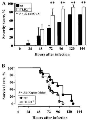
Figure 1. A, Percentage of wild-type (wt;n p 22) and Toll-like re-ceptor-2–deficient (TLR2⫺/⫺;n p 21) mice showing high severity score (including score 4 and score 5) after intracerebral injection of 2⫻ 102 cfu of Streptococcus pneumoniae. Data from 3 independent experiments are represented as the mean percentage of high severity scoreⳲ SD. **P!.02, repeated-measures analysis of variance (ANOVA). B, Sur-vival of TLR2⫺/⫺(n p 21) and wt (n p 22) mice after intracerebral injection of2⫻ 102cfu of S. pneumoniae.P!.02, log-rank test.
(Difco Laboratories), subcultured overnight in new Mueller-Hin-ton broth, and washed in 0.9% sterile saline (12,000 g for 6 min) immediately before use. The inoculum size was calculated from optical density determinations (optical density of 8 0.4 p 1⫻ 10 cfu) and was retrospectively assessed by counting colony-forming units on blood agar plates. L. monocytogenes (strain EGD; pro-vided by R. M. Zinkernagel, University Hospital, Zu¨rich) was grown overnight at 37⬚C, yielding1–2⫻ 109cfu in trypticase soy broth (BBL Microbiology Systems).
Mouse meningitis models. Six- to eight-week-old C57BL/6 wt (RCC) and TLR2⫺/⫺mice (provided by William J. Rieflin, Tularik, South San Francisco, CA; mice had been back-crossed for 6 gen-erations on a C57BL/6 background) were kept under specific path-ogen–free conditions. Mice were anesthetized via intraperitoneal injection of 100 mg/kg ketamine (Ketalar; Warner-Lambert) and 20 mg/kg xylazinum (Xylapan; Graeub) and subsequently inocu-lated intracerebrally into the left forebrain with either 0.9% NaCl, live S. pneumoniae ( 2or 3cfu), or live L.
monocytoge-2⫻ 10 3⫻ 10
nes ( 2cfu) in a 25-mL volume. In addition to the intracerebral 5⫻ 10
inoculation, selected experiments were done by an intracisternal route of infection, as described elsewhere for experimental men-ingitis in infant rats [19]. Mice (n p 10 each C57BL/6 wt and TLR2⫺/⫺) were deeply anesthetized and infected by direct intracis-ternal injection of 10 mL of saline containing 8.5 log10cfu/mL S.
pneumoniae by means of a 32-gauge needle.
The health status of the mice was assessed by the following scores, as described elsewhere [20]: 1, exhibited normal motor ac-tivity and turned upright in!5 s when put on its back; 2, showed decreased spontaneous activity, but still turned upright in!5 s; 3,
turned upright in15 s; 4, did not turn upright; or 5, did not move.
After 6, 12, 24, 48, and 72 h or if they presented with a score of 5, mice were killed by intraperitoneal injection of 100 mg/kg pen-tobarbital (Abbott Laboratories). Blood was obtained by intra-cardiac puncture and collected in EDTA. Animals were perfused with Ringer’s solution (Braun Medical) into the left cardiac ven-tricle until the effluent became clear. Cerebrospinal fluid (CSF) was obtained by puncture of the cisterna magna, as described elsewhere [21]. Because of the small volumes (3–6 mL) obtained from each animal, CSF from 4 mice was pooled.
Selected mice were treated with 80 mg/kg ceftriaxone (Rocephin; Hoffmann–La Roche) dissolved in 0.1 mL of saline via intraper-itoneal injection twice daily for 5 days. Treatment was started 18 h after infection. For the leukocyte depletion treatment, cyclo-phosphamide (Sigma) was reconstituted with sterile PBS and in-jected intraperitoneally (250 mg/kg in 0.2 mL) 48 h before S.
pneu-moniae inoculation.
Real-time in vivo imaging study of meningitis with use of biolumines-cent S. pneumoniae transformed with gram-positive lux transposon.
Mice were intracerebrally injected with 3cfu of luciferase-tagged 3⫻ 10
S. pneumoniae serotype 3 (Xen10) and subsequently shaved for better
imaging. This bacterial strain (provided by L. Chen; Xenogen) was constructed as described elsewhere [22]. After infection, analysis of photons was done repeatedly in mice under isoflurane inhalation an-esthesia in an IVIS CCD (charge-coupled device) camera (Xenogen) coupled to the LivingImage software package (Xenogen).
Determination of bacterial counts and inflammatory parameters.
Blood samples collected in EDTA and pooled CSF samples were serially diluted in 0.9% NaCl to assess the bacterial load after
plating and incubation at 37⬚C for 24 h. Blood and CSF were centrifuged at 10,000 g for 20 min (4⬚C), and CSF was centrifuged at 800 g for 7 min (room temperature), to obtain plasma and cell-free CSF, respectively. Thereafter, they were stored at⫺20⬚C until cytokine determinations were done. The pelleted CSF cells were counted and identified via cytospin or were phenotypically ana-lyzed. Brains were removed, and hemispheres were separated and homogenized with a Polytron homogenizer in 1 mL of 0.1 mol/L PBS. Bacterial titers were determined by plating serial 10-fold di-lutions in 0.9% NaCl on blood agar plates. The concentration of the proinflammatory cytokine tumor necrosis factor (TNF) in plasma and CSF was determined with a bioassay, measuring the degree of cytotoxicity on WEHI cells in the presence of 1 mg/mL actinomycin D, with use of mouse recombinant TNF as a standard. Interleukin (IL)–6 in plasma was measured by a mouse IL-6 ELISA kit (OptEIA; PharMingen).
Evaluation of BBB integrity. BBB permeability was assessed by measuring Evans blue extravasation, according to a method described elsewhere [23]. Evans blue (0.2 mL, 2% in NaCl; Sigma) was injected into the tail vein of infected (24 or 48 h) mice 60 min before death. Mice were perfused as described above, their brains were removed, and the hemispheres were separated. Each
hemi-Table 1. Mean survival time of Listeria meningitis in Toll-like receptor-2–deficient (TLR2⫺/⫺) and wild-type (wt) mice. Mouse strain No. of mice Survival time, mean hⳲ SD P wt 11 91.5Ⳳ 20.4 !.02a TLR2⫺/⫺ 19 71.5Ⳳ 16.5 —
NOTE. Female mice (6–8-weeks old) were infected in-tracerebrally with5⫻ 102cfu of L. monocytogenes in 25 mL of NaCl.
a
Log-rank test in Kaplan-Meier analysis.
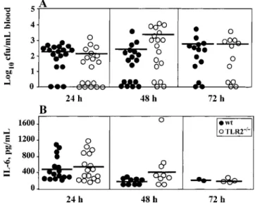
Figure 2. Colony-forming unit counts in blood (A) and interleukin (IL)–6 levels in plasma (B) in wild-type (wt; n p 22) and Toll-like receptor-2–deficient (TLR2⫺/⫺;n p 21) mice after intracerebral injec-tion of3⫻ 103cfu of Streptococcus pneumoniae. Blood was collected 24, 48, and 72 h after infection. Results from individual mice are shown. Horizontal line, mean.P1.05, Mann-Whitney U test.
sphere was homogenized in 1 mL of 0.1 mol/L PBS and then cen-trifuged at 1000 g for 15 min; 0.7 mL of 100% trichloroacetic acid was added to 0.7 mL of the supernatant. The mixture was incubated at 4⬚C for 18 h and then centrifuged at 1000 g for 30 min. The amount of Evans blue in the supernatant was measured spectro-photometrically at 610 nm and compared with a serially diluted standard solution. Results were expressed as micrograms per brain hemisphere.
Statistical analysis. Differences in survival between wt and TLR2⫺/⫺mice were tested by the Kaplan-Meier analysis log-rank test. Data of disease severity scores were assessed by analysis of variance corrected for repeated measurements, followed by posthoc analysis (Fisher’s P least-squares difference, Scheffe, and Bonfer-roni/Dunn tests). Results of the colony-forming unit measurements and IL-6 levels in blood were compared by the Mann-Whitney U test. Differences in CSF parameters between wt and TLR2⫺/⫺mice were analyzed with the nonparametric Wilcoxon signed rank test. The relationship between 2 parameters (parametric variables) was assessed in a linear regression model with the Spearman rank cor-relation test. In all statistical tests,P!.05 was considered to be statistically significant.
Results
TLR2⫺/⫺mice have a higher susceptibility to S. pneumoniae and L. monocytogenes meningitis. To evaluate the in vivo role of TLR2 during gram-positive bacterial meningitis, wt and TLR2⫺/⫺mice were infected intracerebrally with S. pneumoniae or L. monocytogenes. The severity of the disease was monitored by assessing both the clinical score and the survival rate.
Control animals, injected with 0.9% NaCl, showed no altered health status. Infected mice remained free of clinical signs of meningitis at 6 and 12 h after infection but had gradually re-duced spontaneous activity between 12 and 24 h (data not shown). As shown in figure 1A, both wt and TLR2⫺/⫺ mice became severely sick during S. pneumoniae meningitis. The per-centage of severely ill mice was significantly higher among TLR2⫺/⫺than control mice after 72 h (P!.02, repeated-mea-sures analysis of variance). Indeed, only 30% of TLR2⫺/⫺mice survived at 72 h, and all died at 102 h after infection (figure 1B), whereas 60% of the wt mice survived at 72 h and died of S. pneumoniae meningitis later (136 h after infection). TLR2⫺/⫺mice also showed reduced survival time (P!.02,
Kap-lan-Meier analysis), compared with wt mice with L. monocyto-genes meningitis (table 1). These results show that TLR2⫺/⫺ mice had more severe symptoms and earlier death than did wt mice during S. pneumoniae and L. monocytogenes meningitis.
Higher disease severity in TLR2⫺/⫺mice is independent of sep-sis. To investigate whether the high susceptibility of TLR2⫺/⫺ mice to meningitis was due to impaired host defense, colony-forming units in blood were counted 1, 2, and 3 days after in-fection with3⫻ 103cfu of S. pneumoniae. Bacterial counts were similar in wt and TLR2⫺/⫺ mice at all time points (figure 2A). Six and 12 h after infection,!10% of both wt and TLR2⫺/⫺mice showed positive blood culture results at low bacterial density (data not shown).
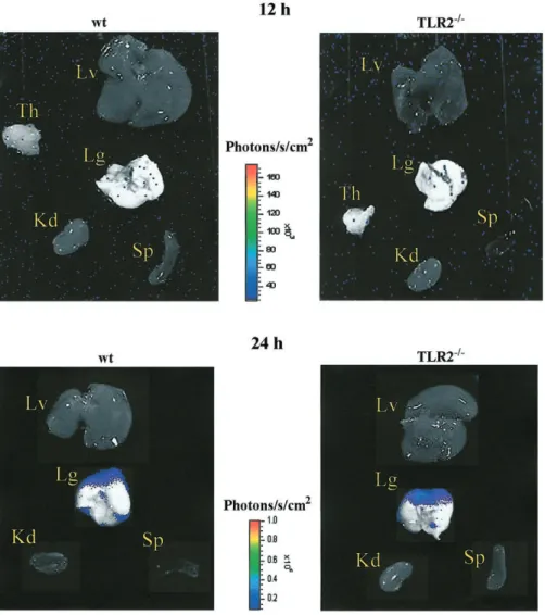
Plasma IL-6, a prognostic marker of sepsis [24], was also analyzed during meningitis. In accordance with bacterial counts, IL-6 levels were not significantly different in wt and TLR2⫺/⫺mice 1–3 days after infection (figure 2B). Twelve and 24 h after intracerebral infection, emitted photons from lucif-erase-tagged S. pneumoniae were analyzed in peripheral organs (figure 3). Twenty-four hours after infection, fluorescence ac-cumulated only in the lung with a similar intensity in wt and TLR2⫺/⫺mice. No other organ showed a detectable level of emitted photons 12 and 24 h after infection. Taken together, these data indicate that wt and TLR2⫺/⫺mice had a similar severity of sepsis and therefore an intact systemic host defense during S. pneumoniae meningitis.
To differentiate between sepsis and inflammation as a cause of death, wt and TLR2⫺/⫺mice received antibiotic treatment
Figure 3. Emitted photons by luciferase-tagged Streptococcus pneumoniae in several organs from wild-type (wt) and Toll-like receptor-2–deficient (TLR2⫺/⫺) mice detected by a highly sensitive CCD (charge-coupled device) camera (IVIS imaging system; Xenogen) 12 and 24 h after intracerebral injection of3⫻ 103cfu of S. pneumoniae. Results from 1 wt and 1 TLR2⫺/⫺mouse are shown. Kd, kidney; Lg, lung; Lv, liver; Sp, spleen; Th, thymus.
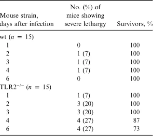
to cure the sepsis. It is known that wt mice treated in the first 24 h after infection with ceftriaxone (80 mg/kg every 12 h for 5 days) survive without apparent signs of meningitis [25]. Delay of treatment beyond 30 h leads to death in150% of the infected mice [25]. We treated infected mice with ceftriaxone after S. pneumoniae infection, and the obtained clinical scores and sur-vival rates of both ceftriaxone-treated and control mice are shown in table 2. With ceftriaxone treatment, all wt mice sur-vived without any clinical signs of disease after 6 days, whereas 4 (27%) of 15 TLR2⫺/⫺mice died despite treatment (P!.04, Kaplan-Meier analysis). As described above, all untreated wt and TLR2⫺/⫺ mice died within 4–6 days (data not shown). Treatment failure in TLR2⫺/⫺mice could be explained by the fact that at the initiation of the treatment (18 h after infection),
7% of TLR2⫺/⫺mice but none of the wt mice were severely sick. By day 6, 27% of the treated TLR2⫺/⫺ mice remained severely ill and subsequently died, whereas all wt mice recovered during treatment.
TLR2⫺/⫺mice have a higher bacterial load in brain and stronger meningeal inflammation after intracerebral infection with S. pneu-moniae than do wt mice. To further explain the high suscep-tibility of TLR2⫺/⫺ mice to S. pneumoniae meningitis, we ex-amined bacterial counts, leukocyte recruitment, and TNF release in the CSF as well as bacterial densities in the brain. Monitoring of meningitis in animals intracerebrally injected with luciferase-tagged S. pneumoniae revealed a higher fluorescence intensity in TLR2⫺/⫺ brains than in wt brains between 2 and 24 h after infection (figure 4A). Twenty-four hours after infection, mice were
Table 2. Effect of ceftriaxone treatment on survival during Streptococcus pneumoniae men-ingitis in wild-type (wt) and Toll-like receptor-2–deficient (TLR2⫺/⫺) mice.
Mouse strain, days after infection
No. (%) of mice showing
severe lethargy Survivors, % wt (n p 15) 1 0 100 2 1 (7) 100 3 1 (7) 100 4 1 (7) 100 6 0 100 TLR2⫺/⫺(n p 15) 1 1 (7) 100 2 3 (20) 100 3 3 (20) 100 4 4 (27) 87 6 4 (27) 73
NOTE. Ceftriaxone treatment (80 mg/kg) was ad-ministered intraperitoneally every 12 h for 5 days, start-ing 18 h after intracerebral injection of 3cfu of
3⫻ 10
S. pneumoniae.P!.04, TLR2⫺/⫺vs. wt mice (log-rank
test in Kaplan-Meier analysis).
killed, and the photon emission was stronger and more widely spread in isolated brains of TLR2⫺/⫺mice than in those of wt mice (figure 4A). In particular, signals from bacteria were dif-ferently distributed in TLR2⫺/⫺and wt mice. In contrast to the picture in wt mice, bacteria were not accumulated in the ventricles of TLR2⫺/⫺mice but concentrated on the ipsilateral and con-tralateral sides of the injection site. Furthermore, a quantitative time course analysis of emitted fluorescence from infected animals showed a more rapid bacterial growth in TLR2⫺/⫺mice, com-pared with wt mice, which was most pronounced already 2 h after S. pneumoniae inoculation (figure 4B). Counting of colony-forming units in ipsilateral and contralateral brain hemispheres 24 h after infection of the left side of the brain confirmed that, in both parts, bacterial numbers were higher in TLR2⫺/⫺than wt mice (median,1.8⫻ 107cfu [left] and1.2⫻ 107cfu [right] in TLR2⫺/⫺mice, and7.4⫻ 105cfu [left] and8.8⫻ 105cfu [right] in wt mice;P!.04). Counting of colony-forming units in brain homogenates 24 h after intracisternal infection with S. pneu-moniae confirmed that bacterial numbers were also significantly higher in TLR2⫺/⫺than wt mice when the intracisternal route of infection was used (median,6.2⫻ 105 cfu and6.8⫻ 104cfu in TLR2⫺/⫺and wt mice, respectively;P!.04, Mann-Whitney U test).
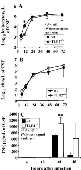
Twelve hours after intracerebral infection, TLR2⫺/⫺ mice showed a lower influx of leukocytes, compared with wt mice (meanⳲ SD 1703 Ⳳ 186, and12,110Ⳳ 3691leukocytes/mL of CSF, respectively; P!.05; figure 5A). After this time point, leukocyte numbers and bacterial counts in the CSF of the 2 groups were almost identical (figure 5B). As expected, infil-trating polymorphonuclear cell numbers and bacterial counts in the CSF were similar in TLR2⫺/⫺and wt mice after
intra-cisternal infection, as we had observed in the intracerebral in-fection model.
Meningeal inflammation, as assessed by TNF activity in the CSF, was significantly higher in TLR2⫺/⫺than in wt mice 24 h after infection with S. pneumoniae (1077Ⳳ 201 and705Ⳳ 4 pg/mL of CSF, respectively; ; figure 5C). Early after
7 P!.02
infection, at 6 and 12 h, TNF activity was low or below de-tection (!20 pg/mL), and, after 48 h, the difference between TLR2⫺/⫺mice and wt mice was no longer statistically signifi-cant, because of high variability (figure 5C).
An association between disease severity and meningeal in-flammation was documented by a significant relationship be-tween TNF activity in the CSF and severity score 24 h after infection (r p 0.589 P; !.001; data not shown). Thus, the level of TNF in CSF might reflect the intensity of clinical signs during the first day of infection.
To investigate whether infiltrating leukocytes contribute to meningeal inflammation during S. pneumoniae meningitis, we analyzed TNF activity in the CSF in infected mice rendered leukopenic by cyclophosphamide. As shown in figure 6, TNF activity was significantly decreased in leukopenic TLR2⫺/⫺mice, compared with immunocompetent mice 24 h after infection (P!.05). In contrast, no difference in TNF activity was observed in wt mice with or without cyclophosphamide pretreatment.
TLR2⫺/⫺ mice have enhanced BBB permeability after intra-cerebral infection with S. pneumoniae. Alteration of the BBB is a consequence of meningeal inflammation. Therefore, we analyzed changes in BBB permeability during S. pneumoniae meningitis with the Evans blue extravasation method.
Measures of BBB permeability and severity of the disease in wt and TLR2⫺/⫺mice are presented in table 3. Infection with
S. pneumoniae induced alteration of the BBB in both wt and TLR2⫺/⫺mice, as evidenced by higher Evans blue concentra-tions in brains of TLR2⫺/⫺mice, compared with control mice. This BBB disruption occurred early in disease (24 h) and was stronger in TLR2⫺/⫺mice than in wt mice. At the same time, TLR2⫺/⫺ mice showed more severe symptoms of meningitis than did wt mice (table 3). In addition, meningeal inflammation, assessed by TNF in the CSF, was significantly related to BBB permeability (r p 0.768;P!.02; data not shown).
Discussion
We have studied the in vivo role of TLR2 in an adult mouse model of S. pneumoniae meningitis. TLR2⫺/⫺mice had more severe clinical symptoms than did wt mice and subsequently showed earlier death. Similar results were obtained with L. monocytogenes as the infecting agent (table 1). This result is in agreement with a previous report on increased mortality of TLR2⫺/⫺ mice from S. aureus sepsis [26]. In that model, TLR2⫺/⫺ mice showed reduced systemic bacterial clearance, compared with wt mice, after a high but not after a low
intra-Figure 4. A, Time course of infection in wild-type (wt) and Toll-like receptor-2–deficient (TLR2⫺/⫺) mice after intracerebral injection of cfu of luciferase-tagged Streptococcus pneumoniae. Images from 1 animal in each group at time of infection (0 h) and 2, 4, 6, 12, and 24 3
3⫻ 10
h after infection and isolated brains 24 h after infection are shown. B, Emitted light was also quantified by LivingImage software (Xenogen) and shown as bacterial growth gradient (calculated as ratio between emitted photons and time elapsed after infection). Results from individual mice are shown, with median (horizontal line). **P!.0001, repeated-measures analysis of variance (ANOVA) (n p 11 andn p 10 at 2 and 4 h,
and at 6 and 12 h, and and at 24 h, for wt and TLR2⫺/⫺mice, respectively).
n p 10 n p 9 n p 6 n p 5
venous inoculum. According to the authors, this was due to failure to recognize invading bacteria [26]. Instead, in our model with a low inoculum in the brain, systemic host defense was not impaired in the absence of TLR2, because similar plasma
levels of IL-6 and numbers of bacteria were observed in blood and organs of TLR2⫺/⫺and wt mice. Moreover, in ceftriaxone-injected animals, in which the infection is successfully treated, 27% of the TLR2⫺/⫺mice died, whereas all wt mice survived.
Figure 5. Time course of cerebrospinal fluid (CSF) parameters in wild-type (wt) and Toll-like receptor-2–deficient (TLR2⫺/⫺) mice (n p 12at each time point in both groups) after intracerebral injection of3⫻ 103cfu of Streptococcus pneumoniae. Pooled CSF from 4 mice was collected at indicated times after infection. Infiltrating leukocytes (A), bacterial numbers (B), and tumor necrosis factor (TNF) levels (C) were analyzed in CSF. Results of 3 independent experiments are shown asmeanⳲ SD. When errors bars are not seen, they fall within symbol. *P!.05and **P!.02, both by the Wilcoxon signed rank test.
Figure 6. Tumor necrosis factor (TNF) levels in cerebrospinal fluid (CSF) in leukocyte-depleted and immunocompetent control mice after intracerebral injection of3⫻ 103cfu of Streptococcus pneumoniae. In each experiment, pooled CSF from 4 mice was collected 24 h after infection. Results of 3 independent experiments are shown as meanⳲ SD. **P!.05, nonparametric Wilcoxon signed rank test. Leukocyte depletion in CSF was81.2%Ⳳ 20.1%in Toll-like receptor-2–deficient (TLR2⫺/⫺) mice and76.2%Ⳳ 10.5%in wild-type (wt) mice.
This indicates that the higher mortality in TLR2⫺/⫺mice was not due to defective bacterial clearance in the periphery.
We observed more bacteria in the brains, but not in the CSF, of TLR2⫺/⫺mice. More rapid growth, which was detectable as early as 2 h after infection, was associated with a protracted accumulation of the bacteria around the site of injection in TLR2⫺/⫺mice. This was in contrast to wt mice, in which the bacteria were mainly in the ventricles after 24 h. It is known that TLR2 is constitutively expressed in the choroid plexus and the lateral ventricle lining [27]. It can be hypothesized that the lack of TLR2 in the choroidal epithelia and/or endothelia and in the ependymal cells contributed to the particular distribution pattern of the bacteria by altering the CSF flow. The different
localization of the bacteria in brains of wt and TLR2⫺/⫺mice probably also explains why the numbers of colony-forming units in CSF were similar in both strains.
Leukocyte infiltration following bacterial invasion into the central nervous system is correlated with brain edema and in-jury in bacterial meningitis [28, 29]. The rapid increased bac-terial growth in brain homogenates of TLR2⫺/⫺mice may, in turn, have contributed to the enhanced inflammation and to the earlier aggravation of the symptoms in these animals. De-spite higher bacterial numbers in brain, pleocytosis was not significantly different in wt and TLR2⫺/⫺ mice, except for a transient delay in leukocyte recruitment 12 h after infection in TLR2⫺/⫺mice. Perhaps the delayed leukocyte influx into the CSF could be explained by a lack of TLR2 action on integrin or intercellular adhesion molecule (ICAM) expression, because it has been shown that blocking of the integrin CD18 and of the adhesion molecule ICAM attenuated meningeal inflam-mation and tissue damage [30, 31]. However, it is also possible that the recruitment of leukocytes may have been delayed in TLR2⫺/⫺mice because of the lack of TLR2 activity on IL-8 [32]. Indeed, a previous study showed that intravenous treat-ment with antibody against IL-8 attenuates the neutrophil pleo-cytosis during experimental pneumococcal meningitis [33].
Despite the slower early influx of leukocytes, we observed higher TNF activity during S. pneumoniae meningitis in CSF of TLR2⫺/⫺ than of wt mice. TNF is elevated in CSF in human and experimental meningitis. It has detrimental effects, because its neutralization decreases meningeal inflammation, as shown in a rabbit pneumococcal meningitis model [34, 35]. However, the total lack of TNF also adversely effects host defense in men-ingitis. Indeed, mice with a targeted deletion of TNF die earlier
Table 3. Blood-brain barrier permeability and clinical score in Toll-like receptor-2–deficient (TLR2⫺/⫺) and wild-type (wt) mice after in-tracerebral injection of3⫻ 103cfu of Streptococcus pneumoniae.
Mouse strain 24 h 48 h Clinical scorea Evans blue stainb Clinical scorea Evans blue stainb wt 1.3 (1–2.8) 1.1Ⳳ 0.1 3.5 (3–4) 3.4Ⳳ 1.9 TLR2⫺/⫺ 1.9 (1–3) 1.3Ⳳ 0.6 4.9 (4.8–5) 7.0Ⳳ 0.9 a
Data are median (range) clinical score. See Materials and Methods for a description of the scoring system.
b
Data aremeanⳲ SDfold increase, compared with uninfected control mice.
than control mice from intracerebral S. pneumoniae infection be-cause of the lack of systemic clearance of bacteria [36]. A systemic protective effect of TNF in our model is unlikely, in view of similar signs of sepsis in TLR2⫺/⫺and wt mice. An important finding of the present study is that, paradoxically, TLR2⫺/⫺mice show an increased inflammation after infection. Thus, it appears that these animals are characterized by inefficient regulation of the inflammatory reaction. Because TLR2⫺/⫺ mice had more TNF activity in the CSF than did wt mice, it is tempting to speculate that the engagement of TLR2 contributes to a down-regulation of the TNF in the CSF. This is not ascribed to an increased production of IL-10 (data not shown) and might be associated with an altered release of TNF from intracellular stores and the cell surface.
The consequence of meningeal inflammation is an alteration of the BBB integrity. In the present study, the BBB disruption was markedly increased in TLR2⫺/⫺mice, compared with con-trol mice, and was related to TNF levels in the CSF. This corroborates the known role of TNF in BBB disruption and consequently in the course of disease [35, 37, 38].
Our observation of a stronger in vivo TNF induction in TLR2⫺/⫺than in wt mice is in contrast to the reduced in vitro TNF production of TLR2⫺/⫺in macrophages in response to S.
aureus [26]. However, an important difference in our experi-mental model is the use of live bacteria, whereas that study [26] was conducted with heat-killed bacteria. We therefore conclude that live S. pneumoniae and their turnover products induce TNF differently than do heat-killed bacteria. It is likely that, during infection, other receptors are activated together with TLR2. The nature of these other receptors is not clear. They could be other TLRs, such as TLR1 and TLR6, which are also triggered by bacterial products [39]. In this case, their stimulation led to the release of large amounts of TNF, which are higher that those induced in the presence of TLR2. This might occur be-cause these TLRs show sequence similarities in their intracy-toplasmic regions and then share common transducing mech-anisms. Other receptors involved in this signaling could be specific for S. pneumoniae products, such as platelet-activating factor receptors [7] or C-reactive protein [40]. Finally, another possibility is that triggering of TLR2 on a particular cell type only might lead to the regulation of TNF. However, in this
case, it is not clear how the engagement of TLR2 would down-regulate the TNF release.
The same accelerated death rate as for those infected with S. pneumoniae was also observed in Listeria-infected TLR2⫺/⫺ mice, yet they and Listeria-infected wt mice had similar TNF activity in the CSF (data not shown). This suggests that dif-ferent mechanisms are involved in S. pneumoniae and L. mono-cytogenes meningitis.
As reported elsewhere, in L. monocytogenes meningitis, monocytes contributed to the leukocyte infiltration in CSF, whereas S. pneumoniae meningitis caused only polymorpho-nuclear leukocyte accumulation [41]. Together with our recent observation that TLR2 protein is constitutively expressed in mouse polymorphonuclear leukocytes, but not in macrophages (authors’ unpublished data), the excess TNF in S. pneumoniae meningitis may be polymorphonuclear leukocyte–derived. To confirm this hypothesis, we investigated which cells contributed to the TNF release in the subarachnoid space during S. pneu-moniae meningitis. Depletion of leukocytes by cyclophospha-mide treatment before infection led to a decrease in TNF level in TLR2⫺/⫺but not in wt mice. This result validated that leu-kocytes participated in TNF production but were not the only source of TNF in TLR2⫺/⫺mice. Brain microvascular endo-thelia have been shown to produce TNF in pneumococcal men-ingitis or after stimulation with cell walls of S. pneumoniae [7, 42]. Furthermore, we and others could show that primary cul-tures of astrocytes, microglial cells, and neurons release TNF after stimulation with live S. pneumoniae (authors’ unpublished data) and pneumococcal cell wall components [43].
In conclusion, the mouse S. pneumoniae meningitis model de-scribed here is fatal for both wt and TLR2⫺/⫺mice. The clinical course of meningitis, the number of bacteria, the level of menin-geal inflammation, and BBB damage are aggravated in mice lacking TLR2. Taken together, these results indicate, for the first time, a contribution of TLR2 in regulation of bacterial clearing and inflammatory response in pneumococcal meningitis. Acknowledgments
We thank Katrin Hafen and Fabrizia Ferracin (University Hospital, Basel), for expert technical help; Zarko Rajacic (University Hospital, Basel) and Ju¨rg Kummer (University of Bern, Bern), for help in animal experiments; Gennaro De Libero (University Hospital, Basel), Beat Mu¨ller (University Hospital, Basel), and Terence O’Reilly (Pharma-Novartis, Basel), for critical manuscript review; and L. Chen (Xenogen, Alameda, CA), for providing bioluminescent Streptococcus pneumoniae Xen10 strain.
References
1. Durand ML, Calderwood SB, Weber DJ, et al. Acute bacterial meningitis in adults: a review of 493 episodes. N Engl J Med 1993; 328:21–8. 2. Bohr V, Paulson OB, Rasmussen N. Pneumococcal meningitis: late neurologic
3. Malipiero UV, Frei K, Fontana A. Production of hemopoietic colony-stim-ulating factors by astrocytes. J Immunol 1990; 144:3816–21.
4. Frei K, Siepl C, Groscurth P, Bodmer S, Schwerdel C, Fontana A. Antigen presentation and tumor cytotoxicity by interferon-gamma–treated micro-glial cells. Eur J Immunol 1987; 17:1271–8.
5. Breder CD, Tsujimoto M, Terano Y, Scott DW, Saper CB. Distribution and characterization of tumor necrosis factor–alpha-like immunoreactivity in the murine central nervous system. J Comp Neurol 1993; 337:543–67. 6. Tuomanen E, Tomasz A, Hengstler B, Zak O. The relative role of bacterial
cell wall and capsule in the induction of inflammation in pneumococcal meningitis. J Infect Dis 1985; 151:535–40.
7. Ring A, Weiser JN, Tuomanen EI. Pneumococcal trafficking across the blood-brain barrier: molecular analysis of a novel bidirectional pathway. J Clin Invest 1998; 102:347–60.
8. Cundell DR, Gerard NP, Gerard C, Idanpaan-Heikkila I, Tuomanen EI.
Streptococcus pneumoniae anchor to activated human cells by the receptor
for platelet-activating factor. Nature 1995; 377:435–8.
9. Medzhitov R, Janeway CA Jr. Innate immunity: the virtues of a nonclonal system of recognition. Cell 1997; 91:295–8.
10. Stein D, Roth S, Vogelsang E, Nusslein-Volhard C. The polarity of the dor-soventral axis in the Drosophila embryo is defined by an extracellular signal. Cell 1991; 65:725–35.
11. Lemaitre B, Nicolas E, Michaut L, Reichhart JM, Hoffmann JA. The dor-soventral regulatory gene cassette spatzle/Toll/cactus controls the potent antifungal response in Drosophila adults. Cell 1996; 86:973–83. 12. Medzhitov R, Preston-Hurlburt P, Janeway CA Jr. A human homologue of
the Drosophila Toll protein signals activation of adaptive immunity. Nature
1997; 388:394–7.
13. Rock FL, Hardiman G, Timans JC, Kastelein RA, Bazan JF. A family of human receptors structurally related to Drosophila Toll. Proc Natl Acad Sci USA 1998; 95:588–93.
14. Akira S, Takeda K, Kaisho T. Toll-like receptors: critical proteins linking innate and acquired immunity. Nat Immunol 2001; 2:675–80.
15. Yoshimura A, Lien E, Ingalls RR, Tuomanen E, Dziarski R, Golenbock D. Cutting edge: recognition of Gram-positive bacterial cell wall components by the innate immune system occurs via Toll-like receptor 2. J Immunol
1999; 163:1–5.
16. Schwandner R, Dziarski R, Wesche H, Rothe M, Kirschning CJ. Peptido-glycan- and lipoteichoic acid–induced cell activation is mediated by toll-like receptor 2. J Biol Chem 1999; 274:17406–9.
17. Brightbill HD, Libraty DH, Krutzik SR, et al. Host defense mechanisms triggered by microbial lipoproteins through toll-like receptors. Science
1999; 285:732–6.
18. Takeuchi O, Hoshino K, Kawai T, et al. Differential roles of TLR2 and TLR4 in recognition of gram-negative and gram-positive bacterial cell wall components. Immunity 1999; 11:443–51.
19. Loeffler JM, Ringer R, Hablutzel M, Tauber MG, Leib SL. The free radical scavenger a-phenyl-tert-butyl nitrone aggravates hippocampal apoptosis and learning deficits in experimental pneumococcal meningitis. J Infect Dis 2001; 183:247–52.
20. Leib SL, Clements JM, Lindberg RL, et al. Inhibition of matrix metallo-proteinases and tumour necrosis factor alpha converting enzyme as ad-juvant therapy in pneumococcal meningitis. Brain 2001; 124:1734–42. 21. Carp RI, Davidson AL, Merz PA. A method for obtaining cerebrospinal
fluid from mice. Res Vet Sci 1971; 12:499.
22. Francis KP, Yu J, Bellinger-Kawahara C, et al. Visualizing pneumococcal infections in the lungs of live mice using bioluminescent Streptococcus
pneumoniae transformed with a novel gram-positive lux transposon. Infect
Immun 2001; 69:3350–8.
23. Chan PH, Yang GY, Chen SF, Carlson E, Epstein CJ. Cold-induced brain edema and infarction are reduced in transgenic mice overexpressing CuZn–superoxide dismutase. Ann Neurol 1991; 29:482–6.
24. Waage A, Halstensen A, Shalaby R, Brandtzaeg P, Kierulf P, Espevik T. Local production of tumor necrosis factor alpha, interleukin 1, and
in-terleukin 6 in meningococcal meningitis: relation to the inflammatory response. J Exp Med 1989; 170:1859–67.
25. Gerber J, Raivich G, Wellmer A, et al. A mouse model of Streptococcus
pneumoniae meningitis mimicking several features of human disease. Acta
Neuropathol (Berl) 2001; 101:499–508.
26. Takeuchi O, Hoshino K, Akira S. Cutting edge: TLR2-deficient and MyD88-deficient mice are highly susceptible to Staphylococcus aureus infection. J Immunol 2000; 165:5392–6.
27. Laflamme N, Soucy G, Rivest S. Circulating cell wall components derived from gram-negative, not gram-positive, bacteria cause a profound induc-tion of the gene-encoding Toll-like receptor 2 in the CNS. J Neurochem
2001; 79:648–57.
28. Weber JR, Angstwurm K, Burger W, Einhaupl KM, Dirnagl U. Anti ICAM-1 (CD 54) monoclonal antibody reduces inflammatory changes in exper-imental bacterial meningitis. J Neuroimmunol 1995; 63:63–8.
29. Pfister HW, Scheld WM. Brain injury in bacterial meningitis: therapeutic implications. Curr Opin Neurol 1997; 10:254–9.
30. Tuomanen EI, Saukkonen K, Sande S, Cioffe C, Wright SD. Reduction of inflammation, tissue damage, and mortality in bacterial meningitis in rab-bits treated with monoclonal antibodies against adhesion-promoting re-ceptors of leukocytes. J Exp Med 1989; 170:959–69.
31. Saez-Llorens X, Jafari HS, Severien C, et al. Enhanced attenuation of men-ingeal inflammation and brain edema by concomitant administration of anti-CD18 monoclonal antibodies and dexamethasone in experimental
Haemophilus meningitis. J Clin Invest 1991; 88:2003–11.
32. Re F, Strominger JL. Toll-like receptor 2 (TLR2) and TLR4 differentially activate human dendritic cells. J Biol Chem 2001; 276:37692–9. 33. Ostergaard C, Yieng-Kow RV, Larsen CG, et al. Treatment with a monoclonal
antibody to IL-8 attenuates the pleocytosis in experimental pneumococcal meningitis in rabbits when given intravenously, but not intracisternally. Clin Exp Immunol 2000; 122:207–11.
34. Mustafa MM, Ramilo O, Saez-Llorens X, Mertsola J, McCracken GH Jr. Role of tumor necrosis factor alpha (cachectin) in experimental and clin-ical bacterial meningitis. Pediatr Infect Dis J 1989; 8:907–8.
35. Saukkonen K, Sande S, Cioffe C, et al. The role of cytokines in the generation of inflammation and tissue damage in experimental gram-positive men-ingitis. J Exp Med 1990; 171:439–48.
36. Wellmer A, Gerber J, Ragheb J, et al. Effect of deficiency of tumor necrosis factor alpha or both of its receptors on Streptococcus pneumoniae central nervous system infection and peritonitis. Infect Immun 2001; 69:6881–6. 37. Sharief MK, Ciardi M, Thompson EJ. Blood-brain barrier damage in patients with bacterial meningitis: association with tumor necrosis factor–a but not interleukin-1b. J Infect Dis 1992; 166:350–8.
38. Geelen S, Bhattacharyya C, Tuomanen E. The cell wall mediates pneumo-coccal attachment to and cytopathology in human endothelial cells. Infect Immun 1993; 61:1538–43.
39. Ozinsky A, Underhill DM, Fontenot JD, et al. The repertoire for pattern recognition of pathogens by the innate immune system is defined by co-operation between toll-like receptors. Proc Natl Acad Sci USA 2000; 97: 13766–71.
40. Kim JO, Romero-Steiner S, Sorensen UB, et al. Relationship between cell surface carbohydrates and intrastrain variation on opsonophagocytosis of Streptococcus pneumoniae. Infect Immun 1999; 67:2327–33. 41. Cauwels A, Wan E, Leismann M, Tuomanen E. Coexistence of
CD14-de-pendent and -indeCD14-de-pendent pathways for stimulation of human monocytes by gram-positive bacteria. Infect Immun 1997; 65:3255–60.
42. Freyer D, Manz R, Ziegenhorn A, et al. Cerebral endothelial cells release TNF-alpha after stimulation with cell walls of Streptococcus pneumoniae and regulate inducible nitric oxide synthase and ICAM-1 expression via autocrine loops. J Immunol 1999; 163:4308–14.
43. Freyer D, Weih M, Weber JR, et al. Pneumococcal cell wall components induce nitric oxide synthase and TNF-alpha in astroglial-enriched cul-tures. Glia 1996; 16:1–6.