HAL Id: pasteur-03263254
https://hal-pasteur.archives-ouvertes.fr/pasteur-03263254
Submitted on 17 Jun 2021
HAL is a multi-disciplinary open access
archive for the deposit and dissemination of
sci-entific research documents, whether they are
pub-lished or not. The documents may come from
teaching and research institutions in France or
abroad, or from public or private research centers.
L’archive ouverte pluridisciplinaire HAL, est
destinée au dépôt et à la diffusion de documents
scientifiques de niveau recherche, publiés ou non,
émanant des établissements d’enseignement et de
recherche français ou étrangers, des laboratoires
publics ou privés.
Distributed under a Creative Commons Attribution| 4.0 International License
Autoantibodies against type I IFNs in patients with
life-threatening COVID-19
Paul Bastard, Lindsey Rosen, Qian Zhang, Eleftherios Michailidis,
Hans-Heinrich Hoffmann, Yu Zhang, Karim Dorgham, Quentin Philippot,
Jérémie Rosain, Vivien Béziat, et al.
To cite this version:
Paul Bastard, Lindsey Rosen, Qian Zhang, Eleftherios Michailidis, Hans-Heinrich Hoffmann, et al..
Autoantibodies against type I IFNs in patients with life-threatening COVID-19. Science, American
As-sociation for the Advancement of Science, 2020, 370 (6515), pp.eabd4585. �10.1126/science.abd4585�.
�pasteur-03263254�
RESEARCH ARTICLE SUMMARY
◥CORONAVIRUS
Autoantibodies against type I IFNs in patients
with life-threatening COVID-19
Paul Bastard
*† and Lindsey B. Rosen† et al.
INTRODUCTION:
Interindividual clinical
vari-ability is vast in humans infected with
severe acute respiratory syndrome
corona-virus 2 (SARS-CoV-2), ranging from silent
in-fection to rapid death. Three risk factors for
life-threatening coronavirus disease 2019
(COVID-19) pneumonia have been identified
—
being male, being elderly, or having other
medical conditions
—but these risk factors
cannot explain why critical disease remains
relatively rare in any given epidemiological
group. Given the rising toll of the COVID-19
pandemic in terms of morbidity and mortality,
understanding the causes and mechanisms of
life-threatening COVID-19 is crucial.
RATIONALE:
B cell autoimmune infectious
phenocopies of three inborn errors of
cyto-kine immunity exist, in which neutralizing
autoantibodies (auto-Abs) against interferon-g
(IFN-g) (mycobacterial disease), interleukin-6
(IL-6) (staphylococcal disease), and IL-17A and
IL-17F (mucocutaneous candidiasis) mimic the
clinical phenotypes of germline mutations of
the genes that encode the corresponding
cyto-kines or receptors. Human inborn errors of
type I IFNs underlie severe viral respiratory
diseases. Neutralizing auto-Abs against type I
IFNs, which have been found in patients with
a few underlying noninfectious conditions,
have not been unequivocally shown to
un-derlie severe viral infections. While
search-ing for inborn errors of type I IFN immunity
in patients with life-threatening COVID-19
pneumonia, we also tested the hypothesis
that neutralizing auto-Abs against type I IFNs
may underlie critical COVID-19. We searched
for auto-Abs against type I IFNs in 987
pa-tients hospitalized for life-threatening
COVID-19 pneumonia, 663 asymptomatic or mildly
affected individuals infected with
SARS-CoV-2, and 1227 healthy controls from whom
samples were collected before the
COVID-19 pandemic.
RESULTS:
At least 101 of 987 patients (10.2%)
with life-threatening COVID-19 pneumonia
had neutralizing immunoglobulin G (IgG)
auto-Abs against IFN-w (13 patients), against
the 13 types of IFN-a (36), or against both (52)
at the onset of critical disease; a few also had
auto-Abs against the other three individual
type I IFNs. These auto-Abs neutralize high
concentrations of the corresponding type I
IFNs, including their ability to block
SARS-CoV-2 infection in vitro. Moreover, all of the
patients tested had low or undetectable serum
IFN-a levels during acute disease. These
auto-Abs were present before infection in the
patients tested and were absent from 663
individuals with asymptomatic or mild
SARS-CoV-2 infection (
P < 10
−16). They were present
in only 4 of 1227 (0.33%) healthy individuals
(
P < 10
−16) before the pandemic. The patients
with auto-Abs were 25 to 87 years old (half
were over 65) and of various ancestries.
No-tably, 95 of the 101 patients with auto-Abs
were men (94%).
CONCLUSION:
A B cell autoimmune phenocopy
of inborn errors of type I IFN immunity
ac-counts for life-threatening COVID-19
pneumo-nia in at least 2.6% of women and 12.5% of men.
In these patients, adaptive autoimmunity
im-pairs innate and intrinsic antiviral immunity.
These findings provide a first explanation for
the excess of men among patients with
life-threatening COVID-19 and the increase in
risk with age. They also provide a means of
identifying individuals at risk of developing
life-threatening COVID-19 and ensuring their
enrolment in vaccine trials. Finally, they pave
the way for prevention and treatment,
includ-ing plasmapheresis, plasmablast depletion,
and recombinant type I IFNs not targeted by
the auto-Abs (e.g., IFN-b).
▪
The full author list and the list of author affiliations is available in the full article online.
*Corresponding authors: Jean-Laurent Casanova (jean-laurent.casanova@rockefeller.edu); Paul Bastard (paul.bastard@institutimagine.org)
†These authors contributed equally to this work. This is an open-access article distributed under the terms of the Creative Commons Attribution license (https://creativecommons.org/licenses/by/4.0/), which permits unrestricted use, distribution, and reproduction in any medium, provided the original work is properly cited. Cite this article as P. Bastardet al., Science 370, eabd4585 (2020). DOI: 10.1126/science.abd4585
READ THE FULL ARTICLE AT
https://doi.org/10.1126/science.abd4585
0/663 (0%)
IFNAR1 IFNAR2 IFNAR1 IFNAR2
TLR3
Type I IFN immunity
ISGs IRF7
TLR3
IRF7
Neutralizing auto-Abs impair type I IFN immunity
pSTATs
Auto-Abs to type I IFNs
ISGs pSTATs
101/987 (10.2%)
Asymptomatic/mild Life-threatening SARS-COV-2
Neutralizing auto-Abs to type I IFNs underlie life-threatening COVID-19 pneumonia. We tested the hypothesis
that neutralizing auto-Abs against type I IFNs may underlie critical COVID-19 by impairing the binding of type I
IFNs to their receptor and the activation of the downstream responsive pathway. Neutralizing auto-Abs are
represented in red, and type I IFNs are represented in blue. In these patients, adaptive autoimmunity impairs innate
and intrinsic antiviral immunity. ISGs, IFN-stimulated genes; TLR, Toll-like receptor; IFNAR, IFN-a/b receptor;
pSTAT, phosphorylated signal transducers and activators of transcription; IRF, interferon regulatory factor.
RESEARCH ARTICLE
◥CORONAVIRUS
Autoantibodies against type I IFNs in patients with
life-threatening COVID-19
Paul Bastard
1,2,3*†, Lindsey B. Rosen
4†, Qian Zhang
3‡, Eleftherios Michailidis
5‡, Hans-Heinrich Hoffmann
5‡,
Yu Zhang
4‡, Karim Dorgham
6‡, Quentin Philippot
1,2‡, Jérémie Rosain
1,2‡, Vivien Béziat
1,2,3‡,
Jérémy Manry
1,2, Elana Shaw
4, Liis Haljasmägi
7, Pärt Peterson
7, Lazaro Lorenzo
1,2, Lucy Bizien
1,2,
Sophie Trouillet-Assant
8,9, Kerry Dobbs
4, Adriana Almeida de Jesus
4, Alexandre Belot
10,11,12, Anne Kallaste
13,
Emilie Catherinot
14, Yacine Tandjaoui-Lambiotte
15, Jeremie Le Pen
5, Gaspard Kerner
1,2, Benedetta Bigio
3,
Yoann Seeleuthner
1,2, Rui Yang
3, Alexandre Bolze
16, András N. Spaan
3,17, Ottavia M. Delmonte
4,
Michael S. Abers
4, Alessandro Aiuti
18, Giorgio Casari
18, Vito Lampasona
18, Lorenzo Piemonti
18, Fabio Ciceri
18,
Kaya Bilguvar
19, Richard P. Lifton
19,20,21, Marc Vasse
22, David M. Smadja
23, Mélanie Migaud
1,2,
Jérome Hadjadj
24, Benjamin Terrier
25, Darragh Duffy
26, Lluis Quintana-Murci
27,28, Diederik van de Beek
29,
Lucie Roussel
30,31, Donald C. Vinh
30,31, Stuart G. Tangye
32,33, Filomeen Haerynck
34, David Dalmau
35,
Javier Martinez-Picado
36,37,38, Petter Brodin
39,40, Michel C. Nussenzweig
41,42, Stéphanie Boisson-Dupuis
1,2,3,
Carlos Rodríguez-Gallego
43,44, Guillaume Vogt
45, Trine H. Mogensen
46,47, Andrew J. Oler
48, Jingwen Gu
48,
Peter D. Burbelo
49, Jeffrey I. Cohen
50, Andrea Biondi
51, Laura Rachele Bettini
51, Mariella D'Angio
51,
Paolo Bonfanti
52, Patrick Rossignol
53, Julien Mayaux
54, Frédéric Rieux-Laucat
24, Eystein S. Husebye
55,56,57,
Francesca Fusco
58, Matilde Valeria Ursini
58, Luisa Imberti
59, Alessandra Sottini
59, Simone Paghera
59,
Eugenia Quiros-Roldan
60, Camillo Rossi
61, Riccardo Castagnoli
62, Daniela Montagna
63,64,
Amelia Licari
62, Gian Luigi Marseglia
62, Xavier Duval
65,66,67,68,69, Jade Ghosn
68,69, HGID Lab
§,
NIAID-USUHS Immune Response to COVID Group
§, COVID Clinicians§, COVID-STORM Clinicians§,
Imagine COVID Group
§, French COVID Cohort Study Group§, The Milieu Intérieur Consortium§,
CoV-Contact Cohort
§, Amsterdam UMC Covid-19 Biobank§, COVID Human Genetic Effort§,
John S. Tsang
70,71, Raphaela Goldbach-Mansky
4, Kai Kisand
7, Michail S. Lionakis
4, Anne Puel
1,2,3,
Shen-Ying Zhang
1,2,3, Steven M. Holland
4¶, Guy Gorochov
6,72¶, Emmanuelle Jouanguy
1,2,3¶,
Charles M. Rice
5¶, Aurélie Cobat
1,2,3¶, Luigi D. Notarangelo
4¶, Laurent Abel
1,2,3¶,
Helen C. Su
4#, Jean-Laurent Casanova
1,2,3,42,73*#
Interindividual clinical variability in the course of severe acute respiratory syndrome coronavirus 2
(SARS-CoV-2) infection is vast. We report that at least 101 of 987 patients with life-threatening
coronavirus disease 2019 (COVID-19) pneumonia had neutralizing immunoglobulin G (IgG) autoantibodies
(auto-Abs) against interferon-
w (IFN-w) (13 patients), against the 13 types of IFN-a (36), or against both
(52) at the onset of critical disease; a few also had auto-Abs against the other three type I IFNs. The
auto-Abs neutralize the ability of the corresponding type I IFNs to block SARS-CoV-2 infection in vitro. These
auto-Abs were not found in 663 individuals with asymptomatic or mild SARS-CoV-2 infection and were
present in only 4 of 1227 healthy individuals. Patients with auto-Abs were aged 25 to 87 years and 95 of
the 101 were men. A B cell autoimmune phenocopy of inborn errors of type I IFN immunity accounts for
life-threatening COVID-19 pneumonia in at least 2.6% of women and 12.5% of men.
M
ycobacteriosis, staphylococcosis, and
candidiasis can be driven by
mono-genic inborn errors of interferon-g
(IFN-g), interleukin-6 (6), and
IL-17A and IL-17F, respectively, or they
can be driven by their genetically driven
auto-immune phenocopies, with the production of
neutralizing autoantibodies (auto-Abs) against
these cytokines (
1
–
8
). Type I IFNs, first
de-scribed in 1957, are ubiquitously expressed
cytokines that contribute to both innate
im-munity (through their secretion by
plasma-cytoid dendritic cells and other leukocytes)
and cell-intrinsic immunity (in most if not all
cell types) against viral infections (
9
–
13
). Their
receptors are ubiquitously expressed and
trig-ger the induction of IFN-stimulated genes
(ISGs) via phosphorylated STAT1-STAT2-IRF9
trimers (STAT, signal transducers and
activa-tors of transcription; IRF, interferon
regula-tory factor) (
14
). Neutralizing immunoglobulin
G (IgG) auto-Abs against type I IFNs can occur
in patients treated with IFN-a2 or IFN-b (
15
)
and exist in almost all patients with
auto-immune polyendocrinopathy syndrome type I
(APS-1) (
16
). They are also seen in women with
systemic lupus erythematosus (
17
).
These patients do not seem to suffer from
unusually severe viral infections, although
hu-man inborn errors of type I IFNs can underlie
severe viral diseases, both respiratory and
otherwise (
18
). In 1984, Ion Gresser described
a patient with unexplained auto-Abs against
type I IFNs suffering from severe chickenpox
and shingles (
19
,
20
). More recently, auto-Abs
against type I IFNs have been found in a few
patients with biallelic, hypomorphic
RAG1 or
RAG2 mutations and viral diseases including
severe chickenpox and viral pneumonias (
21
).
Our attention was drawn to three patients with
APS-1, with known preexisting anti
–type I IFN
auto-Abs, who had life-threatening coronavirus
disease 2019 (COVID-19) pneumonia (
22
) (see
detailed case reports in Methods). While
search-ing for inborn errors of type I IFNs (
18
,
23
), we
hypothesized that neutralizing auto-Abs against
type I IFNs might also underlie life-threatening
COVID-19 pneumonia.
Auto-Abs against IFN-
a2 and/or IFN-w in
patients with critical COVID-19
We searched for auto-Abs against type I IFNs in
987 patients hospitalized for life-threatening
COVID-19 pneumonia. We also examined 663
individuals infected with severe acute
respira-tory syndrome coronavirus 2 (SARS-CoV-2)
presenting asymptomatic infection or mild
disease and 1227 healthy controls whose
samples were collected before the COVID-19
pandemic. Plasma or serum samples were
collected from patients with critical
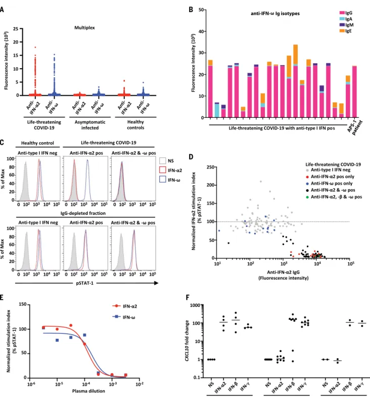
COVID-19 during the acute phase of disease. Multiplex
particle-based flow cytometry revealed a high
fluorescence intensity (FI) (>1500) for IgG
auto-Abs against IFN-a2 and/or IFN-w in 135
patients (13.7%) with life-threatening COVID-19
(Fig. 1A). We found that 49 of these 135
pa-tients were positive for auto-Abs against both
IFN-a2 and IFN-w, whereas 45 were positive
only for auto-Abs against IFN-a2, and 41 were
positive only for auto-Abs against IFN-w.
We also performed enzyme-linked
immuno-sorbent assay (ELISA), and the results
ob-tained were consistent with those obob-tained
with Luminex technology (fig. S1A). We found
that 11 and 14 of 23 patients tested had low
levels of IgM and IgA auto-Abs against IFN-w
and IFN-a2, respectively (Fig. 1B and fig. S1B).
Auto-Abs against type I IFNs were detected in
two unrelated patients for whom we had
plas-ma samples obtained before SARS-CoV-2
in-fection, which indicates that these antibodies
were present before SARS-CoV-2 infection and
were not triggered by the infection. As a
con-trol, we confirmed that all 25 APS-1 patients
tested had high levels of auto-Abs against
IFN-a2 and IFN-w (fig. S1C). Overall, we found that
135 of 987 patients (13.7%) with life-threatening
COVID-19 pneumonia had IgG auto-Abs against
at least one type I IFN.
The auto-Abs neutralize IFN-
a2 and IFN-w in vitro
We then tested whether auto-Abs against
IFN-a2 and IFN-w were neutralizing in vitro. We
incubated peripheral blood mononuclear cells
(PBMCs) from healthy controls with 10 ng/mL
IFN-a2 or IFN-w in the presence of plasma
from healthy individuals or from patients
with auto-Abs. A complete abolition of STAT1
phosphorylation was observed in 101 patients
RESEARCH
with auto-Abs against IFN-a2 and/or IFN-w
(table S1). The antibodies detected were
neu-tralizing against both IFN-a2 and IFN-w in 52
of these 101 patients (51%), against only
a2 in 36 patients (36%), and against only
IFN-w in 13 patients (13%) at the IFN-a2 and IFN-IFN-w
concentrations tested (Fig. 1, C and D). IgG
depletion from patients with auto-Abs restored
normal pSTAT1 induction after IFN-a2 and
IFN-w stimulation, whereas the purified IgG
fully neutralized this induction (Fig. 1C and
fig. S1D). Furthermore, these auto-Abs
neutral-ized high amounts of IFN-a2 (fig. S1E) and
were neutralizing at high dilutions (Fig. 1E
and fig. S1F). Notably, 15 patients with
life-threatening COVID-19 and auto-Abs against
IFN-a2 and/or IFN-w also had auto-Abs against
other cytokines [IFN-g, granulocyte-macrophage
colony-stimulating factor (GM-CSF), IL-6, IL-10,
IL-12p70, IL-22, IL-17A, IL-17F, and/or tumor
necrosis factor
–b (TNFb)], only three of which
(IL-12p70, IL-22, and IL-6) were neutralizing
(in four patients) (fig. S2, A to C). Similar
proportions were observed in the other
co-horts (fig. S2, D to L).
We also analyzed ISG induction after 2 hours
of stimulation with IFN-a2, IFN-b, or IFN-g in
the presence of plasma from healthy
individ-uals or from patients with auto-Abs. With
plas-ma from eight patients with auto-Abs against
IFN-
a2, the induction of ISG CXCL10 was
abo-lished after IFN-a2 stimulation but maintained
after stimulation with IFN-g (Fig. 1F). We then
found that plasma from the five patients with
neutralizing auto-Abs neutralized the
protec-tive activity of IFN-
a2 in Madin–Darby bovine
kidney (MDBK) cells infected with vesicular
stomatitis virus (VSV) (table S2). Overall, we
found that 101 of 987 patients (10.2%)
—including
95 men (94%)
—with life-threatening COVID-19
pneumonia had neutralizing IgG auto-Abs
against at least one type I IFN. By contrast,
auto-Abs were detected in only 4 of 1227 healthy
controls (0.33%) (Fisher exact test,
P < 10
−16)
and in none of the 663 patients with
asymp-tomatic or mild SARS-CoV-2 infection tested
(Fisher exact test,
P < 10
−16).
Auto-Abs against all 13 IFN-a subtypes in
patients with auto-Abs to IFN-a2
We investigated whether patients with
neu-tralizing auto-Abs against IFN-a2 only or those
with neutralizing auto-Abs against IFN-a2 and
IFN-w also had auto-Abs against the other 15
type I IFNs. ELISA showed that all patients
tested (
N = 22) with auto-Abs against IFN-a2
also had auto-Abs against all 13 IFN-a
sub-types (IFN-a1, -a2, -a4, -a5, -a6, -a7, -a8, -a10,
-a13, -a14, -a16, -a17, and -a21), whereas only 2
of the 22 patients tested had auto-Abs against
IFN-b, 1 had auto-Abs against IFN-k, and 2
had auto-Abs against IFN-e (Fig. 2A). The
auto-Abs against IFN-b had neutralizing
activ-ity against IFN-b (Fig. 1D). We confirmed that
all of the patients had auto-Abs against all 13
subtypes of IFN-a by testing the same samples
using luciferase-based immunoprecipitation
as-say (LIPS) (Fig. 2B). For IFN-b, we also screened
the whole cohort in a multiplex assay. We
found that 19 of 987 (1.9%) patients had
auto-Abs against IFN-b and that all of them were in
our cohort of severe COVID-19 individuals
with neutralizing auto-Abs against IFN-a and/
or IFN-w. Of these patients with auto-Abs
against IFN-b, only two were neutralizing
against IFN-b (Fig. 1, D and F).
Ten of the 17 genes encoding type I IFNs
(IFN-a2, -a5, -a6, a8, -a13, -a14, -a21, -b, -w, and
-k), have undergone strong negative selection,
which suggests that they play an essential role
in the general population. By contrast, the
1Laboratory of Human Genetics of Infectious Diseases, Necker Branch, INSERM U1163, Necker Hospital for Sick Children, Paris, France.2University of Paris, Imagine Institute, Paris, France. 3St. Giles Laboratory of Human Genetics of Infectious Diseases, Rockefeller Branch, The Rockefeller University, New York, NY, USA.4Laboratory of Clinical Immunology and Microbiology, Division of
Intramural Research, National Institute of Allergy and Infectious Diseases (NIAID), National Institutes of Health (NIH), Bethesda, MD, USA.5Laboratory of Virology and Infectious Disease, The
Rockefeller University, New York, NY, USA.6Sorbonne Université, INSERM, Centre d’Immunologie et des Maladies Infectieuses, (CIMI-Paris), Paris, France.7Institute of Biomedicine and Translational
Medicine, University of Tartu, Tartu, Estonia.8Hospices Civils de Lyon, Lyon Sud Hospital, Pierre-Bénite, France.9International Center of Research in Infectiology, Lyon University, INSERM U1111,
CNRS UMR 5308, ENS, UCBL, Lyon, France.10International Center of Research in Infectiology, Lyon University, INSERM U1111, CNRS UMR 5308, ENS, UCBL, Lyon, France.11National Referee Centre
for Rheumatic and AutoImmune and Systemic Diseases in Children (RAISE), Lyon, France.12Lyon Immunopathology Federation (LIFE), Hospices Civils de Lyon, Lyon, France.13Internal Medicine
Clinic, Tartu University Hospital, Tartu, Estonia.14Pneumology Department, Foch Hospital, Suresne, France.15Avicenne Hospital, Assistance Publique Hôpitaux de Paris (AP-HP), Bobigny, INSERM
U1272 Hypoxia and Lung, Bobigny, France.16Helix, San Mateo, CA, USA.17Department of Medical Microbiology, University Medical Center Utrecht, Utrecht, Netherlands.18IRCCS San Raffaele
Hospital and Vita-Salute San Raffaele University, Milan, Italy.19Department of Genetics, Yale University School of Medicine, New Haven, CT, USA.20Yale Center for Genome Analysis, Yale University
School of Medicine, New Haven, CT, USA.21Laboratory of Human Genetics and Genomics, The Rockefeller University, New York, NY, USA.22Service de Biologie Clinique and UMR-S 1176, Hôpital
Foch, Suresnes, France.23INSERM UMR-S 1140, Biosurgical Research Laboratory (Carpentier Foundation), Paris University and European Georges Pompidou Hospital, Paris, France.24Laboratory of
Immunogenetics of Pediatric Autoimmune Diseases, INSERM UMR 1163, University of Paris, Imagine Institute, Paris, France.25Department of Internal Medicine, National Referral Center for Rare
Systemic Autoimmune Diseases, Assistance Publique Hôpitaux de Paris-Centre (APHP-CUP), University of Paris, Paris, France.26Translational Immunology Laboratory, Institut Pasteur, Paris, France. 27Human Evolutionary Genetics Unit, Institut Pasteur, CNRS UMR 2000, 75015, Paris, France.28Human Genomics and Evolution, Collège de France, Paris, France.29Amsterdam UMC, University of
Amsterdam, Department of Neurology, Amsterdam Neuroscience, Amsterdam, Netherlands.30Department of Medicine, Division of Infectious Diseases, McGill University Health Centre, Montréal,
Québec, Canada.31Infectious Disease Susceptibility Program, Research Institute, McGill University Health Centre, Montréal, Québec, Canada.32Garvan Institute of Medical Research, Darlinghurst
2010, NSW, Sydney, Australia.33St Vincent’s Clinical School, Faculty of Medicine, University of New South Wales Sydney, Darlinghurst 2010, NSW, Australia.34Department of Paediatric Immunology
and Pulmonology, Centre for Primary Immunodeficiency Ghent (CPIG), PID Research Laboratory, Jeffrey Modell Diagnosis and Research Centre, Ghent University Hospital, Ghent, Belgium.
35Infectious Diseases and HIV Service, Hospital Universitari Mutua Terrassa, Universitat de Barcelona, Fundació Docència i Recerca Mutua Terrassa, Terrassa, Barcelona, Catalonia, Spain.36IrsiCaixa
AIDS Research Institute and Institute for Health Science Research Germans Trias i Pujol (IGTP), Badalona, Spain.37Infectious Diseases and Immunity, Centre for Health and Social Care Research
(CESS), Faculty of Medicine, University of Vic-Central University of Catalonia (UVic-UCC), Vic, Spain.38Catalan Institution for Research and Advanced Studies (ICREA), Barcelona, Spain.39Science
for Life Laboratory, Department of Women's and Children's Health, Karolinska Institutet, Karolinska, Sweden.40Department of Pediatric Rheumatology, Karolinska University Hospital, Karolinska,
Sweden.41Laboratory of Molecular Immunology, The Rockefeller University, New York, NY, USA.42Howard Hughes Medical Institute, New York, NY, USA.43Department of Immunology, Hospital
Universitario de Gran Canaria Dr. Negrín, Canarian Health System, Las Palmas de Gran Canaria, Spain.44Department of Clinical Sciences, University Fernando Pessoa Canarias, Las Palmas de Gran
Canaria, Spain.45Neglected Human Genetics Laboratory, INSERM, University of Paris, Paris, France.46Department of Infectious Diseases, Aarhus University Hospital, Skejby, Denmark.47Department
of Biomedicine, Aarhus University, Aarhus, Denmark.48Bioinformatics and Computational Biosciences Branch, Office of Cyber Infrastructure and Computational Biology, NIAID, NIH, Bethesda, MD,
USA.49Division of Intramural Research, National Institute of Dental Craniofacial Research (NIDCR), NIH, Bethesda, MD, USA.50Laboratory of Infectious Diseases, Division of Intramural Research,
NIAID, NIH, Bethesda, MD, USA.51Pediatric Department and Centro Tettamanti-European Reference Network PaedCan, EuroBloodNet, MetabERN-University of Milano-Bicocca-Fondazione
MBBM-Ospedale, San Gerardo, Monza, Italy.52Department of Infectious Diseases, San Gerardo Hospital - University of Milano-Bicocca, Monza, Italy.53University of Lorraine, Plurithematic Clinical
Investigation Centre INSERM CIC-P 1433, INSERM U1116, CHRU Nancy Hopitaux de Brabois, F-CRIN INI-CRCT (Cardiovascular and Renal Clinical Trialists), Nancy, France.54Intensive Care Unit,
Pitié-Salpétrière Hospital, Paris University, AP-HP, Paris, France.55Department of Clinical Science and K.G. Jebsen Center for Autoimmune Disorders, University of Bergen, Bergen, Norway.56Department
of Medicine, Haukeland University Hospital, Bergen, Norway.57Department of Medicine (Solna), Karolinska Institutet, Stockholm, Sweden.58Human Molecular Genetics Laboratory, Institute of
Genetics and Biophysics,“A. Buzzati-Traverso” Consiglio Nazionale delle Ricerche, Naples, Italy.59Centro di Ricerca Emato-oncologica AIL (CREA) Laboratory, Diagnostic Department, ASST Spedali
Civili di Brescia, Brescia, Italy.60Department of Infectious and Tropical Diseases, University of Brescia and ASST Spedali di Brescia, Brescia, Italy.61Direzione Sanitaria, ASST Spedali Civili di Brescia,
Brescia, Italy.62Department of Pediatrics, Fondazione IRCCS Policlinico San Matteo, University of Pavia, Pavia, Italy.63Laboratory of Immunology and Transplantation, Fondazione IRCCS Policlinico
San Matteo, Pavia, Italy.64Department of Clinical, Surgical, Diagnostic and Pediatric Sciences, University of Pavia, Pavia, Italy.65INSERM CIC 1425, Paris, France.66AP-HP, University Hospital of
Bichat, Paris, France.67University Paris Diderot, Paris 7, UFR de Médecine-Bichat, Paris, France.68Infection, Antimicrobials, Modelling, Evolution (IAME), INSERM, UMRS1137, University of Paris,
Paris, France.69AP-HP, Bichat Claude Bernard Hospital, Infectious and Tropical Diseases Department, Paris, France.70Center for Human Immunology, NIH, Bethesda, MD, USA.71Multiscale Systems
Biology Section, Laboratory of Immune System Biology, NIAID, NIH, Bethesda, MD, USA.72Département d’Immunologie, AP-HP, Hôpital Pitié-Salpétrière, Paris, France.73Pediatric Hematology and
Immunology Unit, Necker Hospital for Sick Children, AP-HP, Paris, France.
*Corresponding author. Email: jean-laurent.casanova@rockefeller.edu (J.-L.C.); paul.bastard@institutimagine.org (P.B.) †These authors contributed equally to this work.
‡These authors contributed equally to this work.
§All collaborators and their affiliations appear at the end of this paper. ¶These authors contributed equally to this work.
Fig. 1. Neutralizing auto-Abs against IFN-
a2 and/or IFN-w in patients with
life-threatening COVID-19. (A) Multiplex particle-based assay for auto-Abs
against IFN-a2 and IFN-w in patients with life-threatening COVID-19 (N = 782), in
patients with asymptomatic or mild SARS-CoV-2 infection (N = 443), and in
healthy controls not infected with SARS-CoV-2 (N = 1160). (B) Anti
–IFN-w Ig
isotypes in 23 patients with life-threatening COVID-19 and auto-Abs to type I
IFNs. (C) Representative fluorescence-activated cell sorting (FACS) plots
depicting IFN-
a2– or IFN-w–induced pSTAT1 in healthy control cells (gated on
CD14
+monocytes) in the presence of 10% healthy control or anti
–IFN-a2 or
anti
–IFN-w auto-Abs–containing patient plasma (top panel) or an IgG-depleted
plasma fraction (bottom panel). Max, maximum; neg, negative; pos, positive;
NS, not stimulated. (D) Plot of anti
–IFN-a2 auto-Ab levels against their
neutralization capacity. The stimulation index (stimulated over unstimulated
condition) for the plasma from each patient was normalized against that of healthy
control plasma from the same experiment. Spearman
’s rank correlation coefficient =
−0.6805; P < 0.0001. (E) Median inhibitory concentration (IC
50) curves
representing IFN-
a2– and IFN-w–induced pSTAT1 levels in healthy donor cells
in the presence of serial dilutions of patient plasma. The stimulation index
(stimulated over unstimulated condition) for patient plasma was normalized
against that of 10% healthy control plasma. IFN-a2: IC
50= 0.016%, R
2
= 0.985;
IFN-w: IC
50= 0.0353%, R
2= 0.926. R
2, coefficient of determination. (F) Neutralizing
effect on CXLC10 induction, after stimulation with IFN-a2, IFN-b, or IFN-g, in the
presence of plasma from healthy controls (N = 4), patients with life-threatening
COVID-19 and auto-Abs against IFN-a2 (N = 8), and APS-1 patients (N = 2).
RESEARCH
|
R E S E A R C H A R T I C L E
other seven IFN loci in the human genome
often carry loss-of-function alleles (
24
).
More-over, the 13 IFN-a subtypes and IFN-w are
more-closely related to each other than they
are to the other three IFNs (IFN-b, IFN-e, and
IFN-k), which are structurally and
phyloge-netically more distant (Fig. 2C). Thus, all
patients with neutralizing auto-Abs against
IFN-
a2 that we tested (N = 22) had auto-Abs
against all 13 IFN-a subtypes, and 3 of the 22
patients tested (14%) had auto-Abs against
14 or more type I IFNs.
The auto-Abs neutralize IFN-
a2 against
SARS-CoV-2 in vitro and IFN-a in vivo
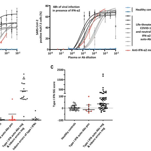
Plasma from eight patients with neutralizing
auto-Abs against type I IFN also neutralized
the ability of IFN-a2 to block the infection of
Huh7.5 cells with SARS-CoV-2 (Fig. 3A).
Plas-ma from two healthy controls or from seven
SARS-CoV-2
–infected patients without
auto-Abs did not block the protective action of
IFN-a2 (Fig. 3A and fig. S3A). These data provide
compelling evidence that the patients
’ blood
carried sufficiently large amounts of auto-Abs
to neutralize the corresponding type I IFNs and
block their antiviral activity in vitro, including
that against SARS-CoV-2.
We also found that all 41 patients with
neu-tralizing auto-Abs against the 13 types of IFN-a
tested had low (one patient) or undetectable
(40 patients) levels of the 13 types of IFN-a in
their plasma during the course of the disease
(Fig. 3B) (
25
,
26
). Type I IFNs may be degraded
and/or bound to the corresponding circulating
auto-Abs. The presence of circulating
neutral-izing auto-Abs against IFN-a is, therefore,
strongly associated with low serum IFN-a levels
(Fisher exact test,
P < 10
−6). Consistently in
patients with neutralizing auto-Abs against
IFN-
a2, the baseline levels of type I IFN–
dependent transcripts were low, whereas they
were normal for nuclear factor
kB (NF-kB)–
dependent transcripts (Fig. 3C and fig. S3B).
Overall, our findings indicate that the
auto-Abs against type I IFNs present in patients
with life-threatening COVID-19 were
neu-tralizing in vitro and in vivo.
Pronounced excess of men in patients with
auto-Abs against type I IFNs
There was a pronounced excess of male patients
(95 of 101; 94%) with critical COVID-19
pneu-monia and neutralizing auto-Abs against type I
Fig. 2. Auto-Abs against the different type I IFN subtypes. (A) ELISA for
auto-Abs against the 13 different IFN-a subtypes, IFN-w, IFN-b, IFN-k, and IFN-e
in patients with life-threatening COVID-19 and auto-Abs against IFN-a2 (N = 22),
APS-1 patients (N = 2), and healthy controls (N = 2). (B) LIPS for the 12 different
IFN-a subtypes tested in patients with auto-Abs against IFN-a2 (N = 22) and
healthy controls (N = 2). (C) Neighbor-joining phylogenetic tree of the 17 human
type I IFN proteins. Horizontal branches are drawn to scale (bottom left, number
of substitutions per site). Thinner, intermediate, and thicker internal branches
have bootstrap support of <50,
≥50, and >80%, respectively. The bootstrap
value for the branch separating IFN-w from all IFN-a subtypes is 100%.
IFNs. This proportion of males was higher than
that observed in patients with critical COVID-19
without auto-Abs (75%; Fisher exact test,
P =
2.5 × 10
−6), and the proportion was much higher
than that in male patients in the asymptomatic
or pauci-symptomatic cohort (28%; Fisher exact
test,
P < 10
−6) (Table 1, Fig. 4A, and fig. S4A).
Further evidence for X-chromosome linkage
was provided by the observation that one
of the seven women with auto-Abs and
life-threatening COVID-19 had X chromosome
–
linked incontinentia pigmenti (IP), in which
cells activate only a single X chromosome (cells
having activated the X chromosome bearing
the null mutation in
NEMO dying in the course
of development) (
27
). The prevalence of
auto-Abs against type I IFNs in the general
pop-ulation was estimated at 0.33% (0.015 to 0.67%)
in a sample of 1227 healthy individuals
—a value
much lower than that in patients with
life-threatening COVID-19 pneumonia, by a factor
of at least 15.
The patients with auto-Abs were also slightly
older than the rest of our cohort (49.5% of
patients positive for auto-Abs were over 65 years
of age versus 38% for the rest of the cohort;
P = 0.024), which suggests that the frequency
of circulating anti
–type I IFNs auto-Abs
in-creases with age (Table 1 and Fig. 4B). However,
auto-Abs were found in patients aged from
25 to 87 years (fig. S4B). Principal components
analysis (PCA) was performed on data from
A
B
C
Fig. 3. Enhanced SARS-CoV-2 replication, despite the presence of IFN-
a2,
in the presence of plasma from patients with auto-Abs against IFN-
a2 and
low in vivo levels of IFN-
a. (A) SARS-CoV-2 replication—measured 24 hours
(left) and 48 hours (right) after infection
—in Huh7.5 cells treated with IFN-a2 in the
presence of plasma from patients with life-threatening COVID-19 and neutralizing
auto-Abs against IFN-
a2 (N = 8); a commercial anti–IFN-a2 antibody; or control plasma
(N = 2). (B) IFN-a levels in the plasma or serum of patients with neutralizing auto-Abs
(N = 41), healthy controls (N = 5), COVID-19 patients without auto-Abs (N = 21), and
patients with life-threatening COVID-19 and loss-of-function (LOF) variants (N = 10),
as assessed by Simoa ELISA. (C) z-scores for type I IFN gene responses in whole
blood of COVID-19 patients with (N = 8) or without (N = 51) neutralizing auto-Abs, or
healthy uninfected controls (N = 22). The median ± interquartile range is shown.
z-scores were significantly lower for patients with neutralizing auto-Abs compared with
patients without auto-Abs (Mann-Whitney test, P = 0.01).
Table 1. Sex and age distribution of patients with critical COVID-19 with and without auto-Abs. Ages
and sexes of the patients and controls and information about auto-Abs against IFN-a2 and IFN-w, presented by
age and sex. Dashes in rightmost column indicate data not available. OR, odds ratio; CI, confidence interval.
Life-threatening
COVID-19
N total
N auto-Abs positive
(percentage)
OR [95% CI]
P value*
Sex
...Female
226
6 (2.6%)
1
–
...Male
761
95 (12.5%)
5.22 [2.27
– 14.80]
2.5 × 10
−6 ...Age
...<65 years
602
51 (8.5%)
1
–
...≥65 years
385
50 (13.0%)
1.61 [1.04
– 2.49]
0.024
...*P values were derived from Fisher’s exact test, as implemented in R (https://cran.r-project.org/).
49 patients: 34 Europeans, 5 North Africans,
4 sub-Saharan Africans, 2 patients from the
Middle East, 2 South Asians, 1 East Asian,
and 1 South American (Fig. 4C). Large-scale
studies will be required to determine the
fre-quency of such auto-Abs in humans of
differ-ent sexes, ages, and ancestries. Finally, the
presence of auto-Abs was associated with a
poor outcome, with death occurring in 37 of
the 101 patients (36.6%) (table S1).
Neutralizing auto-Abs to type I IFNs are
causative of critical COVID-19
There are multiple lines of evidence to suggest
that the neutralizing auto-Abs against type I
IFNs observed in these 101 patients preceded
infection with SARS-CoV-2 and accounted for
the severity of disease. First, the two patients
for whom testing was performed before
COVID-19 were found to have auto-Abs before
infec-tion. Second, three patients with APS-1 known
to have neutralizing auto-Abs against type I
IFN immunity before infection also had
life-threatening COVID-19 (
22
) (supplementary
methods). Third, we screened a series of 32
women with IP and found that a quarter of
them had auto-Abs against type I IFNs,
in-cluding one who developed critical COVID-19
(fig. S1C). Fourth, there is a marked bias in
favor of men, which suggests that the
produc-tion of auto-Abs against type I IFNs
—whether
driven by germ line or somatic genome
—may
be X chromosome
–linked and therefore
pre-existing to infection.
Moreover, IFN-a subtypes were
undetect-able during acute disease in the blood of
patients with auto-Abs against IFN-a, which
suggests a preexisting or concomitant
biolog-ical impact in vivo. It is also unlikely that
patients could break self-tolerance and mount
high titers of neutralizing IgG auto-Abs against
type I IFN within only 1 or even 2 weeks of
infection. Finally, inborn errors of type I IFNs
underlying life-threatening COVID-19 in other
previously healthy adults
—including autosomal
recessive IFN-a/b receptor subunit 1 (IFNAR1)
deficiency
—have also been reported in an
ac-companying paper (
18
). Collectively, these
find-ings suggest that auto-Abs against type I IFNs
are a cause and not a consequence of severe
SARS-Cov-2 infection, although their titers and
affinity may be enhanced by the SARS-CoV-2
–
driven induction of type I IFNs. They also
pro-vide an explanation for the major sex bias seen
in patients with life-threatening COVID-19 and
perhaps also for the increase in risk with age.
Conclusion
We report here that at least 10% of patients
with life-threatening COVID-19 pneumonia
have neutralizing auto-Abs against type I IFNs.
With our accompanying description of patients
with inborn errors of type I IFNs and
life-threatening COVID-19 (
18
), this study
high-lights the crucial role of type I IFNs in protective
immunity against SARS-CoV-2. These auto-Abs
against type I IFNs were clinically silent until
the patients were infected with SARS-CoV-2
—
a poor inducer of type I IFNs (
28
)
—which
sug-gests that the small amounts of IFNs induced
by the virus are important for protection against
severe disease. The neutralizing auto-Abs
against type I IFNs, like inborn errors of type I
IFN production, tip the balance in favor of the
virus, which results in devastating disease with
insufficient, and even perhaps deleterious,
in-nate and adaptive immune responses.
Our findings have direct clinical
implica-tions. First, SARS-CoV-2
–infected patients can
be screened to identify individuals with
auto-Abs at risk of developing life-threatening
pneumonia. Such patients recovering from
life-threatening COVID-19 should also be
ex-cluded from donating convalescent plasma for
ongoing clinical trials, or at least they should
be tested before their plasma donations are
accepted (
29
). Second, this finding paves the
way for preventive or therapeutic intervention,
including plasmapheresis, monoclonal Abs
de-pleting plasmablasts, and the specific
inhibi-tion of type I IFN
–reactive B cells (
30
). Finally, in
this patient group, early treatment with IFN-a
is unlikely to be beneficial; however, treatment
Fig. 4. Demographic and ethnic information about the patients and
controls. (A) Gender distribution in patients with life-threatening COVID-19
and auto-Abs to type I IFNs, patients with life-threatening COVID-19 and
without auto-Abs to type I IFNs, and individuals with asymptomatic or mild
SARS-CoV-2. (B) Age distribution in patients with life-threatening COVID-19
and auto-Abs to type I IFNs, patients with life-threatening COVID-19 and
without auto-Abs to type I IFNs, and individuals with asymptomatic or mild
SARS-CoV-2. yo, years old. (C) PCA on 49 patients with life-threatening
COVID-19 and auto-Abs against type I IFNs. EUR, Europeans; AFR, Africans;
EAS, East-Asians.
with injected or nebulized IFN-b may have
beneficial effects, as auto-Abs against IFN-b
appear to be rare in patients with auto-Abs
against type I IFNs.
Materials and methods
Subjects and samples
We enrolled 987 patients with proven
life-threatening (critical) COVID-19, 663
asympto-matic or pauci-symptoasympto-matic individuals with
proven COVID-19, and 1227 healthy controls
in this study. All subjects were recruited
fol-lowing protocols approved by local Institutional
Review Boards (IRBs). All protocols followed
local ethics recommendations and informed
consent was obtained when required.
COVID-19 disease severity was assessed in
accordance with the Diagnosis and Treatment
Protocol for Novel Coronavirus Pneumonia.
The term life-threatening COVID-19
pneu-monia describes pneupneu-monia in patients with
critical disease, whether pulmonary, with
mechanical ventilation [continuous positive
airway pressure (CPAP), bilevel positive
air-way pressure (BIPAP), intubation, or high-flow
oxygen], septic shock, or damage to any other
organ requiring admission in the intensive care
unit (ICU). The individuals with asymptomatic
or mild SARS-CoV-2 infection were individuals
infected with SARS-CoV-2 who remained
asy-mptomatic or developed mild, self-healing,
am-bulatory disease with no evidence of pneumonia.
The healthy controls were individuals who
had not been exposed to SARS-CoV-2.
Plasma and serum samples from the patients
and controls were frozen at
−20°C immediately
after collection. The fluid-phase LIPS assay was
used to determine the levels of antibodies against
the SARS-CoV-2 nucleoprotein and spike
pro-tein, as has been previously described (
31
).
Detection of anti-cytokine auto-Abs
Multiplex particle-based assay
Serum and plasma samples were screened
for auto-Abs against 18 targets in a multiplex
particle-based assay, in which magnetic beads
with differential fluorescence were covalently
coupled to recombinant human proteins. Patients
with an FI of >1500 for IFN-a2 or IFN-b or
>1000 for IFN-w were tested for blocking
activ-ity, as were patients positive for another cytokine.
ELISA
ELISA was performed as previously described
(
5
). In brief, ELISA plates were coated with
recombinant human interferon-a (rhIFN-a)
or rhIFN-w and incubated with 1:50
dilu-tions of plasma samples from the patients or
controls. A similar protocol was used when
testing for 12 subtypes of IFN-a.
LIPS
Levels of auto-Abs against IFN-a subtypes were
measured with LIPS, as previously described
(
32
). IFN-a1, IFN-a2, IFN-a4, IFN-a5, IFN-a6,
IFN-a7, IFN-a8, IFN-a10, IFN-a14, IFN-a16,
IFN-a17, and IFN-a21 sequences were
trans-fected in HEK293 cells, and the IFN-a-luciferase
fusion proteins were collected in the tissue
culture supernatant. For autoantibody
screen-ing, serum samples were incubated with
protein G agarose beads, and we then added
2 × 10
6luminescence units (LU) of antigen
and incubated. Luminescence intensity was
measured. The results are expressed in
arbi-trary units (AU), as a fold-difference relative to
the mean of the negative control samples.
Functional evaluation of anti-cytokine auto-Abs
The blocking activity of anti
–IFN-a and anti–
IFN-w auto-Abs was determined by assessing
STAT1 phosphorylation in healthy control cells
after stimulation with the appropriate
cyto-kines in the presence of 10% healthy control or
patient serum or plasma.
We demonstrated that the IFN-a and IFN-w
blocking activity observed was due to auto-Abs
and not another plasma factor, by depleting
IgG from the plasma with a protein G column
Without eluting the IgG, the flow-through
fraction (IgG-depleted) was then collected and
compared with total plasma in the
phospho-STAT1 assay.
The blocking activity of anti
–IFN-g,
–GM-CSF,
–IFN-l1, –IFN-l2, –IFN-l3, –IL-6, –IL-10,
–IL-12p70, –IL-22, –IL-17A, –IL-17F, -TNFa, and
-TNFb antibodies was assessed with the
assays outlined in table S3, as previously
reported (
21
).
For the neutralization of ISG induction,
PBMCs were left unstimulated or were
stimu-lated for 2 hours with 10 ng/mL IFN-a or 10 ng/
mL IFN-g in a final volume of 100 mL.
Real-time quantitative polymerase chain reaction
(RT-qPCR) analysis was performed with
Ap-plied Biosystems
Taqman assays for CXCL10,
and the
b-glucuronidase (GUS) housekeeping
gene for normalization. Results are expressed
according to the
DDCt method, as described by
the manufacturer
’s kit.
Phylogenetic reconstruction
Protein sequences were aligned with the online
version of MAFFT v7.471 software (
33
), using
the L-INS-i strategy (
34
) and the BLOSUM62
scoring matrix for amino acid substitutions.
Phylogenetic tree reconstruction was performed
by the neighbor-joining method (
35
) with the
substitution model (
36
). Low-confidence branches
(<50%) are likely to be due to gene conversion
events between
IFNA genes, as previously
re-ported (
24
,
37
). The tree was then visualized
(
38
). Very similar results were obtained with
the corresponding DNA sequences (
37
,
39
).
Statistical analysis
Comparison of proportions were performed
using a Fisher exact test, as implemented in R
(
https://cran.r-project.org/
). PCA was performed
with Plink v1.9 software on whole-exome and
whole-genome sequencing data with the 1000
Genomes (1kG) Project phase 3 public
data-base as a reference.
Simoa
Serum IFN-a concentrations were determined
with Simoa technology, as previously described
(
40
,
41
), with reagents and procedures
ob-tained from the Quanterix Corporation.
VSV assay
The seroneutralization assay was performed
as previously described (
42
). In brief, the
incu-bation of IFN-a2 with MDBK cells protects the
cultured cells against the cytopathic effect of
VSV. The titer of anti
–IFN-a antibodies was
defined as the last dilution causing 50% cell
death.
SARS-CoV-2 experiment
SARS-CoV-2 strain USA-WA1/2020 was obtained
from BEI Resources and amplified in Huh7.5
hepatoma cells at 33°C. Viral titers were
mea-sured on Huh7.5 cells in a standard plaque
assay. Plasma samples or a commercial anti
–
IFN-a2 antibody were serially diluted and
incubated with 20 pM recombinant IFN-a2
for 1 hour at 37°C (starting concentrations:
plasma samples = 1/100 and anti
–IFN-a2
antibody = 1/1000). The cell culture
me-dium was then removed and replaced with
the plasma
– or antibody–IFN-a2 mixture. The
plates were incubated overnight, and the
plasma
– or antibody–IFN-a2 mixture was
removed by aspiration. The cells were washed
once with phosphate-buffered saline (PBS) to
remove potential anti
–SARS-CoV-2
neutral-izing antibodies, and fresh medium was then
added. Cells were then infected with
SARS-CoV-2 by directly adding the virus to the wells.
Cells infected at a high multiplicity of
infec-tion (MOI) were incubated at 37°C for 24 hours,
whereas cells infected at a low MOI were
incubated at 33°C for 48 hours. The cells
were fixed with 7% formaldehyde, stained for
SARS-CoV-2 with an anti-N antibody, imaged,
and analyzed as previously described (
43
).
Nanostring
For the NanoString assay, total RNA was
ex-tracted from whole blood samples collected in
PaxGene tubes. The expression of selected
genes was determined by NanoString
meth-ods and a 28-gene type I IFN score was
calcu-lated (
44
).
REFERENCES AND NOTES
1. J.-L. Casanova, L. Abel, The human genetic determinism of life-threatening infectious diseases: Genetic heterogeneity and physiological homogeneity? Hum. Genet. 139, 681–694 (2020). doi:10.1007/s00439-020-02184-w; pmid:32462426
2. R. Döffinger et al., Autoantibodies to interferon-g in a patient with selective susceptibility to mycobacterial infection and
organ-specific autoimmunity. Clin. Infect. Dis. 38, e10–e14 (2004). doi:10.1086/380453; pmid:14679469
3. C. Höflich et al., Naturally occurring anti–IFN-g autoantibody and severe infections with Mycobacterium cheloneae and Burkholderia cocovenenans. Blood 103, 673–675 (2004). doi:10.1182/blood-2003-04-1065; pmid:12947000
4. B. Kampmann et al., Acquired predisposition to mycobacterial disease due to autoantibodies to IFN-g. J. Clin. Invest. 115, 2480–2488 (2005). doi:10.1172/JCI19316; pmid:16127458
5. A. Puel et al., Recurrent staphylococcal cellulitis and subcutaneous abscesses in a child with autoantibodies against IL-6. J. Immunol. 180, 647–654 (2008). doi:10.4049/ jimmunol.180.1.647; pmid:18097067
6. A. Puel et al., Autoantibodies against IL-17A, IL-17F, and IL-22 in patients with chronic mucocutaneous candidiasis and autoimmune polyendocrine syndrome type I. J. Exp. Med. 207, 291–297 (2010). doi:10.1084/jem.20091983; pmid:20123958
7. K. Kisand et al., Chronic mucocutaneous candidiasis in APECED or thymoma patients correlates with autoimmunity to Th17-associated cytokines. J. Exp. Med. 207, 299–308 (2010). doi:10.1084/jem.20091669; pmid:20123959
8. C.-L. Ku, C.-Y. Chi, H. von Bernuth, R. Doffinger, Autoantibodies against cytokines: Phenocopies of primary
immunodeficiencies? Hum. Genet. 139, 783–794 (2020). doi:10.1007/s00439-020-02180-0; pmid:32419033
9. A. Isaacs, J. Lindenmann, Virus interference. I. The interferon. Proc. R. Soc. Lond. B 147, 258–267 (1957). doi:10.1098/ rspb.1957.0048; pmid:13465720
10. A. Isaacs, J. Lindenmann, R. C. Valentine, Virus interference. II. Some properties of interferon. Proc. R. Soc. Lond. B 147, 268–273 (1957). doi:10.1098/rspb.1957.0049; pmid:13465721
11. I. Gresser, Wherefore interferon? J. Leukoc. Biol. 61, 567–574 (1997). doi:10.1002/jlb.61.5.567; pmid:9129205
12. H.-H. Hoffmann, W. M. Schneider, C. M. Rice, Interferons and viruses: An evolutionary arms race of molecular interactions. Trends Immunol. 36, 124–138 (2015). doi:10.1016/ j.it.2015.01.004; pmid:25704559
13. N. A. de Weerd, J. P. Vivian, S. S. Lim, S. U.-S. Huang, P. J. Hertzog, Structural integrity with functional plasticity: What type I IFN receptor polymorphisms reveal. J. Leukoc. Biol. 108, 909–924 (2020). doi:10.1002/JLB.2MR0420-152R
14. J. E. Darnell Jr., STATs and gene regulation. Science 277, 1630–1635 (1997). doi:10.1126/science.277.5332.1630; pmid:9287210
15. A. Vallbracht, J. Treuner, B. Flehmig, K. E. Joester, D. Niethammer, Interferon-neutralizing antibodies in a patient treated with human fibroblast interferon. Nature 289, 496–497 (1981). doi:10.1038/289496a0; pmid:6162104
16. A. Meager et al., Anti-interferon autoantibodies in autoimmune polyendocrinopathy syndrome type 1. PLOS Med. 3, e289 (2006). doi:10.1371/journal.pmed.0030289; pmid:16784312
17. S. Panem, I. J. Check, D. Henriksen, J. Vilcek, Antibodies to alpha-interferon in a patient with systemic lupus erythematosus. J. Immunol. 129, 1–3 (1982). pmid:6177744
18. Q. Zhang et al., Inborn errors of type I IFN immunity in patients with life-threatening COVID-19. Science 370, eabd4570 (2020). doi:10.1126/science.abd4570
19. B. Pozzetto, K. E. Mogensen, M. G. Tovey, I. Gresser, Characteristics of autoantibodies to human interferon in a patient with varicella-zoster disease. J. Infect. Dis. 150, 707–713 (1984). doi:10.1093/infdis/150.5.707; pmid:6238105
20. J.-L. Casanova, Ion Gresser. J. Interferon Cytokine Res. 39, 317–320 (2019). doi:10.1089/jir.2018.29015.mem
21. J. E. Walter et al., Broad-spectrum antibodies against self-antigens and cytokines in RAG deficiency. J. Clin. Invest. 125, 4135–4148 (2015). doi:10.1172/JCI80477; pmid:26457731
22. G. Beccuti et al., A COVID-19 pneumonia case report of autoimmune polyendocrine syndrome type 1 in Lombardy, Italy: Letter to the editor. J. Endocrinol. Invest. 43, 1175–1177 (2020). doi:10.1007/s40618-020-01323-4; pmid:32519200
23. J.-L. Casanova, H. C. Su, COVID Human Genetic Effort, A Global Effort to Define the Human Genetics of Protective Immunity to SARS-CoV-2 Infection. Cell 181, 1194–1199 (2020). doi:10.1016/j.cell.2020.05.016; pmid:32405102
24. J. Manry et al., Evolutionary genetic dissection of human interferons. J. Exp. Med. 208, 2747–2759 (2011). doi:10.1084/ jem.20111680; pmid:22162829
25. S. Trouillet-Assant et al., Type I IFN immunoprofiling in COVID-19 patients. J. Allergy Clin. Immunol. 146, 206–208.e2 (2020). doi:10.1016/j.jaci.2020.04.029; pmid:32360285
26. J. Hadjadj et al., Impaired type I interferon activity and inflammatory responses in severe COVID-19 patients. Science 369, 718–724 (2020). doi:10.1126/science.abc6027; pmid:32661059
27. A. Harris, J. Collins, D. Vetrie, C. Cole, M. Bobrow, X inactivation as a mechanism of selection against lethal alleles: Further investigation of incontinentia pigmenti and X linked lymphoproliferative disease. J. Med. Genet. 29, 608–614 (1992). doi:10.1136/jmg.29.9.608; pmid:1404291
28. D. Blanco-Melo et al., Imbalanced Host Response to SARS-CoV-2 Drives Development of COVID-19. Cell 181, 1036–1045. e9 (2020). doi:10.1016/j.cell.2020.04.026; pmid:32416070
29. S. L. Klein et al., Sex, age, and hospitalization drive antibody responses in a COVID-19 convalescent plasma donor population. J. Clin. Invest. 142004 (2020). doi:10.1172/JCI142004; pmid:32764200
30. T. T. Wang, J. V. Ravetch, Functional diversification of IgGs through Fc glycosylation. J. Clin. Invest. 129, 3492–3498 (2019). doi:10.1172/JCI130029; pmid:31478910
31. P. D. Burbelo et al., Sensitivity in Detection of Antibodies to Nucleocapsid and Spike Proteins of Severe Acute Respiratory Syndrome Coronavirus 2 in Patients With Coronavirus Disease 2019. J. Infect. Dis. 222, 206–213 (2020). doi:10.1093/infdis/ jiaa273; pmid:32427334
32. S. Meyer et al., AIRE-Deficient Patients Harbor Unique High-Affinity Disease-Ameliorating Autoantibodies. Cell 166, 582–595 (2016). doi:10.1016/j.cell.2016.06.024; pmid:27426947
33. K. Katoh, J. Rozewicki, K. D. Yamada, MAFFT online service: Multiple sequence alignment, interactive sequence choice and visualization. Brief. Bioinform. 20, 1160–1166 (2019). doi:10.1093/bib/bbx108; pmid:28968734
34. K. Katoh, K. Kuma, H. Toh, T. Miyata, MAFFT version 5: Improvement in accuracy of multiple sequence alignment. Nucleic Acids Res. 33, 511–518 (2005). doi:10.1093/nar/gki198; pmid:15661851
35. N. Saitou, M. Nei, The neighbor-joining method: A new method for reconstructing phylogenetic trees. Mol. Biol. Evol. 4, 406–425 (1987). doi:10.1093/oxfordjournals.molbev.a040454; pmid:3447015
36. D. T. Jones, W. R. Taylor, J. M. Thornton, The rapid generation of mutation data matrices from protein sequences. Comput. Appl. Biosci. 8, 275–282 (1992). doi:10.1093/bioinformatics/ 8.3.275; pmid:1633570
37. C. H. Woelk, S. D. W. Frost, D. D. Richman, P. E. Higley, S. L. Kosakovsky Pond, Evolution of the interferon alpha gene family in eutherian mammals. Gene 397, 38–50 (2007). doi:10.1016/j.gene.2007.03.018; pmid:17512142
38. M. V. Han, C. M. Zmasek, phyloXML: XML for evolutionary biology and comparative genomics. BMC Bioinformatics 10, 356 (2009). doi:10.1186/1471-2105-10-356; pmid:19860910
39. S. Pestka, C. D. Krause, M. R. Walter, Interferons, interferon-like cytokines, and their receptors. Immunol. Rev. 202, 8–32 (2004). doi:10.1111/j.0105-2896.2004.00204.x; pmid:15546383
40. D. M. Rissin et al., Single-molecule enzyme-linked immunosorbent assay detects serum proteins at subfemtomolar concentrations. Nat. Biotechnol. 28, 595–599 (2010). doi:10.1038/nbt.1641; pmid:20495550
41. A. Mathian et al., Monitoring Disease Activity in Systemic Lupus Erythematosus With Single-Molecule Array Digital Enzyme-Linked Immunosorbent Assay Quantification of Serum Interferon-a. Arthritis Rheumatol. 71, 756–765 (2019). doi:10.1002/art.40792; pmid:30507062
42. P. Lebon, G. Ponsot, J. Aicardi, F. Goutières, M. Arthuis, Early intrathecal synthesis of interferon in herpes encephalitis. Biomedicine 31, 267–271 (1979). pmid:94549
43. D. F. Robbiani et al., Convergent antibody responses to SARS-CoV-2 in convalescent individuals. Nature 584, 437–442 (2020). doi:10.1038/s41586-020-2456-9; pmid:32555388
44. H. Kim et al., Development of a Validated Interferon Score Using NanoString Technology. J. Interferon Cytokine Res. 38, 171–185 (2018). doi:10.1089/jir.2017.0127; pmid:29638206
45. J. P. Ferreira et al., Cohort Profile: Rationale and design of the fourth visit of the STANISLAS cohort: a familial longitudinal population-based cohort from the Nancy region of France. Int. J. Epidemiol. 47, 395–395j (2018). doi:10.1093/ije/ dyx240; pmid:29220499
ACKNOWLEDGMENTS
We thank the patients, their families, and healthy donors for placing their trust in us. We thank the French Incontinentia pigmenti association for their help and support. We thank Y. Nemirovskaya, D. Papandrea, M. Woollett, D. Liu, C. Rivalain, and C. Patissier for administrative assistance; D. Kapogiannis (National Institute on Aging) for providing healthy donor samples; and S. Xirasager, J. Barnett, X. Cheng, S. Weber, J. Danielson, B. Garabedian, and H. Matthews for their assistance in this study. We also thank R. Apps, B. Ryan, and Y. Belkaid of the CHI for their assistance. We thank the CRB-Institut Jérôme Lejeune, CRB-BioJeL, Paris, France, for their assistance. We thank
M. C. García Guerrero; I. Erkizia; E. Grau; M. Massanella from IrsiCaixa AIDS Research Institute, Badalona, Spain; and J. Guitart from the Department of Clinical Genetics, University Hospital Germans Trias i Pujol, Badalona, Spain, for providing samples. We also thank J. Dalmau from IrsiCaixa for assistance. Funding: The Laboratory of Human Genetics of Infectious Diseases is supported by the Howard Hughes Medical Institute, The Rockefeller University, the St. Giles Foundation, the National Institutes of Health (NIH) (R01AI088364), the National Center for Advancing Translational Sciences (NCATS), NIH Clinical and Translational Science Award (CTSA) program (UL1 TR001866), a Fast Grant from Emergent Ventures, the Mercatus Center at George Mason University, the Yale Center for Mendelian Genomics and the GSP Coordinating Center funded by the National Human Genome Research Institute (NHGRI) (UM1HG006504 and U24HG008956), the French National Research Agency (ANR) under the Investments for the Future program (ANR-10-IAHU-01), the Integrative Biology of Emerging Infectious Diseases Laboratory of Excellence (ANR-10-LABX-62-IBEID), the French Foundation for Medical Research (FRM) (EQU201903007798), the FRM and ANR GENCOVID project (ANRS-COV05), the Square Foundation, Grandir– Fonds de solidarité pour l’enfance, the SCOR Corporate Foundation for Science, the Institut Institut National de la Santé et de la Recherche Médicale (INSERM), and the University of Paris. Samples from San Raffaele Hospital were obtained through the Covid-BioB project and by healthcare personnel of San Raffaele Hospital, San Raffaele Telethon Institute for Gene Therapy (SR-TIGET) clinical laboratory and clinical research unit, funded by the Program Project COVID-19 OSR-UniSR and Fondazione Telethon. The French COVID Cohort Study Group was sponsored by INSERM and supported by the REACTing consortium and by a grant from the French Ministry of Health (PHRC 20-0424). The Cov-Contact Cohort was supported by the REACTing consortium, the French Ministry of Health, and the European Commission (RECOVER WP 6). The Milieu Intérieur Consortium was supported by the French Government’s Investissement d’Avenir program, Laboratoire d’Excellence Milieu Intérieur grant (ANR-10-LABX-69-01) (primary investigators: L.Q.-M. and D.Du.). The Simoa experiment was supported by the PHRC-20-0375 COVID-19 grant “DIGITAL COVID” (primary investigator: G.G.). S.G.T. is supported by a Leadership 3 Investigator Grant awarded by the National Health and Medical Research Council of Australia and a COVID19 Rapid Response Grant awarded by UNSW Sydney. C.R.-G. and colleagues were supported by the Instituto de Salud Carlos III (COV20_01333 and COV20_01334, Spanish Ministry of Science and Innovation RTC-2017-6471-1; AEI/FEDER, UE) and Cabildo Insular de Tenerife (CGIEU0000219140 and“Apuestas científicas del ITER para colaborar en la lucha contra la COVID-19”). S.T.-A. and A.B. were supported by ANR-20-COVI-0064 (primary investigator: A.Be.). This work is supported by the French Ministry of Health“Programme Hospitalier de Recherche Clinique Inter regional 2013,” by the Contrat de Plan Etat-Lorraine and FEDER Lorraine, and by a public grant overseen by the French National Research Agency (ANR) as part of the second Investissements d’Avenir program FIGHT-HF (reference no. ANR-15-RHU-0004) and by the French PIA project“Lorraine Université d’Excellence” (reference no. ANR-15-IDEX-04-LUE) (45); and biobanking is performed by the Biological Resource Center Lorrain BB-0033-00035. This study was supported by the Fonds IMMUNOV, for Innovation in Immunopathology; by a grant from the Agence National de la Recherche (ANR-flash Covid19“AIROCovid” to F.R.-L.); and by the FAST Foundation (French Friends of Sheba Tel Hashomer Hospital). Work in the Laboratory of Virology and Infectious Disease was supported by NIH grants P01AI138398-S1, 2U19AI111825, and R01AI091707-10S1; a George Mason University Fast Grant; and the G. Harold and Leila Y. Mathers Charitable Foundation. The Amsterdam UMC Covid-19 Biobank was supported by grants from the Amsterdam Corona Research Fund, the Dr. C.J. Vaillant Fund, and the Netherlands Organization for Health Research and Development [ZonMw; NWO-Vici-Grant (grant no. 918·19·627 to D.v.d.B.)]. This work was also supported by the Division of Intramural Research of the National Institute of Dental Craniofacial Research and the National Institute of Allergy and Infectious Diseases, National Institutes of Health, and by Regione Lombardia, Italy (project“Risposta immune in pazienti con COVID-19 e comorbidita”). The opinions and assertions expressed herein are those of the author(s) and do not necessarily reflect the official policy or position of the Uniformed Services University or the Department of Defense. J.H. holds an Institut Imagine M.D.-Ph.D. fellowship from the Fondation Bettencourt Schueller. J.R. is supported by the INSERM Ph.D. program (“poste d’accueil Inserm”). P.Ba. was supported by the French Foundation for Medical Research (FRM, EA20170638020) and the M.D.-Ph.D. program