HAL Id: inserm-00991784
https://www.hal.inserm.fr/inserm-00991784
Submitted on 16 May 2014
HAL is a multi-disciplinary open access
archive for the deposit and dissemination of
sci-entific research documents, whether they are
pub-lished or not. The documents may come from
teaching and research institutions in France or
abroad, or from public or private research centers.
L’archive ouverte pluridisciplinaire HAL, est
destinée au dépôt et à la diffusion de documents
scientifiques de niveau recherche, publiés ou non,
émanant des établissements d’enseignement et de
recherche français ou étrangers, des laboratoires
publics ou privés.
High viral load of Merkel cell polyomavirus DNA
sequences in Langerhans cell sarcoma tissues.
Ichiro Murakami, Michiko Matsushita, Takeshi Iwasaki, Satoshi Kuwamoto,
Masako Kato, Yasushi Horie, Kazuhiko Hayashi, Jean Gogusev, Francis
Jaubert, Shu Nakamoto, et al.
To cite this version:
Ichiro Murakami, Michiko Matsushita, Takeshi Iwasaki, Satoshi Kuwamoto, Masako Kato, et al.. High
viral load of Merkel cell polyomavirus DNA sequences in Langerhans cell sarcoma tissues.. Infectious
Agents and Cancer, BioMed Central, 2014, 9 (1), pp.15. �10.1186/1750-9378-9-15�. �inserm-00991784�
S H O R T R E P O R T
Open Access
High viral load of Merkel cell polyomavirus DNA
sequences in Langerhans cell sarcoma tissues
Ichiro Murakami
1*, Michiko Matsushita
2, Takeshi Iwasaki
1, Satoshi Kuwamoto
1, Masako Kato
1, Yasushi Horie
3,
Kazuhiko Hayashi
1, Jean Gogusev
4, Francis Jaubert
5, Shu Nakamoto
6, Mitsunori Yamakawa
7, Hirokazu Nakamine
8,
Katsuyoshi Takata
9, Takashi Oka
9and Tadashi Yoshino
9Abstract
Background: Langerhans cell (LC) sarcoma (LCS) is a high-grade neoplasm with overtly malignant cytologic
features and an LC phenotype. We very recently suggested that LC behaves as a reservoir for common dermotropic
Merkel cell polyomavirus (MCPyV) and determined the relationship between LC histiocytosis (LCH), which has an
underlining oncogenic capacity, and MCPyV as a trigger for a reactive process rather than a neoplastic process.
We propose LC to be a reservoir for MCPyV and hypothesize that some LCS subtypes may be related to the MCPyV
agent.
Findings: We examined seven LCS tissues using multiplex quantitative PCR (Q-PCR) and immunohistochemistry
with anti MCPyV large-T (LT) antigen antibody. High viral loads of MCPyV DNA sequences (viral load = relative levels
of MCPyV) were detected (0.328–0.772 copies/cell (Merkel cell carcinoma (MCC) = 1.0)) using Q-PCR in 43% (3/7)
tissues, but LT antigen expression was not observed (0/7).
Conclusions: Frequent MCPyV-DNA amplification suggests that LCS in some patients may be related to MCPyV
infection. Moreover, the higher viral load of LCS (median, 0.453 copies/cell) than low load of LCH (0.003, median of
12 cases) (P < 0.01) may suggest a virally induced tumorigenic process in some LCS. Although the absence of LT
antigen expression may indicate a different role for MCPyV in this pathology, some subtypes of LCS may develop in
the background of MCPyV-infected LC. To the best of our knowledge, this is the first report on the relationship
between MCPyV and LCS. The recent discovery of MCPyV opened new therapeutic avenues for MCC. These data
open novel possibilities for therapeutic interventions against LCS.
Keywords: Merkel cell polyomavirus, Langerhans cell sarcoma, Langerhans cell, Multiplex quantitative PCR
Findings
Background
Merkel cell polyomavirus (MCPyV) was discovered in
2008 and was linked to the pathogenesis of Merkel cell
carcinoma (MCC), which is a rare and aggressive skin
cancer occurring in the dermis of individuals aged
60 years or older [1,2]. Approximately 80% MCC
har-bors MCPyV, indicating its prominent role in the
devel-opment of the disease. Mechanistically, MCPyV-induced
oncogenesis is considered to be induced by MCPyV
large T (LT) antigen through molecular binding with the
retinoblastoma protein [1]. Several tumorigenic
pro-cesses leading to MCC were proposed. One was that the
induced mutations of MCPyV due to long exposure to
ultraviolet light leads to integration of the cytoplasmic
viral sequences into the DNA of originating MCC cells.
MCPyV primarily resides on the skin, and we have
de-tected MCPyV-DNA in the organs of autopsy cases, with
the highest prevalence (53%) in the skin [3].
Because Langerhans cells (LCs) exist above the middle
of the spinous zone of epidermis [4] and can capture
ex-ternal pathogens [5] and because of their ability to play
roles as antigen-presenting cells [6,7], we proposed that
external pathogens may be initially recognized by LC
and may subsequently infect Merkel cells which are
mostly located at the basal cell layer of the epidermis.
* Correspondence:[email protected]
1Division of Molecular Pathology, Faculty of Medicine, Tottori University, Yonago 683-8503, Japan
Full list of author information is available at the end of the article
© 2014 Murakami et al.; licensee BioMed Central Ltd. This is an Open Access article distributed under the terms of the Creative Commons Attribution License (http://creativecommons.org/licenses/by/2.0), which permits unrestricted use, distribution, and reproduction in any medium, provided the original work is properly credited. The Creative Commons Public Domain Dedication waiver (http://creativecommons.org/publicdomain/zero/1.0/) applies to the data made available in this article, unless otherwise stated.
Therefore, we hypothesized that LC were a reservoir for
MCPyV and demonstrated this phenomenon by showing
MCPyV-DNA in microdissected LC of dermatopathic
lymphadenopathy (DLA) [8].
In this study, we hypothesized that LC sarcoma (LCS)
may originate from a long standing reservoir cell for
MCPyV and showed some prevalence of MCPyV-DNA
in LCS with high viral load compared with that in
non-affected LC.
Results
Quantitative PCR (Q-PCR) for MCPyV-DNA in LCS tissues
The results of Q-PCR tissue analysis for MCPyV are
shown in Table 1 with a positive (MCC = 1.0) and
nega-tive control (water = −). MCPyV-DNA sequences in
three of seven tissues from LCS patients were detected
corresponding to high viral loads (0.328–0.772).
Immunohistochemistry for MCPyV-LT antigen
Neither cytoplasmic nor nuclear immuno-reactivity for
this protein was observed in LCS cells as reported in
Figure 1 and Table 1. MCC as a positive control is
shown in Figure 1.
Discussion
LC is an epidermal dendritic cell (DC) with
antigen-presenting cell capacities. Immature DC typically
re-sponds to pathogen exposure by specific maturation
processes that facilitate induction of further innate and
adaptive immune responses [6,9]. However, some
vi-ruses, such as HIV, vaccinia, measles, and dengue,
inter-fere with DC function and maturation to escape the
immune surveillance [9-13].
Buffy coats of healthy donors above 20 years of age
ex-amined for MCPyV-DNA showed a 22% prevalence [14].
In this study [14] the cellular reservoir was not shown.
In another investigation [15], CD14+ activated
mono-cytes in peripheral blood were shown to serve as a
reser-voir for MCPyV. Recently we have described the
prevalence of MCPyV in human tissues from 41 autopsy
cases, i.e., skin (53%), lymph node (0%, excluding DLA),
and lung (8%), with low viral load (viral load = 0.00026–
0.22) [3]. Our data indicated the possibility that MCPyV
is a dermotropic virus. We have previously shown the
prevalence of MCPyV in LC in DLA tissues (viral load =
0.001–0.006) and in LC histiocytosis (LCH) lesions (viral
load = 0.0001–0.033) [8]. Our data using DLA tissue, in
which epidermal LCs with MCPyV migrate into lymph
nodes [16,17], suggest that LC is a candidate reservoir of
MCPyV in the epidermis. MCPyV may also interfere
with LC functions similar to some viruses, as mentioned
above.
In this study, we have shown the presence of MCPyV
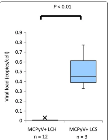
in LCS with high viral loads (Table 1, viral load = 0.328–
0.772). MCPyV-positive LCS samples were compared
with MCPyV-positive LCH samples [8] in the point of
MCPyV viral load in Figure 2 with significant difference
(P < 0.01). The presence of MCPyV in LCS lesions
sug-gests three possibilities: a) viral infection as a
conse-quence of LCS development, b) LCS as a bystander, and
c) a viral causal agent of LCS. Though MCPyV is
typic-ally an asymptomatic infection in adults [18], the
pres-ence of MCPyV with high viral loads denies the
possibility it is a simple bystander within LCS lesions.
Whether MCPyV-DNA is integrated into the nuclei of
LCS cells as an oncogenic virus warrants further
investi-gations, but the high viral loads of MCPyV in LCS lesions
may suggest an important oncogenic factor in LCS cells.
We are now trying specific PCR as Sastre-Garau et al.
Table 1 Clinical characteristics and lesional MCPyV data
of patients with LCS
Patient Age (years) Sex Tissue Q-PCR (Viral load)* MCPyV large T antigen (CM2B4) LCS1 81 F Skin - -LCS2 73 M LN - -LCS3 72 F Salivary gland + (0.453) -LCS4 61 M Lung + (0.772) -LCS5 62 M LN + (0.328) -LCS6 40 M LN - -LCS7 81 F LN --The median age of the LCS patients (n = 7) was 72 years (range, 42–81 years of age). *Positive control (MCC):+ (Viral load = 1.0), Negative control (double distilled water): −. Viral load, relative levels of MCPyV. Abbreviations: LCS Langerhans cell sarcoma, LN lymph node, MCC Merkel cell carcinoma, MCPyV Merkel cell polyomavirus, Q-PCR multiplex quantitative PCR for MCPyV.
Figure 1 No immunoreactivity for MCPyV-LT using CM2B4 antibody in LCS tissue lesion (LCS4). Insert: MCC tissue lesion as a positive control. Scale bar, 50 μm. Abbreviations: CM2B4, a
monoclonal antibody to MCPyV-LT; LCS, Langerhans cell sarcoma; MCC, Merkel cell carcinoma; MCPyV, Merkel cell polyomavirus; MCPyV-LT, large T antigen protein of MCPyV.
Murakami et al. Infectious Agents and Cancer 2014, 9:15 Page 2 of 4 http://www.infectagentscancer.com/content/9/1/15
did [19]. Immunohistochemical negativity for
MCPyV-LT (CM2B4) may indicate a different tumorigenetic
mechanism that is involved in MCPyV-LT positive MCC
[1,20] or under the limit of detection for MCPyV-LT.
Conclusion
To the best of our knowledge, this is the first report
in-dicating a relationship between MCPyV infection and
LCS. Thus, we suggest that MCPyV may play some role
as an oncogenic factor in particular subtypes of LCS.
Based on the foregoing, we propose an LCS
tumorigen-esis model that MCPyV may be a cause of LCS. The
re-cent discovery of MCPyV opened new therapeutic
avenues for MCC [21]. Although MCPyV-LT expression
was not detected, the origin of some forms of LCS may
be MCPyV-infected LC. When confirmed, these findings
will open novel possibilities for therapeutic interventions
against LCS.
Methods
Patients and tissue samples of LCS
This study was approved by the Institutional Review
Board of Faculty of Medicine, Tottori University,
Yonago, Japan.
A total of seven tissues from patients with LCS were
analyzed. All tissues of LCS were obtained as
formalin-fixed paraffin-embedded (FFPE) samples.
Confirmation of accurate diagnosis of LCS
Diagnostic accuracy of all collected samples, histological
sections and immunohistochemistry for CD1a, S100
pro-tein (S100), and CD207 (langerin) of all specimens was
confirmed by two pathologists on the basis of the
diag-nostic criteria [22].
DNA extraction from LCS tissues
DNA was extracted from each FFPE sample using the
QIAamp DNA FFPE Tissue Kit and Mini Kit (QIAGEN
GmbH, Hilden, Germany).
Multiplex quantitative PCR (Q-PCR) for MCPyV detection
Q-PCR was performed in a 10-μl reaction mix
contain-ing TaqMan® Copy number reference assay RNase P
(Applied Biosystems, Foster City, CA, USA) as internal
control [8]. A primer pair targeting the position 859–
934 (MCPyV-LT) on MCC350 (GenBank EU375803) was
[20] used. To determine the MCPyV-DNA ratio relative
to MCPyV-DNA of the reference MCC (MCC = 1.0) for
each case, Q-PCR was performed using an ABI PRISM
7900HT Sequence Detection System (Applied
Biosys-tems) as previously described [20]. The ratio of the virus
was determined using the viral signal in a positive MCC
sample as a reference (viral load = relative levels of
MCPyV, MCC = 1.0 copy/cell). Thresholds were plotted
against each standard sample. All reactions of samples
and controls were performed in triplicate, and the
aver-age was reported. The MCPyV-DNA ratio in each
sam-ple was determined on the basis of corresponding
standard curves.
Immunohistochemistry for detection of MCPyV-LT antigen
For detection of MCPyV-LT expression,
immunohisto-chemistry was performed using monoclonal antibody
CM2B4 (mouse monoclonal IgG2b, 200 μg/ml, sc-136172,
Santa Cruz Biotechnology, CA, USA) generated against a
peptide fragment of MCPyV-LT as immunogen [23,24].
MCC samples were used as controls throughout.
Statistical analysis
Comparisons of MCPyV viral load between
MCPyV-positive LCH and MCPyV-MCPyV-positive LCS were performed
using the Mann–Whitney U test. Differences between
values were considered statistically significant at P < 0.05.
Figure 2 Q-PCR data comparing MCPyV-positive LCH samples and MCPyV-positive LCS samples. Q-PCR provides the viral load of MCPyV (relative level of MCPyV (MCC = 1)) for MCPyV-positive LCH samples and MCPyV-positive LCS samples plotted as box-whisker plots (Mann–Whitney U test, P < 0.01). The median viral load data are 0.003 and 0.453 in MCPyV-positive LCH and MCPyV-positive LCS, respectively. Abbreviations: LCH, Langerhans cell histiocytosis; LCS, Langerhans cell sarcoma; MCC, Merkel cell carcinoma; MCPyV, Merkel cell polyomavirus; Q-PCR, multiplex quantitative PCR.
Abbreviations
DC:Dendritic cell; DLA: Dermatopathic lymphadenopathy; FFPE: Formalin-fixed paraffin-embedded; LC: Langerhans cell; LCH: Langerhans cell histiocytosis; LCS: Langerhans cell sarcoma; LN: lymph node; MCC: Merkel cell carcinoma; MCPyV: Merkel cell polyomavirus; MCPyV-LT: Large T antigen protein of MCPyV; Q-PCR: Multiplex quantitative PCR.
Competing interests
The authors declare no competing financial interests. Authors’ contributions
IM, KH, and HN conceived the initial study proposal. IM, KH, and JG were responsible for writing the manuscript. All authors were involved in the design of the research. MM, TI, and SK were involved in the analysis of the data. All authors have critically reviewed and approved the manuscript. Acknowledgements
This work was partly supported by a Grant-in-aid for Scientific Research (C) 23590426 from the Japanese Ministry of Education, Science, Sports and Culture.
We thank Dr. Toshiharu Maeda (Ehime Prefectural Central Hospital) for providing clinical information and specimens.
Author details
1Division of Molecular Pathology, Faculty of Medicine, Tottori University, Yonago 683-8503, Japan.2Department of Pathobiological Science and Technology, School of Health Science, Faculty of Medicine, Tottori University, Yonago 683-8503, Japan.3Department of Pathology, Tottori University Hospital, Yonago 683-8503, Japan.4Inserm U507 and U1016, Institut Cochin, Paris 75014, France.5University of Paris Descartes (Paris V), Paris 75006, France.6Department of Pathology, Tottori Prefectural Central Hospital, Tottori 680-0901, Japan.7Department of Pathological Diagnostics, Yamagata University School of Medicine, Yamagata 990-9585, Japan.8Department of Laboratory Medicine, The Japan Baptist Medical Foundation, Kyoto 606-8273, Japan.9Department of Pathology, Okayama University Graduate School of Medicine, Dentistry and Pharmaceutical Sciences, Okayama 700-8530, Japan.
Received: 7 January 2014 Accepted: 28 March 2014 Published: 6 May 2014
References
1. Feng H, Shuda M, Chang Y, Moore PS: Clonal integration of a polyomavirus in human Merkel cell carcinoma. Science 2008, 319:1096–1100.
2. Kuwamoto S: Recent advances in the biology of Merkel cell carcinoma. Hum Pathol 2011, 42:1063–1077.
3. Matsushita M, Kuwamoto S, Iwasaki T, Higaki-Mori H, Yashima S, Kato M, Murakami I, Horie Y, Kitamura Y, Hayashi K: Detection of merkel cell polyomavirus in the human tissues from 41 Japanese autopsy cases using polymerase chain reaction. Intervirology 2013, 56:1–5. 4. Kanik A, Li M, Uramacher CD: Normal skin. In Histology for pathologist.
Edited by Mills SE. Philadelphia: Lippincott Williams & Wilkins; 2012:3–28. 5. Kubo A, Nagao K, Yokouchi M, Sasaki H, Amagai M: External antigen
uptake by Langerhans cells with reorganization of epidermal tight junction barriers. J Exp Med 2009, 206:2937–2946.
6. Banchereau J, Steinman RM: Dendritic cells and the control of immunity. Nature 1998, 392:245–252.
7. Weitzman S, Egeler RM: Histiocytic disorders of children and adults: introduction to the problem, overview, historical perspective and epidemiology. In Histiocytic disorders of children and adults. Edited by Weitzman S, Egeler RM. Cambridge: Cambridge University Press; 2005:1–13. 8. Murakami I, Matsushita M, Iwasaki T, Kuwamoto S, Kato M, Horie Y, Hayashi K, Imamura T, Morimoto A, Imashuku S, Gogusev J, Jaubert F, Takata K, Oka T, Yoshino T: Merkel cell polyomavirus DNA sequences in peripheral blood and tissues from patients with Langerhans cell histiocytosis. Hum Pathol 2014, 45:119–126.
9. da Costa CET, Annels NE, Egeler RM: The immunological basis of Langerhans cell histiocytosis. In Histiocytic disorders of children and adults. Edited by Weitzman S, Egeler RM, Cambridge UK. Cambridge, UK: Cambridge University Press; 2005:66–82.
10. Grosjean I, Caux C, Bella C, Berger I, Wild F, Banchereau J, Kaiserlian D: Measles virus infects human dendritic cells and blocks their
allostimulatory properties for CD4+ T cells. J Exp Med 1997, 186:801–812. 11. Engelmayer J, Larsson M, Subklewe M, Chahroudi A, Cox WI, Steinman RM, Bhardwaj N: Vaccinia virus inhibits the maturation of human dendritic cells: a novel mechanism of immune evasion. J Immunol 1999, 163:6762–6768.
12. Tortorella D, Gewurz BE, Furman MH, Schust DJ, Ploegh HL: Viral subversion of the immune system. Annu Rev Immunol 2000, 18:861–926. 13. Izmailova E, Bertley FM, Huang Q, Makori N, Miller CJ, Young RA, Aldovini A:
HIV-1 Tat reprograms immature dendritic cells to express
chemoattractants for activated T cells and macrophages. Nat Med 2003, 9:191–197.
14. Pancaldi C, Corazzari V, Maniero S, Mazzoni E, Comar M, Martini F, Tognon M: Merkel cell polyomavirus DNA sequences in the buffy coats of healthy blood donors. Blood 2011, 117:7099–7101.
15. Mertz KD, Junt T, Schmid M, Pfaltz M, Kempf W: Inflammatory monocytes are a reservoir for Merkel cell polyomavirus. J Invest Dermatol 2010, 130:1146–1151.
16. Shamoto M, Osada A, Shinzato M, Kaneko C, Yoshida A: Do epidermal Langerhans cells, migrating from skin lesions, induce the paracortical hyperplasia of dermatopathic lymphadenopathy? Pathol Int 1996, 46:348–354.
17. O'Malley DP, George TI, Orazi A, Abbondanzo SL: Dermatopathic lymphadenitis. In Atlas of nontumor pathology, First series, Fascicle 7, Benign and reactive conditions of lymph node and spleen. Edited by King DW. Washington, DC: American registry of pathology; 2009:143–145. 18. Tolstov YL, Pastrana DV, Feng H, Becker JC, Jenkins FJ, Moschos S, Chang Y,
Buck CB, Moore PS: Human Merkel cell polyomavirus infection II. MCV is a common human infection that can be detected by conformational capsid epitope immunoassays. Int J Cancer 2009, 125:1250–1256. 19. Sastre-Garau X, Peter M, Avril MF, Laude H, Couturier J, Rozenberg F,
Almeida A, Boitier F, Carlotti A, Couturaud B, Dupin N: Merkel cell carcinoma of the skin: pathological and molecular evidence for a causative role of MCV in oncogenesis. J Pathol 2009, 218:48–56. 20. Kuwamoto S, Higaki H, Kanai K, Iwasaki T, Sano H, Nagata K, Kato K, Kato M,
Murakami I, Horie Y, Yamamoto O, Hayashi K: Association of Merkel cell polyomavirus infection with morphologic differences in Merkel cell carcinoma. Hum Pathol 2011, 42:632–640.
21. Schrama D, Ugurel S, Becker JC: Merkel cell carcinoma: recent insights and new treatment options. Curr Opin Oncol 2012, 24:141–149. 22. Jaffe R, Weiss LM, Facchetti F: Tumours derived from Langerhans cells. In
WHO Classification of Tumours of Haematopoietic and Lymphoid Tissues. Edited by Swerdlow SH, Campo E, Harris NL, Jaffe ES, Pileri SA, Stein H, Thiele J, Vardiman JW. Lyon, France: IARC; 2008:358–360.
23. Busam KJ, Jungbluth AA, Rekthman N, Coit D, Pulitzer M, Bini J, Arora R, Hanson NC, Tassello JA, Frosina D, Moore P, Chang Y: Merkel cell polyomavirus expression in merkel cell carcinomas and its absence in combined tumors and pulmonary neuroendocrine carcinomas. Am J Surg Pathol 2009, 33:1378–1385.
24. Shuda M, Arora R, Kwun HJ, Feng H, Sarid R, Fernandez-Figueras MT, Tolstov Y, Gjoerup O, Mansukhani MM, Swerdlow SH, Chaudhary PM, Kirkwood JM, Nalesnik MA, Kant JA, Weiss LM, Moore PS, Chang Y: Human Merkel cell polyomavirus infection I. MCV T antigen expression in Merkel cell carcinoma, lymphoid tissues and lymphoid tumors. Int J Cancer 2009, 125:1243–1249.
doi:10.1186/1750-9378-9-15
Cite this article as: Murakami et al.: High viral load of Merkel cell polyomavirus DNA sequences in Langerhans cell sarcoma tissues. Infectious Agents and Cancer 2014 9:15.
Murakami et al. Infectious Agents and Cancer 2014, 9:15 Page 4 of 4 http://www.infectagentscancer.com/content/9/1/15