HAL Id: hal-03131495
https://hal.sorbonne-universite.fr/hal-03131495
Submitted on 4 Feb 2021
HAL is a multi-disciplinary open access
archive for the deposit and dissemination of
sci-entific research documents, whether they are
pub-lished or not. The documents may come from
teaching and research institutions in France or
abroad, or from public or private research centers.
L’archive ouverte pluridisciplinaire HAL, est
destinée au dépôt et à la diffusion de documents
scientifiques de niveau recherche, publiés ou non,
émanant des établissements d’enseignement et de
recherche français ou étrangers, des laboratoires
publics ou privés.
regeneration
Francesco Girardi, Anissa Taleb, Majid Ebrahimi, Asiman Datye, Dilani
Gamage, Cécile Peccate, Lorenzo Giordani, Douglas Millay, Penney Gilbert,
Bruno Cadot, et al.
To cite this version:
Francesco Girardi, Anissa Taleb, Majid Ebrahimi, Asiman Datye, Dilani Gamage, et al.. TGFβ
signaling curbs cell fusion and muscle regeneration. Nature Communications, Nature Publishing
Group, 2021, 12 (1), �10.1038/s41467-020-20289-8�. �hal-03131495�
TGF
β signaling curbs cell fusion and muscle
regeneration
Francesco Girardi
1
, Anissa Taleb
1
, Majid Ebrahimi
2,3
, Asiman Datye
2,3
, Dilani G. Gamage
4
, Cécile Peccate
1
,
Lorenzo Giordani
1
, Douglas P. Millay
4,5
, Penney M. Gilbert
2,3,6
, Bruno Cadot
1
& Fabien Le Grand
1,7
✉
Muscle cell fusion is a multistep process involving cell migration, adhesion, membrane
remodeling and actin-nucleation pathways to generate multinucleated myotubes. However,
molecular brakes restraining cell–cell fusion events have remained elusive. Here we show that
transforming growth factor beta (TGF
β) pathway is active in adult muscle cells throughout
fusion. We
find TGFβ signaling reduces cell fusion, regardless of the cells’ ability to move and
establish cell-cell contacts. In contrast, inhibition of TGF
β signaling enhances cell fusion and
promotes branching between myotubes in mouse and human. Exogenous addition of TGF
β
protein in vivo during muscle regeneration results in a loss of muscle function while inhibition
of TGF
βR2 induces the formation of giant myofibers. Transcriptome analyses and functional
assays reveal that TGF
β controls the expression of actin-related genes to reduce cell
spreading. TGF
β signaling is therefore requisite to limit mammalian myoblast fusion,
deter-mining myonuclei numbers and myo
fiber size.
https://doi.org/10.1038/s41467-020-20289-8
OPEN
1Sorbonne Université, INSERM UMRS974, Association Institut de Myologie, Centre de Recherche en Myologie, 75013 Paris, France.2Institute of Biomedical Engineering, University of Toronto, Toronto, ON M5S3G9, Canada.3Donnelly Centre for Cellular and Biomolecular Research, Toronto, ON M5S3E1, Canada. 4Division of Molecular Cardiovascular Biology, Cincinnati Children’s Hospital Medical Center, Cincinnati, OH 45229, USA.5Department of Pediatrics, University of Cincinnati College of Medicine, Cincinnati, OH 45229, USA.6Department of Cell and Systems Biology, University of Toronto, Toronto, ON M5S3G5, Canada.7Present address: Institut NeuroMyoGène, Université Claude Bernard Lyon 1, CNRS UMR 5310, INSERM U1217, 69008 Lyon, France. ✉email:fabien.le-grand@inserm.fr
123456789
T
he adult skeletal muscle cell is a syncytial myofiber that
contains hundreds of myonuclei. Formation and
regenera-tion of the myofiber requires fusion of mononuclear
pro-genitors (myoblasts) to form multinucleated myotubes. Located in a
niche around the myofibers are quiescent muscle stem cells
1, called
satellite cells, which can activate and proliferate to give rise to adult
myoblasts. Myoblasts differentiate into myocytes competent to fuse
with each other and with myofibers
2. As such, cell fusion plays
essential roles in the adult, allowing physiological muscle
hypertrophy
3,4and muscle regeneration following injury
5,6.
When induced to fuse, adult myoblasts exit the cell cycle, commit
to terminal differentiation, and migrate toward each other
7. They
then adhere through membrane integrins
8and cadherins
9. The later
stages of fusion are controlled by the muscle-specific protein
Myo-maker
10and peptide Myomerger
11–13(also known as MINION,
Myomixer). Myomaker and Myomerger function in hemifusion and
pore formation activity, respectively. Recent studies demonstrated
that muscle cell fusion is promoted by actin-based structures
14generating protrusive forces
15and membrane stress before
coales-cence
16. The fusion process must be tightly controlled to ensure that
fusogenic myoblasts do not form aberrant hypertrophic syncytia or
fuse with non-muscle cells. However, while it is known that muscle
cell fusion can be prevented by tetraspanins at the cell membrane
17,
few signaling pathways that can limit this process and prevent
unscheduled cell fusion has been identified.
Canonical Wnt/β-catenin signaling is a crucial regulator of
satellite cells and adult muscle regeneration
18,19. Interestingly,
β-catenin activation in adult myoblasts promotes their progression
through the myogenic lineage
20, but limits differentiated muscle cell
fusion
6. This contrasting regulation of different steps of adult
myogenesis can be explained by the fact that
β-catenin signaling
induces the expression of transforming growth factor beta (TGFβ)
ligands and receptors by adult muscle cells
20,21. As such, a large
body of evidence, built on seminal work from three decades ago,
clearly demonstrated that TGFβ signaling inhibits muscle cell
terminal differentiation
22,23. Mechanistically, TGFβ signaling has
been shown, mainly in vitro, to negatively regulate differentiation
through functional repression of the myogenic regulatory factors
Myod1
24and Myogenin
25. However, TGFβ signaling has a broader
function in muscle cells, including quiescence
26and activation
27.
The impact of TGFβ signaling specifically in muscle cell fusion
has not been investigated. This gap in our knowledge mainly
comes from the fact that the primary effects of TGFβ
over-expression in skeletal muscles are the development of endomysial
fibrosis, due to its role as a potent growth factor for connective
tissue cells
28,29.
We thus asked whether TGFβ signaling regulates the fusion of
differentiated muscle cells. Here, we present a detailed analysis of its
role during muscle cell differentiation and tissue regeneration. We
observe that the activation of TGFβ signaling leads to a strong
reduction of myotube formation, independently of previous
differ-entiating steps such as myoblast motility and Myogenin expression.
Importantly, neither myoblast proliferation nor mortality is altered by
the signaling modulation. Inhibition of TGFβ signaling results in the
formation of large syncytia with an elevated number of nuclei and
aberrant shapes. These results are confirmed in vivo, where
phar-macological stimulation of the signaling pathway dramatically
reduces
fiber size and myonuclear number, while its inhibition
generates giant myofibers with multiple myonuclei. Collectively, our
results demonstrate that TGFβ signaling controls syncytia formation
and muscle regeneration by limiting muscle cell fusion.
Results
TGF
β signaling is active in adult muscle progenitor cells. TGFβ
ligands bind to TGFBR2 that will recruit a type I receptor dimer.
The receptor complex will then phosphorylate SMAD2 and
SMAD3 that will accumulate in the nucleus where they act as
transcription factors
30. To evaluate TGFβ isoforms expression by
adult muscle progenitor cells, we purified limb muscle satellite
cells and grew them in vitro as primary myoblasts. We observed
that Tgfb1 and Tgfb2 expression levels were high in proliferating
cells, and diminished following induction of differentiation, while
Tgfb3 expression pattern showed an opposite trend (Fig.
1
a),
suggesting that TGFβ signaling may be still active at later
dif-ferentiation time points. Of note, the expression levels of the
TGFβ receptors (Alk5, also known as Tgfbr1; and Tgfbr2) did not
significantly change during the course of in vitro myogenesis
(Fig.
1
b). We next investigated the state of TGFβ signaling in
primary myoblasts, differentiated myocytes, and multinucleated
myotubes. We observed that the expression level of the TGFβ/
SMAD2/3 target gene Smad7 diminished during myogenic
pro-gression (Fig.
1
c). While immunolocalization of
phosphorylated-SMAD2/3 (p-phosphorylated-SMAD2/3) proteins showed that the canonical
TGFβ pathway is active at all studied stages (Fig.
1
d), quantitative
western blotting experiments demonstrated that the intensity of
TGFβ signaling decreases during muscle cell differentiation, but is
not abrogated in multinucleated cells (Fig.
1
e). Previous work has
established that TGFβ ligands are secreted during muscle tissue
repair
31. Gene expression analysis of regenerating tibialis anterior
(TA) muscles demonstrated that the three TGFβ isoforms are
dynamically expressed following injury (Supplementary Fig. 1a).
Evaluation of protein levels further validated that TGFβ1 and
TGFβ3 expressions peak during the acute phase of tissue repair
~4 days post injury (d.p.i.), while TGFβ2 protein levels are higher
at later time points (7 d.p.i.) (Supplementary Fig. 1b).
The dynamic expression profiles of TGFβ isoforms during
adult myogenesis both in vitro and in vivo prompted us to
evaluate the activity of TGFβ signaling in vivo at the cellular level.
We performed co-immunolocalization for p-SMAD3 proteins
and markers of muscle cells on cryosections of uninjured and
regenerating TA muscles. In the resting muscle, p-SMAD3
proteins are detected in quiescent satellite cells and myonuclei
(Fig.
2
a). In acutely regenerating tissues, we found it to be active
in proliferating myogenic stem and progenitor cells expressing
PAX7 and in centrally located myonuclei of newly formed
myofibers (Fig.
2
b). In contrast, TGFβ signaling is inactive
(p-SMAD3 staining at background levels) in MYOGENIN+
differentiating myocytes (Fig.
2
d). At 7 d.p.i., when muscle
architecture is restored, p-SMAD3 expression is detected in the
nuclei of satellite cells and myonuclei, as well as in interstitial cells
(Fig.
2
c). While the presence of an active TGFβ signaling in
PAX7-expressing cells was expected, the p-SMAD3
immunor-eactivity of recently fused myonuclei was both surprising and
fitting with the in vitro data. Together, these observations suggest
that TGFβ pathway is involved in regulating the assembly of
multinucleated muscle cells. We thus aimed to investigate the role
of TGFβ in the fusion process.
Fusion defects in adult muscle cells stimulated by TGF
β. Since
all three TGFβ ligands are expressed during adult myogenesis, we
evaluated the impact of recombinant protein treatments on
pri-mary myoblast differentiation and fusion (Supplementary
Fig. 2a). After 72 h of differentiation, muscle cells aggregated to
form multinucleated myotubes, while the addition of
recombi-nant TGFβ proteins forced muscle cells to remain mostly
mononucleated (Supplementary Fig. 2b, e). Quantification of
Myh3 gene expression, which codes for the embryonic myosin
heavy-chain isoform, further indicated that the cells in
TGFβ-treated cultures were in a less mature state than control cultures
(Supplementary Fig. 2c). However, quantification of the
percentage of differentiated nuclei expressing Pan-MYOSIN
HEAVY-CHAIN (Pan-MyHC) proteins revealed that the vast
majority (>90%) of myoblasts did undergo differentiation in all
conditions (Supplementary Fig. 2d), suggesting that TGFβ
sig-naling does not primarily block muscle cell differentiation.
Likewise, quantification of Pan-MyHC staining intensity of single
multinucleated muscle cells demonstrated that TGFβ-treated
myotubes, while containing less nuclei, expressed similar levels of
the differentiation marker (Supplementary Fig. 3a).
To test this hypothesis, we adapted the protocol used by Saclier
et al.
32to uncouple differentiation and fusion of primary muscle
cells. In this experimental setup, primary myoblasts were
differentiated for 2 days at a low density that does not allow
contact between cells. The cells were then split and re-plated at a
high density and cultured for an additional 2 days to evaluate
muscle cell fusion (Fig.
3
a). Following re-plating, almost all
muscle cells were terminally differentiated and expressed
MYOGENIN (>94%) (Fig.
3
b). To assert that TGFβ signaling
does not impact the differentiation state of re-plated cells, we
stimulated them with recombinant TGFβ1 protein and validated
that the treatment did not result in changes in Myogenin gene
expression, but did activate expression of the TGFβ target gene
Smad7 (Fig.
3
c). We thus evaluated the effect of TGFβ protein
stimulations on mononucleated differentiated muscle cells
(myocytes) and observed that all three TGFβ isoforms strongly
inhibited cell fusion (Fig.
3
d), despite the muscle cells progressing
down the differentiation pathway (Fig.
3
e; ~100% MyHC+).
Activation of the TGFβ pathway reduced the fusion index
(Fig.
3
f) and completely blocked the formation of large myotubes
(Fig.
3
g), thus demonstrating that TGFβ signaling limits muscle
cell fusion independently of myogenic differentiation. Moreover,
quantification of Pan-MyHC staining intensity of single myotubes
did not reveal any differences in the maturation states between
control and TGFβ-stimulated cells (Supplementary Fig. 3b).
Importantly, the addition of TGFβ proteins to adult muscle cells
did not alter myoblast proliferation (Supplementary Fig. S4a), did
not induce programmed cell death in myoblasts and myocytes
(Supplementary Fig. 4b), and did not modify myoblasts and
myocytes ability to migrate in scratch-wound assays
(Supple-mentary Fig. 4c).
TGF
β signaling does not impact cell–cell contacts. To fuse,
myocytes must undergo cell–cell recognition and adhesion
33,34.
Fig. 1 TGFβ signaling pathway remains active during myoblast differentiation. a qRT-PCR analysis of Tgfb1, 2, and 3 transcripts expression during in vitro differentiation of primary muscle cells shows different profiles. N = 3 biologically independent experiments for each time point. b qRT-PCR analysis of Alk5 and Tgfbr2 transcript expressions describes a constant expression of the receptors during primary muscle cell differentiation. N= 6 biologically independent experiments for each time point.c qRT-PCR analysis of the TGFβ target gene Smad7 transcript expression reveals a decreased activity of the pathway alongside in vitro primary muscle cell differentiation. N= 3 biologically independent experiments for each time point. d p-SMAD2/3
immunofluorescent staining of proliferating, differentiating, and differentiated primary myoblasts reveals a constant and basal activation of the pathway. N= 3 primary cell cultures. e p-SMAD2/3 and SMAD2/3 western-blot analysis of proliferating, differentiating, and differentiated primary myoblasts confirms a decrease in SMAD2/3 phosphorylation during differentiation. N = 3 biologically independent experiments. Scale bars: d 200 μm. Data are presented as mean ± SEM. Source data are provided as a Source Datafile.
Cell motility is therefore required to bring pre-fusing cells into
proximity. We aimed to evaluate if TGFβ signaling impacts
muscle cell motility and contact using live imaging. To this aim,
we did not use the replating technique since high cell density
would have impeded a clear tracking of cells’ motions and
con-tacts. We stimulated primary muscle cells with a single pulse of
recombinant TGFβ1 protein (Fig.
4
a) and validated that this
treatment does not significantly change the proportion of
MYOD1+ nuclei at 48 h in the differentiation medium (Fig.
4
b)
and the expression of Myogenin at 60 h in the differentiation
medium (Fig.
4
c). These observations suggest that the inhibitory
action of TGFβ signaling on myoblast differentiation is partially
compensated in culture, at least for the analyzed markers. We
then recorded primary muscle cells at two different time frames
(early differentiation: before cell fusion; late differentiation:
dur-ing syncytium formation) to determine the effect of exogenous
TGFβ1 protein (Fig.
4
d) (Supplementary Movies 1–4). During
early differentiation, TGFβ1 stimulation did not impact cell
motility and did not change the frequency of cell–cell contacts
(Fig.
4
e and Supplementary Movies 1 and 2). Interestingly, during
the late differentiation, TGFβ1-treated cells could still contact
each other with a frequency similar to control cells, but these
contacts were infructuous (Supplementary Movies 3 and 4). As
such, we observed that TGFβ signaling prevented contacting cells
from fusing and strongly increased the proportion of paired cells
that remained mononucleated (Fig.
4
e). Together, our
observa-tions indicate that TGFβ signaling does not impact “pre-fusion”
cell dynamics, but prevents the merging of cells into syncytia.
Inhibition of TGF
β receptor function promotes muscle cell
fusion. We next asked if inhibition of TGFβ signaling in fusing
myocytes could enhance the formation of multinucleated
myo-tubes. To this aim, we selected ITD-1, a highly selective TGFβ
inhibitor that triggers proteasomal degradation of TGFBR2
35.
ITD-1 clears TGFBR2 from the cell surface and selectively
inhibits intracellular signaling. ITD-1 treatment of primary
myocytes resulted in reduced expression of TGFβ target genes
(Fig.
5
a) and blocked the phosphorylation of nuclear SMAD2/3
proteins induced by TGFβ1 treatment (Fig.
5
b). Importantly,
treatment of differentiated mononucleated muscle cells re-plated
at high density (as in Fig.
3
a) with ITD-1 enhanced the fusion
process (Fig.
5
c). As such, ITD-1-treated cultures showed a higher
fusion index (Fig.
5
e) and were composed of myotubes of larger
diameter (Fig.
5
f), containing more nuclei (Fig.
5
d) and
char-acterized by an aberrant branched shape (Fig.
5
c, g). Since ITD-1
does not fully inhibit p-SMAD2/3 signaling in our assays, we
further investigated SB-431542, another TGFβ inhibitor that
targets ALK5/TGFBR1
36. Treatment of re-plated myocytes with
SB-431542 strongly increased the percentage of large myotubes
containing high numbers of nuclei, suggesting that both type 1
and type 2 receptors are mediating the effects of TGFβ on muscle
cell fusion (Supplementary Fig. 5). Taken together, our data
demonstrate that TGFβ receptors regulate fusion and suggest that
the levels of TGFβ signaling must be tightly controlled to ensure
proper syncytia formation.
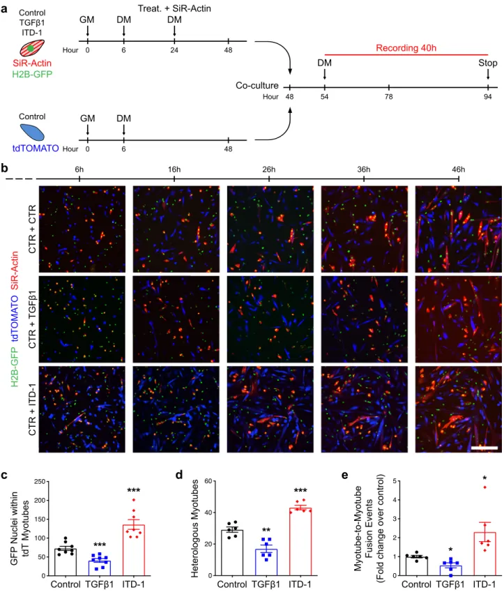
To test if TGFβ signaling controls fusion cell autonomously, we
expanded primary myoblasts from satellite cells expressing
either H2B-GFP or membrane tdTomato (pseudocolored in
blue). Both primary cell types were pre-differentiated at low
density, but only the GFP-expressing myocytes were treated
with either TGFβ1 or ITD-1 and their F-actin content stained
with SiR-Actin (pseudocoloured in red) (Fig.
6
a). Cells were
then mixed, re-plated at high density, and fusion events were
imaged live (Fig.
6
b). We observed that fusion of GFP-labeled
myonuclei into tdTomato myotubes was controlled by the
intrinsic state of TGFβ signaling in the fusing cells (Fig.
6
c).
As such the incidence of heterologous fusion was controlled by
TGFβ signaling (Fig.
6
d). Interestingly, we noticed that
multi-nucleated myotubes could fuse together and that TGFβ signaling
regulates the frequency of myotube-to-myotube fusion (Fig.
6
e)
Hoechst Pax7 Dystrophin p-SMAD3 Merge 4 d.p.i. TA muscle
b
c
7 d.p.i. TA muscle
d
4 d.p.i. TA musclea
Uninjured TA muscle H o ec h st Myogenin Dystrophin p-SMAD3 MergeFig. 2 The state of TGFβ signaling during in vivo muscle regeneration. a–c p-SMAD3, PAX7, and DYSTROPHIN immunofluorescent stainings on 0-, 4-, and 7-day post injury (d.p.i.) regenerating TA muscle cryosections. SMAD3 signaling is active in interstitial cells and PAX7+ cells (white arrows) during muscle regeneration. p-SMAD3 is strongly expressed by myonuclei of the regenerating myofibers marked by DYSTROPHIN at 4 d.p.i. N = 8 cryosections. d p-SMAD3, MYOGENIN, and DYSTROPHIN immunofluorescent staining on 4 d.p.i. regenerating TA muscle cryosections. Myocytes do not express p-SMAD3 at the time of differentiation. N= 8 cryosections. Scale bars: low magnification, 400 μm; high magnification, 100 μm.
(Supplementary Movies 5–7). These results suggest that TGFβ
acts cell autonomously to limit the fusion between muscle cells,
and to prevent fusion between syncytia.
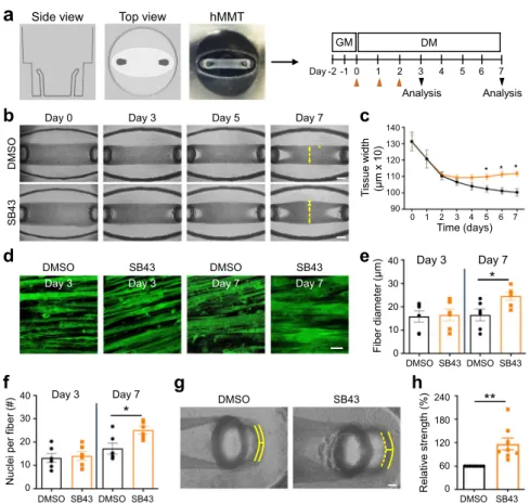
TGF
β signaling regulates the formation of human muscle
microtissues (hMMTs). To determine the human relevance of the
hyper-fusion phenotype we observed with mouse cells, we
eval-uated the influence of TGFβ pathway inhibition on human muscle
cell differentiation in a 3D culture format
37(Fig.
7
a). In this case,
immortalized human muscle progenitor cells were embedded
within a
fibrin scaffold containing reconstituted basement
mem-brane, which was spread within a custom polydimethylsulfoxane
rubber device in which at either end of a cell seeding depression is
a vertical post (also cast using
flexible rubber). Within hours of
plating the cell-scaffold slurry within the depression, the cells
self-organize across the axis of uni-axial tension supported by the
vertical posts to generate a compact 3D tissue. By quantifying the
tissue remodeling and compaction process (Fig.
7
b, c), we found
that inhibition of TGFBR1 function by the small-molecule
SB-431542 did not change the thickness of hMMTs at early
differ-entiation time points (days 0–4), but that as the hMMTs further
matured, there was a bifurcation and TGFBR1 inhibitor-treated
hMMTs were significantly thicker than control hMMTs. In this
system, hMMT thickening was due to an increase in the width of
individual
fibers (Fig.
7
d, e). Notably, SB-431542-treated human
muscle
fibers contained more nuclei than control fibers,
con-firming increased muscle cell fusion as the underlying cellular
mechanism (Fig.
7
f). In parallel, we ensured that TGFβ signaling
does not interact with the anabolic Akt/mTOR anabolic pathway
that drives myotube hypertrophy
38in differentiated muscle cells
(Fig. S6). Lastly, by treating hMMTs with acetylcholine (ACh) to
induce tissue contraction and capturing short videos to visualize
the magnitude of vertical rubber post deflections, we found that
TGFBR1 inhibition renders the hMMTs stronger than their
con-trol counterpart (Fig.
7
g, h). Together, these data demonstrate that
TGFβ signaling regulates human cell fusion.
Pharmacological modulation of TGF
β pathway in vivo
per-turbs muscle regeneration. To evaluate the impact of TGFβ
signaling on muscle cell fusion in vivo, we injured TA muscle of
adult mice and injected either TGFβ1 protein or ITD-1
com-pound at 3 d.p.i., the time point when fusion begins (Fig.
8
a).
Evaluation of regenerating tissues at 7 d.p.i. revealed that both
treatments lead to striking changes in myofiber size and
mor-phology (Fig.
8
b). TGFβ1 addition resulted in a robust decrease in
nuclear number in newly formed myofibers (Fig.
8
e), resulting in
a dramatic drop in
fiber cross-sectional area (Fig.
8
c, d). In
contrast, ITD-1 induced a large increase in myonuclear accretion
(Fig.
8
e) resulting in the formation of larger
fibers (Fig.
8
c, d). To
further elucidate if modulating TGFβ signaling in vivo affects
regenerated muscle tissue structure and function, we performed
TA muscle injury, followed by three successive injections of
TGFβ1 or ITD-1, and evaluated the regenerated muscles 2 weeks
after injury (Fig.
8
f). In this setup, the effects of modifying TGFβ
signaling were more pronounced (Fig.
8
g). Activation of TGFβ
signaling induced the formation of very small myofibers, while
ITD-1 treatment generated giant myofibers (Fig.
8
h, i). We next
performed in situ force measurement of regenerated TA muscles.
As suggested by the severe myofiber atrophy observed in
TGFβ1-injected muscles, ectopic activation of TGFβ signaling during
tissue regeneration lead to a strong reduction of muscle-specific
force (Fig.
8
j). Despite being composed of larger myofibers,
ITD-1-treated muscles did not show any improvement in force
gen-eration. These observations indicate that TGFβ signaling
deter-mines the numbers of fusion events occurring during tissue
regeneration in vivo.
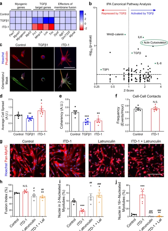
Expression of actin-related genes is controlled by TGFβ
naling. To identify the genetic networks regulated by TGFβ
sig-naling in muscle cells, we performed transcriptome analysis of
primary myocytes differentiated for 24 h and stimulated with
either TGFβ1 or ITD-1 for another 24 h (Supplementary Data 1).
Analysis of the transcriptomic dataset revealed that the relative
expression levels of known TGFβ target genes (such as
Fibro-nectin, Lysyl oxidase, etc.) were strongly modulated by either
treatment, while the expression levels of myogenic transcription
factors (such as Pax7, Myod1, etc.) were not significantly changed,
confirming that the modulation of TGFβ signaling does not act
a
b
72 96 54 48 GM DM Treat. Stop Hour 0 6 48 GM DM Control TGFβ1 Hoec hs t Pan-MyHCe
f
g
d
*
#c
Hoechst Myogenin Cells in*
Myogenin + (%) # Cells inRelative expression Relative expression
TGFβ2 TGFβ3
Treat.
Fig. 3 TGFβ signaling limits cell fusion. a Experimental scheme. Primary myoblasts seeded at low density (5000 cells/cm2) were differentiated for 2 days, split, and re-plated at high density (75,000 cells/cm2) and cultured for 2 more days.b Immunofluorescent staining for MYOGENIN of primary myocytes pre-differentiated for 48 h and re-plated at high density confirms that >90% of cells express Myogenin. N= 7 biologically independent experiments.c qRT-PCR analysis for Myogenin and Smad7 transcript expression of re-plated primary myocytes cultured for 24 h with or without TGFβ1 recombinant protein. Although TGFβ1 stimulation activates Smad7 expression, it does not affect Myogenin transcript levels. N= 6 primary cell cultures.d Immunofluorescent staining for the MYOSIN HEAVY-CHAIN isoforms (Pan-MyHC) of re-plated primary myocytes cultured for 48 h. e Percentage of Pan-MyHC-expressing cells of re-plated myotubes shows that cells were differentiated in all conditions. N= 5 biologically
independent experiments.f Fusion index of re-plated myotubes reveals that TGFβ stimulation inhibits fusion. N = 5 biologically independent
experiments.g Percentage of nuclei in the smallest and largest myotube classes. TGFβ-treated myotubes are characterized by less nuclei per myotube. N= 11 (control) and 15 (TGFβ1) biologically independent experiments. Scale bars:b 400μm; d 200 μm. Data are presented as mean ± SEM. Unpaired two-tailed Student’s t tests were used to compare between data. ** and *** denote a significant difference with the Control group of P < 0.01 and P < 0.001, respectively. NS not significant. Source data are provided as a Source Datafile.
a
d
e
Hours 0 DM Treat. 12 24 36 GM 48 60 Stop Start Rec. -12 Early Differentiation Late Differentiationf
Early Differentiation Late Differentiation
12h 24h 36h 48h 60h Differentiation Control TGFβ1
b
MYOD1 + nuclei at 24 hours (%) Control TGFβ1c
Myogenin expression at 60 hours Control TGFβ1 Frequency (Events/Hour) Control TGFβ1 Cell-Cell Contacts Control TGFβ1 Distance (μm) Covered Distance Control TGFβ1 Speed (μm/min) Cell SpeedCells that make contacts but do not fuse Contacts that
results in fusion Cell-Cell Contacts
Frequency
(Events/Hour) Percentage (%) Percentage (%)
Control TGFβ1 Control TGFβ1 Control TGFβ1
Fig. 4 TGFβ signaling does not affect cell motility and cell–cell contact frequency. a Experimental scheme. Primary myoblasts seeded at 10,000 cells/ cm2were induced to differentiate with or without TGFβ1 recombinant protein. After 12 h, cells were recorded live for 12 h during early differentiation and for 24 h during late differentiation.b Quantification of the percentage of MYOD1+ nuclei of primary myocytes cultured for 48 h with or without TGFβ1 recombinant protein. N= 3 biologically independent experiments. c qRT-PCR analysis for Myogenin transcript expression of primary myotubes cultured for 60 h with or without TGFβ1 recombinant protein. N = 5 biologically independent experiments. d Brightfield live-imaging frames of differentiating myoblasts confirm that TGFβ stimulation reduces fusion. N = 6 primary cell cultures. e Early differentiation movies were used to quantify cell speed, covered distance and cell–cell contact frequency. None of these parameters were significantly different between control and TGFβ1-treated myoblasts. N = 240 cells for each condition examined over six biologically independent experiments.f Late differentiation movies were used to quantify cell–cell contact frequency, the percentage of cell–cell contacts that result in fusion events and the percentage of cells that make contacts but do not fuse. Although TGFβ1 stimulation does not impair cell–cell contact frequency, many TGFβ1-treated contacting myoblasts make contacts but do not accomplish fusion. N = 240 cells for each condition examined over six biologically independent experiments. Scale bars:d 400μm. Data are presented as mean ± SEM. Unpaired two-tailed Student’s t tests were used to compare between data. *** denote a significant difference with the Control group of P < 0.0001. NS not significant. Source data are provided as a Source Datafile.
on myogenesis in these experimental conditions. Likewise,
recently identified fusion master regulatory factors (Myomaker
and Myomerger) were also unaffected by TGFβ signaling (Fig.
9
a).
We then used Ingenuity Pathway Analysis (IPA) to reveal the
pathways affected by TGFβ signaling. Interestingly, we found that
“Actin Cytoskeleton” was among the top-regulated pathways
(Fig.
9
b). This is significant, since actin remodeling and the
for-mation of
finger-like actin protrusions are essential for myoblast
fusion
39. IPA further revealed changes in the transcription of
numerous genes implicated in actin dynamics following TGFβ
treatment (Supplementary Fig. 7a). Visualization of the F-ACTIN
network and measurements of the local orientation of actin
fila-ments in differentiated muscle cells showed that the level of TGFβ
signaling negatively correlates with cytoskeleton reorganization
(Fig.
9
c). TGFβ signaling inhibited cell spreading (Fig.
9
d) and the
coherency of actin
filament alignment (Fig.
9
e). Importantly, the
increased cell size in ITD-1-stimulated cells did not result in any
changes in the frequency of cell–cell contacts (Fig.
9
f).
To better understand actin remodeling during muscle cell
fusion, we performed live-imaging experiments to visualize the
accumulation of F-ACTIN foci in invasive podosome-like
structures at the sites of fusion between cells
40. By mixing cells
stained with SiR-Actin and unstained cells, we observed dynamic
actin remodeling in untreated cells (Supplementary Fig. 7b, top)
and cells stimulated with ITD-1 (Supplementary Fig. 7b, Bottom).
TGFβ1 stimulation prevented the formation of actin-rich invasive
structures and promoted the maintenance of a rounded cell shape
(Supplementary Fig. 7b, middle). We then asked if the fusogenic
effect of ITD-1 can be suppressed by treating cells with
Latrunculin, which prevents actin polymerization (Fig.
9
g). We
observed that Latrunculin treatment resulted in a decrease in
fusion index (Fig.
9
h), increased the proportion of small
myotubes in both control and ITD-1-treated cultures (Fig.
9
i),
and completely prevented the formation of large myotubes
containing high numbers of nuclei in ITD-1-treated cultures
(Fig.
9
j). These data demonstrate that inhibition of TGFβ
signaling does not promote fusion in cells that are capable of
fusion, but have perturbed actin dynamics.
To gain more mechanistic insights into the regulation of fusion
by TGFβ, we followed a candidate–gene approach among the
c
Control H oechst Pan-MyHC ITD-1d
f
e
g
a
b
Smad7 Klf10Relative expression Relative expression
p-SMAD2 SMAD2 Histone 3 Histone 3 p-SMAD3 SMAD3 60 17 52 kDa 60 17 52 CTR TGFβ 1 ITD-1TGFβ 1+ITD-1 Control Nuclei (%) ITD-1
Fusion Index (%) Myotube Diameter
(A.U.)
Myotube Branching (%)
Control ITD-1
Fig. 5 Inhibition of TGFBR2 function in differentiated muscle cell enhances fusion. a qRT-PCR analysis of TGFβ target genes transcript expression in primary myocytes treated with TGFβ1 protein or ITD-1 compound proves that Smad7 and Klf10 are over-expressed when the signaling pathway is activated and inhibited when TGFβ cascade is blocked. N = 10 (Control), 8 (TGFβ1), and 8 (ITD-1) biologically independent experiments. b Nuclear p-SMAD2/3 and SMAD2/3 western blot analysis of primary myoblast treated with TGFβ1 protein, ITD-1 compound, or both combined. The intracellular mediators SMAD2/ 3 are phosphorylated upon TGFβ stimulation, while ITD-1 is able to reduce their phosphorylation. N = 3 biologically independent experiments. c Immunofluorescent staining for Pan-MyHC of re-plated myocytes cultured for 48 h. N = 8 primary cell cultures. d Aggregation index of re-plated myocytes shows that ITD-1 treatment leads to the formation of myotubes with higher numbers of nuclei compared to the control. N= 8 biologically independent experiments.e Fusion index of re-plated myocytes confirms the enhanced fusion when TGFβ cascade is inhibited. N = 8 biologically independent experiments. Diameter of re-plated myotubes (f) and of the distribution of branched-myotubes (g) of re-plated cells highlight aberrant morphology of syncytia treated with ITD-1 N= 4 (f) and 6 (g) biologically independent experiments. Scale bars: c 200 μm. Data are presented as mean ± SEM. Unpaired two-tailed Student’s t tests were used to compare between data. * and *** denote a significant difference with Control group of P < 0.05 and P < 0.001, respectively. Source data are provided as a Source Datafile.
6h 16h 26h 36h 46h Hour 0 6 48 48 54 78 94 Hour 0 6 48 Hour
tdTOMATO
GM
DM
DM
Stop
Recording 40h
a
b
c
d
e
24GM
DM
Treat. + SiR-Actin
DM
Control TGFβ1 ITD-1SiR-Actin
H2B-GFP
ControlCo-culture
H2B-GFP
tdT
OMA
T
O
SiR-Actin
CTR +
TGFβ1
C
TR + CTR
CTR + ITD-1
TGFβ1 ITD-1
Control
GFP Nuclei within
tdT Myotubes
TGFβ1 ITD-1
Control
Heterologous Myotubes
TGFβ1 ITD-1
Control
Myotube-to-Myotube
Fusion Events
(Fold change over control)
Fig. 6 Live imaging of myoblast fusion. a Experimental scheme. H2B-GFP primary myoblasts were seeded at low density (5000 cells/cm2), treated with TGFβ1 protein or ITD-1 compound, stained with SiR-Actin, and differentiated for 2 days. Membrane-tdTOMATO primary myoblasts seeded at low density (5000 cells/cm2) and were differentiated for 2 days. Both populations were split and co-cultured (50/50) at high density (75,000 cells/cm2) for 2 more days. In the last 40 h, cells were recorded live by confocal microscopy.b Live-imaging frames of co-cultured pre-differentiated myocytes confirm the phenotype previously observed. TGFβ activation inhibits fusion, while ITD-1 enhance fusion. N = 8 biologically independent primary co-cultures. c Quantification of H2B-GFP nuclei within tdTOMATO myotubes. N = 8 biologically independent experiments. d Quantification of heterologous myotubes (double positive for SiR-Actin and tdTOMATO). N= 6 biologically independent experiments. e Quantification of Myotube-to-Myotube events. ITD-1 treatment allows more myotube-to-myotube events compared to the control. N= 6 biologically independent experiments. Scale bars: b 200 μm. Data are presented as mean ± SEM. Unpaired two-tailed Student’s t tests were used to compare between data. *, **, and *** denote a significant difference with the Control group of P < 0.05, P < 0.01, and P < 0.001 respectively. Source data are provided as a Source Datafile.
transcripts with expression most impacted by both TGFβ1 and
ITD-1. We selected Timp1 (induced by TGFβ, Supplementary
Fig. 8b), an inhibitor of matrix metalloproteinases, which are
known to promote actin-rich structures in invadopodias
41, and
Arhgef6 (inhibited by TGFβ, Supplementary Fig. 8e), which acts
as a RAC1 guanine nucleotide exchange factor (GEF)
42, a crucial
regulator of actin cytoskeleton. We observed that either
exogenous TIMP1 protein addition (Supplementary Fig. S8c) or
small interfering RNA (siRNA)-mediated silencing of Arhgef6
expression (Supplementary Fig. 8f) slightly reduced the fusion of
muscle cells, without impacting their ability to differentiate.
Taken together, our data indicate that TGFβ signaling limits cell
fusion by preventing actin cytoskeleton remodeling.
Lastly, we asked whether the TGFβ-driven effect on fusion is
conserved in a non-muscle context. To do so, we took advantage
of a
fibroblast dox-inducible cell fusion reconstitution system
43.
Myomaker- and Myomerger-transduced 10T1/2
fibroblasts were
seeded and stimulated with dox to induce fusion (Supplementary
Fig. 9a). Recombinant TGFβ1 recombinant protein was
adminis-tered at multiple time points (from days 0 to 3), but none of these
treatments led to a reduction of fusion (Supplementary Fig. 9b, c).
These results show that TGFβ1 protein is unable to inhibit the
fusion process in a non-muscle cell type and that its signaling
cascade is unable to physically prevent fusion through Myomaker
and Myomerger, confirming the evidence obtained from the
transcriptome analysis (Fig.
9
a). Finally, our data suggest that
TGFβ acts on the fusion process before the final step mediated by
Myomaker/Myomerger.
Discussion
The data presented here identify an unexpected negative role for
TGFβ in the fusion of adult myocytes to form myotubes. TGFβ
signaling has previously been shown to play a major role in
c
b
DMSO
SB43
Day 0 Day 3 Day 5 Day 7
g
DMSO SB43h
60 240 120 0 Re la ti v e str e ng th (%) DMSO**
SB43 180 F ib er di amete r (μm) DMSO 0 10 20 30 40 SB43 Day 3 Day 7*
DMSO SB43f
SB43 Nuclei p er fi b e r ( # ) DMSO 0 10 20 30 40 SB43 Day 3 Day 7*
DMSOd
DMSO SB43 Day 7 Day 3 SB43 DMSO Day 3 Day 7 Analysis Day -2 -1 0 1 2 3 4 GM DM 5 6 7 Analysise
Ti s s u e w id th (μm x 10) Time (days) 0 1 2 3 4 5 6 7 * * 90 100 110 130 140 120 *a
Side view Top view hMMTFig. 7 TGFβ inhibition induces human myotube fusion in 3D culture resulting in increased microtissue strength. a Schematic representation (left) and timeline (right) of 3D human muscle cell experimental approach utilized in panels (b–h). Briefly, immortalized human myoblasts are suspended in a fibrin/ reconstituted basement membrane protein scaffold and seeded into the bottom of a custom rubber 96-well plate culture device. A side view depicts the vertical posts across which the cells remodel the protein scaffold, align, and fuse to form a 3D human muscle microtissue (hMMT). For thefirst 2 days of culture (Day−1, Day −2), tissues are maintained in growth media (GM). On Day 0, GM is removed from wells and replaced with differentiation media (DM). TGFBR1 inhibitor SB-431542 (SB43, 10μM) was included in the DM on Days 0–2 (orange arrowheads) of culture. b Representative bright-field images of 3D hMMT culture over the time course of differentiation treated with SB43 as compared to DMSO-treated control. White arrows demarcate the region of tissues that are assessed in panel (c). c Line graph quantifying hMMT width over the time course of differentiation in DMSO (black line) or SB43 (orange line) conditions. N= 14 (DMSO) and 15 (SB43) biologically independent experiments. d Representative confocal slices of hMMT cultures immunostained for SARCOMERICα-ACTININ (green) on Days 3 and 7 of culture. e, f Bar graph quantifying muscle fiber diameter (e) and average number of nuclei perfiber (f) at Days 3 and 7 of culture. N = 6 biologically independent experiments for each time point. g Representative brightfield images of hMMTs. Micro-post position before (solid yellow line) and after (dashed yellow line) acetylcholine stimulation is represented.h Bar graph quantifying relative strength of SB43-treated hMMTs compared to DMSO-treated hMMTs. N= 8 biologically independent experiments. Scale bars: b, 500 μm; d 50 μm; g 100 μm. A minimum of 30 microscopic images per culture condition was analyzed. Data are presented as mean ± SEM. Unpaired two-tailed Student’s t tests were used to compare between data. * and ** denote a significant difference with Control group of P < 0.05 and P < 0.01, respectively. Source data are provided as a Source Datafile.
Day 0 1 2 Injection 3 4 CTX 5 6 7 8 9 10 11 12 13 14 Stop Injection Injection
Hoechst
Laminin
Control
TGFβ1
ITD-1
Day 0 1 2 Injection 3 4 CTX 5 6 7 Stopa
b
f
g
h
e
c
d
j
i
Cross-Section Area (μm²) TGFβ1 ITD-1 ControlHoechst
Laminin
Control
TGFβ1
ITD-1
Myofibers (%)Cross-Section Area Classes (μm²)
Control TGFβ1 ITD-1 Myofibers (%) Control TGFβ1 ITD-1
Myonuclei per Fiber
Cross-Section Area (μm²)
TGFβ1 ITD-1
Control
Myofibers (%)
Cross-Section Area Classes (μm²)
Control TGFβ1 ITD-1 Specific Force (g/mg) TGFβ1 ITD-1 Control
Fig. 8 TGFβ signaling regulates muscle cell fusion in vivo. a Experimental scheme. Adult murine tibialis anterior (TA) muscles were subjected to CTX injury and regenerating tissues were injected intramuscularly with either TGFβ1 proteins or ITD-1 compound 3 days after damage. b Immunofluorescent staining for LAMININ of 7 days regenerating TA muscles.c Quantification of myofiber size (cross-sectional area, CSA). While the injection of TGFβ strongly reducesfibers size, ITD-1 administration increases fibers size. d Distribution of myofiber CSA. N = 10 (Control), 5 (TGFβ1), and 5 (ITD-1) biologically independent TA muscles.e Distribution of myonuclei perfiber shows that the inhibition of TGFβ cascade leads to the formation of multinucleated myofibers, while TGFβ activation reduces the number of myonuclei per fibers. N = 6 (Control), 3 (TGFβ1), and 3 (ITD-1) biologically independent TA muscles.f Experimental scheme. Adult murine TA muscles were subjected to CTX injury and regenerating tissues were injected with either TGFβ proteins or ITD-1 compound 3, 6, and 9 days after damage. Fourteen days after injury, force measurements were performed, and TA muscles were collected.g Immunofluorescent staining for LAMININ of 14-day regenerating TA muscles. h Quantification of myofiber size confirms the phenotypes observed at 7 d.p.i. N= 8 (Control), 4 (TGFβ1), and 4 (ITD-1) biologically independent TA muscles. i Distribution of myofiber CSA. N = 8 (Control), 4 (TGFβ1), and 4 (ITD-1) biologically independent TA muscles. j Specific force measurement of regenerating muscles. While TGFβ1-treated muscles are weaker compared to the control, ITD-1-injected muscles show no differences. N= 18 (Control), 9 (TGFβ1), and 9 (ITD-1) biologically independent TA muscles. Scale bars:b, g, 100μm. Data are presented as mean ± SEM. Unpaired two-tailed Student’s t tests were used to compare between data. *, **, and *** denote a significant difference with control group of P < 0.05, P < 0.01, and P < 0.001, respectively. Control represents mock-treated contralateral TA muscle. Source data are provided as a Source Datafile.
f
c
Control TGFβ1 ITD-1OrientationJ
Mask
d
e
Control ITD-1 Latrunculin ITD-1 + Latrunculin
g
h
i
j
Myogenic genes TGFβ target genes Effectors of membrane fusion 4 2 0 -2 Control TGFβ1 ITD-1 Pax7Myod1 Myogenin Myh3 Fn1Mmp14 Lox Tnc Dysf Itgb1Gm7325Mymk
Hoechst Phalloidin
a
b
IPA Canonical Pathway AnalysisRepressed by TGFβ Activated by TGFβ
Frequency
(Events/Hour)
Coherency (A.U.)
Average Cell Spread
(A.U.)
TGFβ1 ITD-1
Control ControlTGFβ1 ITD-1 Control ITD-1
Cell-Cell Contacts Hoechst Pan-MyHC Con trol ITD-1
LatrunculinITD-1 + Lat
Control ITD-1
LatrunculinITD-1 + Lat
Cont rol
ITD-1
LatrunculinITD-1 + Lat
Fusion Index (%)
Nuclei in 2-Nucleated
Myotubes (%)
Nuclei in 16+-Nucleated
Myotubes (%)
Fig. 9 Fusogenic actin remodeling is controlled by TGFβ signaling. Transcriptomic analysis was performed on differentiated myocytes treated with either TGFβ1 or ITD1. N = 3 biologically independent experiments. a Heatmaps of TGFβ target genes, myogenic genes, and fusion genes. b Volcano plot showing the Ingenuity Pathway Analysis (IPA). Among the top modulated pathways, Actin Signaling Pathway is highlighted.c Phalloidin staining of 1-day differentiated myocytes. These pictures were analyzed with OrientationJ (ImageJ Plug-in) to obtain a color-coded orientation mask. N= 8 primary cell cultures.d Average cell spread quantification. TGFβ1 treatment reduces cell size; ITD-1 promotes cell spreading. N = 8 primary cell cultures.
e Quantification of orientation coherency of the actin fibers. Both treatments reduce coherency compared to the control. N = 150 cells for each condition examined over six biologically independent experiments.f Quantification of cell–cell contact frequency. N = 240 cells for each condition examined over six biologically independent experiments.g Immunofluorescent staining for Pan-MyHC of re-plated primary myotubes cultured for 48 h with ITD-1, Latrunculin, or both. N= 5 primary cell cultures. h Fusion index of re-plated myotubes shows that Latrunculin significantly reduces the parameter when administrated. N= 5 biologically independent experiments. i Percentage of nuclei in the smallest myotube classes. ITD-1-treated myotubes are characterized by a lower number of nuclei in the smallest myotubes, while Latrunculin increases the percentage of nuclei in small myotubes when administrated alone or together with ITD-1. N= 5 biologically independent experiments. j Percentage of nuclei in the biggest myotube classes. ITD-1 strongly increases the number of nuclei in big myotubes, but Latrunculin blunts this effect, reducing the percentage. N= 5 biologically independent experiments. Scale bars:c 40μm; g 200 μm. Data are presented as mean ± SEM. Unpaired two-tailed Student’s t tests were used to compare between data. *, **, and *** denote a significant difference with the Control group of P < 0.05, P < 0.01, and P < 0.001 respectively.#,##, and###denote a significant difference with ITD-1 group of P < 0.05, P < 0.01, and P < 0.001, respectively. NS not significant. Source data are provided as a Source Data file.
skeletal muscle morphogenesis. Throughout development, TGFβ
ligands are expressed mostly by connective tissue cells and in
close proximity to growing muscle tissue
44. While it is known
that connective tissue cells provide a pre-pattern for limb muscle
patterning
45and control the amount of myofibers within the
developing muscle masses
46, we propose that TGFβ is the main
signal limiting muscle cell fusion. As such, TGFβ could regulate
muscle homeostasis and the proper shape of myofibers.
During adult tissue repair, TGFβ1 and TGFβ3 are mainly
expressed by inflammatory macrophages invading the
regener-ating muscle tissue
47,48, while TGFβ2 is secreted by activated
satellite cells and differentiating myotubes
47. This sequential
expression of ligands by different cell types may be instrumental
in preventing the premature fusion of transient amplifying
myoblasts, inappropriate fusions between syncytia, and the
for-mation of aberrant branched myotubes. Indeed, we speculate that
it is the lack of other cell types in 3D human skeletal muscle
microtissue that stunts the maturation of myotubes. It is only
upon introducing a missing signal to induce
myotube-to-myotube fusion that we release a developmental brake that
pre-vents the next phase of tissue maturation.
Moreover, we found that proliferating primary muscle cells
express both TGFβ1 and TGFβ2. Interestingly, while the
expression levels of these two isoforms dramatically decrease
during the differentiation/fusion process, TGFβ3 expression is
increased. Since we observed that p-SMAD3 signaling is reduced
but not abrogated in fusing muscle cells, we propose that TGFβ3
is instrumental in maintaining a basal level of TGFβ signaling
activity to prevent excessive fusion. The persistence of p-SMAD3
signaling in newly formed (centrally-nucleated) myofibers during
muscle tissue regeneration suggest that TGFβ signaling prevents
additional cells from fusing into these still immature myofibers.
This concept is strengthened by our observation that inhibition of
remaining TGFβ signaling enhances fusion. TGFβ3 may be an
autocrine signal for myotubes to keep fusion
“in check.”
In the context of disease, myofibers with branches are found in
muscular dystrophy
49. They present morphological
malforma-tions, as well as alterations in calcium signaling
50, and arise from
asynchronous myofiber remodeling. Aberrant fusion events
dri-ven by chronic elevation of TGF-β signaling in muscle
patholo-gies
51, associated with impaired regeneration, may contribute to
disease progression and the severity of disease phenotypes.
Knowledge of the signaling pathways regulating muscle cell
fusion may help design therapeutic strategies to decrease
myofi-ber branching in dystrophic patients.
Our in vivo experiments indicate that TGFβ signaling must be
tightly regulated in muscle progenitor cells during tissue repair.
Treatment of regenerating muscle tissues with TGFβ1 protein
strongly blocks muscle progenitor cell fusion and impairs the
function of regenerated tissue. Interestingly, ITD-1
administra-tion leads to the formaadministra-tion of giant myofibers containing more
nuclei. However, while functional, the regenerated muscle did not
generate higher force compared to mock-treated regenerated
tissues. While appealing, it remains to be demonstrated if
enhancing cell fusion might improve regenerated muscle
func-tion. As such,
“bigger” does not always means “stronger”, and this
is exemplified by our previous analysis showing that lack of Rspo1
results in the formation of larger myofibers containing
super-numerary nuclei following regeneration
6. Further to that point,
the end goal of coordinated muscle tissue regeneration is to
restore a functional tissue architecture and mechanical properties
in accordance with the other components of the musculoskeletal
system.
Work in Drosophila melanogaster previously demonstrated
that actin polymerization drives muscle cell fusion
39. Our
demonstration that TGFβ stimulation breaks down actin
architecture link extracellular cues to cell mechanics. We show
that the state of TGFβ signaling in pre-fusing cells controls their
shape, and the formation of actin-based protrusions that are
necessary for the fusion of mammalian cells
40. Importantly, TGFβ
signaling may regulate multiple points of the fusion pathway
through actin nucleation. Our results also demonstrate that the
blockade of actin polymerization blunts the over-fusion
pheno-type induced by inhibition of TGFβ signaling. Interestingly, TGFβ
stimulation of Myomaker- and Myomerger-expressing
fibroblast
enhanced fusion of these cells. This can be explained by the fact
that TGFβ induces local cytoskeletal reorganization and promote
F-ACTIN polymerization in
fibroblasts
52,53in the opposite way
as we observed in muscle cells. Future work should investigate
why TGFβ signaling has a different effect on actin dynamics,
depending on the cell identity.
In the present state of our knowledge, most of the
fusion-promoting factors have been discovered through in vivo studies
in the
fly embryo. As such, the concept of the fusogenic synapse,
the site where an attacking fusing cell propels an actin-rich
membrane protrusion towards a receiving cells, is poorly
char-acterized in mammalian cells
54. Here, we identify numerous
actin-related transcripts that are regulated by TGFβ signaling and
may be integral parts of the molecular fusion machinery. While
our data hints that TIMP1 and ARHGEF6, among others, may be
effectors of TGFβ signaling at the level of cell fusion, further work
should thus be dedicated to the study of the TGFβ-regulated
genes, and their specific roles in actin dynamics to increase our
understanding of how muscle cell membranes are brought
toge-ther for fusion.
In conclusion, the elucidation of TGFβ signaling as a brake for
myoblast fusion opens new avenues to study this fundamental
cellular process at a molecular level and to understand how fusion
is perturbed in neuromuscular diseases. Our demonstration that
blockade of TGFβ signaling enhances human cell fusion may lead
to new therapeutic approaches for pathologies associated with
defects in muscle stem cell fusion to their host myofibers.
Methods
Mice. Wild-type (WT) mice used in this project were 2 to 5 months-old C57Bl6/N mice purchased from Janvier Laboratories. Experiments were performed at the Centre d’Expérimentation Fonctionnelle (UMS28) Animal Facility following the European regulations for animal care and handling. Experimental animal protocols were performed in accordance with the guidelines of the French Veterinary Department and approved by the Sorbonne Université Ethical Committee for Animal Experimentation. Cardiotoxin (CTX) injection in TA muscle and hindlimb muscle dissection were performed following the protocol described in ref.55.
Skeletal muscle injury. Mice were anesthetized by intraperitoneal injection of ketamine at 0.1 mg/g body weight and xylazine at 0.01 mg/g body weight diluted in saline solution. 30μl of CTX (12 mM in saline, Latoxan) was injected into hindlimb TA muscles to induce injury, and mice were euthanized 0, 1, 2, 3, 4, 5, 7, or 14 days afterward. Recombinant mouse TGFβ1 (R&D Systems) was diluted in saline and 250 ng (25μl) was injected into the TA every injection. ITD-1 compound was diluted in saline and 2000 ng (25μl) was injected into the TA every injection. Muscles were freshly frozen in OCT Embedding Matrix compound (CellPath) and cut transversally at 10μm with a Leica cryostat.
In situ physiological assay. TA muscles were evaluated by the measurement of in situ isometric muscle contraction in response to nerve stimulation. Mice were anesthetized intraperitoneal injection of ketamine at 0.1 mg/g body weight and xylazine at 0.01 mg/g body weight diluted in saline solution. Feet werefixed with clamps to a platform and knees were immobilized using stainless-steel pins. The distal tendons of muscles were attached to an isometric transducer (Harvard Bioscience) using a silk ligature. The sciatic nerves were proximally crushed and distally stimulated by bipolar silver electrode using supramaximal square wave pulses of 0.1 ms duration. All data provided by the isometric transducer were recorded and analyzed on a microcomputer, using PowerLab system (4SP, AD Instruments). All isometric measurements were made at an initial length L0(length at which maximal tension was obtained during tetanus). Responses to tetanic stimulation (pulse frequency from 75 to 143 Hz) were successively recorded. The
maximal tetanic force was determined. Muscle masses were measured to calculate specific force.
Murine cell cultures. Skeletal muscle-derived primary myoblasts were isolated from WT mice using the Satellite Cell Isolation Kit MACS protocol (Miltenyi Biotec). Briefly, hindlimb muscles were dissected out, placed in a sterile Petri dish, and minced to a pulp with a curved scissor. The pulp was then incubated in a CollagenaseB/DispaseII/CaCl2solution at 37 °C for 40 min with two trituration steps. The enzymes are then blocked by the addition of fetal bovine serum (FBS) and the muscle extract was treated with Red Blood Cell Lysis Solution to remove erythrocytes. After this step, magnetic labeling is performed by adding Buffer (5% bovine serum albumin (BSA), 2 mM EDTA, phosphate-buffered saline (PBS)) and Satellite Cell Isolation Kit (a mixture of antibodies specific for non-satellite cells conjugated with magnetic beads). The cell suspension is then poured into the column in the magneticfield. Unlabeled cells (satellite cells) flow through the column while magnetically labeled cells are retained within the column. Satellite cells were resuspended in the growth medium (Ham’s F10 with 20% FBS, 1% penicillin/streptomycin (P/S), and 2.5 ng/ml of basicfibroblast growth factor) and plated into a collagen-coated 60 mm Petri dish. Cells were maintained in the growth medium until cells reached 80% confluence. To induce myogenic differ-entiation and fusion, myoblasts were plated at different concentrations depending on the experimental design (5000, 20,000, or 75,000 cells/cm2) onto
Matrigel-coated plates in the growth medium. Once adherent, cells were incubated in the differentiation medium (Dulbecco’s modified Eagle’s medium (DMEM) with 2% horse serum and 1% P/S) for up to 3 days.
Cell treatments and RNA interference. For recombinant protein treatments, TGFβ1 (Thermo Fisher Scientific), TGFβ2 (R&D System), and TGFβ3 (R&D System) were administered at afinal concentration of 20 ng/ml. ITD-1 (R&D System) and SB-431542 (Sigma-Aldrich) compounds were administered at afinal concentration of 5 µM. TIMP1 (R&D System) recombinant protein was used at a final concentration of 500 ng/ml. Latrunculin B (Santa Cruz BioTechnology) was administered at afinal concentration of 500 nM. For RNA interference experi-ments, primary cells were seeded in collagen-coated plate in antibiotic-free growth medium. siRNA transfection was performed using Lipofectamine 2000 and Opti-MEM according to the manufacturer’s protocol. Both siArhgef6 (Thermo Fisher Scientific; Cat#4390771; ID: s91693) and siControl (Thermo Fisher Scientific; Cat#4390844) were used at a 33 mM concentration.
Polydimethylsiloxane (PDMS) mold fabrication for 3D hMMTs. To generate 3D hMMTs, we employed a second-generation micro-molded device in a 96-well format made of PDMS (monomer/cross-linker ratio= 15:1) in a single simple molding process56. At the bottom of each well of the 96-well microfabricated
device, an oval-shaped pool was designed with a verticalflexible PDMS post on each side of it. PDMS culture plates were sterilized via an autoclave. Just prior to use, wells were further sterilized by a overnight incubation with a 5% pluronic acid solution (100 µl/well) at 4 °C, which also served to create a non-adhesive surface to support tissue self-organization.
hMMT culture. The 3D hMMTs were generated using human immortalized myoblast lines obtained from V. Mouly (AB1167 from fascia lata muscle of a healthy 20-year-old male, AB1190 from the paravertebral muscle of a healthy 16-year-old male, and KM155 from thigh muscle of a healthy 25-16-year-old male)57.
Immortalized myoblasts were cultured in Skeletal Muscle Cell Basal Medium with Skeletal Muscle Cell Growth Medium Supplement Mix (PromoCell) supplemented with 20% FBS and 1% P/S. Myoblasts were harvested by trypsinization and resuspended (1.5 × 10⁵cells/tissue or 1.0 × 10⁷cells/ml) in a hydrogel mixture
consisting offibrinogen (4 mg/ml, 40% v/v; Sigma-Aldrich) and Geltrex (20% v/v; Thermo Fisher Scientific) in DMEM (40% v/v) in the absence of thrombin. Then, 0.2 U of thrombin (Sigma-Aldrich) per each mg offibrinogen was added just before seeding the cell–hydrogel mixture into the wells and left for 5 min in an incubator at 37 °C to allow optimalfibrin gel formation. Subsequently, 200 µl of growth medium consisted of Skeletal Muscle Cell Growth Medium lacking supplement mix (Skeletal Muscle Cell Basal Medium; PromoCell) supplemented with 20% FBS, 1% P/S, and 1.5 mg/ml 6-aminocaproic acid (ACA; Sigma-Aldrich) was added to each well. The hMMTs were cultured in the growth medium for 2 days, and then the medium was replaced with the differentiation medium (DMEM supplemented with 2% horse serum, 1% P/S, and 10 µg/ml human recombinant insulin) con-taining 2 mg/ml ACA to induce differentiation. SB-431542 (Sigma-Aldrich) was added at afinal concentration of 10 µM into the differentiation medium of hMMTs for Days 0–2 of differentiation, while an equivalent volume of DMSO was added to control samples. On Day 3 of differentiation, afinal full media exchange was performed to remove SB-431542 or DMSO. Half of the culture medium was replaced every other day for the remaining differentiation period.
3D tissue compaction and tissue remodeling analysis. The effect of SB-431542 treatment on hMMT compaction was evaluated by measuring the tissue diameter as an indication for tissue self-organization and remodeling over the differentiation period. Phase-contrast ×4 magnification images were captured over time using an
inverted microscope (Olympus) to analyze 3D tissue compaction. In each image, four-width measurements were done across the length of the tissue using ImageJ software and the average diameter was calculated. The data were shown as absolute diameter change over time and the result was compared between SB-431542- and DMSO-treated control tissues.
hMMT myofiber width and nuclear index analyses. Myofiber width was mea-sured using ×40 magnification stack images of SB-431542- and DMSO-treated hMMTs at Days 3 and 7 of the differentiation period. Analysis of SAA immu-nostained images of 3D muscle tissues was facilitated using the NIH ImageJ software. We analyzed a total of three tofive images per tissue, to determine the diameter of each musclefiber. Myofibers were only qualified for fiber diameter analysis if they were visible across the length of the stacked image. In this work, myotubes were defined as multinucleated cells comprising at least three fused nuclei. Three-width measurements were done across the length of each qualified fiber to ensure that the thickest and the thinnest parts were included in the measurements, and, subsequently, the averagefiber diameter per condition was calculated. To determine the average number of nuclei perfibers, we quantified the total number of Hoechst+ nuclei contained within each SAA+ muscle fiber in each hMMT culture condition.
hMMT relative force quantification. To evaluate the effect of TGFβ inhibition on the function of hMMTs, we evaluated hMMT contraction in response to ACh (Sigma-Aldrich) stimulation and tissue contraction. Briefly, ACh solution (in DMEM) was directly added (1 mMfinal concentration) into the wells containing hMMTs after 7 days of differentiation. We then captured short-phase-contrast videos at ×10 magnification to visualize the movement of the flexible PDMS posts. Post displacement was quantified using the ImageJ software. Relative hMMT strength data were evaluated by normalizing the post displacement of SB-431542-treated tissues to DMSO-SB-431542-treated control tissues.
Myomaker- and Myomerger-expressing 10T1/2fibroblasts. Myomaker- and Myomerger-expressingfibroblasts were used and described previously16.
Myo-maker- and Myomerger-transduced 10T1/2fibroblasts were seeded in 8-chamber Ibidi slides with a 3 × 10³cell density per well (Day 0). After 8 h of post seeding
(Day 0), Myomerger expression was induced by treating the cells with dox-containing culture medium (1μg/ml) and replaced every 24 h. Each experimental chamber was treated with human TGFβ1 recombinant protein (20 ng/ml; Thermo Fisher Scientific) as specified. Four days after seeding, cells were fixed and fusion was evaluated by analyzing the number of nuclei in GFP+ cells. The experiment was performed three times in duplicate and at least three images per well were quantified.
RNA extraction and quantitative real-time PCR (qRT-PCR). Total RNA was isolated from cultured cells and TA muscles using TRIzol Reagent (Thermo Fisher Scientific) or Direct-zol RNA Kit (Zymo Research) according to the manufacturer’s protocol. TA muscle tissue was destroyed using the MagNa Lyser System (Roche). RNA concentration was evaluated with Nanodrop. After subsequent DNAse treatment, complementary DNA (cDNA) was generated using High Capacity Reverse Transcription Kit (Thermo Fisher Scientific). cDNA was then used for quantitative PCR (qPCR) done with LightCycler 480 SYBR Green Master Mix (Roche) and run in LC480 for 40 cycles. Primers are reported in Supplementary Table 1. All samples were duplicated, and transcripts levels were normalized for a housekeeping gene relative abundance (TBP, transcription regulator).
Microarray and bioinformatics. The RNA from primary myoblasts were isolated using Direct-zol RNA Kit (Zymo Research) according to the manufacturer’s pro-tocol. After validation of the RNA quality with Bioanalyzer 2100 (using Agilent RNA6000 Nano Chip Kit), 100 ng of total RNA is reverse transcribed following the GeneChip® WT Plus Reagent Kit (Affymetrix). Briefly, the resulting double-stranded cDNA is used for in vitro transcription with T7 RNA polymerase (all these steps are included in the WT cDNA synthesis and amplification kit of Affymetrix). After purification according to Affymetrix protocol, 5.5 μg of Sens Target DNA are fragmented and biotin labeled. After control of fragmentation using Bioanalyzer 2100, cDNA is then hybridized to GeneChip® MouseGene2.0ST (Affymetrix) at 45 °C for 17 h. After overnight hybridization, chips are washed on thefluidic station FS450 following specific protocols (Affymetrix) and scanned using the GCS3000 7G. The scanned images are then analyzed with the Expression Console software (Affymetrix) to obtain raw data (celfiles) and metrics for Quality Controls. Data were normalized using RMA algorithm in Bioconductor with the custom CDF vs 22. Statistical analyses were carried out with the use of Partek® GS. First, variations in gene expression were analyzed using unsupervised hierarchical clustering and principal component analysis to assess data from technical bias and outlier samples. Tofind differentially expressed genes, we applied a one-way analysis fo variance for each gene. Then, we used unadjusted P value and fold changes tofilter and select differentially expressed genes. In TGFβ1- or ITD-1-treated myocytes, the genes were selected with P < 0.05 significance and at least 50% difference. Gene networks and canonical pathways representing key genes were identified using the curated IPA®database.