R E V I E W
Open Access
Impact of airborne particulate matter on
skin: a systematic review from
epidemiology to in vitro studies
Irini M. Dijkhoff
1, Barbara Drasler
1, Bedia Begum Karakocak
1, Alke Petri-Fink
1, Giuseppe Valacchi
2,3,
Marc Eeman
4and Barbara Rothen-Rutishauser
1*Abstract
Background: Air pollution is killing close to 5 million people a year, and harming billions more. Air pollution levels remain extremely high in many parts of the world, and air pollution-associated premature deaths have been reported for urbanized areas, particularly linked to the presence of airborne nano-sized and ultrafine particles. Main text: To date, most of the research studies did focus on the adverse effects of air pollution on the human cardiovascular and respiratory systems. Although the skin is in direct contact with air pollutants, their damaging effects on the skin are still under investigation. Epidemiological data suggested a correlation between exposure to air pollutants and aggravation of symptoms of chronic immunological skin diseases. In this study, a systematic literature review was conducted to understand the current knowledge on the effects of airborne particulate matter on human skin. It aims at providing a deeper understanding of the interactions between air pollutants and skin to further assess their potential risks for human health.
Conclusion: Particulate matter was shown to induce a skin barrier dysfunction and provoke the formation of reactive oxygen species through direct and indirect mechanisms, leading to oxidative stress and induced activation of the inflammatory cascade in human skin. Moreover, a positive correlation was reported between extrinsic aging and atopic eczema relative risk with increasing particulate matter exposure.
Keywords: Particulate matter, Air pollution, Urban pollution, Skin models, Skin, In vitro studies, In vivo studies, Oxidative stress, Inflammation, Barrier dysfunction
Introduction
Over 91% of the world’s population lives in areas of poor air quality with air pollutant concentrations exceeding the World Health Organization reference limits [1]. The State of Global Air 2019 report published by the Health Effects Institute (HEI) indicated that air pollution (Par-ticulate Matter 2.5 (PM2.5), ozone, and household air
pollution) is the fifth leading risk factor for mortality
worldwide and in 2017, air pollution is estimated to have contributed to close to 5 million deaths globally [2].
Air pollutants can be grouped into gaseous pollutants (e.g., sulfur dioxide, nitrogen oxides, carbon monoxide, ozone, and volatile organic compounds), persistent or-ganic pollutants (e.g., dioxins), heavy metals (e.g., cad-mium, lead, mercury), and particulate matter (PM).
Among many sources, the primary source of PM is due to anthropogenic activities, arising from the com-bustion of fossil fuels used for transport and the gener-ation of energy [3]. Traffic is one of the most significant contributors to urban PM [4], resulting in a ubiquitous © The Author(s). 2020 Open Access This article is licensed under a Creative Commons Attribution 4.0 International License, which permits use, sharing, adaptation, distribution and reproduction in any medium or format, as long as you give appropriate credit to the original author(s) and the source, provide a link to the Creative Commons licence, and indicate if changes were made. The images or other third party material in this article are included in the article's Creative Commons licence, unless indicated otherwise in a credit line to the material. If material is not included in the article's Creative Commons licence and your intended use is not permitted by statutory regulation or exceeds the permitted use, you will need to obtain permission directly from the copyright holder. To view a copy of this licence, visithttp://creativecommons.org/licenses/by/4.0/. The Creative Commons Public Domain Dedication waiver (http://creativecommons.org/publicdomain/zero/1.0/) applies to the data made available in this article, unless otherwise stated in a credit line to the data.
* Correspondence:barbara.rothen@unifr.ch
1Adolphe Merkle Institute, University of Fribourg, Chemin des Verdiers 4,
CH-1700 Fribourg, Switzerland
air distribution. PM is a complex mixture, and the concen-tration, particle size, and chemical properties of PM vary widely in space and time. PM consists of primary particles, which are emitted directly to the atmosphere as a result of incomplete combustion processes or are produced by the abrasion of tires, of brakes, road surfaces, and the gener-ation of fugitive dust. Additionally, PM contains secondary particles, which are chemically formed in the atmosphere from gaseous precursors [5]. Because of their fissured struc-ture, these particles offer an ideal surface for the attach-ment of other (toxic) substances. According to the US Environmental and Protection Agency (EPA), PM can be classified according to particle size (diameter) as follows, PM0.1(ultrafine particles,≤ 0.1 μm), PM2.5(fine particles,≤
2.5μm), PM10(coarse particles,≤ 10 μm) [6].
The PM10 fraction consists primarily of crustal
mate-rials, sea salt, biological factors, including bacteria and fragments of pollen, and are generated by mechanical processes rather than combustion. On the other hand, PM2.5 and PM0.1 are predominantly produced by
com-bustion processes and consist primarily of metals, hydro-carbons, and secondary particles formed by chemical reactions with gaseous compounds in the atmosphere [7, 8]. The majority of the particle mass is in a fraction of less than 2.5μm, with the largest number of particles in the fraction in less than 0.1μm. PM0.1 with the greater
surface area can carry a large amount of adsorbed pol-lutants, oxygen gases, organic compounds, and transition metals [9]. An example of hydrocarbons that are fre-quently adsorbed on the PM’s surface is polycyclic aro-matic hydrocarbons (PAHs) [10]. PAHs are considered to cause one of the most relevant health hazards due to their ability to induce the formation of reactive oxygen species (ROS) [11], as mediators of cardiovascular effects [12] and increases the risks of cancer in humans [13].
Long-term effects of air pollution on the pulmonary and cardiovascular system have been extensively studied, and include a strong correlation between different levels of air pollution and mortality, exacerbation of asthma, chronic bronchitis, respiratory tract infections, ischemic heart disease and stroke [14]. Although most of the at-tention of airborne PM has focused on the impact on human respiratory and cardiovascular systems [15, 16], many other primary and secondary organs (e.g., skin, gut, liver, kidney) are affected upon repetitive exposure to PM [17]. Indeed, because of its peculiar location, hu-man skin acts as a biological shield against air pollutants, and prolonged and repetitive exposure to high levels of airborne PM has been shown to have profound adverse effects on cutaneous tissue [18–21].
This review highlights the current understanding of the impact of airborne PM on the skin and the under-lying physiological mechanisms that are affected. Fur-thermore, it provides an in-depth overview of in vivo
and in vitro studies of airborne PM exposure to skin and lists the biomarkers of interest and findings. The follow-ing search engines and databases, PubMed, ScienceDir-ect, Google Scholar, and Web of Science, were used to identify peer-reviewed research articles and reviews on the effects of airborne PM on skin. From among 540 ar-ticles identified, 75 arar-ticles were included for further consideration after manual screening of the articles. The articles that were excluded either did not contain data or were classified as editorials or conference abstracts. Fi-nally, articles in languages other than English or German were also omitted.
The structure and function of human skin
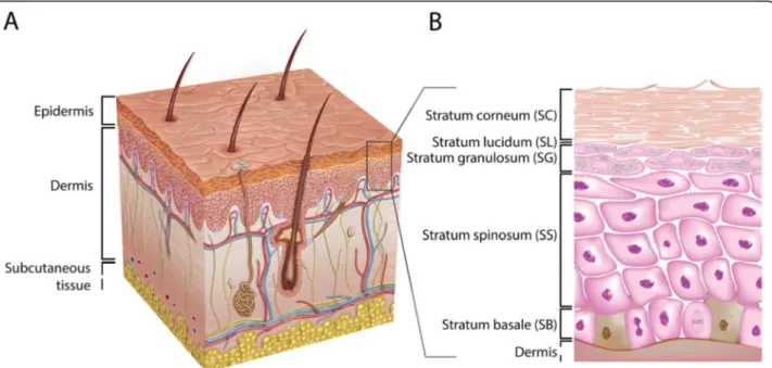
Mammalian skin is the largest organ in the human body and can be divided into two major layers, the epidermis and the dermis (Fig. 1). The dermis, the lower layer of the skin, is made of connective tissue and has a thick-ness of up to 3 mm. Fibroblasts are the primary cell type in this layer [23, 24]. These cells secrete collagen and elastin, which contribute to the generation of the extra-cellular matrix, providing the dermis with strain resist-ance and elasticity, respectively [23, 25, 26]. Embedded within the dermis are structures such as nerve endings, sebaceous glands, hair follicles, and blood and lymphatic vessels [24]. The vascular network provides nutrients and oxygen to the upper skin layers and their surround-ing tissues. Additionally, the vascular network takes part in detoxification, regulation of temperature, and wound repair. Besides fibroblasts, there are various other cell types found in the dermis: endothelial cells, mast cells, macrophages, dendritic cells called Langerhans cells, T cells, and neutrophils. Numerous dermal immune cells provide an effective defense mechanism against the inva-sion of pathogens and exogenous substances [27,28].
The layer above the dermis, the epidermis, is divided into four to five layers and contains mainly keratinocytes [29]. The lower layer, the stratum basale (SB), is made of a single layer of undifferentiated keratinocytes. These cells are continually dividing and upon differentiation migrating up to the adjacent layer, the stratum spinosum (SS), where they start a maturation process, such as changing their shape from columnar to polygonal. The layer above the SS is the stratum granulosum (SG) that contains differentiated keratinocytes with a typical accu-mulation of cytoplasmic materials, e.g., keratohyalin granules. They also produce lamellar bodies (LBs) that secrete specific lipid compounds, which are essential for the formation of the uppermost layer of the skin, the stratum corneum (SC) [30,31]. In thick skin, such as on the palms of the hands and soles of the feet, there is an additional thin layer named the stratum lucidum (SL), located between the SG and SC [32]. The SC is a mul-tiple layered tissue (e.g., 15–20 layers), which generally
has a thickness of 10–20 μm [33]. This layer consists of corneocytes that are terminally differentiated keratino-cytes lacking both nucleus and cytoplasmic organelles [33–35], surrounded by a lipid matrix providing a per-meability barrier. The described differentiation process is marked by the expression of early (e.g., cytokeratin 5 and 14) and late (e.g., cytokeratin 1 and 10) differenti-ation markers [36]. Other cells found in the epidermis are melanocytes, dendritic cells (i.e., Langerhans cells), T cells (e.g., CD8+ cells), and Merkel cells [29].
Epidemiology
Exposure to airborne PM is associated with several skin diseases and disorders and has been extensively reviewed elsewhere [20,21,37–39].
In healthy skin, exposure to urban PM has been associ-ated with accelerassoci-ated extrinsic aging [40–42], its clinical symptoms being pigment spot formation, coarse wrinkle development, and elastosis [43]. Further evidence from epi-demiological, clinical studies, and in vitro studies, and aging mechanisms have been reviewed [19,44–46], all indicating the generation of free radicals, the activation of aryl hydro-carbon receptor signaling, the induction of inflammatory cascade, and finally the impairment of the skin barrier.
In Germany, a higher incidence of skin melanoma among women was reported for women living in rural areas compared to women living in non-rural areas [47]. In another semi-individual cohort study, an increase in PM10 resulted in a 52% increase in the relative risk of
non-melanoma skin cancer [48]. The genotoxic potential
of PM was further investigated in an in vitro study that showed (i) a positive dose-response relationship for PM-induced DNA reactivity, and (ii) the highest DNA re-activity for PM2.5[7].
The chronic relapsing inflammatory skin disease, atopic eczema (AE), is characterized by skin barrier defects, in-tense pruritus, and immunological dysregulation [49–51]. This disease is not only caused by genetic predisposition (i.e., filaggrin mutation) and a dysregulated immune sys-tem but also triggered by several environmental factors, including air pollution, as suggested by recent epidemio-logical reports [20, 52]. A meta-analysis on skin diseases due to PM exposure included 13 studies and showed cor-relations between PM10 and PM2.5-exposure and several
skin diseases [53]. It reported on a significant association between PM2.5-exposure of younger people (e.g., 2–30
years), which was not the case for PM10exposure.
How-ever, with an odds ratio of 1.05 and the small quantity of observational, cohort studies, and individual studies, the statistical power of this meta-analysis is limited. Several new studies have been published that were not included in this meta-analysis [54–60]. More recently, Krämer and co-workers have reviewed this evidence by combining data from 57 environmental epidemiological studies in a sys-tematic review [61]. No sufficient evidence for a higher AE incidence upon large-scale exposure assessments of air pollution (PM10 and sulfur dioxide (SO2)) was reported.
Contrary to the small-scale exposure assessments (PM2.5),
where in most of the studies (23 out of 30), a significant association between AE and traffic-related emissions was
Fig. 1 Schematic overview of the human skin. (A) The skin consists of the epidermis and the dermis. (B) The epidermal layer consists, from top to bottom, of the stratum corneum (SC), the stratum lucidum (SL), the stratum granulosum (SG), the stratum spinosum (SS), and the stratum basale (SB). Figure adopted with permission from van Smeden et al. [22]
determined. Additionally, in all of the panel studies, there was a strong correlation between the AE symptom sever-ity and outdoor pollution. Interestingly, an association be-tween maternal exposure to traffic-related pollution and AE incidence in the offspring was found. From the pro-spective birth cohort study, Cohort for Childhood Origin of Asthma and Allergic Diseases (COCOA) [62], 468 one-year-olds were included with measurements of outdoor PM10and PM2.5 and transepidermal water loss (TEWL),
which is a measure for skin barrier dysfunction [63]. The effects of prenatal PM exposures were studied during three trimesters on skin barrier dysfunction and AE [64]. A positive correlation was found between prenatal PM ex-posure during the first trimester and the skin barrier dys-function and AE in the offspring: early-onset AE and higher AE severity at age three. It is suggested that PM-induced oxidative stress could induce epigenetic changes in fundamental DNA repair genes in the placenta, which could affect fetal immune development. Pulmonary and intranasal exposure to PM10, PM2.5, and PM0.1have been
extensively studied for their ability to enhance Th2-related immune responses, their role as an immune adjuvant, and their effects on the development of atopic disease [65–67]. Dong and co-workers reviewed reports on PM expos-ure and skin inflammation in China and concluded that, indeed, PM emissions aggravate the symptoms of in-flammatory skin diseases like AE [68]. The aforemen-tioned results have been supported by a recent review on the impact of different air pollutants on AE, which included a total of 21 in vitro studies, animal studies, clinical trials, and case studies [69].
The exact mechanisms through which PM can aggra-vate AE symptoms are yet to be further investigated. Add-itionally, determining the synergistic effects of different air pollutants while taking solar radiation into account can be challenging and is often not addressed when studying the effect of a single pollutant on the skin [70]. In a study from Japan, biopsies from 75 AE patients were investi-gated and a correlation was found between several oxida-tive stress markers in the SC and the severity of the disease [71]. It is hypothesized that environmentally gen-erated reactive oxygen species can induce oxidative dam-age to proteins in the SC. Eventually, this may lead to the disruption of the skin barrier and further exacerbation of AE. Additionally, the aryl hydrocarbon receptor may play a key role in PM-induced AE aggravation. It is suggested that PM and AE are linked by the activation of the aryl hydrocarbon receptor signaling pathway and transactiva-tion of neurotrophic factor artemin, by ligand species that are adsorbed onto airborne PM [72,73].
Thus far, the studies mostly investigated the link be-tween AE and PM. However, it is hypothesized that pa-tients with other inflammatory skin diseases with an impaired barrier function, such as the rare genetic skin
disease Netherton syndrome [74] and psoriasis [75], are more prone to the effects of PM.
Simulating biological responses of the skin to airborne PM in vivo and in vitro
The effects of airborne PM on the skin and underlying mechanisms will be critically evaluated, i.e., by reviewing studies on exposure of in vitro human two-dimensional (2D) and three-dimensional (3D) models, and finally in vivo animal models. The studies used either standard reference materials or in-house diesel emission and urban particulate matter extracts as exposure constituents.
Airborne PM for experimental studies
As aforementioned, the composition of PM is complex and includes metals, organic compounds (e.g., organic carbon and materials of biological origin), inorganic carbonaceous material, sulfate, and nitrate, among others. To assess their health effects, different approaches have been applied, such as epidemiological studies, controlled exposure studies to a defined source, or collection of particles on filters which can then be used for further experiments. It is important to add that the thorough characterization of the particles, in-cluding particle size and chemical analysis of the adsorbed chemicals onto the particles to correlate observed effects, is challenging but highly relevant [5].
The most common particles used in in vitro and in vivo studies to simulate the effects of airborne PM on the skin are Standard Reference Materials® available from the Na-tional Institute of Standards and Technology (NIST) [76]. NIST offers three different diesel PM extracts: SRM® 1650b, SRM® 1975, and SRM® 2975. Both SRM® 1975 and SRM® 2975 contain diesel PM collected from a filtering system of a diesel-powered forklift engine. SRM® 1650b contains ma-terial that is obtained from the heat exchangers of a dilution tube connected to diesel engines. This standard reference material is considered representative of particulate emis-sions of heavy-duty diesel engines and can be categorized as PM2.5. Besides using certified standards, diesel particulate
extracts from several different diesel engine emissions col-lected on filters have been extracted and tested on skin models. Additionally, NIST offers two airborne PM stan-dards named SRM® 1649b and SRM® 1648a that have been collected in the Washington, DC area and the St. Louis, MO area, respectively, between 1976 and 1977. Other stan-dards for fine atmospheric PM are SRM® 2787 (i.e., PM10)
and SRM® 2786 (i.e., PM≤ 4 μm) collected in Prague, the Czech Republic in 2005.
The Joint Research Centre (JRC) of the European Commission produces European Reference Materials (ERM) that are used to simulate airborne PM [77]. JRC is accredited to ISO 17034, ensuring the appropriate quality of ERMs. They offer CZ100 and ERM-CZ120 fine urban dust particles that are processed in a
way that resembles PM10-like urban dust without (e.g.,
ERM-CZ120) or with PAHs (ERM-CZ100). The National Institute for Environmental Studies (NIES) in Japan offers PM10-like urban dust certified reference material (CRM)
no. 28 that have been collected from a central ventilating system in a building in Beijing, China [78].
Several studies reported self-collection of airborne PM or PM from seasonal dust storms in Asia and West Af-rica, e.g., on building rooftops in Asia, using an in-house filtering system, yielding PM2.5. Others reported usage of
concentrated ambient air particles (CAPs), which are air-borne particles that are concentrated to PM2.5 for skin
model exposure.
Skin models used to study airborne PM effects Animal models
The main in vivo approaches to study skin responses to ei-ther drug permeation or toxicological outcome are eiei-ther rodents (i.e., rats or mice) or pigs [79]. Rodent skin differs significantly from human skin, due to considerably thinner skin layers and a higher hair follicle density, leading to higher substance permeation [80]. On the contrary, por-cine skin more closely resembles the structural properties of the human skin [81–84]; however the animals are more challenging to handle and have higher fat storage [85].
Animal models are employed to compare PM effects on skin with a healthy and compromised barrier func-tion. A disrupted barrier can be created by the conven-tional tape stripping method, which consists essentially of removing the SC of the animal skin model layer by layer [86–92]. Another approach is to use the NC/Nga mouse model that develops skin lesions that are compar-able to human AE lesions [93–96].
Animal models have various disadvantages, besides their different physiological structures. Animal studies are highly time and cost-ineffective; they are restricted to many regulations and countless ethical concerns [97]. To effectively comply with and implement the three R’s (i.e., reduction, refinement, and replacement) [98] for the laboratory use of animals, alternatives to animal test-ing like 2D and 3D in vitro human skin models should be considered to comply with the reduction and replace-ment concept [99].
In vitro human 2D models
Human adult low calcium high temperature (HaCaT) cells are an immortalized keratinocyte cell line that is frequently used for 2D in vitro studies [100]. However, a significant limitation of this cell line is their limited re-sponse to major T helper cell cytokines [101]. It was shown that HaCaT cells respond differently to these pro-inflammatory cytokines compared to primary cells, resulting in different expression profiles of genes related to the development of the skin barrier [102]. Therefore,
a model using HaCaT cells that focuses on the patho-physiological response towards pro-inflammatory cyto-kines should necessitate careful interpretation. The cells that are considered more biologically relevant are primary cells, which are explanted directly from healthy donors. The primary skin cells that are most often studied con-cerning the effects of PM are normal human epidermal keratinocytes (NHEK) or normal human dermal fibro-blasts (NHDF) monocultures. Human primary cells are ei-ther isolated from the neonatal foreskin or adult skin. In a study that investigated the proteome in primary keratino-cytes with respect to gender and age, it was determined that there were only minor differences [103]. Primary skin cells are cultured on specific substrates in an optimized cell culture medium and receive PM treatment directly in their culture medium, referred to as systemic treatment. Although the use of 2D models allows for easy, fast, and low-cost experiments, they lack a physiological skin bar-rier with a competent SC layer. Without this protective layer, the viable cells are in immediate contact with the tested substance. Therefore, systemic treatment of a 2D model consisting of living cells could result in an overesti-mation of the impact of the testing material and not re-semble the real-life exposure situation. It was found that the keratinocytes cultured in 3D, even upon a systemic treatment, are more resistant to cytotoxic agents such as hydrogen peroxide compared to keratinocytes cultured in 2D [104]. A recent study compared the effect of silver nanoparticles on oxidative stress and inflammation in 2D and 3D skin models [105]. Cell viability was reduced, and the silver content was approximately 9-fold higher in 2D keratinocytes compared to a 3D model. Additionally, the oxidative stress and pro-inflammatory response were sub-stantially stronger in a 2D model compared to a 3D model. For this reason, the use of a 3D skin model to bet-ter simulate the real-life topical exposure condition and assessment of the skin’s barrier function upon the treat-ment is recommended.
In vitro human 3D models
The two main types of 3D models used to mimic human skin are human epidermal equivalents (HEE) and human skin equivalents (HSE) [106,107].
Cultivation of HEE models starts with one cell type: ei-ther primary keratinocytes isolated from the human epi-dermis, or immortalized cell lines (e.g., N/TERT). These keratinocytes are grown in typical culture inserts on a supporting substrate, such as an inert membrane with a defined pore size or collagen matrix, using the air-liquid culture technique. This technique includes removing the cell culture medium from the upper side to expose the cells to air on one side, allowing the cells to‘feed’ from the medium in the chamber underneath [108–110]. Upon stimulation with growth factors, insulin, ascorbic
acid, and calcium, these keratinocytes can terminally dif-ferentiate into corneocytes, forming the upper layers of the epidermis over a time span of one to two weeks.
HSE models are comprised of both a dermal and an epidermal compartment consisting of fibroblasts embed-ded in a collagen matrix and keratinocytes, respectively. This two-compartmental model creates an interplay be-tween the two skin layers, resulting in a better resem-blance to human skin.
Although 3D skin models like the HSE and HEE are practical models that well-resemble the human skin, their barrier properties are less defined, due to a differ-ent SC lipid composition and a higher barrier permeabil-ity compared to that of native human skin [80, 111– 115]. In 3D models exposed to the air-liquid interface, PM can be applied either directly in the culture medium (systemic treatment) or on the topical side of the skin model either as aerosol or suspension (topical treat-ment). Topical treatment is considered more physiolo-gically relevant compared to real-life exposures as opposed to systemic treatment.
Effects of PM in vivo and in vitro
PM-induced skin barrier dysfunction and particle penetration
There are four different barrier compartments in the epidermis: physical, microbial, chemical, and immuno-logical compartments [106]. In this section, the effects of PM on the physical, microbial, and chemical barrier compartments will be reviewed.
Microbial barrier
The microbial barrier compartment consists of a diverse range of microorganisms, called the commensal skin microbiome that colonizes the skin. This includes colonization by millions of bacteria, fungi, and viruses, and the composition varies depending on the location of the skin [116]. These commensals protect the skin by preventing infection by pathogenic microbes. Although the research field of the skin’s microbiome has been given rise to many investigations over the last years [116, 117], studies on PM and the skin’s microbiome are scarce. It was shown that the composition of children’s skin microbiome was dependent on their age and living environment (e.g., rural versus urban), indicating a pos-sible link between PM exposure and the skin micro-biome composition [118]. Nevertheless, the role of the microbial barrier in PM-induced damages remains yet to be unraveled.
Physical barrier
Reported findings on the effects of PM on the skin’s bar-rier function in vivo and in vitro are summarized (Table1).
The physical barrier compartment of the skin can be seen as a so-called“outside-in” and “inside-out” barrier. The “outside-in” barrier consists of the SC, which pro-tects against the penetration of pathogens, allergens, and other exogenous substances such as PM [146, 147]. Sev-eral distinct proteins in the epidermis are crucial to the terminal differentiation program and the barrier function of the skin, such as involucrin, loricrin, and profilaggrin [148, 149]. Keratin filaments in the SG keratinocytes interact with profilaggrin, leading to aggregation of these filaments. Additionally, the intracellular components are degraded, and the desmosomes are transformed into corneodesmosomes, linking the corneocytes together that results in a densely packed layer [150]. The corni-fied envelope is formed around the plasma membrane and composed of structural proteins like involucrin and loricrin that are cross-linked by calcium-dependent transglutaminases [147]. Another protein that is crucial to the cornified envelope structure is the members of the small proline-rich (SPRR) family. SPRR3 was shown to be upregulated in keratinocytes treated with PM2.5
[132]. SPRR3 upregulation has been shown to decrease ciliogenesis in vitro [140], which is responsible for sup-pressing skin pigmentation in melanocytes [151].
The “inside-out” barrier, mainly provided by tight junctions, prevents excessive TEWL and, therefore, pro-tects the skin against dehydration [146, 147, 152]. It is likely that PM disrupts the barrier function of the skin, by modulating or even degrading the tight junctions. In other epithelial cells like lung cells or nasal cells, PM is shown to disrupt the barrier by modulating and even de-grading tight junction proteins such as occludin, zona occludens-1 (ZO-1), and claudin-1 [153, 154]. Also, in-creased endothelial barrier permeability was shown to be PM-induced, due to ZO-1 degradation [155].
In animal models, PM treatment mainly involved repetitive topical treatment for a longer time period (up to 2 weeks) of a relatively high dose (0.1–8 mg/ m2), resulting in decreased expression of structural barrier protein like keratins and filaggrin [120–122]. In porcine skin, deterioration of the skin barrier by PM due to disrupted tight junctions led to increased skin permeability [120]. In mice, penetration of PM was observed in both healthy skin and to a greater extent in the skin with a decreased barrier function (e.g., tape-stripped skin or skin of an AE mice model) [121]. In 2D cultures, e.g., in keratinocytes and fibro-blasts, cellular internalization of PM was observed after short-term exposures (i.e., 24 h) [121, 123, 135, 138, 145]. It is important to note that engineered nano-particles (e.g., single nano-particles with a diameter less than 100 nm [156]) have been reported to penetrate the first layers of the epidermis (i.e., SC and SG) and few reports on penetration into deeper layers of the viable epidermis
Table 1 Review of reported effects on the barrier function, such as proliferation, differentiation, and PM penetration in varying skin models upon exposure to PM
Model PM type Dose and
application
Exposure time Main findings Ref.
Ex vivo human skin
SRM® 1649b 2 mg/cm2,
topical
24 h - Pyknotic nuclei.
- Decreased collagen-1 protein expression.
[119] Pig SRM® 1648a and 1649b 100μg/m2,
topical
5 d - Decreased E-cadherin, cytokeratin, and filaggrin protein expression. [120] Mice, BALB/c with disrupted barrier PM≤1 μm from Seoul, Korea 8μg/cm2, topical 6 h and 2 w repetitive exposures, 10 times total
- Healthy barrier: PM remained in follicles. - Disrupted barrier: penetration of PM in SS. - Repeated exposure: dermal penetration of PM.
[121]
Mice, BALB/c SRM® 1649b 100μg/m2,
topical
24 h repetitive exposures, 5 d total
- Decreased filaggrin protein expression. [122]
Mice, HR-1 SRM® 1650b 100μg/mL,
topical
7 d - PM internalization. [123]
SRM® 1650b 100μg/mL,
topical
7 d - Increased keratin 10 and PCNA protein expression. [124]
HSE SRM® 2975 200μg/mL,
systemic
2 d repetitive exposures, 6 d total
- Decreased keratin 16, keratin 17, and Ki67 protein expression.
[125]
ERM-CZ120 200μg/mL,
systemic
48 h - Altered morphology of epidermis. [126]
HEE Diesel PM or vapor 0.05% (v/v), systemic
20 d - Decreased protein expression of cornifin A, suprabasin, and antileukoproteinase.
[127]
CAPs, PM2.5 0.5 and
2.0μg/cm2, topical
24 and 48 h - PM penetration in SC after 24 h.
- PM penetration in deeper layers after 48 h. - No alterations in morphology.
[128]
CRM no. 28 25 mg,
topical
6 h - Claudin 4 gene expression is upregulated. - No changes in mRNA expression of filaggrin, loricrin,
involucrin, transglutaminase 1, keratin 1, keratin 10, keratin 5, and keratin 14.
[129]
SRM® 1648a 200 ppm,
systemic
96 h - Altered morphology.
- Decreased PCNA and filaggrin protein expression.
[130]
SRM® 1648a 2.2, 8.9, and
17.9μg/cm2, topical
24 and 48 h - Decreased cytokeratin 10, involucrin, and loricrin protein expression.
- Increased barrier permeability for 17.9μg/cm2.
- Increased protein expression of aquaporin 3.
[131]
PM2.5from Seoul, Korea 50μg/mL,
topical
24 h - Decreased keratin-10, claudin-1, desmocollin-1, and IFN-γ protein expression.
- Increased S100A7 and S100A8 protein expression.
[132]
PM0.3–2.5from Benin, West-Africa
30μg/cm2, topical
24 h - Decreased loricrin protein expression. [133]
PM2.5from Xi’an, China 50μg/mL,
systemic
24 h repetitive exposures, 2, 4, or 6 d total
- Increased cholesterol levels. - Reduced squalene levels.
[134]
NHDF SRM®2787 30μg/cm2,
systemic
24 h - PM internalization into autolysosomes. [135]
ERM-CZ100 200μg/mL,
systemic
24 h - Increased elastase and collagenase activity. - Increased procollagen protein expression.
[136] The pre-conditioned
medium of HaCaT treated with CRM no. 28
125μg/mL, systemic
30 m, 48 h post-incubation
- Increased elastase and collagenase activity. [137]
NHEK PM2.5from Xi’an, China 50μg/mL,
systemic
24 h - Top upregulated genes of transcriptomics analysis are S100A8, S100A9, keratin 6B, keratin 19, serpin B3, and mostly the genes are involved in cholesterol metabolism.
[134]
Diesel PM or vapor 0.05% (v/v), systemic
20 d - Decreased envoplakin, suprabasin, filaggrin, involucrin, sciellin, caspase-14, cornifin-A protein, 1, and keratin-10 protein expression.
or even dermal layers [157,158]. However, the penetration of PM depending on size and agglomeration into the epi-dermis in healthy and diseased skin must be evaluated in future studies.
In 3D HSE models, a systemic treatment with PM (200μg/mL) induced alterations in skin morphology and expression levels of structural proteins after a long time exposure (i.e., 48 h up to 6 days) [125, 126]. In 3D HEE models, PM usually in concentrations of up to 50μg/mL was mainly applied topically and short-term exposures (i.e., 24 to 48 h) were performed [128,130,132,133]. PM was shown to affect the physical epidermal barrier in 3D
models, as indicated by altered expression of proteins im-portant for epidermal differentiation, such as filaggrin, involucrin, loricrin, and keratins [125,127,129,130,132, 133]. Additionally, markers for proliferation, such as Ki67, 5-bromo-2′-deoxyuridine (BrdU) incorporation, and pro-liferating cell nuclear antigen (PCNA), were shown to be decreased upon PM exposure [125,130]. Reduced expres-sion of differentiation and proliferation markers was also shown in 2D models, i.e., HaCaT and NHEK cells [122, 125,127,140,141,143,144].
PM was also reported to affect the skin’s cholesterol metabolism and increased cholesterol levels in 2D and
Table 1 Review of reported effects on the barrier function, such as proliferation, differentiation, and PM penetration in varying skin models upon exposure to PM (Continued)
Model PM type Dose and
application
Exposure time Main findings Ref.
Diesel PM 30 and
60μg/mL, systemic
24 h - PM internalization. [138]
Asian dust storm particles from Seoul, Korea
25μg/mL, systemic
24 h - Increased caspase 14 and ITGA6 mRNA expression. [139] PM≤1 μm from Seoul,
Korea
40μg/cm2,
systemic
24 h - PM internalization. [121]
PM2.5from Seoul, Korea 50μg/mL,
systemic
2 or 4 d - Decreased number and length of cilia. - Increased SPRR3 protein expression.
- Decreased keratin 1, keratin 10, and IFT88 mRNA expression. [140]
SRM®2786 1 mg/mL,
systemic
6 h - RNA-Seq analysis: Upregulation of keratin-associated protein 2–3, keratin 13, keratin 34, and claudin 4. Downregulation of growth differentiation factor 15.
[141]
PM2.5from Seoul, Korea 25μg/mL,
systemic
24 h - Top upregulated genes from transcriptomics analysis are SPRR family members and other cornified envelope-related genes, transglutaminase 3, involucrin, filaggrin, keratin 10, keratin 15, S100A7, S100A8, S100A9, and S10012.
[132]
HaCaT CRM no. 28 125μg/mL,
systemic
30 m, 24 h post-incubation
- Increased elastase and collagenase activity. [137]
SRM® 1649b 50μg/cm2,
systemic
24 h - Decreased aquaporin-3 protein expression. [142]
SRM® 1649b 50μg/cm2,
systemic
24 h - Decreased filaggrin, repetin, involucrin, and loricrin protein expression after 24 h. [143] PM2.5from Shanghai, China 10–100 μg/ mL, systemic
24 h - Increased involucrin, repetin, and filaggrin (highest dose) protein expression.
- No changes in loricrin protein expression.
[144]
SRM® 1649b 25 and
50μg/cm2,
systemic
4 and 24 h - Decreased filaggrin protein expression. [122]
SRM® 1650b 50μg/mL, systemic 1, 4, 8, 12, and 24 h - PM internalization. [123] SRM® 2975 100 and 200μg/mL, systemic
24 h - No changes in loricrin protein expression. - Increased involucrin protein expression. - Decreased keratin 16, keratin 17, and Ki67 protein
expression.
- Decreased BrdU incorporation.
[125]
CAPs, PM2.5 5–25 μg/mL,
systemic
1, 3, 6, and 24 h - PM internalization. [145]
Abbreviations: w, week; d, day; h, hour; m, minute; HSE, human skin equivalent; HEE, human epidermal equivalent; NHEK, normal human epidermal keratinocyte; SRM, standard reference material; PM, particulate matter; CAP, concentrated ambient particles; SS, stratum spinosum; ppm, parts per million; CRM, certified reference material; BrdU, 5-bromo-2′-deoxyuridine
3D skin models [134]. Cholesterol is one of the main lipid components of the matrix surrounding the corneo-cytes in the SC, thereby actively contributing to the per-meability barrier on the epidermis. Altered cholesterol metabolism in the skin due to PM exposure could indi-cate an altered barrier function of the skin.
Chemical barrier
Keratinocytes are crucial to the production of antimicro-bial peptides (AMPs), serving as the chemical barrier compartment, preventing microbial invasion at sites of damaged epithelium by directly terminating the patho-gen. These molecules can destroy bacteria either by cre-ating holes in their cellular walls or by sequestering iron, a crucial nutrient for bacterial growth [159]. Examples of AMPs are cathelicidin (i.e., LL-37) [160], defensins, and the S100 protein family [161]. The expression of numer-ous genes that are encoding AMPs is differentially regu-lated by several pro-inflammatory cytokines [162]. Increased AMP expression was observed in a 3D HEE and primary keratinocytes treated with PM [132, 134], indicating an alteration in the chemical epidermal barrier.
PM-induced oxidative stress
One of the primary mechanisms of the adverse effects of PM is through the generation of ROS. ROS are a group of highly reactive chemical species that are derived from the oxygen metabolism characterized by an unpaired electron (e.g., hydroxyl radical and superoxide anion) [163]. When the production of ROS overwhelms the skin’s antioxidant defense, tissues incur an oxidative stress condition [164]. ROS can damage lipids, proteins, and DNA, leading to oxidative injury via stress on several cellular organelles, resulting in tissue damage [165]. Reports on PM-induced oxidative stress in the skin have been reviewed and sum-marized (Table 2). In brief, PM can induce ROS gener-ation via direct and indirect mechanisms, as further discussed in this section.
PM triggers exogenous and endogenous ROS formation
PM can trigger the formation of exogenous ROS levels through the formation of free radicals as a result of the high particle surface reactivity [188,189]. Diesel PM and its organic extract were shown to both possess high re-activity by generating ROS via the catalyzation of oxygen reduction by various reductants [190]. In addition, PAHs and quinones that are bound to the PM’s surface have shown strong redox activity in vitro [191]. Transition metals (e.g., copper and iron) present in PM can gener-ate ROS through Fenton reactions [192]. In keratino-cytes, redox-active components such as copper and quinones were shown to induce oxidative stress by means of inducing the nuclear translocation of a
redox-sensitive protein complex, causing mitochondrial stress and finally leading to apoptosis [193]. The redox activity of PM is correlated to the size and the adsorbed species on the surface [194]. Higher redox activity per PM mass was measured for ultrafine particles, compared to parti-cles of a larger size. Moreover, a strong correlation was found between PM’s redox activity and the mass frac-tions of species they carry (i.e., metals and PAHs).
Indirectly, PM or its adsorbed species (e.g., PAHs, qui-nones, metals) can increase endogenous ROS generation by inducing mitochondrial stress and increasing the ac-tivity of ROS producing enzymes. The mitochondria are commonly considered as the main source of ROS pro-duction in the cell. PM induces mitochondrial dysfunc-tion, by inducing major structural damage [123, 135, 195]. Typically, mitochondrial dysfunction is indicated by reduced ATP levels, as determined in PM2.5-treated
keratinocytes and fibroblasts [175]. This dysfunction leads to the generation of mitochondrial ROS, and to in-creased radical production and mitochondrial Ca2+levels in vitro [123, 143, 174, 175, 182]. As a result, the intra-cellular ROS levels were shown to increase substantially after 30 min of PM-treatment up to 24 h (e.g., 50μg/mL) in 2D skin models [119, 121–124, 126, 130, 137, 138, 142,143,168,171,173,175,176,179–186].
The endoplasmic reticulum (ER) is responsible for most of the intracellular Ca2+, and, as a result, changes in the cellular Ca2+-balance activate ER stress [196]. PM was shown to induce ER stress, as measured by in-creased levels of intracellular Ca2+ [123, 168, 175, 184– 186]. Additionally, the ER stress sensor proteins for un-folded or misun-folded proteins, protein kinase-like ER kin-ase (PERK), inositol-requiring 1 alpha (IRE1α), and activating transcription factor 6 (ATF6) [197], were acti-vated in vitro [123,175]. In vivo, mitochondrial and ER stress was observed after longer (e.g., 7 days) topical PM-exposure, as indicated by mitochondrial and ER swelling [123].
Sensing exogenous chemical substances is paramount to stimulating an immune response as the first line of defense against these compounds. An important sensor of environ-mental chemicals is the aryl hydrocarbon receptor (AhR), a transcription factor that is highly expressed in all skin cells [198]. Besides the xenobiotic metabolism and preserving the skin’s homeostasis, the AhR plays a role in epidermal differentiation and barrier function of the skin [199, 200]. Common synthetic exogenous ligands are PAHs and halo-genated aromatic hydrocarbons (HAHs) such as 2,3,7,8-tet-rachlorodibenzodioxin (TCDD), dimethylbenz [a] anthracene (DMBA), methylcholanthrene or benzo[a]pyr-ene (BaP), coming from environmental air pollutants, such as PM [201]. Upon binding to its ligand, the AhR translo-cates from the cytoplasm to the nucleus, where it forms a dimer with the AhR nuclear translocator (ARNT) [202]. PM
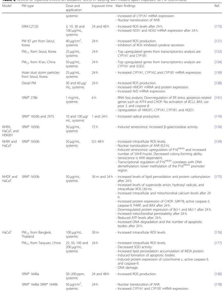
Table 2 Review of reported effects on oxidative stress in varying skin models upon exposure to PM
Model PM type Dose and
application
Exposure time Main findings Ref.
Ex vivo human skin SRM® 1649b 2 mg/cm2, topical 24 h - Lipid peroxidation. [119]
Mice, C-57 Diesel PM 4.5, 11.1 and 26.7 mg/cm2, topical
80 h - Increased DNA adduct formation. [166]
Mice, FVB/ N
SRM® 1650b 1 mg/time
(PAH extracted), topical
12 h - Increased CYP1B1 mRNA expression. [167]
Mice, HR-1 SRM® 1650b 100μg/mL,
topical
7 d - Increased protein carbonylation and lipid peroxidation. [168]
SRM® 1650b 100μg/mL,
topical
7 d - ER stress: upregulation protein expression of CHOP and GRP78. - Lipid peroxidation: increased expression of HNE protein. - Mitochondrial and ER swelling.
- Increased protein carbonylation.
- Apoptosis: increased protein expression of BAX, active caspase-3, and caspase-9, and DNA breakage.
- Autophagy: increased protein expression of LC3B-II.
[123]
SRM® 1650b 100μg/mL,
topical
7 d - Increased protein carbonylation. - Increased NOX4 protein expression.
[169] HSE SRM® 2975 200μg/mL, systemic 2 d repetitive exposure, 6 d total
- Increased protein expression of cleaved caspase-3. [125]
HEE CAPs, PM2.5 0.5 and 2.0μg/
cm2, topical
24 and 48 h - Increased isoprostanes protein level. - Increased HNE protein expression. - Increased CYP1A1 protein expression. - Increased DNA fragmentation.
[128]
CAPs, PM2.5 25μg/mL,
topical
24 and 48 h - Increased protein carbonylation. [170] PM0.3–2.5from Benin,
West-Africa
30μg/cm2,
topical
24 h - Increased HNE protein expression.
- Increased HMOX1, metallothionein 1G and 1E, cyclin dependent kinase inhibitor 2A, and caspase 3 mRNA expression. - Decreased BIRC5 mRNA expression.
[133]
CRM no. 28 25 mg, topical 6 h - Upregulated mRNA expression of CYP1A1, CYP1B1, and SOD2. [129]
SRM® 1975 5 mg/mL,
topical
48 h - Increased level of carbonylated proteins [171]
SRM® 1648a 2.2, 8.9, and
17.9μg/cm2, topical
24 and 48 h - Decreased AhR and increased NOTCH1 protein expression. [131]
NHDF SRM® 2787 30μg/cm2,
systemic
24 h - Autophagy: accumulation of LC3-II. - Mitochondrial stress: deformed mitochondria. - Increased CYP1A1 and CYP1B1 mRNA expression.
[135]
ERM-CZ100 50–400 μg/mL,
systemic
3.3 h - Increased levels of intracellular ROS. [136] The pre-conditioned
medium of HaCaT treated with CRM no. 28 125μg/mL, systemic 30 m, 48 h post-incubation
- Increased levels of intracellular ROS. - Increased number of apoptotic bodies.
[137]
SRM® 1649b 50μg/mL,
systemic
24 h - Increased intracellular ROS levels. - Activation of AhR (XRE activity).
- Upregulation of CYP1A1 mRNA expression.
[119]
SRM® 1649b 100–400 μg/
mL, systemic
24 h - Nuclear translocation of AhR. - Increased mRNA expression of CYP1A1. - Induced apoptosis.
[172]
NHEK Diesel PM or vapour 0.05% (v/v), systemic
20 d - Increased Nrf2 protein expression.
- Mitochondrial dysfunction: overexpression of proteins from mitochondrial complex I and IV.
[127]
Table 2 Review of reported effects on oxidative stress in varying skin models upon exposure to PM (Continued)
Model PM type Dose and
application
Exposure time Main findings Ref.
systemic - Increased of CYP1A1 mRNA expression. - Nuclear translocation of AhR.
ERM-CZ120 3, 10, 30 and
100μg/mL, systemic
24 and 48 h - Increased ROS levels after 24 h.
- Increased NOX1 and NOX2 mRNA expression after 24 h.
[173]
PM≤1 μm from Seoul, Korea
40μg/cm2, systemic
24 h - Increased ROS production.
- Inhibition of ROS inhibited cytokine secretion.
[121] PM2.5from Seoul, Korea 25μg/mL,
systemic
24 h - Top upregulated genes from transcriptomics analysis are CYP1A1 and CYP1B1.
[132] PM2.5from Xi’an, China 50μg/mL,
systemic
24 h - Top upregulated genes from transcriptomics analysis are CYP1A1 and SOD2.
[134] Asian dust storm particles
from Seoul, Korea
25μg/mL, systemic
24 h - Increased CYP1A1, CYP1A2, and CYP1B1 mRNA expression. [139]
Diesel PM 30 and 60μg/
mL, systemic
24 h - Increased ROS production.
- Increased HMOX1 mRNA and protein expression. - Increased Nrf2 mRNA expression.
[138]
SRM® 2786 1 mg/mL,
systemic
6 h - RNA-Seq analysis: Downregulation of ER stress apoptosis-related genes such as ATF4 and CHOP. No activation of BCL2, BAX, cas-pase 3, and cascas-pase 8.
- Upregulation of HMOX1, CYP1A1, CYP1B1, and NQO1.
[141]
SRM® 1650b and 2975 10 and 100μg/ mL, systemic
1 and 24 h - Increased radical production. [174]
NHEK, HaCaT, and HEK001
SRM® 1650b 50μg/mL,
systemic
72 h - Induced senescence: increasedβ-galactosidase activity. [124]
NHEK and HaCaT
SRM® 1650b 50μg/mL,
systemic
0.5–48 h - Increased intracellular ROS levels. - Nuclear translocation of AhR (0.5 h).
- Induced senescence: upregulation of P16INK4Aand increased number of SAHF/nuclei. Decreased colony-forming ability. - Senescence is AhR dependent.
- Transcriptional regulation of P16INK4Acorrelates with DNA
demethylation: lower methylation of the P16INK4Apromoter region. [124] NHDF and HaCaT SRM® 1650b 50μg/mL, systemic
30 m and 24 h - Increased levels of lipid peroxidation and protein carbonylation after 24 h.
- Increased levels of superoxide anion, hydroxyl radicals, and intracellular ROS (30 m).
- Increased intracellular and mitochondrial calcium levels after 24 h.
- Increased protein expression of CHOP, GRP78, active caspase-3, caspase-9, PARP, and BAX after 24 h.
- Downregulated protein expression of Bcl-1 and Mcl-1 after 24 h. - Increased mitochondrial permeability after 24 h.
- Reduced ATP levels after 24 h.
- Increased DNA degradation and the number of apoptotic bodies after 24 h.
[175]
HaCaT PM2.5from Bangkok,
Thailand
100μg/mL, systemic
30 m - Increased intracellular ROS levels. [176]
PM2.5from Taoyuan, China 25, 50, 100 and
200μg/mL, systemic
24 h - Increased intracellular ROS levels. - Decreased SOD activity.
- Increased lipid peroxidation: accumulation of MDA protein. - Induced formation of apoptotic bodies.
- Induced protein expression of cytochrome c, active caspase-3, and caspase-9.
- DNA damage.
[177]
SRM® 1648a 50–200 ppm,
systemic
24 and 48 h - Increased ROS production. [130]
SRM® 1648a SRM® 1649b 50μg/cm2,
systemic
24 h - Nuclear translocation of AhR.
- Increased CYP1A1 and CYP1B1 mRNA expression.
Table 2 Review of reported effects on oxidative stress in varying skin models upon exposure to PM (Continued)
Model PM type Dose and
application
Exposure time Main findings Ref.
SRM® 1649b 50μg/cm2,
systemic
1 h - Increased levels of intracellular ROS. [142]
SRM® 1649b 50μg/cm2,
systemic
2 and 24 h - Increased cellular and mitochondrial ROS levels after 2 h. - Increased HMOX1 protein expression after 24 h.
[143]
SRM® 1649b 25 and 50μg/
cm2, systemic
4 and 24 h - Increased ROS production. - Increased NOX activity.
[122] SRM® 1649b 25–100 μg/cm2,
systemic
4 and 24 h - Increased NOX2 protein expression. - Increased ROS production.
[179]
SRM® 1649b 25μg/mL,
systemic
2 and 24 h - Increased ROS production. [180]
SRM® 1649b 50μg/cm2,
systemic
1 and 4 h - Increased ROS production. - Increased NOX2 activity.
[181]
SRM® 1649b 25 and 50μg/
cm2, systemic
4 and 24 h - Increased ROS production - Increased NOX activity.
[122]
SRM® 1649b 100–400 μg/
mL, systemic
24 h - Nuclear translocation of AhR. - Increased CYP1A1 mRNA expression. - Induced apoptosis.
[172]
SRM® 1650b 50μg/mL,
systemic
24 h - Increased cellular and mitochondrial ROS levels and mitochondrial stress.
- Increased lipid peroxidation and protein carbonylation. - Increased cleaved caspase-3 and BAX and decreased Bcl-2
pro-tein expression. - Induced DNA damage.
- Increased number of apoptotic bodies.
[182]
SRM® 1650b 50μg/mL,
systemic
1 and 24 h - Increased intracellular ROS and superoxide anion levels. - Increased levels of protein carbonylation and lipid peroxidation
after 24 h.
- Induced DNA damage and apoptotic body formation. - Increased mitochondrial permeability after 24 h.
- Increased BAX, active caspase-3, and PARP and decreased Bcl-2 protein expression.
[183]
SRM® 1650b 50μg/mL,
systemic
24 h - Increased intracellular ROS and calcium levels.
- Increased levels of lipid peroxidation and protein carbonylation. - DNA damage.
- Increased mitochondrial ROS, calcium, and permeability. - Apoptosis: increased protein expression of ATF6, GRP78, p-IRE1,
BAX, and active caspase-3 and caspase-9. Decreased protein ex-pression of Bcl2. Increased number of apoptotic bodies. - Autophagy: autophagic lysosomes. Increased protein expression
of LC3B-II and beclin-1.
[168]
SRM® 1650b 50μg/mL,
systemic
24 h - Increased intracellular ROS and superoxide levels. - Induced NOX activity.
- Increased intracellular calcium levels and mitochondrial membrane permeability.
- Induced lipid peroxidation and protein carbonylation. - DNA damage.
- Increased number of apoptotic bodies.
[184]
SRM® 1650b 50μg/mL,
systemic
1, 4, 8, 12, and 24 h
- Increased intracellular ROS.
- Increased levels of intracellular calcium.
- ER stress: Increased protein expression of CHOP, GRP78, and p-PERK.
- Increased mitochondrial permeability. - DNA damage.
- Increased lipid peroxidation and protein carbonylation. - Apoptosis: Increased protein expression of BAX, DNA breakage,
apoptotic body formation, and increased expression of active caspase-3 and caspase-9.
- Autophagy: Increased protein expression of LC3B-II.
[123]
SRM® 1650b 50μg/mL,
systemic
30 m, 1 h, and 24 h
- Increased ROS production.
- Increased levels of intracellular calcium.
induces the nuclear translocation of AhR in vitro [119,124, 171,172,178]. The AhR/ARNT complex binds to conserved promoter regions containing the xenobiotic response element (XRE), promoting the transcription of several groups of target genes, such as from the phase I metabolism (e.g., cytochrome P450 family 1 subfamily A member 1, CYP1A1, CYP1A2, and CYP1B1), the phase II metabolism (e.g., UDP glucuronosyl-transferase family 1 member A complex locus, UGT1A and glutathione S-transferase A1, GSTA1) and a gene for the aryl-hydrocarbon receptor repressor (AhRR) [203]. CYP enzymes can metabolize PAHs, and the formed metabolites can induce cell damage either by the formation of DNA, protein adducts, or the generation of ROS [195,204–208]. Indeed, PM upre-gulates the CYP enzyme mRNA and protein expression in vitro and in vivo [119,128,129,134,135,139,141,167, 171,172,178,187].
Other ROS producing enzymes are the members of the nicotinamide adenine dinucleotide phosphate (NADPH) oxidase (NOX) family [209]. Once activated by PM [122, 169, 173, 179, 181, 184], these enzymes generate high levels of ROS.
PM-induced oxidative stress results in lipid, protein, and DNA damage
The skin is covered by a thin layer of sebum, that func-tions as a barrier against water evaporation (i.e., TEWL)
[210]. Sebum lipids, such as squalene, wax, cholesterol (esters), triglycerides, and free fatty acids, are a target for peroxidation by ROS. In addition to oxidation by ex-ogenous ROS, endex-ogenous ROS can target polyunsatur-ated fatty acids (PUFAs) from cell membranes, leading to the generation of reactive aldehyde byproducts like malondialdehyde (MDA) and 4-hydroxy-2-nonenal (HNE) [211], commonly used as biomarkers for oxida-tive stress [212,213]. Skin exposure to PM was found to induce lipid peroxidation, as well as elevated levels of re-active aldehyde byproducts [119,123,128,133,145,168, 175, 177, 182–184]. These reactive aldehydes can, in turn, react with amino acid residues in proteins, result-ing in the formation of carbonylated proteins [214], which were also detected in the skin upon treatment with PM [123, 168–171, 175, 182–184]. The introduc-tion of carbonyl groups (e.g., aldehydes and ketones) in proteins can also be caused by oxidative cleavage or the direct oxidation of amino acid residues [214]. The for-mation of these highly stable carbonyl groups results in conformational changes and irreversible damage to the polypeptide chain. Subsequent unfolding and complete inactivation of the protein allows for protein crosslink-ing, leading to the formation of adducts [215]. Also, DNA can be a target for ROS, and PM was shown to in-duce the formation of DNA adducts in mice skin [166].
Table 2 Review of reported effects on oxidative stress in varying skin models upon exposure to PM (Continued)
Model PM type Dose and
application
Exposure time Main findings Ref.
- Induced senescence.
SRM® 1650b 50μg/mL,
systemic
24 h - Increased ROS production. - Increased lipid peroxidation.
- Increased number of apoptotic bodies.
[186]
SRM® 1650b and 2975 10 and 100μg/ mL, systemic
1 and 24 h - No changes in radical production. [174]
SRM® 2975 100 and
200μg/mL, systemic
24 h - Increased protein expression of cleaved caspase-3 and PARP. - Increased protein expression of BAX and p53.
[125] CRM no. 28 125μg/mL, systemic 30 m, 24 h post-incubation
- Increased levels of intracellular ROS. [137]
ERM-CZ120 100μg/mL,
systemic
30 m - Increased intracellular ROS production. [126]
ERM-CZ120 25–100 μg/mL,
systemic
3 and 24 h - Increased CYP1A1 protein expression. - Decreased AhR protein expression. - Increased LC3-II and p62 protein expression.
[187]
CAPs, PM2.5 5–25 μg/mL,
systemic
1, 3, 6, and 24 h
- Increased HNE protein adduct formation. - Increased nuclear translocation of Nrf2.
- No changes in GPX, GR, and NPQO1 mRNA expression.
[145]
Abbreviations: h, hour; d, day; m, minute; HSE, human skin equivalent; HEE, human epidermal equivalent; NHEK, normal human epidermal keratinocyte; SRM, standard reference material; PM, particulate matter; CAP, concentrated ambient particles; ppm, parts per million; CRM, certified reference material; ROS, reactive oxygen species; PARP, poly (ADP-ribose) polymerase; Nrf2, nuclear factor erythroid 2-related factor 2; AhR, aryl hydrocarbon receptor; LC3-II, light-chain 3 II; BIRC5, baculoviral IAP repeat containing 5; NQO1, NAD(P) H quinone dehydrogenase 1; HMOX1, heme oxygenase 1; NOX, NADPH oxygenase; HNE, 4-hydroxy-2-nonenal; ER, endoplasmic reticulum; CYP1A1, cytochrome P450 family 1 subfamily A member 1
Counteracting PM-induced oxidative stress
The first line of defense against ROS is the cornified en-velope, especially the SPRR family [216]. It was shown that cysteine residues within the SPRR proteins are re-sponsible for ROS quenching, most likely due to its loca-tion on the outer layer of the skin [217]. Additionally, it was shown that common skin bacteria secrete specific proteins to counteract oxidative stress [218].
Other defense mechanisms, to abrogate oxidative stress processes, are identified like the upregulation of antioxidant proteins and detoxifying enzymes that are activated by transcription factors via antioxidant re-sponse elements (ARE) [165]. The expression and the subsequent nuclear translocation of one of these tran-scription factors, namely the nuclear factor erythroid 2-related factor 2 (Nrf2), is increased in vitro upon expos-ure to PM [127, 138, 145]. In turn, Nrf2 target genes and their protein expression such as heme oxygenase 1 (HMOX1), NAD(P) H quinone dehydrogenase 1 (NQO1), glutathione S-transferases (GSTs) are upregu-lated [127,133,138,141,143].
Activation of the AhR induces the nuclear transloca-tion of Nrf2 by a mechanism that needs to be elucidated, but evidence suggests it involves protein kinases that in-duce transcription of detoxifying enzymes, leading to a decrease of ROS levels [219].
PM-induced apoptosis and autophagy
Oxidative stress can lead to the activation of programmed cell death, i.e., apoptosis [220]. Protein levels of CCAAT-enhancer-binding protein homologous protein (CHOP), a transcription factor that mediates ER-stress induced apoptosis [221] and endoplasmic reticulum chaperone BiP (GRP78), a key element in normal ER function [222], were determined to be upregulated by PM2.5 [123, 168, 175].
Mitochondrial dysfunction leading to mitochondria-dependent apoptosis is characterized by increased permeability of the mitochondrial membrane [223]. Mitochondria-dependent apoptosis (e.g., intrinsic pathway of apoptosis) is mediated by the release of cytochrome c and caspase activation (e.g., caspase-3 and caspase-9) [223]. It was shown that PM induces mitochondrial-dependent apoptosis, indicated by an increased permeability of the mitochondrial membrane [123, 168, 175, 177, 183, 184], increased cellular cytochrome c [177], and activation of caspase-3, caspase-9 and poly (ADP-ribose) polymerase (PARP) [123, 125, 133, 168, 175, 177, 182–184, 186]. Furthermore, PM-induced apoptosis was observed by and upregulation of apoptosis regulator BAX, a downregulation of anti-apoptotic proteins Bcl-2 and Mcl-1 [123, 168,175, 182], DNA fragmentation [123, 128, 168, 175, 177, 182– 184], and the formation of apoptotic bodies [123,137,168, 172,175,177,182–184,186].
ER stress is also related to autophagy, and PM expos-ure was shown to promote autophagy in vitro. Proteins involved in the initiation of autophagosome formation, such as light-chain 3B II (LC3B-II) and Beclin 1, were shown to be upregulated [123,135,168,175,224].
PM-induced senescence
Another mechanism that can be activated upon ROS-induced DNA damage is cellular senescence. Senescent cells are in irreversible arrest, thereby limiting prolifera-tion, that is implicated in skin aging [225]. It was shown that PM2.5 induces senescence in human keratinocytes
(i.e., NHEK, HaCaT, and HEK001) [124,172,185]. PM-induced senescence was shown to be dependent on ROS formation and subsequent AhR activation [124]. The senescence inducer protein p16INK4A was found to be upregulated upon PM2.5 treatment, and its transcription
was shown to be epigenetically regulated through pro-moter methylation [124]. In mice, topical PM exposure led to a hyperkeratotic epidermis. Epidermal hyperproli-feration might result from the activation of the AhR, which is known to induce increased keratinocyte prolif-eration and differentiation [200].
Activation of the inflammatory cascade
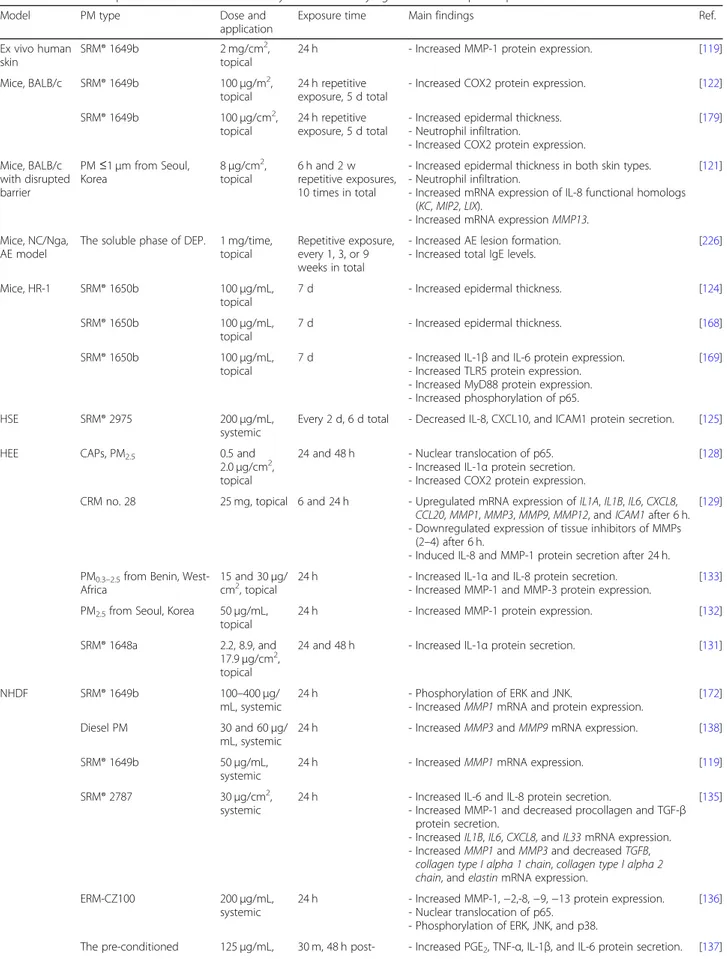
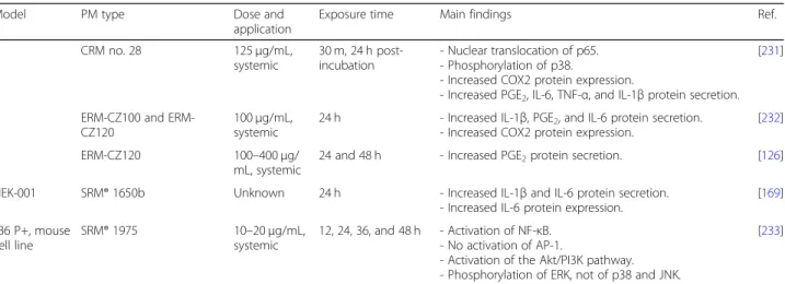
The skin plays a major role in protecting the body from harmful environmental factors. The keratinocytes and immune cells contribute to the first and second lines of defense of the skin’s immune system and are an indis-pensable part of the immunological barrier compart-ment. Additionally, the dermal fibroblasts play an important role in the sensing of pathogens, to protect the body further from harmful intrusive species. The re-ported findings on the effects of PM on the activation of the inflammatory cascade in skin models have been summarized (Table3).
Activation of toll-like receptors
Keratinocytes in the viable epidermis (i.e., SG, SS, and SB) and dermal fibroblasts defend the human body by sensing pathogens and mediating an immune response by differ-entiating between harmful and harmless pathogens [28]. They do so by the recognition of pathogen-associated mo-lecular patterns (PAMPs), which are evolutionary con-served small molecular motifs within microbes, including lipopolysaccharide (LPS), peptidoglycan, flagellin and nu-cleic acids [234]. These PAMPs trigger responses once they are recognized by pattern recognition receptors (PRR) [235], such as members of the Toll-like receptor (TLR) family, located at the cell surface or in the cyto-plasm [236]. Keratinocytes constitutively express TLR1–6 and TLR9 [237] whereas fibroblasts express all TLRs (i.e., 1–10) [238]. Their activation is critical for provoking dis-tinct immune responses in the skin [236].
Table 3 Review of reported effects on inflammatory cascade in varying skin models upon exposure to PM
Model PM type Dose and
application
Exposure time Main findings Ref.
Ex vivo human skin
SRM® 1649b 2 mg/cm2,
topical
24 h - Increased MMP-1 protein expression. [119]
Mice, BALB/c SRM® 1649b 100μg/m2, topical
24 h repetitive exposure, 5 d total
- Increased COX2 protein expression. [122]
SRM® 1649b 100μg/cm2,
topical
24 h repetitive exposure, 5 d total
- Increased epidermal thickness. - Neutrophil infiltration.
- Increased COX2 protein expression.
[179] Mice, BALB/c with disrupted barrier PM≤1 μm from Seoul, Korea 8μg/cm2, topical 6 h and 2 w repetitive exposures, 10 times in total
- Increased epidermal thickness in both skin types. - Neutrophil infiltration.
- Increased mRNA expression of IL-8 functional homologs (KC, MIP2, LIX).
- Increased mRNA expression MMP13.
[121]
Mice, NC/Nga, AE model
The soluble phase of DEP. 1 mg/time, topical
Repetitive exposure, every 1, 3, or 9 weeks in total
- Increased AE lesion formation. - Increased total IgE levels.
[226]
Mice, HR-1 SRM® 1650b 100μg/mL,
topical
7 d - Increased epidermal thickness. [124]
SRM® 1650b 100μg/mL,
topical
7 d - Increased epidermal thickness. [168]
SRM® 1650b 100μg/mL,
topical
7 d - Increased IL-1β and IL-6 protein expression. - Increased TLR5 protein expression. - Increased MyD88 protein expression. - Increased phosphorylation of p65.
[169]
HSE SRM® 2975 200μg/mL,
systemic
Every 2 d, 6 d total - Decreased IL-8, CXCL10, and ICAM1 protein secretion. [125]
HEE CAPs, PM2.5 0.5 and
2.0μg/cm2, topical
24 and 48 h - Nuclear translocation of p65. - Increased IL-1α protein secretion. - Increased COX2 protein expression.
[128]
CRM no. 28 25 mg, topical 6 and 24 h - Upregulated mRNA expression of IL1A, IL1B, IL6, CXCL8, CCL20, MMP1, MMP3, MMP9, MMP12, and ICAM1 after 6 h. - Downregulated expression of tissue inhibitors of MMPs
(2–4) after 6 h.
- Induced IL-8 and MMP-1 protein secretion after 24 h.
[129]
PM0.3–2.5from Benin,
West-Africa
15 and 30μg/ cm2, topical
24 h - Increased IL-1α and IL-8 protein secretion. - Increased MMP-1 and MMP-3 protein expression.
[133] PM2.5from Seoul, Korea 50μg/mL,
topical
24 h - Increased MMP-1 protein expression. [132]
SRM® 1648a 2.2, 8.9, and
17.9μg/cm2, topical
24 and 48 h - Increased IL-1α protein secretion. [131]
NHDF SRM® 1649b 100–400 μg/
mL, systemic
24 h - Phosphorylation of ERK and JNK.
- Increased MMP1 mRNA and protein expression.
[172]
Diesel PM 30 and 60μg/
mL, systemic
24 h - Increased MMP3 and MMP9 mRNA expression. [138]
SRM® 1649b 50μg/mL,
systemic
24 h - Increased MMP1 mRNA expression. [119]
SRM® 2787 30μg/cm2,
systemic
24 h - Increased IL-6 and IL-8 protein secretion.
- Increased MMP-1 and decreased procollagen and TGF-β protein secretion.
- Increased IL1B, IL6, CXCL8, and IL33 mRNA expression. - Increased MMP1 and MMP3 and decreased TGFB,
collagen type I alpha 1 chain, collagen type I alpha 2 chain, and elastin mRNA expression.
[135]
ERM-CZ100 200μg/mL,
systemic
24 h - Increased MMP-1,−2,-8, −9, −13 protein expression. - Nuclear translocation of p65.
- Phosphorylation of ERK, JNK, and p38.
[136]
Table 3 Review of reported effects on inflammatory cascade in varying skin models upon exposure to PM (Continued)
Model PM type Dose and
application
Exposure time Main findings Ref.
medium of HaCaT treated with CRM no. 28
systemic incubation - Increased COX2 protein expression. - Nuclear translocation of p65. - Phosphorylation of p38, ERK, and JNK.
- Increased MMP-1 and MMP-2 protein expression.
Diesel PM 30 and 60μg/
mL, systemic
6 h - Increased MMP2 and MMP9 mRNA expression. [227] NHDF and HaCaT co-culture SRM® 1648a and SRM® 1649b 50μg/cm2, systemic 24 h - Phosphorylation of p38.
- Increased IL1A, IL1B, IL6, and CXCL8 mRNA expression and protein secretion (HaCaT).
- No changes in TNF mRNA expression and protein secretion (HaCaT).
- Increased MMP1 and COX2 mRNA expression (NHDF). [178]
NHEK PM2.5from Xi’an, China 50μg/mL,
systemic
24 h - Top upregulated genes from transcriptomics analysis are CXCL1, IL1A, and IL1B.
[134]
Diesel PM 30 and 60μg/
mL, systemic
24 h - Increased IL1A, IL6, CXCL8, and TNF mRNA expression and protein secretion.
[138]
ERM-CZ120 3, 10, 30 and
100μg/mL, systemic
24 and 48 h - Increased IL1B, IL6, CXCL8, and TNF mRNA expression after 24 h.
- Increased IL-1β, IL-6, IL-8, and TNF-α protein secretion after 48 h.
- Increased MMP1 mRNA expression and protein secretion after 24 h.
[173]
SRM® 1650b Unknown 24 h - Increased IL-1β and IL6 protein secretion. - Increased IL-6 protein expression.
[169] PM≤1 μm from Seoul,
Korea
40μg/cm2, systemic
24 h - Increased CXCL8 mRNA expression and protein secretion. - Increased MMP1 mRNA expression and protein
secretion.
[121]
SRM® 2786 1 mg/mL,
systemic
6 h - RNA-Seq analysis: Upregulation of IL1B, IL36G, CXCL3, CXCL8, and IL1R2. Downregulation of MMP3 and MMP28.
[141]
SRM® 1649b 50μg/cm2,
systemic
24 h - Increased COX2 protein expression. [181]
SRM® 1649b 100μg/mL,
systemic
24 and 48 h - Increased IL-8 protein secretion. [228]
Diesel PM 20μg/mL,
systemic
48 h - Increased IL-1β and IL-8 protein secretion. [229]
Diesel PM 30 and 60μg/
mL, systemic
6 h - Increased IL1A, IL6, CXCL8, and TNF mRNA expression. [227] Asian dust storm particles
from Seoul, Korea
25μg/mL, systemic
24 h - Increased IL6, CXCL8, and CSF2 mRNA expression. - No changes in IL1B, IFNG, and IL18 mRNA expression.
[139] PM2.5from Seoul, Korea 25μg/mL,
systemic
24 h - Top upregulated genes from transcriptomics analysis are IL1B, IL36G, IL1A, IL1R2, PTGS2, IRAK2, MMP1, MMP9, and MMP10.
- Downregulated gene is CXCL14. - Induced IL-1α protein secretion. - Phosphorylation of p65 and p38. [132] HaCaT CRM no. 28 125μg/mL, systemic 30 m, 48 h post-incubation
- Increased PGE2, TNF-α, IL-1β, and IL6 protein secretion.
- Increased COX2 protein expression. - Nuclear translocation of p65. - Phosphorylation of p38, ERK, and JNK.
- Increased MMP-1 and MMP-2 protein expression.
[137]
PM2.5from Shanghai,
China
10–100 μg/ mL, systemic
24 h - No changes in CSF2 protein secretion.
- Increased TSLP, TNF-α, IL-1α, and IL-8 protein secretion. [144]
SRM® 1648a 50–200 ppm,
systemic
24 and 48 h - Increased TRPV1 mRNA and protein expression (via p38/ MAPK and NF-κB pathway).
- Increased IL-1β, IL-8, and TNF-α protein secretion.
[130] SRM® 1648a and SRM® 1649b 50μg/cm2, systemic 24 h - Induced phosphorylation of p-38. [178]