412 Zatout et al.
RESEARCH PAPER
OPEN ACCESS
Antibacterial and antibiofilm activity of the phenazine extract
of a fluorescent
Pseudomonas on coagulase-negative
staphylococci isolated from the Anti-Cancer Center of Batna,
Algeria
Zatout Asma
*1, Djibaoui Rachid
1, Dahah
Hicham
1, Mazari
Hibat Errahmen
2,
Benkredda Fatima
2, Kassah-Laouar Ahmed
31
Laboratory of Microbiology and Plant Biology, Department of Biological Sciences,
Faculty of Natural Sciences and Life, University of Abdlhamid Ibn Badis, Mostaganem, Algeria
2
Department of Biological Sciences, Faculty of Natural Sciences and Life,
University of Abdlhamid Ibn Badis, Mostaganem, Algeria
3
Laboratoire Central de Biologie, Anti-Cancer Centre, Batna, Algérie
Key words:Coagulase-negative staphylococci, Pseudomonas, Inhibition, Biofilm, Phenazine compounds
http://dx.doi.org/10.12692/ijb/15.3.412-424
Article published on September 30, 2019
Abstract
Faced with many failures in the elimination of CoNS pathogens, we aimed to find an effective agent to eradicate these bacteria. In the present study we tested 20 isolates of CoNS collected from the Anti-Cancer Center of Batna (Algeria). As antistaphylococcal agents we used in our experiment 29 Pseudomonas isolates selected from the roots of plant (Hordeum murinum). The results of crossed streak method on MHA medium showed that the 29 isolates of Pseudomonas inhibited 19 CoNS. The strain with the greater inhibitory effect PK inhibited 14 CoNS. The high performing Pseudomonas PK has been used for the production of phenazine compounds. The dry compound of PK from ethyl acetate extraction has been shown to be effective against 13 isolates of staphylococci. The percentage of biofilm inhibition by the PK phenazine extract was between (32.2%) and (77.56%). The analysis of the compound obtained by UV-visible and infrared spectrum showed that it was similar to hydroxyphenazine.
* Corresponding Author: Zatout Asma asma.zatout@univ-mosta.dz
International Journal of Biosciences | IJB |
ISSN: 2220-6655 (Print) 2222-5234 (Online) http://www.innspub.net Vol. 15, No. 3, p. 412-424, 2019
413 Zatout et al.
IntroductionCoagulase-negative staphylococci (CoNS) are among the most frequently isolated microorganisms in microbiological samples. These bacteria, long regarded as contaminants, are currently recognized as true pathogens (Bertrand et al., 2002; Koksal et al., 2009) including species S. epidermidis, S. haemolyticus and S. saprophyticus, CoNS are causative agents of conjunctivitis, endophthalmitis, skin infections, urinary tract infections, endocarditis, peritonitis, bone and joint infections, post-neurosurgical meningitis, infections originated from equipments and valves as well as septicemia whose starting point can be a catheter (Garnier and Denis, 2007).
Many antibiotics are available for the treatment of various pathogenic bacteria. However, the increase in antibiotic resistance has led to more severity of diseases caused by CoNS. In addition, low immunity in hosts and the ability of bacteria to develop antibiotic resistance associated with biofilm have further increased the number of life-threatening bacterial infections in humans (Raut and Karuppayil, 2014). Consequently, treatment choices for resistant CoNS infections extend to natural antibacterial (Singh et al., 2015; Moloney, 2016). The capacity of the antibacterial compounds obtained from other microorganisms to inhibit CoNS has also been tested (El Amraoui et al., 2014). Among the metabolites studied produced by various groups of microorganisms those of Pseudomonas (Chain and Mellows, 1977). The members of this bacterium are found in the different environments, including soil, water, plant surfaces, animals and plants. They are also competitive colonizers in the rhizosphere and are well adapted to different biotic and a biotic stresses. Pseudomonas is well known for its ability to use a variety of organic compounds and for its production of antimicrobial compounds some of them are used in the treatment of bacterial diseases (Laine et al., 1996). It also shows activity against staphylococci (Cardozo et al., 2013; Hotterbeekx et al., 2017). Pseudomonas fluorescens is a non-pathogenic saprophyte that colonizes water, soil
and plants surfaces. It lives in a commensally relationship with plants, allowing them to acquire essential nutrients, degradation of chemicals and biological pollutants species (Igbinosa et al., 2014). P. fluorescens also produce a large number of secondary metabolites that could play a role in the antagonistic effect in soil (Jacques et al., 1993) making the bacteria a performing biocontrol agent (Igbinosa et al., 2014). Among its metabolites produced are phenazines which have long been recognized for their importance in microbial destruction and their positive physiological roles for the bacteria that produce them are increasingly appreciated (Grahl et al., 2013).The aim of this study is to isolate, from plant rhizospheres, a set of fluorescent Pseudomonas to examine their antagonist and antibiofilm activity against clinical isolates of CoNS. This step will lead us to search for an extracellular metabolite effective against staphylococci of medical interest.
Materials and methods
Bacterial material
20 CoNS isolates were collected between January 1 and February 28, 2017 from hospitalized patients in different
departments (Onco-Pediatric, Onco-Hematology,
intensive care, Carcinological Surgery) of the Anti-Cancer Center of Batna (Algeria) and non-hospitalized patients who came for external consultation.
Clinical strains of CoNS were grown on Chapman medium and identified using standard bacteriological methods. CoNS were used as target microorganisms to determine the antagonistic activities and spectrum of action of Pseudomonas isolates.
Antibiotic susceptibility and minimum inhibitory concentrations (MIC) of CoNS strains
Antibiotic sensitivity of CoNS isolates was achieved by the MHA disk diffusion method, as described by Bauer et al. (1966). The diameters of the inhibition zones were measured with caliper. The results were interpreted according to the recommendations of Clinical and Laboratory Standards Institute (2014). MICs were determined for Oxacillin and Vancomycin on MHA using E-test method and interpreted
414 Zatout et al.
according to Clinical and Laboratory Standards Institute (2014).
Study of biofilm formation by the in vitro crystal violet staining method of on microplates (TCP) Polystyrene microplates with 96 wells were used to study adherence and biofilm formation in CoNS isolates. The quantitative determination of biofilm formation in the microplates was evaluated according to Christensen et al. (1985) method with some modifications. Briefly, the CoNS were grown in nutrient agar medium for 18-24h at 37°C; one colony of each isolate was suspended in 5ml of TSB and incubated at 37°C for 24 h. Each suspension is diluted 1 / 100th in the same TSB + 1% glucose and each well of the microplate was filled with 200μL of this dilution (three independent cultures for each species were used). A sterile TSB + 1% glucose was used as a control. These microplates were covered and incubated for 24 h at 37°C. The contents of the wells were gently poured and washed three times with sterile physiological water, dried in an inverted position in an oven at 60°C for 60 min, adherent cells are stained with 200μl of 1% crystal violet (w/v). After 30 min of incubation, excess of crystal violet was removed by 5 successive washes with sterile distilled water. The dye incorporated by the adhered or biofilm-forming cells was solubilised with 200μL of 95% ethanol (v/v).The amount of solubilised crystal violet was measured by reading the OD at 550nm (Rodrigues et al., 2010). The interpretation of the results was performed according to Stepanovic et al., (2007) recommendations.
Isolation of Pseudomonas
Several samples were taken from different points in the vineyards of two regions of the state of Mostaganem, one of Oued Elkhire and the other of Sidi Lakhdar on February 2018. Samples are wild grass roots (barley of rats = Hordeum murinum). The isolated strains were identified using conventional bacteriological methods. Only the colonies giving a fluorescent yellow-green pigment on King B medium were selected (Bossis et al., 2000).
In Vitro Antagonism Test
A cross streak method as described by Selvin et al., (2009) was used. It consists in seeding the
Pseudomonas antagonist strain in a single streak at the edge of the surface of MHA plate. After incubation for 48h at 30°C, Staphylococcus isolates are seeded perpendicular to Pseudomonas. After 24h further incubation, the antimicrobial interactions are analyzed by measuring the inhibition zone size using a caliper.
Inhibitory effect of Pseudomonas culture
supernatants
The method used in the present work described by Veerendra kumar and Janakiram (2015) is based on the diffusion of the inhibitory agent into wells made in an agar containing an indicator strain. The culture supernatant of Pseudomonas was obtained after centrifugation of a culture in MHB, the supernatant was filtered through a millipore filter of 0.22µm. 0.1ml of a pure culture of 24 h indicator strain, whose optical density located between 0.08 and 0.1 at ʎ = 625nm was placed in each Petri dish containing 20ml of MHA. Wells of 4 mm of diameter were made, and then each well was filled with 50μL of supernatant and incubated at 37°C for 24 h. The inhibition zones were revealed around the wells.
Production and extraction of phenazines
The phenazine compounds were extracted according to the method described by by Bonsall et al., (1997) and Dahah et al., (2016). The performing isolate PK was inoculated on solid King B medium and incubated at 30°C for 24h. Flasks containing 50ml of Nutrient Broth Yeast Extract (NBY) were inoculated with the PK isolate, and then incubated at 30°C for 72 h with continuous shaking of 180rpm. Then cultures were centrifuged and the supernatant was acidified to pH 2 with concentrated HCl (Delaney et al., 2001). Phenazines were extracted from the supernatant by ethyl acetate 1(v/v). The organic phase was treated with anhydrous ammonium sulfate, filtered and then concentrated to dryness under reduced pressure using a rotary evaporator at 55°C (Mezaache-Aichour et al., 2012).The dry substrate is solubilised separately in methanol and in DMSO 10% for subsequent utilizations (Delaney et al., 2001).
Study of the antimicrobial activity of the extract by the disk method
415 Zatout et al.
Antibacterial activity of the extract obtained, was determined using a diffusion disc method of Hazalin et al., (2009). Two solutions of 0.25g/ml and 0.5g/ml of the dry substrate were prepared in DMSO 10%. 0.1ml of inoculum of each of the 20 isolates of CoNS was swabbed on MHA plate. The sterile disks (6mm in diameter) were deposited and impregnated with 10µl of the extract. The Vancomycin disk (30μg) was used as a positive control and the DMSO 10% disk as a negative control. Then, the dishes were incubated at 37°C for 24 h.
MICs of phenazine extract
The determination of the MICs of The phenazine extract against the CoNS was carried out according to the dilution technique in liquid medium described by
(Bazargani and Rohloff, 2016) with some
modifications. The bacterial strains were cultured on MHA and incubated at 37°C for 12 h, after incubation, 5-7 isolated colonies were inoculated in tubes containing 5ml MHB and incubated at 37°C for 8 to 12 h, then, the bacterial suspension was diluted 1: 100 in
sterile MHB (106 CFU/ml). For each microplate well,
100μL of MHB were added, then 100μL of the extract was placed in the first microplate well and two-fold serially diluted was performed in MHB. Finally, 100μL of the diluted bacterial strain was added to obtain a final concentration between 250 and 1.95mg /ml. 100μL of DMSO 10% was used as a negative control with 100μL of MHB. S. aureus ATCC 25923 was used as a control.
After incubation for 18 to 24h at 37°C, the MIC was determined macroscopically via turbidity observation (Nostro et al., 2016). Minimum bactericidal concentration (MBC) was determined by inoculating 10μL of all MICs on MHA (Marino et al., 2010). Effect of phenazine extract on biofilm formation The extract at the MICs concentration was evaluated for their inhibitory potential against cell attachments described by Bazargani and Rohloff (2016). 100 µl of the extract at the MICs value were added to each well of a 96-well microplate. The negative control contained 100 µl of TSB + 1% Glu. Finally, 100 µl of
each bacterial culture (106 CFU/ml) were put into each
well. 200µl of TSB + 1% Glu + DMSO 10 % were added without bacterial culture and a blank control (TSB + 1% glucose + extract) were included. The microplates were incubated at 37°C for 24h. Then, the biomass biofilm was assayed using the crystal violet staining test as described above. The mean absorbance of the samples was determined, the absorbance in blank well was subtracted from absorbance reading and percentage inhibition and efficiency were determined. UV-visible spectrophotometer analysis
Phenazine analysis by UV-visible spectrophotometer described by Veselova et al., (2008) was assayed. The dry phenazine extract was put into a methanol solution and then put into a vat for spectrophotometer analysis in order to characterize the absorption spectrum of this metabolite.
Fourier Transform Infrared Identification (FTIR) A sample of the extract was analyzed by a Fourier Transform Infrared spectroscopy technique in order to characterize partially the structure of the extracted phenazine (Dahah et al., 2016).
Results and discussion
Identification of CoNS
We identified 20 isolates of CoNS belonging to 5 different species following conventional methods. (45%) of CoNS studied were belonged to, S. epidermidis, (30%) to S. haemolyticus, (15%) to S. xylosus, (5%) to S. hominis and finally, (5%) too to S. cohnii. Similar study on blood culture samples reported by Koksal et al. (2009). Ehlersson et al. (2017) found different results. A dozen of species and subspecies, most of which part of the commensal flora, are potentially pathogenic for man through the breaking of the cutaneous -mucous barrier. S. epidermidis may be responsible for vascular or joint prosthetic infections, heart valves, CSF bypass valves; it is also isolated from peritoneal dialysis, peritonitis, subacute endocarditis treatment and from intravenous drug users (Abalain-Colloc et al., 2014). S. haemolyticus is the second most commonly isolated of CoNS. It is more frequently associated with endocarditis on native valves, bacteraemia, peritonitis and central venous catheter infections (Herard et al., 1998). It can be responsible for urinary tract infections (Gaucherie and
416 Zatout et al.
Avril 2005; Nauciel and Vildé 2005). S. xylosus is rarely isolated in blood cultures, rare reports of human infections have been reported, including endocarditis, pyelonephritis, and intra-abdominal infection (Mack et al., 2006).
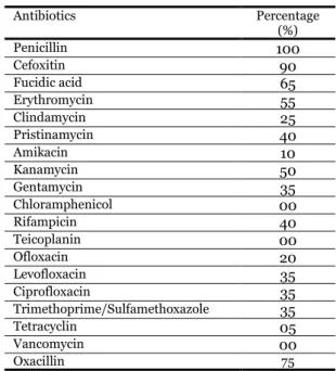
Antibiotic resistance of CoNS
The isolates are resistant to antibiotics with different percentages (Table 1). Different results were found by Pinna et al. (1999) of which (67%) CoNS are Penicillin-resistant. No strain of CoNS showed resistance to Vancomycin. Similar results are also reported by Jain et al. (2004). Different findings are reported by Soumya et al. (2017) with a (7%) resistance to Vancomycin. According to Leclercq (2002), the strains with diminished susceptibility to glycopeptides appear to be rare but may be underestimated because of the difficulty of in vitro detection of resistance. Results showing deference with our findings have been reported by Shrestha et al. (2017) for Amikacin with a sensitivity of (67%) and for Chloramphenicol (12.6%) and (8.9%) were reported by Cuevas et al. (2004) in Spain by statistical studies of the years 1996 and 2002 successively.
Table 1. Antibiotic resistance and MIC of CoNS.
Percentage (%) Antibiotics 100 Penicillin 90 Cefoxitin 65 Fucidic acid 55 Erythromycin 25 Clindamycin 40 Pristinamycin 10 Amikacin 50 Kanamycin 35 Gentamycin 00 Chloramphenicol 40 Rifampicin 00 Teicoplanin 20 Ofloxacin 35 Levofloxacin 35 Ciprofloxacin 35 Trimethoprime/Sulfamethoxazole 05 Tetracyclin 00 Vancomycin 75 Oxacillin
Detection of CoNS biofilm formation by in vitro microplate crystal violet staining (TCP) method In the TCP method, biofilm production was detected in all isolated CoNS (100%) with different intensities:
(35%) are highly biofilm producing, (25%) are moderate and (40%) are low. Soumya et al. (2017) found that (11%) of CoNS strains were strongly biofilm forming and (6%) were moderate. Other different results have been reported by Nasr et al. (2012).
Isolation of Pseudomonas isolates and identification of PK isolate
In our study we selected 29 isolates cultivated on King B medium. Their preliminary identification was essentially based on morphological characteristics, Gram stain, mobility, catalase search and oxidase. These characters allowed us to classify them with the group of fluorescent Pseudomonas (Bossis et al., 2000). Results of the in vitro antagonism tests Showed that inhibition was variable depending on the Pseudomonas isolate and the strain of CoNS tested (Fig. 1).
Fig. 1. Pseudomonas antagonistic activity against
CoNS isolates.
Overall, the results of this test showed that all 29 Pseudomonas showed a growth inhibition of 19 of the targeted Staphylococci (Table 2). Clear areas between Pseudomonas and the most of Staphylococci tested were observed; these areas vary from 3 to 55mm, the histograms given by (Fig. 2) show the different inhibition zone distances in mm between Pseudomonas isolates and Staphylococci. The largest area was noted by the PK isolate against S. epidermidis 94C. The second histogram given by (Fig. 3) shows the number of Staphylococci inhibited by each Pseudomonas isolate. 14 different Staphylococci were inhibited by the PK isolate,
417 Zatout et al.
while the PR isolate inhibited only 6 Staphylococcus could have an effect on S. epidermidis 203CAC which is Resistant to all other isolates of Pseudomonas, reflecting the difference of the inhibitory substances
responsible for antagonistic activity in each strain. The identification of PK isolate by API NE Gallery directs us towards the species P. fluorescens.
Table 2. Antagonism test results by crossing technique.
S. epidermidis S. haemolyticus S. xylosus cohnii S.
S. Homini s 29 mg 94 C CAC 170 893 203G 173C 82 C 128 123 C 805 182H 467 1076 316 H 14:15 25 293 347 H CAC 106 132 T PK + + - + - + + + - + + + + - - + + + + - 14 P25 + - + - - + + + - + - + - - + - - - + + 10 P14 + - - - + - + - + + - - - - + + - + - 8 PI + + + - - + + - - - + - - - - + - - - - 7 P19 + - - - + - + + - + - - - + 6 P10 + - + - - + - - + - - + - - - + - 6 PA + + - - - + + - + - - - + - - - - 6 PR - + + - - + + + - - - + - - - 6 P26 + - - - + - - + - - - + + 5 PL + + + - - - + - + 5 PB - + - - - + - + - - - + - - - - 4 PE - + - + - - - + - - - + - - - 4 PN - + - - - + - + - - - + - - - - 4 PQ - + + - - + - + - - - 4 PG + - - - - + + - - - + - - 4 P15 + - - - + - - - + - 4 P9 + - - - + - - - - + - - - 3 P11 + - - - + - - - + - 3 P3 - - + - - + - - + - - - 3 PJ - - + - - + - - + - - - 3 PS + - - - + - - - + - - - 3 P22 + - - - + - 2 P1 + - - - + - - - 2 PH + + - - - 2 P28 - - + - - - 1 P21 + - - - 1 P12 + - - - 1 P17 + - - - 1 P2 + - - - 1
Fig. 2. Distances of inhibition zones between
Pseudomonas isolates and Staphylococci.
Fig. 3. Number of Staphylococci Inhibited by each
418 Zatout et al.
Some strains of fluorescent Pseudomonas that colonize agricultural soils have several intrinsic characteristics that make them particularly attractive for use as biological control agents (Haas and Keel, 2003). In addition to competition for carbon sources, antagonism can be attributed largely to the production of secondary metabolites (antibiotics, siderophores, hydrogen cyanide, enzymes etc.) (Jacques et al., 1993). Indeed, P. aeruginosa produces more than 55 quinolones/quinolines in addition to 2-heptyl-3-hydroxy-4 (1H) -quinolone, and these have significant antibiotic activity against gram-positive bacteria. Antimicrobial quinolones can be inserted in extracellular membrane vesicles to cause a S. epidermidis lysis (Mashburn et al., 2005). Another extracellular protein secreted by P. aeruginosa which has notable staphylolytic activity is the LasA protease.
Extracellular polysaccharides secreted by P.
aeruginosa may be a promising strategy for use against staphylococcal biofilms in future applications (Qin et al., 2007).
The inhibitory effect of the culture supernatant of Pseudomonas
Among the 29 isolates with antagonistic activity, 12 isolates namely: PK, P14, PG, PL, PN, PR, P3, PE, P25, PJ, PQ and PA were selected because of their inhibition spectrum against Staphylococcus and the zones of their inhibition. The 12 isolates selected were tested for the inhibitory effect of their supernatants. However, they did not show any antagonist activity. This may be justified by the low concentration of inhibitory agents in the supernatants. According to studies by Emmerich and Löwthe cell-free culture fluid of P. aeruginosa should be concentrated to one tenth of its initial volume to be effective (Leisinger and Margraff, 1979). The antimicrobial effect, MICs and BICs of phenazines extract
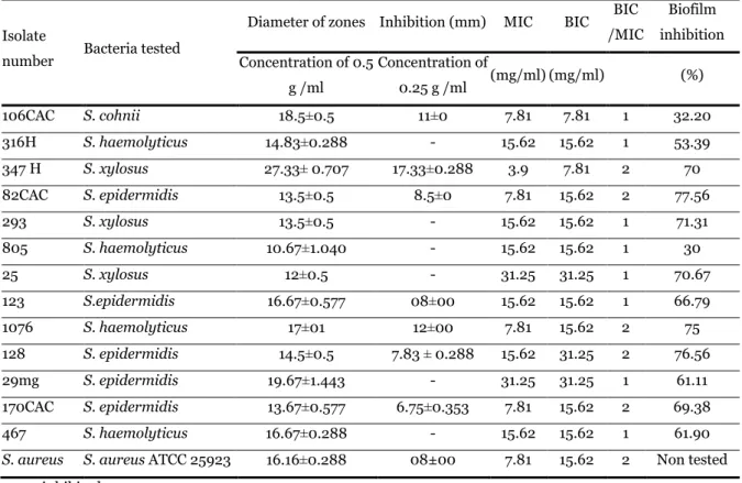
Phenazine production by PK isolate was performed on NBY medium, and extraction was by ethyl acetate. The dry phenazine extract was then melted in a (DMSO 10%) solution to test its antimicrobial effect . The results obtained showed that our extract exhibits anti staphylococcal activity, as shown in (Fig. 4) clear areas around the disks impregnated with the phenazine
extract were observed. These zones of inhibition vary according to the strain tested between 6.75 ±0.353 and
27.33
±0.707 mm (Table 3). The largest zone was found
against isolate 347 H (S. xylosus).
Pseudomonas produce a range of phenazine compounds that differ widely in their antibiotic properties, depending on the nature and position of the side groups attached to the phenazine nucleus (Saleem et al., 2010). Other biological activities of phenazine include natural products such as anti tumorals, anti malarials and antiparasitics have been reported (Laursen and Nielsen, 2004). Our results are in agreement with the studies of several authors, who showed that phenazine substances are known for their antifungal and antibacterial activities (Shahid et al., 2017).
In the present study, the MICs were tested to know the ability of phenazines to inhibit the growth of 13 CoNS strains. This MIC of phenazine extract was from 3.9mg/ml to 31.25mg/ml. The MBC was ranging from 7.81mg/ml to 31.25mg/ml (Table 3).
According the studies of Nansathit et al. (2009), phenazine-1-carboxylic acid showed more potent inhibition against some strains such us: A. avenae subsp citrulli, B. subtilis, C. albicans, E. coli and X. campestris pv. vesicatoria. The inhibitory mechanism of phenazines was the result of the toxicity of the superoxide radical and hydrogen peroxide, described in a report by Dwivedi and Johri (Nansathit et al., 2009). According to other studies conducted by Borrero et al., (2014), 13 various phenazine compounds, five of which are of natural origin, possess inhibitory activity against S. aureus and S.
epidermidis. Phenazine-1-carboxylic acid and
chlororaphine showed only weak antibiotic activity against S. aureus (MIC> 5μg/ml) (Laursen and Nielsen, 2004).
Effect of phenazine extract on CoNS biofilm formation In our study, the phenazine extract showed antibiofilm activity with significant reduction between (32.2%) and (77.56%) (Table 3). Staphylococci commonly colonize the skin and are
419 Zatout et al.
frequently found in wounds and implants. Interestingly, S. epidermidis was not considered an opportunistic pathogen until the widespread use of medical devices. Biofilm formation, then, can be thought of a virulence factor a bacterial strategy that contributes to its ability to cause an infection (Hall-Stoodley et al., 2004). Qin et al. (2009) found that P.
aeruginosa extracellular products, mainly
polysaccharides, disrupted established S.
epidermidis biofilms and these extracellular products are important microbial competition factors that overcome competition with S. epidermidis, and the results may provide clues for the development of a novel strategy for controlling S. epidermidis biofilms.
Table 3. Diameters of the zones of inhibition (mm), MICs, BICs and biofilm inhibition of phenazine extract of PK strain against isolates of CoNS.
Isolate
number Bacteria tested
Diameter of zones Inhibition (mm) MIC BIC BIC
/MIC Biofilm inhibition Concentration of 0.5 g /ml Concentration of 0.25 g /ml (mg/ml) (mg/ml) (%) 106CAC S. cohnii 18.5 0.5 ± 11 0 ± 7.81 7.81 1 32.20 316H S. haemolyticus 14.83±0.288 - 15.62 15.62 1 53.39 347 H S. xylosus 27.33 0.707 ± 17.33 0.288 ± 3.9 7.81 2 70 82CAC S. epidermidis 13.5±0.5 8.5 0 ± 7.81 15.62 2 77.56 293 S. xylosus 13.5 0.5 ± - 15.62 15.62 1 71.31 805 S. haemolyticus 10.67±1.040 - 15.62 15.62 1 30 25 S. xylosus 12±0.5 - 31.25 31.25 1 70.67 123 S.epidermidis 16.67±0.577 08 00 ± 15.62 15.62 1 66.79 1076 S. haemolyticus 17±01 12 00 ± 7.81 15.62 2 75 128 S. epidermidis 14.5±0.5 7.83 ± 0.288 15.62 31.25 2 76.56 29mg S. epidermidis 19.67 1.443 ± - 31.25 31.25 1 61.11 170CAC S. epidermidis 13.67 0.577 ± 6.75 0.353 ± 7.81 15.62 2 69.38 467 S. haemolyticus 16.67 0.288 ± - 15.62 15.62 1 61.90
S. aureus S. aureus ATCC 25923 16.16 0.288 ± 08±00 7.81 15.62 2 Non tested
-: non inhibited
C1: concentration 0.5 g /ml, C2: concentration 0.25g /ml, C (-): negative control (DMSO 10%), C (+): positive control (Van 30µg).
Fig. 4. Antimicrobial effect of the phenazine extract on the target bacteria.
Characterization and identification of the extracted metabolite
The dry phenazine extract was disolved in a methanol solution, and after spectrophotometric analysis by UV-visible spectrophotometer we
-
-
-
-
420 Zatout et al.
obtained a single peak. The peak obtained is shown in (Fig. 5). The extract showed absorbtion characteristics at about 220nm.
Fig. 5. Spectrum of phenazine identification by
UV-visible spectrophotometer.
Fourier Transform Infrared Identification (FTIR)
The IR spectrum obtained represents the
characteristic vibrations of the various bonds
constituting the structure of the extract studied (Fig. 6). The majority of peaks appear in the 400 to
4000cm-1 range. The absorption bands correspond to
the molecule whose details are as follows:
A band at 3664cm-1 corresponds to the elongation of the O-H group for phenols;
A band located at 2924cm-1 corresponds to the elongation of the aromatic C-H group;
A band located between 1664cm-1 corresponds to the elongation of the aromatic C = N group;
A band at 1401cm-1 corresponds to the elongation of the C-N group;
A band at 1228cm-1 corresponds to the C-C elongation; A band at 1072cm-1 corresponds to the elongation = C-O - . The different bands detected correspond to the molecule of hydroxyphenazine.
Fig. 6. Spectrum of phenazine identification by FTIR. Conclusion
The aim of the present work is to isolate a group of fluorescent Pseudomonas from rhizosphere soil (which is known for its high ability to synthesize secondary metabolites) to control CoNS. Isolates from the Anti-Cancer Center belong to the following species: S. epidermidis, S. haemolyticus, S. xylosus, S. hominis and S. cohnii. These isolates are all susceptible to these antibiotics: Chloramphenicol and Vancomycin. The 29 Pseudomonas from the root samples of the wild Hordeum murinum were antagonized on MHA, in order to evaluate their antistaphylococcal capacity against 20 clinical isolates of CoNS. Thus,
Pseudomonas isolates tested, revealed a power of inhibition on 19 CoNS. These zones of inhibition vary from 3 to 55mm, the largest zone has been observed in the PK isolate against S. epidermidis isolate No. 94C. 13 different CoNS were inhibited by the PK isolate. The other Pseudomonas inhibited a significant number of Staphylococcus with diversity in their spectrum. Therefore all CoNS (except one isolate) were inhibited by all Pseudomonas isolates combined. This reflects the difference of the inhibitory substances responsible for the antagonist activity in each strain. The obtained dry compound of PK has been shown to be effective against staphylococci and. The MICs and MBCs values
421 Zatout et al.
of the phenazine extract showed a remarkable activity. The percentages of its inhibition of the CoNS biofilms were relatively elevated. Analysis of the compound by UV-visible spectrum and infrared has shown that it is probably close to hydroxyphenazine.
References
Abalain-Collocml, Béraud J, Lay-Roguès GL, Tandé D, Tran Minoui A. 2014. Bacttériologie. In:
Béraud J, Ed. Le technicien d'analyses biomédicales.
2ème édition, 936-1067.
Bauer AW, Kirby WMM, Sherris JC, Turck M.
1966. Antibiotic Susceptibility Testing by a Standardized Single Disk Method. American Journal of Clinical Pathology 45, 493-496.
Bazarganimm and Rohloff J. 2016. Antibiofilm
activity of essential oils and plant extracts against Staphylococcus aureus and Escherichia coli biofilms. Food Control 61, 156-164.
Bertrand X, Lallemand S, Thouverez M, Boisson K, Talon D. 2002. Bactériémies liées aux
staphylocoques à coagulase négative : incidence, niveau de résistance à la teicoplanine et épidémiologie moléculaire. Pathologie Biologie 50, 552-559.
Bonsall RF, Weller DM, Thomashow LS. 1997.
Quantification of 2,4diacetylphloroglucinol produced by fluorescent Pseudomonas spp. in vitro and in the rhizosphere of wheat. Applied and Environmental Microbiology 3, 951-955.
Borrero NV, Bai F, Perez C, Duong BQ, Rocca JR, Jin S, Huigens III RW. 2014. Phenazine
Antibiotic Inspired Discovery of Potent
Bromophenazine Antibacterial Agents against
Staphylococcus aureus and Staphylococcus
epidermidis. Organic & Biomolecular Chemistry 1-6.
Bossis E, Lemanceau P, Latour X, Gardan L.
2000. The taxonomy of Pseudomonas fluorescens, Pseudomonas putida: current status and need for revision. Agronomie 20, 51-63.
Cardozo VF, Oliveira AG, Nishio EK, Perugini MRE, Andrade CGTJ, Silveira WD, Durán N,
Andrade G, Kobayashi RKT, Nakazato G. 2013.
Antibacterial activity of extracellular compounds produced by a Pseudomonas strain against methicillinresistant Staphylococcus aureus (MRSA) strains. Annals of Clinical Microbiology and Antimicrobials 12, 1-8.
Chain EB, Mellows G. 1977. Pseudomonic acid. Part 1:
The structure of Pseudomonic acid A, a novel antibiotic produced by Pseudomonas fluorescens. Journal of the Chemical Society, Perkin Transactions 1, 294.
Christensen GD, Simpson WA, Yonger JJ, Baddor LM, Barrett FF, Melton DM, Beachey
EH. 1985. Adherence of coagulase-negative
staphylococci to plastic tissue culture plates: a quantitative model for the adherence of staphylococci to medical devices. Journal of Clinical Microbiology
22, 996-1006.
Clinical and Laboratory Standard Institute.
2014. Performance standards for Antimicrobial
Susceptibility; Twenty-Fourth informational
supplement.
Cuevas O, Cercenado E, Vindel A, Guinea J, Sa´nchez-Conde M, Sa´nchez-Somolinos M, Bouza E, and the Spanish Group for the Study of
Staphylococcus. 2004. Evolution of the Antimicrobial
Resistance of Staphylococcus spp. In Spain: Five Nationwide Prevalence Studies, 1986 to 2002. Antimicrobial Agents and Chemotherapy 4240-4245.
Dahah H, Djibaoui R, Nemmiche S. 2016.
Antimicrobial, antioxidant and hemolytic effects of Pyocyanin produced by Pseudomonas aeruginosa isolated from saline soil of Mina river, Algeria. International Journal of Biosciences 9, 134-143.
Delaney SM, Mavrodi DV, Bonsall RF, Thomashow LS. 2001. phzO, a gene for biosynthesis
of 2-hydrolyated phenazine compounds in
Pseudomonas aureofaciens 30–84. Journal of Bacteriology 183, 318-327.
422 Zatout et al.
Ehlersson G. Hellmark B, Svartström O, Stenmark B, Söderquist B. 2017. Phenotypic
characterisation of coagulase-negative staphylococci isolated from blood cultures in newborn infants, with a special focus on Staphylococcus capitis. Acta paediatrica 106, 1576-1582.
El Amraoui B, El Amraoui M, Cohen N et al.
2014. Antifungal and antibacterial activity of marine microorganisms Annales Pharmaceutiques Françaises
72, 107-111.
Fauchère JL, Avril JL. 2005. Bactériologie générale
et médicale. Ed. Ellipses 213-218.
Garnier F, Denis F. 2007. Cocci à Gram positif. In:
Denis F., Ploy M. C., Martin C., Bingen E et Quentin R, Bactériologie médicale (Techniques usuelles). Ed. Elsevier Masson p. 252-257.
Grahl N, Kern SE, Newman DK, Hogan DA.
2013. The Yin and Yang of Phenazine Physiology. In: Chincholkar S, Thomashow L, Ed. Microbial Phenazines Biosynthesis, Agriculture and Health, Springer p. 48.
Haas D, Keel C. 2003. Regulation of antibiotic
production in root-colonizing Pseudomonas spp. and relevance for biological control of plant disease. Annual Review of Phytopathology 41, 117-153.
Hall-Stoodley L, Costerton JW, Stoodley P.
2004. Bacterial biofilms: from the natural environment to infectious diseases. Nature Reviews Microbiology 2, 95-108.
Hazalin NA, Ramasamy K, Meng Lim SS, Abdul Wahab I, Cole ALJ, Abdul Majeed AB. 2009.
Cytotoxic and antibacterial activities of endophytic fungi isolated from plants at the National Park, Pahang, Malaysia. BMC Complementary and Alternative Medicine 09, 1-5.
Herard A, Brasme L, Jaussaud R, Colin J, Vernet-Garnier V, Lardennois B. 1998. Place
actuelle des staphylocoques à coagulase négative en urologie. Progrès en Urologie 8, 579-585.
Hotterbeekx A, Kumar-Singh S, Goossens H, alhotra-Kumar S. 2017. In vivo and In vitro
Interactions between Pseudomonas aeruginosa and Staphylococcus spp. Frontiers in Cellular and Infection Microbiology 7, 1-13
Igbinosa IH, Igbinosa EO, Okoh AI. 2014.
Molecular Detection of Metallo-β- Lactamase and Putative Virulence Genes in Environmental Isolates of
Pseudomonas Species. Polish Journal of
Environmental Studies. 23, 2327-2331.
Jacques P, Delfosse P, Ongena M, Lepoivre P, Cornélis P, Koedam N, Neirinckx L, Thonart P.
1993. Les mécanismes biochimiques développés par les Pseudomonas fluorescents dans la lutte biologique contre les maladies des plantes transmises par le sol. Cahiers Agricultures 2, 301-307.
Jain A, Agarwal J, Bansal S. 2004. Prevalence of
methicillin-resistant, coagulase negative staphylococci in neonatal intensive care units: findings from a tertiary care hospital in India. Journal of Medical Microbiology
53, 941-944
Koksal F, Yasar H, Samasti M. 2009. Antibiotic
resistance patterns of coagulase negative
Staphylococcus strains isolated from blood cultures of septicemic patients in Turkey. Microbiological Research 164, 404-410.
Laine MH, Karwoski MT, Raaska LB, Sandholm TM. 1996. Antimicrobial activity of Pseudomonas spp.
against food poisoning bacteria and moulds. Letters in Applied Microbiology 22, 214-218.
Laursen JB, Nielsen J. 2004. Phenazine Natural
Products: Biosynthesis, Synthetic Analogues, and Biological Activity. Chemical Reviews 104, 1663-1685.
Leclercq R. 2002. Résistance des staphylocoques aux
antibiotiques Annales francaises d'anesthesie et de réanimation 21, 375-383.
423 Zatout et al.
Leisinger T, Margraff R. 1979. Secondary
Metabolites of the Fluorescent Pseudomonads. Microbiological Reviews 43, 422-442.
Mack D, Horstkotte MA, Rohde H, Knobloch JK-M. 2006. Coagulase-Negative Staphylococci. In:
Pace JL, Rupp ME, Finch RG, Ed. Biofilms, Infection, and Antimicrobial Therapy. Taylor and Francis p.115.
Marino A, Bellinghieri V, Nostro A, Miceli N, Taviano MF, Güvenç A, Bisignano G. 2010. In
vitro effect of branch extracts of Juniperus species from Turkey on Staphylococcus aureus biofilm. FEMS Immunology & Medical Microbiology 470-476.
Mashburn LM, Jett AM, Akins DR, Whiteley M.
2005. Staphylococcus aureus serves as an iron source for Pseudomonas aeruginosa during in vivo co-culture. Journal of Bacteriology 187, 554-566.
Mezaache-Aichour S, Guechi A, Nicklin J, Drider D, Prevost H, Strange RN. 2012. Isolation,
identification and antimicrobial activity of
pseudomonads isolated from the rhizosphere of potatoes growing in Algeria. Journal of Plant Pathology 94, 89-98.
Moloneymg. 2016. Natural products as a source for
novel antibiotics. Trends in pharmacological sciences
37, 689-701.
Nansathit A, Apipattarakul S, Phaosiri C, Pongdontri P, Chanthai S, Ruangviriyachai C.
2009. Synthesis, Isolation of Phenazine Derivatives and Their Antimicrobial Activities. Walailak Journal of Science and Technology 6, 79-91.
Nasr RA, AbuShady HM, Hussein HS. 2012.
Biofilm formation and presence of icaAD gene in clinical isolates of staphylococci. The Egyptian Journal of Medical Human Genetics 13, 269-274.
Nauciel C, Vildé JL. 2005. Bactériologie médicale
connaissances et pratique. 2ème édition Masson Paris p.
53- 80.
Nostro A, Guerrini A, Marino A, Tacchini M, Di Giulio M, Grandini A, Akin M, Cellini L, Bisignano G, Saracoglu HT. 2016. In vitro activity
of plant extracts against biofilm-producing food-related bacteria. International Journal of Food Microbiology 6, 79-91.
Pinna A, Zanetti S, Sotgiu M, Sechi LA, Fadda G, Carta F. 1999. Identification and antibiotic
susceptibility of coagulase negative staphylococci isolated in corneal/external infections. British Journal of Ophthalmology 83, 771-773.
Qin Z, Yang L, Qu D, Molin S, Tolker-Nielsen T.
2009. Pseudomonas aeruginosa extracellular
products inhibit staphylococcal growth, and disrupt established biofilms produced by Staphylococcus epidermidis. Microbiology 155, 2148-2156.
Qin Z, Yang X, Yang L, Jiang J, Ou Y, Molin S, et
al. 2007. Formation and properties of in vitro biofilms of
ica-negative Staphylococcus epidermidis clinical isolates. Journal of Medical Microbiology 56, 83-93.
Raut JS, Karuppayil SM. 2014. A status review on
the medicinal properties of essential oils. Industrial Crops and Products 62, 250-264.
Rodrigues LB, Santos LRD, Tagliari VZ, Rizzo NN, Trenhago G, de Oliveira AP, Goetz F, do Nascimento VP. 2010. Quantification of biofilm
production on polystyrene by Listeria, Escherichia coli and Staphylococcus aureus isolated from a poultry slaughterhouse. Brazilian Journal of Microbiology 41, 1082-1085.
Saleem M, Nazir M, Ali MS, Hussain H, Lee YS, Riaz N, Jabbar A. 2010. Antimicrobial natural
products: an update on future antibiotic drug candidates. Natural Product Reports 27, 238-254.
Selvin J, Shanmughapriya S, Gandhimathi R, Seghal-Kiran G, Rajeetha RT, Natarajaseenivasan K, Hema TA. 2009.
424 Zatout et al.
agents from sponge associated marine actinomycetes
Nocardiopsis dassonvillei MAD08. Applied
Microbiology and Biotechnology 83, 435-445.
Shahid I, Rizwan M, Baig DN, Saleem RS, Malik KA, Mehnaz S. 2017. Secondary Metabolites
Production and Plant Growth Promotion by Pseudomonas chlororaphis and P. aurantiaca Strains Isolated from Cactus, Cotton, and Para Grass. Journal of Microbiology and Biotechnology 27, 480-491.
Shrestha LB, Bhattarai NR, Khanal B. 2017.
Antibiotic resistance and biofilm formation among coagulase-negative staphylococci isolated from clinical samples at a tertiary care hospital of eastern Nepal. Antimicrobial Resistance and Infection Control 6, 89.
Singh RP, Kunari P, Reddy CR. 2015.
Antimicrobial compounds from sea-weeds associated bacteria and fungi. Applied Microbiology and Biotechnology 99, 1571-1586.
Soumya KR, Philip S, Sugathan S, Mathew J, Radhakrishnan EK. 2017. Virulence factors
associated with Coagulase Negative Staphylococci isolated from human infections. 3 Biotech 7, 1-10.
Stepanovic S, Vukovi D, Hola V, Bonaventura G, Djukic S, C´irkovic´ I, Ruzicka F. 2007.
Quantification of biofilm in microtiter plates: overview of testing conditions and practical recommendations for assessment of biofilm production by Staphylococci. Journal of Pathology, Microbiology and Immunology
115, 891-899.
Veerendrakumar M, Janakiram P. 2015.
Antagonistic Activity Exhibited by Crude Extracts of Pseudomonas aeruginosa (PIC-4) against Aeromona shydrophila and E. coli. International Journal of Life Sciences Biotechnology and Pharma Research 4, 36-41.
Veselova A, Klein SH, Bass IA, Lipasova VA, Metlitskaya AZ, Ovadis MI, Chernin LS, Khmel IA. 2008. Quorum sensing systems of regulation,
synthesis of phenazine antibiotics, and antifungal activity in rhizospheric bacterium Pseudomonas chlororaphis 449. Russian Journal of Genetics 44, 1400-1408.

![[PDF] Utilisation des Algorithmes dans le Calcul Formel | Cours informatique](data:image/gif;base64,R0lGODlhAQABAIAAAP///wAAACH5BAEAAAAALAAAAAABAAEAAAICRAEAOw==)