HAL Id: hal-01968710
https://hal.archives-ouvertes.fr/hal-01968710
Submitted on 3 Jan 2019
HAL is a multi-disciplinary open access archive for the deposit and dissemination of sci-entific research documents, whether they are pub-lished or not. The documents may come from teaching and research institutions in France or abroad, or from public or private research centers.
L’archive ouverte pluridisciplinaire HAL, est destinée au dépôt et à la diffusion de documents scientifiques de niveau recherche, publiés ou non, émanant des établissements d’enseignement et de recherche français ou étrangers, des laboratoires publics ou privés.
in human cerebral organoids to model Alzheimer’s
disease associated phenotypes
Serena Pavoni, Rafika Jarray, Ferid Nassor, Anne-Cécile Guyot, Steve Cottin,
Jessica Rontard, Jacqueline Mikol, Aloïse Mabondzo, Jean-Philippe Deslys,
Frank Yates
To cite this version:
Serena Pavoni, Rafika Jarray, Ferid Nassor, Anne-Cécile Guyot, Steve Cottin, et al.. Small-molecule induction of Aβ-42 peptide production in human cerebral organoids to model Alzheimer’s disease associated phenotypes. PLoS ONE, Public Library of Science, 2018, 13 (12), pp.e0209150. �hal-01968710�
Small-molecule induction of Aβ-42 peptide
production in human cerebral organoids to
model Alzheimer’s disease associated
phenotypes
Serena PavoniID1, Rafika Jarray1,2, Ferid Nassor1, Anne-Ce´cile Guyot3, Steve CottinID1,
Jessica Rontard1, Jacqueline Mikol1, Aloïse Mabondzo3, Jean-Philippe Deslys1,
Frank YatesID1,2*
1 Service d’Etude des Prions et Infections Atypiques (SEPIA), Institut Franc¸ois Jacob, CEA, Universite´ Paris-Saclay, Fontenay-aux-Roses, France, 2 Sup’Biotech, Villejuif, France, 3 Laboratory of Drug Metabolism Research (LEMM), Institute of Biology and Technology Saclay (IBITECS), CEA, Universite´ Paris-Saclay, Gif-sur-Yvette, France
*frank.yates@supbiotech.fr
Abstract
Human mini-brains (MB) are cerebral organoids that recapitulate in part the complexity of the human brain in a unique three-dimensional in vitro model, yielding discrete brain regions reminiscent of the cerebral cortex. Specific proteins linked to neurodegenerative disorders are physiologically expressed in MBs, such as APP-derived amyloids (Aβ), whose physio-logical and pathophysio-logical roles and interactions with other proteins are not well established in humans. Here, we demonstrate that neuroectodermal organoids can be used to study the Aβaccumulation implicated in Alzheimer’s disease (AD). To enhance the process of protein secretion and accumulation, we adopted a chemical strategy of induction to modulate post-translational pathways of APP using an Amyloid-βForty-Two Inducer named Aftin-5. Secreted, soluble Aβfragment concentrations were analyzed in MB-conditioned media. An increase in the Aβ42fragment secretion was observed as was an increased Aβ42/Aβ40ratio
after drug treatment, which is consistent with the pathological-like phenotypes described in vivo in transgenic animal models and in vitro in induced pluripotent stem cell-derived neural cultures obtained from AD patients. Notably in this context we observe time-dependent Aβ
accumulation, which differs from protein accumulation occurring after treatment. We show that mini-brains obtained from a non-AD control cell line are responsive to chemical com-pound induction, producing a shift of physiological Aβconcentrations, suggesting that this model can be used to identify environmental agents that may initiate the cascade of events ultimately leading to sporadic AD. Increases in both Aβoligomers and their target, the cellu-lar prion protein (PrPC), support the possibility of using MBs to further understand the patho-physiological role that underlies their interaction in a human model. Finally, the potential application of MBs for modeling age-associated phenotypes and the study of neurological disorders is confirmed. a1111111111 a1111111111 a1111111111 a1111111111 a1111111111 OPEN ACCESS
Citation: Pavoni S, Jarray R, Nassor F, Guyot A-C, Cottin S, Rontard J, et al. (2018) Small-molecule induction of Aβ-42 peptide production in human cerebral organoids to model Alzheimer’s disease associated phenotypes. PLoS ONE 13(12): e0209150.https://doi.org/10.1371/journal. pone.0209150
Editor: Stephen D. Ginsberg, Nathan S Kline Institute, UNITED STATES
Received: June 21, 2018 Accepted: December 2, 2018 Published: December 17, 2018
Copyright:© 2018 Pavoni et al. This is an open access article distributed under the terms of the Creative Commons Attribution License, which permits unrestricted use, distribution, and reproduction in any medium, provided the original author and source are credited.
Data Availability Statement: All relevant data are within the paper and its Supporting Information files.
Funding: This work was supported by the French national PIA2 program under contract No. P112331-3422142 (3DNS project) and the EU Joint Programme Neurodegenerative Diseases Research (JPND; 3DMiniBrain). The funders had no role in study design, data collection and
Introduction
Although much effort has been made to establish in vitro and in vivo models capable of reca-pitulating pathological AD phenotypes to study pathophysiological mechanisms and test drug candidates [1], more than 99% of drug candidates fail in clinical trials [2]. It has been shown that APP-derived Aβ42oligomers are neurotoxic in vivo in mice [3] and that increasing Aβ42
fosters an oligomerization reaction [4] that eventually leads to amyloid plaque deposition. However, the failure at the clinical level of anti-β-amyloid strategies initially validated in genetic mouse models (familial Alzheimer’s disease (FAD) mutations) illustrates the complex-ity of this human pathology and the difficulty of reproducing all its pathological hallmarks [5,6]. In practice, no animal model appears to develop true AD; a critical need exists for better experimental models that can provide more predictive and physiologically relevant results reflecting the complexity of human brain tissue. [7].
Recently, in vitro models based on iPSC technology have made it possible to study patient-specific phenotypes. In particular, patient-derived neurons can be used to investigate patholog-ical markers and pathways implicated in neurologpatholog-ical disorders in vitro [8,9]. This provides an opportunity to overcome limitations linked to the inaccessibility of brain samples and may help to highlight unknown mechanisms and to discover and validate new therapeutic strate-gies. Two-dimensional (2D) cell culture models of familial (FAD) [10] and sporadic AD (SAD) [11] obtained with iPSC-derived patients’ neurons have been shown to reproduce many of the pathological features that characterize AD pathology, notably extracellular or intracellular soluble Aβ accumulation or insoluble Aβ aggregation [8,10,12]. Human iPSC-derived AD neurons, at least in FAD, can successfully recapitulate the hallmarks of early stage of AD pathogenesis [11], but 2D cultures do not allow robust extracellular ß-amyloid aggrega-tion. Indeed, even with the most aggressive AD familial mutations, low levels of Aß species are produced, and the lack of interstitial compartment in 2D cultures prevents ß-amyloid aggrega-tion [13]. Moreover, these 2D models are not able to recapitulate in vivo-like cytoarchitectural organization and the brain’s synaptic connections [14].
Breakthroughs in three-dimensional (3D) cell culture systems have been proposed as a way to more closely recapitulate in vivo CNS architecture: more realistic 3D models could fulfill the existing gap between 2D cell culture and animal models as 3D cerebral organoids can be cultured in the long term and provide a microenvironment more prone to Aß deposition and aggregation in high concentrations [13]. They may better predict in vivo functions and pheno-types [13] through specific gene and protein expression, cell-to-cell and cell-ECM (Extra-Cel-lular Matrix) network interactions, resulting in higher spatial and chemical complexity [15]. However, several potential difficulties have been identified in studying disorders that require significant maturation and aging [16]. Most notably, the persistent embryonic nature of iPS cell derivatives represents a major hurdle and could compromise their use in modeling age-related neurodegenerative diseases. A variety of protocols are available for differentiating iPSCs into 3D neural cell aggregates, which provide the potential for neurodegenerative dis-ease modeling. However, the lack of cytoarchitectural organization or cellular diversity in these 3D models impedes the recapitulation of complex brain development processes that are dependent on cell-cell interactions across different brain regions. Recently, more complex 3D arrangements of cells resembling human brain tissue, termed brain organoids, which use plu-ripotent stem cells derived from FAD patients (harboring amyloid precursor protein–APP-duplication or presenilin1 –PSEN1-mutation), have shown the spontaneous emergence of hallmark AD pathologies such as increases in Aβ42soluble fragment secretion and amyloid
aggregation, when compared to control organoids [17,18]. However, Alzheimer-related phe-notypes obtained in these studies with 3D culture models [18] can vary from those observed in
analysis, decision to publish, or preparation of the manuscript.
Competing interests: The authors have declared that no competing interests exist.
2D models even if they use the same iPSC cell line, notably in the absence of an increase in the Aβ42/Aβ40ratio, which is one of the best biomarkers observed in the human AD brain. It has
been shown that small alterations of Aβ concentrations that affect the Aβ42/Aβ40ratio
ulti-mately lead to changes in biophysical and biological properties of the Aβ. Variations in the Aβ pool influence the kinetics of aggregation and the capacity to form amyloid fibrils in vitro and in vivo [19]. Moreover, no 3D model of sporadic AD has been described yet, even though it represents almost 95% of human cases[20].Importantly, iPSC generation involves complete cell reprogramming, which eliminates the phenotypes associated with cellular aging, such as mitochondrial function and telomere length, which are returned to a “embryonic-like” state [21,22]: this means that even if iPSC cell lines originate from sporadic AD patients [13], repro-gramming may have erased a variable proportion of the epigenetic marks responsible for a favorable genetic background for the disease, allowing full recapitulation of AD hallmarks.
With all of that in mind, our aim was to change the paradigm by defining a novel induction strategy for a typical AD signature in control iPSCs from healthy people. We aimed to repro-duce the Aβ fluctuations observed in AD in vivo and in vitro, which support the amyloid cas-cade hypothesis [4,23,24], in brain organoids from control iPSCs, to establish a new model of sporadic AD. Physiological maturation of the MBs in vitro was studied through the expression of Aβ amyloids and PrPc, which is the main receptor for Aβ oligomers, during the first months of culture. An Amyloid-β Forty-Two Inducer, a pharmacological compound also known as Aftin-5, was used on MBs to influence APP post-translational pathways and to alter the gener-ation of Aβ forms, producing an imbalance between Aβ42and Aβ40in 2D culture models[25].
The results reported here show for the first time that after chemical induction, MBs can repro-duce the phenotype of the increased Aβ42/Aβ40ratio, which is observed in both familial and
sporadic AD patients.
Results
Generation and characterization of mini-brains
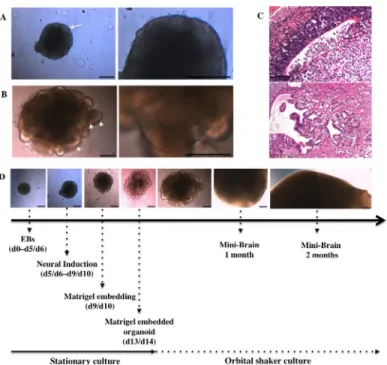
The MB protocol previously described by Lancaster et al. [26,27] was reproduced with minor modifications (see Methods) from human iPSCs (S1 Fig) to achieve discrete brain regionaliza-tion in vitro as confirmed after analysis of the expression of several neural markers. We observed 3D neuroectodermal differentiation towards neuroepithelium and self-organization leading to growth and development of specific brain regions representative of whole brain tis-sues. Neural ectoderm acquired apicobasal polarity and developed as a continuous layer dur-ing neural induction (NI) along the outer surface of EBs (Fig 1A). Neural differentiation and encapsulation of 3D free-floating cell aggregates within the extracellular matrix provided by Matrigel enabled neuroepithelia to organize radially (Fig 1B, higher magnification image on the right). Moreover, the use of an external scaffold that provided a structural support allowed the neuroepithelial buds to expand (Fig 1B, arrowhead) along with further growth, regionaliza-tion and differentiaregionaliza-tion of neuroepithelial tissue (Fig 1C). Finally the MB protocol supported the growth of neural tissue and its continuous expansion, achieving maximal size after 2 months (Fig 1C) as confirmed by previous studies [27,28] as well as the development of 3D neural microenvironments, which allowed the identification of discrete brain regions (Fig 1D).
Immunohistochemical analyses were used to characterize the presence of different neural cell types. Ventricle-like cavities reminiscent of brain ventricles (Fig 1D) expressing specific markers such as Nestin, PAX6, Ki67 and SOX2 suggest the proliferation of neural stem cells (NSCs) (S2A Fig) and real-time PCR analysis confirms increased marker expression associated with regionalization and neural progenitors, along with a decrease in other germ layer markers
(S2B Fig). The presence of more mature neurons was shown by the expression of neuronal proteins such as NeuN, NCAM, MAP2, TUJ1, and CTIP2 indicating differentiation, migration and maturation of newly formed neurons (S2C Fig). Moreover, staining for intermediate fila-ment proteins GFAP and Vifila-mentin shows the existence of glial cell types (S2D Fig).
Chemical induction of A
β accumulation
MBs develop discrete brain regions after one month of culture [28] and reach their maximal size of 3–4 mm in diameter after 2 months (Fig 1. and [27]). To investigate whether MBs can be used to present an increased Aβ42/Aβ40ratio, MBs were treated with Aftin-5, (Amyloid-β
Forty-Two Inducer) a chemical compound which increases the production of extracellular sol-uble secreted amyloid peptides in vitro [25]. We measured Aβ42and Aβ40concentrations in
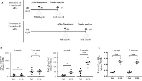
conditioned media after Aftin-5 induction in 1-month old and 2-month old MBs (Fig 2A). Chemical treatment of MBs was found to alter APP catabolism, leading to changes in extra-cellular Aβ42production without alteration of Aβ40concentration (Fig 2B). Statistical data
analysis showed significantly higher Aβ42mean levels in 1 month- (�p = 0.02) and 2-month
old (���p = 0.009) MBs following treatment with Aftin-5 when compared to vehicle-treated
control MBs. An increase in the Aβ42/Aβ40ratio was observed consequently (Fig 2C),
consis-tent with previously published in vitro and in vivo 2D models. Interestingly, higher Aβ42
pro-duction was observed after Aftin-5 treatment of 2-month old MBs compared to 1-month old MBs. This could be a consequence of the maturation of neuroectodermal organoids occurring between 1 and 2 months. Moreover, after treatment with aftin, organoids displayed no
Fig 1. Mini-Brain maturation through neural induction and differentiation. (A) Neural induction matures the neuroectodermic layers at the outer surface of the EB, the edge of which appears optically translucent (arrow). (B) Neural differentiation in Matrigel droplets expands neuroepithelial buds (arrowheads) and increases their size (primitive organoid). (C) Organoids develop in size and morphology for 2 months, reaching their maximal size of 3–4 mm in diameter at the end of this period. (D) Hematoxylin and eosin staining shows the presence of discrete brain regions such as ventricle like-cavities (upper panel, 1-month old MBs) and choroid plexus-like structures (lower panel, 4-month old MBs). Scale bars 250μm (A, B, C) and 100 μm (D).
observable phosphorylation of tau protein, nor extracellularβ-amyloid as shown by immuno-histochemical staining (S4 Fig).
Time-dependent physiological expression of Aβ oligomers during culture
To establish the physiological expression of Aβ during MB culture, Aβ levels were compared at different timepoints. The concentration of soluble Aβ peptides measured in conditioned media harvested from 1-month old MBs was compared to that harvested from 2-month old MBs. During physiological maturation of MBs, Aβ levels increased with age (S3 Fig): A signifi-cant increase in concentrations of secreted Aβ fragments was observed in 2-month-old MBs when compared to 1-month-old cultures. Both Aβ40and Aβ42concentrations increased during
MB maturation without alteration in Aβ42/Aβ40ratios (S3 Fig). These data suggest that during
MB maturation there is an accumulation of Aβ peptides, while the ratio remains stable, which is consistent with a physiological condition.
APP and PrP
Cexpression analysis throughout maturation of MB culture
We have shown that cerebral organoid maturation and cellular type enrichment occur in a time-dependent manner during up to 6 months of culture [28]. We investigated the expression level of relevant genes during MB maturation. Research for a specific Aβ42cellular surface
receptor has led to the identification of a highly specific interaction with the cellular prion pro-tein (PrPC) [29]. Once the expression pattern of secreted Aβ oligomers was established in the
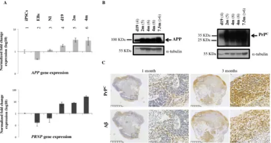
MB model, we chose to study the expression and metabolism of APP and PrPCthroughout the maturation of MB (Fig 3A and 3B). Transcript and protein expression were analyzed during 7.5 months of culture (Fig 3A and 3B). The expression levels of APP and PrPCwere modulated in a time-dependent manner during culture.APP and PRNP gene expression increased after
Fig 2. Chemical induction of APP cleavage in vitro upregulates both Aβ42and Aβ42/Aβ40ratio without altering
Aβ40levels. (A) Schematic representation of treatments with Aftin-5; 1- and 2-month-old MBs (upper and lower
representations respectively) were treated once with Aftin-5 for 4 days before collecting conditioned media. (B) Aβ40
(left) and Aβ42(right) mean concentration levels in conditioned media measured by ELISA (MSD); concentrations of
soluble Aβ40and Aβ42peptides from supernatant of vehicle-treated with DMSO (Ctrl) were compared with
concentrations obtained from MBs treated with Aftin-5 (concentration of 150μM (A150)). (C) Aβ42/Aβ40ratios
corresponding to 1 month- and 2-month old organoids measured by ELISA (MSD) in conditioned media of MBs treated with control vehicle or with Aftin-5. Statistical analysis: unpaired nonparametric Mann-Whitney test. On charts�: p = 0.02;���: p = 0.009; ns: not significant.
neural induction (NI) (Fig 3A) and protein analysis confirmed time-dependent production of APP and PrPC(Fig 3B). Finally, immunohistochemistry of MBs was used to localize PrPCand Aβ expression and to compare 1-month and 2-month old MBs. Immunohistochemical analy-sis confirmed the existence of increased immunoreactivity between 1 and 3 months using an N-terminal specific antibody for PrPC(SAF32) and an antibody directed against the N-termi-nal part of Aβ peptide sequence (clone BAM10) (Fig 3C). Aβ and PrPc were expressed in the
same regions of the organoid (Fig 3C), suggesting a coexpression of the two biomarkers. These data correlate with the parallel expression signatures of these two genes during the early phases of brain development (see human transcriptome available online:http://hbatlas.org). These data confirm that time-dependent changes in APP metabolism, which correspond to increased Aβ production found in conditioned media (S3 Fig) as well as in MB sections (Fig 3C), corre-late with the increase in PrPCexpression, confirming a potential role for these proteins during cerebral tissue maturation in vitro.
Discussion
Human MBs have been shown to be a highly valuable and predictive in vitro tool to study human brain physiological pathways during development as well as a powerful means of reca-pitulating brain-associated pathologies [17,18,27,30]. The MB model is considered to be very promising in the future study of neurological and neurodegenerative disorders [31]. One remaining question is whether human neural cells issued from iPSCs provide an adequate model for recapitulating phenotypes associated with neurodegenerative diseases. Because
Fig 3. APP and PrPCexpression in MBs during culture. (A) Real-time PCR analysis showsAPP and PRNP gene
expression at different times during culture. Expression during MB generation and differentiation was normalized with iPSCs expression, which was used as the baseline. (1) iPSCs; (2) embryoid bodies, (EBs); (3) embryoid bodies after neural induction (NI); (4) MBs after 19 days of culture (d19); (5) 2-month old MBs (2m); (6) 4-month old MBs (4m). (B) Western Blot analysis of APP and PrPCusing specific antibodies: PrPCantibody SAF32 is specific for the
octapeptide repeat region at the N-terminal site which contains the octarepeat region; full-length diglycosylated PrPC appears at * 35 kDa (arrow) whereas unglycosylated and truncated forms appear at a lower molecular weight. 22C11 antibody recognizes amino acid sequence 66–81 and allows identification of a full-length APP695 isoform at * 100 kDa (arrow) as well as an N-truncated form of APP protein. The Bradford method was used to measure the protein load (5μg of total protein per well); α-tubulin (* 55 kDa) was used as loading control. (4) MBs after 19 days of culture (d19); (5) 2-month old MBs (2m); (6) 4-month old MBs (4m); (>6) 7-month old MBs (7m). (C) Tissue sections of 1 month- (left panel) and 3-month old MBs (right panel), immunostained with specific antibodies recognizing PrPC (SAF32 antibody, specific for an N-terminal epitope of protein, upper panel) and Aβ peptides (BAM10 antibody, lower panel).
reprogramming to iPSC reverts cells to a fetal state, this could represent a problem in model-ling age-related neurological disorders. However, several studies have demonstrated and vali-dated the use of iPSCs to model pathologies such as AD. Cultures of patient-derived neurons have shed light on AD pathogenic mechanisms confirming the utility and the substantial potential of patient-derived iPSCs obtained from sporadic or familial forms of the disease [32]. These models show high variability, where a single cell line can give rise to different results depending on the differentiation protocol. For instance, the same FAD cell line may demon-strate contrasting results when used in 2D neuronal assays compared 3D brain organoid assays (S1 Table). Critical improvements are needed to obtain more reproducible models of AD pathology based on these systems, especially concerning the sporadic form of AD, which rep-resents most clinical cases.
We followed the secretion of Aβ oligomers by MBs obtained from a control IPS cell line to evaluate the possibility of provoking an AD-associated phenotype through chemical induction. It has been shown that increased production of the longer Aβ species (such as Aβ42or Aβ43)
makes aggregation more likely, and leads to the formation of toxic oligomers which confers synaptic dysfunction and ultimately may give rise to amyloid plaque formation [33]. A shorter and more hydrophilic Aβ40form, which is the most abundantly produced Aβ fragment, is
thought to contribute directly to small changes observed in Aβ pool concentrations [4,34,35]. The Aβ42/Aβ40ratio has been shown in many preclinical and clinical models to be more
bio-logically relevant for AD than Aβ42level alone [33,36,37].
In this work, a normal control IPS cell line differentiated in neuroectodermal organoids dis-played a physiological time-dependent increase in Aβ secretion and a stable Aβ42/Aβ40ratio
over the first two months of culture, reminiscent of the natural maturation occurring in humans. This suggests that although IPS-derived organoids remain embryonic in nature, mat-uration occurs in vitro. A chemical induction strategy was adopted to modulate APP cleavage and to increase the susceptibility of MBs to Aβ accumulation mechanisms. Induction with Aftin-5 reproducibly led to disruption in Aβ42/Aβ40ratios due to a rapid increase of soluble
Aβ42accumulation after 4 days of treatment, thus acquiring the specific AD signature. Our
data shows that the MB model can be used for the specific purpose of studying AD phenotypes, on a normal genetic background, reminiscent of the signature of sporadic AD.
Some accounts evaluate that sporadic AD affects up to 50% of the population by the age of 85 [20]. This evidence suggests that the basic genetic component of sporadic AD is not the main etiological factor in most of these cases and orientates to alternative hypotheses as non-sequence-based sources of variability in the genome, notably epigenetic modifications (which encompass the different conformational changes in DNA and/or chromatin). It has been reported that >30% of chromatin is dramatically reorganized in senescent cells [38]. More-over, multiple aging models strongly suggest a massive alteration in histone modification pat-terns, while multiple genes are directly affected by an altered DNA methylation (global DNA hypomethylation and local hypermethylation that may activate specific transcriptional pro-grams). If old cells accumulate aggregated proteins, genomic damage and display telomere ero-sion and mitochondrial dysfunction, their reprogramming into iPSC can be considered to mimic the “resetting” that occurs during meiosis and fertilization, which allows the formation of a new individual from two older individuals. [21]. In other words, the most critical epige-netic modifications expected from the cells from AD patients are potentially neutralized dur-ing the reprogrammdur-ing process to generate iPSC cell lines. Moreover, dependdur-ing on the differentiation protocols used to obtain 2D or 3D neuronal or more complex models such as MBs, different epigenetic marks will be created, i.e. a different impact on the underlying genetic component.
The consequence of Aβ peptide production, accumulation and interaction with other pro-teins is still under study, but prion-like mechanisms are known to underlie most human neurodegenerative disorders including AD and Parkinson’s disease [39]. Because PrPc acts as a high-affinity receptor for Aβ oligomers [29] and this interaction produces a neurotoxic effect in mice [29], we investigated if PrPc was expressed during the first stages of development of the organoid. We found that the expression of APP and PrPc are both increased during the first two months of culture.
It has been suggested that PrPc extracted from AD brains contains multiple specific binding sites depending on the C-terminal of the Aβ fragment as well as its biophysical conformation at multiple sites on the protein [40,41], but the consequences of the strong interaction between PrPc and Aβ species remain to be established in the AD context. Moreover very little is known about the physiological roles of these proteins in brain development and physiology [42–45]. This study has shown for the first time a spatial and temporal correlation between PrPc and APP/Aβ expression during normal MB maturation, which suggests physiological interactions and opens up new avenues for further investigations.
Confronted with current limits linked to the use of transgenic mouse models that reflect neither the complexity of the human brain nor all the different aspects of AD pathology, MBs constitute a unique human brain model describing time-dependent expression and metabo-lism of APP and PrPCduring long-term culture as well as a model in which Aβ42amyloid
secretion can be chemically induced.
Conclusion
To surmount prevailing obstacles inhibiting the development of efficient strategies in the treat-ment of AD, the pharmaceutical industry requires alternative experitreat-mental models. Although progress has been made using transgenic mouse models, to date no in vivo model reflects the complexity of the human brain or reproduces the full extent of the pathology found in patients. Comparing the phenotypes obtained using various 2D in vitro cell culture methods highlights discrepancies between the models as well as their inability to coherently recapitulate pathologi-cal events. 3D-based models may constitute a more physiologipathologi-cally relevant model, although the embryonic nature of IPS derivatives may hinder this type of approach. The aim of this work was to take advantage of the research prospects offered by neuroectodermal organoids in the field of neuroscience. To validate the use of MB models to recapitulate AD-associated phe-notypes, the ability of the chemical inductor Aftin-5 to modulate the APP post-transcriptional pathway towards a pathological outcome was tested on organoids obtained from a control IPS cell line without any described mutations. MBs were chosen for this study to investigate the proteins implicated in pathophysiological mechanisms linked to sporadic forms of AD. Treat-ment of MBs with Aftin-5 leads to a reproducible disruption of the physiological balance of the Aβ42/Aβ40ratio observed during normal maturation. The secreted Aβ42concentration and
Aβ42/Aβ40ratio rose over 4 days of treatment whereas the secreted Aβ40concentration
remained stable. In this timeframe, neither phosphorylation of tau protein nor extracellular β-amyloid were revealed by immunohistochemistry in the aftin-treated organoids (S4 Fig). Our study supports the pioneering work of Raja et al., who aimed to closer emulate human pathol-ogy using a 3D in vitro model of the brain based on FAD iPSC to study neurological disease, although further work is still needed in order to recapitulate all the hallmarks of AD in vitro. Our aim was to test the capacity of the MBs to model sporadic AD even when the initial iPSC cell line originated from a non-AD patient. We used a strategy of chemical induction to reproduce the fluctuations in Aβ observed in AD which support the amyloid cascade hypothe-sis in brain organoids issued from control iPSCs [4,23,24]. We confirmed that human MBs
obtained from a normal patient can reproduce a relevant phenotype in vitro, opening new per-spectives in the understanding of physiological and pathological brain mechanisms. We further demonstrate the possibility of studying the expression of biomarkers during neural differentia-tion and maturadifferentia-tion over several months in vitro, validating a physiological expression profile for APP and PrPCover time. This is consistent with human brain development and maturation in vivo. In addition, we were able to identify a physiological Aβ accumulation in vitro which dif-fers from that obtained using Aftin-5. In particular, Aβ40and Aβ42levels were found to increase
with time while maintaining a stable ratio during the first two months of culture.
Through the observation of APP metabolism and PrPCexpression, we show that after chemical induction, the MB model of 3D maturing human cells, capable of long-term culture, can be used to study phenotypes associated with sporadic cases of neurodegenerative patholo-gies. This concept can also be tested in AD derived IPSCs to compensate for critical phenotypi-cal signatures that could have been lost during reprogramming. Future improvements to this model such as the integration of vascularization and the combination of genetic and molecular strategies to accelerate normal aging processes may contribute to the identification of novel biomarkers or potential targets in AD. Chemical induction of AD-related proteins in neuroec-todermal organoids provides a biochemical signature that is closer to the most frequent spo-radic forms of AD, which could provide a path towards more predictive future models applicable in the field of drug discovery.
Experimental procedure
Maintenance of iPSCs culture and 3D differentiation of mini-brains
BJ primary fibroblasts were obtained from ATCC (CRL-2522). Fibroblasts were repro-grammed using the Sendai virus reprogramming method as recommended by the manufac-turer (Life Technologies, cat. no. A16517). Briefly, BJ cells were infected on day 0, and plated on inactivated mouse embryonic fibroblasts (MEF, GlobalStem) 48 hours after infection. Stem cell medium composed of DMEM/F12 (Life Technologies), supplemented with 20% KOSR (Life Technologies), glutamine, non-essential amino-acids and bFGF (10 ng/ml, Stem Cell Technologies,) was changed daily. Clones were picked starting 20 days post infection, and expanded on murine embryonic fibroblasts (MEF, GlobalStem) before being adapted to feeder-free conditions. Assessment of pluripotency by semi-quantitative PCR was performed periodically to confirm the expression of pluripotent markers in iPSC cultures (S1 Fig). iPSCs used to obtain cerebral organoids were maintained in feeder-free condition on Matrigel-coat-ing (CornMatrigel-coat-ing), fed usMatrigel-coat-ing mTeSR1 medium (StemCell Technologies) and checked to confirm the absence of mycoplasma. iPSC colonies were cultured up to 80% of confluence. We fol-lowed the protocol published by Lancaster et al. with minor modifications [26]. At day 0 of the differentiation protocol cells were dissociated to allow cell counting as described previously. The first step of the previously published protocol was modified using a more homogeneous method to obtain embryoid bodies (EBs) [46]. Briefly, hanging drops of 20μl of EBs medium containing 9000 cells per drop were cultured on a Petri dish cover (10 cm in diameter) for 2 days in order to allow them to aggregate before being collected (S5A Fig). At day 2 each EB was harvested manually using a cut micropipette tip and placed in a well of a non-treated 24-well plate (S5B Fig) (Corning). Media changes, neural induction and neural differentiation were performed following the published protocol [26].
Immunohistochemical analysis
MBs were washed twice with calcium and magnesium-free D-PBS (Sigma-Aldrich) and fixed in formaldehyde (4%) (VWR) solution for 15 minutes at 4˚C and then 72 hours at RT. MBs
were washed again in D-PBS and prepared for paraffin embedding (Leica TP 1050). Histologi-cal paraffin sections of 5μm were prepared using a microtome (Leica) and collected on glass microscope slides (Thermo scientific). For immunostaining, paraffin was removed in a Med-ite-Medixintechnik Tissue Stainer TST 40 device and pretreated for epitope retrieval. We per-formed HIER (heat-induced epitope retrieval) pretreatment combining a heat treatment at 95˚C with a citrate buffer solution (pH 6.0) for 40 minutes. Slides were then recovered, washed in MilliQ warm water, blocked, washed twice with MilliQ water, once with PBS solution 1X (Sigma-Aldrich) and once with TBS-Tween 0.05% solution (Sigma-Aldrich). Sections were then incubated with 200μl of primary antibody solution overnight at 4˚C. The antibody solu-tion was removed and secsolu-tions received three washes of TBS-Tween 0.05% solusolu-tion (Sigma-Aldrich) with agitation using an orbital shaker before the successive incubation with the sec-ondary antibody (ImmPress Reagent Kit peroxidase) for 30 minutes at RT. TBS-Tween 0,05% washes were repeated and 200μl of DAB reagent per section (ImmPACT Peroxidase substrate kit) was incubated (for 5 to 10 minutes) before being subjected to nuclear staining procedure in hematoxylin solution (600μl per section for 5 minutes). Slides were processed automatically (Medite-Medixintechnik Tissue Stainer) to allow tissue dehydration. Finally sections were cov-erslipped using Eukitt (Sigma-Aldrich) mounting medium and allowed to dry overnight at RT. The primary antibodies used for MB characterization as well as for the study of APP and PrPCexpression were diluted as follows: anti-Nestin antibody (Abcam), 1:500; anti-PAX6 (Covance), 1:300; anti-Ki67 (Dako), 1:200; anti-SOX2 (Epitomics), 1:300; anti-NeuN (Chemi-con), 1:1000; anti-NCAM (Santa Cruz), 1:100; anti-MAP2 (Sigma), 1:250; anti-TUJ1 (Cov-ance), 1:5000; anti-CTIP2 (Abcam), 1:100; anti-GFAP (Dako), 1:4000; anti-Vimentin (Dako), 1:2000; antibody directed against N-terminal residues of APP 66–81 (22C11, Millipore), 1:100; antibody directed against residues 1–12 of Aβ peptide (BAM10, Sigma-Aldrich), 1:1000; anti-body directed against octapeptide region of PrPC(SAF32, provided by J. Grassi, Commissariat a´ l’Energie Atomique/Saclay, Gif sur Yvette, France), 1:1000.
Analysis of gene expression
Total RNA was extracted from iPSCs and organoids using a Nucleospin RNA II Kit (Macherey-Nagel) according to the manufacturer’s instructions. Reverse transcription (RT) was carried out using an Iscript cDNA synthesis kit (Biorad) following the instructions provided by the manu-facturer. A total of 1μg of RNA was used in RT reactions. PCR amplifications were performed using the Real-Time PCR Mini-Opticon System (Biorad, USA) and the IQSYBR Green Super-mix kit (Biorad, USA) according to the manufacturer’s protocol. Two microlitres of the first strand cDNA (1:5 diluted) were used for amplification in triplicate in a 10μL reaction contain-ing SYBR Green and 10 pmol of each primer. The followcontain-ing PCR program was used: 95˚C for 3 min, 40 amplification cycles at 95˚C for 10 s, 60˚C for 30 s. Serial dilutions of each studied tran-script were used to determine the amplification efficiency of each target and housekeeping gene (TBP or RPLP0). Data were obtained by analysis with BioRad MFX Software 2.0 and are pre-sented as the fold-change in target gene expression normalized to the internal control gene. The average threshold cycle (CT) was calculated for both the target gene and housekeeping genes, andΔCT was determined as [the mean of the triplicate CT values for the target gene]-[the mean of the triplicate CT values for TBP or RPLP0].ΔΔCT represented the difference between the paired samples, as calculated by the formulaΔΔCT = (ΔCT of sample—ΔCT of control).
Aβ quantification
The concentration of soluble Aβ42, Aβ40and Aβ38fragments produced after Aftin-5 treatment
and imager manufactured by Meso Scale Diagnostic (MSD Multi-Spot Assay System) and compared to the control (DMSO). After four days of treatment the medium was collected and a multiplex kit containing Aβ peptide panel (4G8 and 6E10 Aβ antibodies) was applied accord-ing to the manufacturer’s protocol.
Statistical analysis
Data were analyzed using PRISM 7 (GraphPad) and SPSS (IBM Analytics) analysis software and values are expressed as the means± SD. Means of concentrations and ratios were calcu-lated from independent experiments and values corresponding to each mini-brain were repre-sented by a circle (controls) or a triangle (treated). Each point represents a conditioned medium obtained from the culture of a single organoid. Statistical significance was tested using a nonparametric Mann-Whitney test to compare the differences between groups. Differ-ences were retained as statistically significant using p-values of < 0.05 (confidence level 95%).
Supporting information
S1 Fig. Assessment of pluripotency. Assessment of pluripotency by semi-quantitative PCR to confirm the expression of pluripotent markers by iPSC line BJ. PCR analysis shows the expres-sion of pluripotency markersOCT4, SOX2, NANOG and REX-1. BJ fibroblasts were used as a control andRPLP0 as a housekeeping gene.
(TIF)
S2 Fig. Characterization of MBs (A) Immunohistochemical (IHC) staining of neurogenic niches shows the expression of markers associated with neural stem cell maintenance (Nestin, PAX6, SOX2) and proliferation (Ki67) at the apical surface of ventricular-like cavities. (B) Quantitative PCR analysis of neural markers which compare undifferentiated iPSCs with dif-ferentiated EBs and MBs (after 19 days of differentiation); increased genetic expression of ecto-dermal (NES and OLIG2), associated markers accompanied by a decrease of endoderm (AFP) and mesoderm (BRACH) markers after 19 days culture.
OLIG2 expression indicates also ventral and dorsal regionalization of the spinal cord as well as the presence of oligodendrocytic precursor identities. The expression of forebrain (FOXG1) and hindbrain (ISL-1) markers confirms MB regionalization during differentiation. (C) IHC staining allows the identification of neural marker expression which indicates the presence of more mature neurons suggesting the differentiation and migration of NSCs from a neurogenic niche. NeuN, neuron specific nuclear protein; NCAM, neural cell adhesion molecule; MAP2, microtubule associated protein 2; TUJ1, neuron specific class IIIβ-tubulin; CTIP2, newly born deep layer neurons. (D) Glia identity was established using intermediate filament specific anti-bodies GFAP (glial fibrillary acidic protein) and Vimentin.
(TIF)
S3 Fig. Analysis of Aβ40, Aβ42and Aβ42/Aβ40ratio in conditioned media after 1 and 2
months of MB culture; Statistical analysis: unpaired nonparametric Mann-Whitney test. On charts�: p = 0.002;���: p = 0.009; ns: not significant.
(TIF)
S4 Fig. Immunohistochemical staining of Aftin 5-treated MB. IHC with 12F4 (anti-Aß42) and AT8 (anti-phospho-Tau(Ser202,Thr205). A- Staining controls: IHC stained sections with 12F4 antibody of postmortem brain samples: on the left non-human primate, adult sample (1), on the right human late stage AD sample (2) B- Staining of organoids: IHC stained sections with 12F4 antibody of Minibrains: on the left MB treated with vehicle (DMSO) (1), on the right MB treated with Aftin-5 (2) C- Staining controls: IHC stained sections with AT8
antibody of postmortem brain samples: on the left healthy non-human primate adult sample (1), on the right human late-stage AD sample (2) D- IHC stained sections with AT8 of Mini-brains: on the left MB treated with vehicle (DMSO) (1), on the right MB treated with Aftin-5 (2). Scale bars: 100μm.
(TIF)
S5 Fig. Generation of MBs using hanging drop method. (A) iPSC colony cultured on Matri-gel before dissociation (corresponding to day 0 of the MB protocol, on the left), hanging drop culture of iPS cell suspension on a petri dish cover (day 0—day 2, second picture); iPSCs were maintained in drop culture for 2 days allowing cells to aggregate, forming the EB in the center of the hanging drop and each drop contains one EB (third picture) which is harvested at day 2 (right). (B) Representation of the two step hanging drop method; EBs were cultured in the hanging drops for 2 days; then, each EB was recovered manually with a cut micropipette tip and placed into a 24-well plate containing EB medium.
(TIF)
S1 Table. Comparison of iPSC lines used to reproduce AD-associated phenotypes in 2D and 3D neural culturesin vitro. In the study of Raja et al. 2016, all four FAD lines were used
to create 3D brain organoids. APPDp 1–1, APPDp 2–3 (EOAD) carrying APP duplication were used in the study of Israel et al. 2012 whereas FAD lines ND34732 (PSEN1 M146I) and AG06840 (PSEN1 A264E) were used in other studies as indicated in the respective lines. Results indicated in the table refer to extracellular measures of Aβ peptide concentrations (Duan et al. 2014; Yagi et al. 2014; Liu et al. 2014; Sproul et al. 2014).(□) Increase in the con-centration of Aβ compared to control lines; (=) no change in the concon-centration observed between FAD and control lines; (/) unknown.
(TIF)
Acknowledgments
We are very thankful to Franc¸oise Raynaud, Dolorès Jouy, Emmanuel Comoy, Je´rome Del-motte, Elodie Sanfins and Laure Caruana in our laboratory, Ouerdia Arkoun (Sup’Biotech) for the statistical analysis support provided, and Charlotte Leuxe (CEA) for the contribution to the MSD analysis. This work was supported by the French national PIA2 program under con-tract No. P112331-3422142 (3DNS project) and the EU Joint Programme Neurodegenerative Diseases Research (JPND; 3DMiniBrain).
Author Contributions
Conceptualization: Serena Pavoni, Jacqueline Mikol, Aloïse Mabondzo, Frank Yates. Formal analysis: Serena Pavoni.
Funding acquisition: Jean-Philippe Deslys.
Investigation: Serena Pavoni, Rafika Jarray, Ferid Nassor, Anne-Ce´cile Guyot, Steve Cottin, Jessica Rontard.
Methodology: Serena Pavoni, Aloïse Mabondzo, Frank Yates.
Project administration: Serena Pavoni, Jean-Philippe Deslys, Frank Yates. Supervision: Jean-Philippe Deslys.
Visualization: Serena Pavoni, Frank Yates.
Writing – original draft: Serena Pavoni, Jacqueline Mikol, Jean-Philippe Deslys, Frank Yates. Writing – review & editing: Rafika Jarray, Ferid Nassor, Frank Yates.
References
1. Kitazawa M, Medeiros R M. LaFerla F. Transgenic Mouse Models of Alzheimer Disease: Developing a Better Model as a Tool for Therapeutic Interventions. Curr Pharm Des. 2012; 18: 1131–1147.https:// doi.org/10.2174/138161212799315786PMID:22288400
2. Cummings J. Lessons Learned from Alzheimer Disease: Clinical Trials with Negative Outcomes. Clin Transl Sci. 2018; 11: 147–152.https://doi.org/10.1111/cts.12491PMID:28767185
3. Lesne´ S, Ming TK, Kotilinek L, Kayed R, Glabe CG, Yang A, et al. A specific amyloid-βprotein assembly in the brain impairs memory. Nature. 2006; 440: 352–357.https://doi.org/10.1038/nature04533PMID:
16541076
4. Haass C, Selkoe DJ. Soluble protein oligomers in neurodegeneration: lessons from the Alzheimer’s amyloidβ-peptide. Nat Rev Mol Cell Biol. 2007; 8: 101–112.https://doi.org/10.1038/nrm2101PMID:
17245412
5. Cavanaugh SE. Animal models of Alzheimer disease: historical pitfalls and a path forward. ALTEX. 2014; 31: 279–302.https://doi.org/10.14573/altex.1310071PMID:24793844
6. De Strooper B. Lessons from a failedγ-secretase Alzheimer trial. Cell. Elsevier Inc.; 2014; 159: 721– 726.https://doi.org/10.1016/j.cell.2014.10.016PMID:25417150
7. Goldstein LSB, Reyna S, Woodruff G. Probing the secrets of Alzheimer’s disease using human-induced pluripotent stem cell technology. Neurotherapeutics. 2015;https://doi.org/10.1007/s13311-014-0326-6
PMID:25534395
8. Freude K, Pires C, Hyttel P, Hall V. Induced Pluripotent Stem Cells Derived from Alzheimer’s Disease Patients: The Promise, the Hope and the Path Ahead. J Clin Med. 2014; 3: 1402–1436.https://doi.org/ 10.3390/jcm3041402PMID:26237610
9. Parr CJC, Yamanaka S, Saito H. An update on stem cell biology and engineering for brain development. Mol Psychiatry. 2017; 22: 808–819.https://doi.org/10.1038/mp.2017.66PMID:28373686
10. Muratore CR, Srikanth P, Callahan DG, Young-Pearse TL. Comparison and optimization of hiPSC fore-brain cortical differentiation protocols. PLoS One. 2014; 9: 1–18.https://doi.org/10.1371/journal.pone. 0105807PMID:25165848
11. Israel M a., Yuan SH, Bardy C, Reyna SM, Mu Y, Herrera C, et al. Probing sporadic and familial Alzhei-mer’s disease using induced pluripotent stem cells. Nature. 2012; 482: 216–220.https://doi.org/10. 1038/nature10821PMID:22278060
12. Kondo T, Asai M, Tsukita K, Kutoku Y, Ohsawa Y, Sunada Y, et al. Modeling Alzheimer’s disease with iPSCs reveals stress phenotypes associated with intracellular Aβand differential drug responsiveness. Cell Stem Cell. Elsevier Inc.; 2013; 12: 487–496.https://doi.org/10.1016/j.stem.2013.01.009PMID:
23434393
13. Choi SH, Kim YH, Hebisch M, Sliwinski C, Lee S, D’Avanzo C, et al. A three-dimensional human neural cell culture model of Alzheimer’s disease. Nature. Nature Publishing Group; 2014; 515: 274–278.
https://doi.org/10.1038/nature13800PMID:25307057
14. D’Avanzo C, Aronson J, Kim YH, Choi SH, Tanzi RE, Kim DY. Alzheimer’s in 3D culture: Challenges and perspectives. BioEssays. 2015;https://doi.org/10.1002/bies.201500063PMID:26252541
15. Fatehullah A, Tan SH, Barker N. Organoids as an in vitro model of human development and disease. Nat Cell Biol. Nature Publishing Group; 2016; 18: 246–254.https://doi.org/10.1038/ncb3312PMID:
26911908
16. Brennand KJ, Marchetto MC, Benvenisty N, Bru¨stle O, Ebert A, Izpisua Belmonte JC, et al. Creating Patient-Specific Neural Cells for the in Vitro Study of Brain Disorders. Stem Cell Reports. 2015; 5: 933– 945.https://doi.org/10.1016/j.stemcr.2015.10.011PMID:26610635
17. Gonzalez C, Armijo E, Bravo-alegria J, Mays ABCE, Soto C. Modeling amyloid beta and tau pathology in human cerebral organoids. Mol Psychiatry. Springer US; 2018; https://doi.org/10.1038/s41380-018-0229-8PMID:30171212
18. Raja WK, Mungenast AE, Lin Y-T, Ko T, Abdurrob F, Seo J, et al. Self-Organizing 3D Human Neural Tissue Derived from Induced Pluripotent Stem Cells Recapitulate Alzheimer’s Disease Phenotypes. PLoS One. 2016; 11: e0161969.https://doi.org/10.1371/journal.pone.0161969PMID:27622770
19. Kuperstein I, Broersen K, Benilova I, Rozenski J, Jonckheere W, Debulpaep M, et al. Neurotoxicity of Alzheimer’s disease Aβpeptides is induced by small changes in the Aβ42 to Aβ40 ratio. EMBO J. 2010; 29: 3408–3420.https://doi.org/10.1038/emboj.2010.211PMID:20818335
20. Hirtz D, Thurman DJ, Gwinn-Hardy K, Mohamed M, Chaudhuri AR, Zalutsky R. How common are the “common” neurologic disorders? Neurology. 2007; 68: 326–337.https://doi.org/10.1212/01.wnl. 0000252807.38124.a3PMID:17261678
21. Mahmoudi S, Brunet A. Aging and reprogramming: A two-way street. Current Opinion in Cell Biology. 2012.https://doi.org/10.1016/j.ceb.2012.10.004PMID:23146768
22. Sen P, Shah PP, Nativio R, Berger SL. Epigenetic Mechanisms of Longevity and Aging. Cell. 2016; 166: 822–839.https://doi.org/10.1016/j.cell.2016.07.050PMID:27518561
23. Hardy JA, Higgins GA. Alzheimer’s Disease: The Amyloid Cascade Hypotesis. Science (80-). 1992; 256: 184–5.
24. Tanzi RE, Bertram L. Twenty years of the Alzheimer’s disease amyloid hypothesis: A genetic perspec-tive. Cell. 2005; 120: 545–555.https://doi.org/10.1016/j.cell.2005.02.008PMID:15734686
25. Hochard A, Oumata N, Bettayeb K, Gloulou O, Fant X, Durieu E, et al. Aftins Increase Amyloid-β42, Lower Amyloid-β38, and Do Not Alter Amyloid-β40 Extracellular Production in vitro: Toward a Chemical Model of Alzheimer’s Disease? J Alzheimer’s Dis. 2013; 35: 107–120. https://doi.org/10.3233/JAD-121777PMID:23364140
26. Lancaster MA, Knoblich JA. Generation of cerebral organoids from human pluripotent stem cells. Nat Protoc. 2014; 9: 2329–2340.https://doi.org/10.1038/nprot.2014.158PMID:25188634
27. Lancaster MA, Renner M, Martin CA, Wenzel D, Bicknell LS, Hurles ME, et al. Cerebral organoids model human brain development and microcephaly. Nature. Nature Publishing Group; 2013; 501: 373– 379.https://doi.org/10.1038/nature12517PMID:23995685
28. Quadrato G, Nguyen T, Macosko EZ, Sherwood JL, Min Yang S, Berger DR, et al. Cell diversity and network dynamics in photosensitive human brain organoids. Nature. 2017;https://doi.org/10.1038/ nature22047PMID:28445462
29. Laure´ n J, Gimbel D a, Nygaard HB, Gilbert JW, M S. Cellular Prion Protein Mediates Impairment of Syn-aptic Plasticity by Amyloid-βOligomers-Supplementary Information. Nature. 2009; 457: 1128–1132.
https://doi.org/10.1038/nature07761PMID:19242475
30. Qian X, Nguyen HN, Song MM, Hadiono C, Ogden SC, Hammack C, et al. Brain-Region-Specific Orga-noids Using Mini-bioreactors for Modeling ZIKV Exposure. Cell. 2016; 165: 1238–1254.https://doi.org/ 10.1016/j.cell.2016.04.032PMID:27118425
31. Brawner AT, Xu R, Liu D, Jiang P. Generating CNS organoids from human induced pluripotent stem cells for modeling neurological disorders. Int J Physiol Pathophysiol Pharmacol. 2017; 9: 101–111. PMID:28694921
32. Ghaffari LT, Starr A, Nelson AT, Sattler R. Representing Diversity in the Dish: Using Patient-Derived in Vitro Models to Recreate the Heterogeneity of Neurological Disease. Front Neurosci. 2018; 12.https:// doi.org/10.3389/fnins.2018.00056PMID:29479303
33. Lewczuk P, Matzen A, Blennow K, Parnetti L, Molinuevo JL, Eusebi P, et al. Cerebrospinal Fluid Aβ42/ 40 Corresponds Better than Aβ42 to Amyloid PET in Alzheimer’s Disease. J Alzheimer’s Dis. 2017; 55: 813–822.https://doi.org/10.3233/JAD-160722PMID:27792012
34. Fagan AM, Head D, Shah AR, Marcus D, Mintun M, Morris JC, et al. Decreased CSF Aβ42 correlates with brain atrophy in cognitively normal elderly. Ann Neurol. 2009; 65: 176–183.https://doi.org/10.1002/ ana.21559PMID:19260027
35. Collins-Praino LE, Francis YI, Griffith EY, Wiegman AF, Urbach J, Lawton A, et al. Soluble amyloid beta levels are elevated in the white matter of Alzheimer’s patients, independent of cortical plaque severity. Acta Neuropathol Commun. 2014; 2: 83.https://doi.org/10.1186/s40478-014-0083-0PMID:25129614
36. Lue L, Kuo Y, Roher AE, Brachova L, Shen Y, Sue L, et al. Soluble AmyloidβPeptide Concentration as a Predictor of Synaptic Change in Alzheimer’s Disease. Am J Pathol. 1999; 155: 853–862.https://doi. org/10.1016/S0002-9440(10)65184-XPMID:10487842
37. Spies PE, Slats D, Sjo¨gren JMC, Kremer BPH, Verhey FRJ, Rikkert MGMO, et al. The cerebrospinal fluid amyloid beta42/40 ratio in the differentiation of Alzheimer’s disease from non-Alzheimer’s demen-tia. Curr Alzheimer Res. 2010; 7: 470–6.https://doi.org/10.2174/156720510791383796PMID:
20043812
38. Shah PP, Donahue G, Otte GL, Capell BC, Nelson DM, Cao K, et al. Lamin B1 depletion in senescent cells triggers large-scale changes in gene expression and the chromatin landscape. Genes Dev. 2013;
39. Goedert M. Alzheimer’s and Parkinson’s diseases: The prion concept in relation to assembled Aβ, tau, andα-synuclein. Science (80-). 2015; 349: 601–702.https://doi.org/10.1126/science.1255555PMID:
26250687
40. Gunther E, Strittmatter S.β-amyloid oligomers and cellular prion protein in Alzheimer’s disease. J Mol Med. 2010; 88: 331–338.https://doi.org/10.1007/s00109-009-0568-7PMID:19960174
41. Zou WQ, Xiao X, Yuan J, Puoti G, Fujioka H, Wang X, et al. Amyloid-β42 interacts mainly with insoluble prion protein in the Alzheimer brain. J Biol Chem. 2011; 286: 15095–15105.https://doi.org/10.1074/jbc. M110.199356PMID:21393248
42. Manson J, West JD, Thomson V, McBride P, Kaufman MH, Hope J. The prion protein gene: a role in mouse embryogenesis? Development. 1992; 115: 117–22. PMID:1353438
43. Kovacs GG, Zerbi P, Voigtla¨nder T, Strohschneider M, Trabattoni G, Hainfellner JA, et al. The prion pro-tein in human neurodegenerative disorders. Neurosci Lett. 2002; 329: 269–272.https://doi.org/10. 1016/S0304-3940(02)00668-7PMID:12183028
44. Adle-Biassette H, Verney C, Peoc’h K, Dauge M-C, Razavi F, Choudat L, et al. Immunohistochemical Expression of Prion Protein (PrPC) in the Human Forebrain During Development. J Neuropathol Exp Neurol. 2006; 65: 698–706.https://doi.org/10.1097/01.jnen.0000228137.10531.72PMID:16825956
45. Steele AD, Emsley JG, Ozdinler PH, Lindquist S, Macklis JD. Prion protein (PrPc) positively regulates neural precursor proliferation during developmental and adult mammalian neurogenesis. Proc Natl Acad Sci. 2006; 103: 3416–3421.https://doi.org/10.1073/pnas.0511290103PMID:16492732
46. Sheridan SD, Surampudi V, Rao RR. Analysis of embryoid bodies derived from human induced pluripo-tent stem cells as a means to assess pluripotency. Stem Cells Int. 2012;https://doi.org/10.1155/2012/ 738910PMID:22550517