HAL Id: hal-01405905
https://hal.archives-ouvertes.fr/hal-01405905
Submitted on 28 Jun 2018
HAL is a multi-disciplinary open access
archive for the deposit and dissemination of
sci-entific research documents, whether they are
pub-lished or not. The documents may come from
teaching and research institutions in France or
abroad, or from public or private research centers.
L’archive ouverte pluridisciplinaire HAL, est
destinée au dépôt et à la diffusion de documents
scientifiques de niveau recherche, publiés ou non,
émanant des établissements d’enseignement et de
recherche français ou étrangers, des laboratoires
publics ou privés.
Cécile Mathieu, Ines Li de la Sierra-Gallay, Romain Duval, Ximing Xu, Angélique Cocaign, et al..
Insights into Brain Glycogen Metabolism. Journal of Biological Chemistry, American Society for
Biochemistry and Molecular Biology, 2016, 291, pp.18072 - 18083. �10.1074/jbc.M116.738898�.
�hal-01405905�
Insights into Brain Glycogen Metabolism
THE STRUCTURE OF HUMAN BRAIN GLYCOGEN PHOSPHORYLASE
*□SReceived for publication, May 18, 2016, and in revised form, July 7, 2016 Published, JBC Papers in Press, July 8, 2016, DOI 10.1074/jbc.M116.738898
Ce´cile Mathieua1, Ines Li de la Sierra-Gallayb, Romain Duvala2, Ximing Xua3, Ange´lique Cocaigna1, Thibaut Le´gerc, Gary Woffendind, Jean-Michel Camadroc, Catherine Etchebeste,f,g,h,i, Ahmed Haouzj, Jean-Marie Dupreta,i,
and Fernando Rodrigues-Limaa,i4
From theaUniversite´ Paris Diderot, Sorbonne Paris Cite´, Unite´ BFA, CNRS UMR 8251, 75013 Paris, France, thebFonction et Architecture des Assemblages Macromole´culaires, Institut de Biologie Inte´grative de la Cellule, Universite´ Paris Sud, UMR 9198 Orsay, 91405 France, thecUniversite´ Paris Diderot, Sorbonne Paris Cite´, Institut Jacques Monod, CNRS UMR 7592, 75013 Paris, France,dThermo Fisher Scientific, Hemel Hempstead HP2 7GE, United Kingdom,eINSERM, UMR S1134, Universite´ Paris Diderot, 75015 Paris, France,fUniversite´ Paris Diderot, Sorbonne Paris Cite´, 75004 Paris, France,gInstitut National de la Transfusion Sanguine, 75015 Paris, France,hLaboratoire d’Excellence GR-Ex, 75015 Paris, France,iUFR Sciences du Vivant, Universite´ Paris Diderot, 75013 Paris, France, and thejInstitut Pasteur, Plateforme de Cristallographie, CNRS UMR 3528, 75015 Paris, France
Brain glycogen metabolism plays a critical role in major brain functions such as learning or memory consolidation. However, alteration of glycogen metabolism and glycogen accumulation in the brain contributes to neurodegeneration as observed in Lafora disease. Glycogen phosphorylase (GP), a key enzyme in glycogen metabolism, catalyzes the rate-limiting step of glyco-gen mobilization. Moreover, the allosteric regulation of the three GP isozymes (muscle, liver, and brain) by metabolites and phosphorylation, in response to hormonal signaling, fine-tunes glycogenolysis to fulfill energetic and metabolic requirements. Whereas the structures of muscle and liver GPs have been known for decades, the structure of brain GP (bGP) has remained elusive despite its critical role in brain glycogen metabolism. Here, we report the crystal structure of human bGP in complex with PEG 400 (2.5 A˚ ) and in complex with its allos-teric activator AMP (3.4 A˚ ). These structures demonstrate that bGP has a closer structural relationship with muscle GP, which is also activated by AMP, contrary to liver GP, which is not. Importantly, despite the structural similarities between human bGP and the two other mammalian isozymes, the bGP struc-tures reveal molecular feastruc-tures unique to the brain isozyme that provide a deeper understanding of the differences in the activa-tion properties of these allosteric enzymes by the allosteric effector AMP. Overall, our study further supports that the dis-tinct structural and regulatory properties of GP isozymes con-tribute to the different functions of muscle, liver, and brain glycogen.
Glycogen performs various functions depending on its loca-tion in the body. In muscle, glycogen provides energy via the glycolysis pathway for skeletal muscle contraction (1, 2). Liver glycogen is metabolized and subsequently released as glucose into systemic circulation during fasting periods (1, 2). Brain glycogen has long been considered to be an emergency glucose store (1, 2). The key roles that brain glycogen and its metabo-lism play in the mammalian nervous system have only emerged over the last 20 years (3–5). For instance, astrocyte function and neuronal activity are critically dependent on glycogen stores and glycogenolysis, especially during episodes of stress includ-ing hypoglycemia and ischemia (6 –9). Astrocytic glycogen breakdown and subsequent astrocyte-neuron lactate transport have also been proposed to support high cognitive processes such as learning and memory consolidation (10 –16). In addi-tion, glycogen metabolism in neurons participates in neuronal tolerance to hypoxic stress (17). Glycogen itself is also impor-tant for brain function. However, alteration of its metabolism and overaccumulation in the brain, such as in Lafora disease, contributes to neurodegeneration (16, 18, 19).
Glycogen phosphorylase (EC 2.4.1.1; GP)5is the key enzyme
that regulates glycogen mobilization. This complex allosteric enzyme catalyzes the rate-limiting step of glycogenolysis by releasing glucose-1-phosphate (1). In mammals, GP comprises a family of three isozymes named according to the tissue of their highest abundance: muscle isozyme (mGP), liver isozyme (lGP), and brain isozyme (bGP). The three isozymes form homodimers comprised of identical subunits that are encoded by distinct but structurally related genes (20 –22). GPs are allos-teric enzymes that are regulated by both phosphorylation of
Ser14 and binding of allosteric effectors (such as AMP) that
control their transition between inactive and active conforma-tions (1). Although GPs display high similarity and catalyze the same reaction, these three isozymes differ in their sensitivity to allosteric activation and phosphorylation (1, 23, 24): lGP is essentially regulated by phosphorylation, mGP responds coop-eratively to activation by AMP and to phosphorylation, and
*This work was supported by running grants from University Paris Diderot, CNRS, and Institut Pasteur. The authors declare that they have no conflicts of interest with the contents of this article.
□S This article containssupplemental Tables S1 and Figures S1 and S2.
The atomic coordinates and structure factors (codes5IKPand5IKO) have been deposited in the Protein Data Bank (http://wwpdb.org/).
1Supported by Ph.D. fellowships from the French Ministry of Research (Ecole
Doctorale BioSPC).
2Supported by a Ph.D. fellowship from Re´gion Ile de France (appel hors DIM
2013).
3Supported by a fellowship from the China Scholarship Council. Present
address: Institut Pasteur, Unite´ de Pathoge´ne`se des Infections Vasculaires, 75015 Paris, France.
4To whom correspondence should be addressed. E-mail: fernando.
5The abbreviations used are: GP, glycogen phosphorylase; mGP, muscle GP;
lGP, liver GP; bGP, brain GP; PDB, Protein Data Bank.
THE JOURNAL OF BIOLOGICAL CHEMISTRY VOL. 291, NO. 35, pp. 18072–18083, August 26, 2016 © 2016 by The American Society for Biochemistry and Molecular Biology, Inc. Published in the U.S.A.
by guest on June 28, 2018
http://www.jbc.org/
bGP responds strongly and non-cooperatively to activation by AMP (23, 24). These regulatory differences likely represent dis-tinct functions of glycogen metabolism. Activation by AMP provides a response to intracellular energy demand, whereas the enzymatic cascade leading to phosphorylation of GP is the result of extracellular signals (1). Crystallographic determina-tion of the structures of mGP in 1976 and 1992 and lGP in 2000 led to a better understanding of the structural bases for the regulation of these isoenzymes and the development of drugs (25–32). The structure of a mammalian bGP has not been yet determined even though glycogen and bGP are increasingly recognized to play key roles in brain function. Knowledge of the structure of bGP would allow a better understanding of the molecular basis of brain glycogen metabolism, its regulation, and its role in physiological and pathological brain processes. In addition, the structure of bGP could be helpful to design isozyme-specific drugs.
Here, we report the first structures of human bGP in complex with PEG 400 and in complex with AMP, its allosteric activator. These structures demonstrate that bGP has a closer structural relationship with mGP, which is also activated by AMP, con-trary to liver GP, which is not. More importantly, the bGP structures suggest the molecular bases responsible for the dis-tinct responses of mGP and bGP to allosteric activation by AMP. More broadly, our study further supports that the dis-tinct structural and regulatory properties of GP isozymes con-tribute to the different functions of muscle, liver, and brain glycogen.
Results
Biochemical Characterization of bGP—Human bGP was overexpressed in Escherichia coli expressing the chaperonin complex GroES. The co-expression of the GroEL-GroES complex and human bGP substantially increased the production and purification yield of the functional recombi-nant enzyme by improving the correct folding of human bGP. The enzyme was expressed as an N-terminal
polyhisti-dine-tagged fusion protein and purified using Ni2⫹ affinity
chromatography. The polyhistidine tag was removed by thrombin digestion to ensure structural integrity and full biological activity of the protein. We assessed the protein for purity, enzymatic activity, and full-length sequence (Fig. 1,
a– c, andsupplemental Fig. S1).
GPs are allosteric enzymes activated by both
phosphoryla-tion of Ser14by phosphorylase kinase and binding of allosteric
effectors, such as AMP. We carried out biochemical and enzy-matic characterization of the recombinant bGP to ensure that the purified enzyme was functional. The glycogen saturation curve (Fig. 1d) and the activation of the enzyme by AMP, as well as following phosphorylation by phosphorylase kinase (Fig. 1e), confirmed the functionality of the protein. We assessed the phosphorylation state of the enzyme by Western blotting, using an antibody raised against phosphorylated serine (Fig. 1f) and native mass spectrometry analysis and confirmed the presence
of two phosphate groups per dimer (supplemental Fig. S1).
Phosphorylation of bGP promoted the activation of the enzyme
FIGURE 1. Biochemical characterization of recombinant human bGP. a, SDS-PAGE analysis of purified bGP. 1, 2, and 5g of purified bGP were subjected to SDS-PAGE under reducing conditions and stained with Coomassie Blue. b, Western blotting analysis of untagged and His6-tagged bGP. Western blotting
analysis was first conducted using a polyclonal antibody against the six-histidine tag and then reprobed using a monoclonal antibody against bGP. c, plot of the activity of glycogenolysis as a function of the purified bGP concentration. Various concentrations of purified protein were assayed for enzymatic activity in the direction of glycogen breakdown in the presence of 1 mMAMP and 0.25% glycogen. The data represent the averages of initial velocities (expressed as mAbs/min) of three experiments⫾ S.D. The curve was fitted to the data using linear regression. d, glycogen saturation curve of purified bGP. Purified recombinant bGP (100 nM) activity was measured in the direction of glycogen breakdown in the presence of a saturating concentration of AMP (1 mM) and various concentrations of glycogen (0 – 0.35%). The data shown represent the means of initial velocities of three different experiments (expressed as mean Abs/min⫾ S.D.). e, activation of bGP by phosphorylation and AMP. Specific phosphorylation of bGP Ser14was performed prior to the assay by incubating the
recombinant enzyme with phosphorylase kinase in the presence of ATP for 2 h at room temperature. bGP and phosphorylated bGP was then assayed for activity with or without 1 mMAMP. Activity was expressed as the percentage of the control (bGP activity in the presence of AMP alone). The data shown represent the means of initial velocities of three different experiments⫾ S.D. ***, p ⬍ 0.001. f, Western blotting of phosphorylated and non-phosphorylated bGP. Recombinant bGP was incubated with (lane 1) or without (lane 2) phosphorylase kinase and subjected to Western blotting analysis using an anti-phosphoserine antibody. The presence of purified protein was assessed by Ponceau red staining. g, non-cooperative binding of AMP. bGP activity was measured in the presence of various concentrations (0 –3 mM) of AMP and a saturating concentration of glycogen (0.25%). Initial velocities were determined and fitted to the Hill equation. The resulting nHillwas equal to 0.994, indicating non-cooperative binding of AMP to bGP as previously reported (23). MW,
molecular weight.
by guest on June 28, 2018
http://www.jbc.org/
to 60% of that of the control with AMP. The addition of AMP to phosphorylated bGP resulted in a similar level of enzyme acti-vation as that by AMP alone (Fig. 1e). We incubated the recom-binant enzyme with increasing concentrations of AMP and fit-ted the initial velocities to the Hill equation to determine whether the binding of AMP to bGP is cooperative. The
result-ing nHillcoefficient of 0.994 indicated that AMP binding to bGP
was not cooperative (Fig. 1g). All these characteristics are in accordance with previously published data (23, 24). We thus used this functional enzyme for further crystallographic studies.
The Overall Structure of bGP—We performed several trials to obtain the human bGP structure in its active and inactive con-formations. We first attempted to crystallize the enzyme alone and complexed with allosteric effectors such as AMP, ATP, caffeine, glucose, or ammonium sulfate. We obtained bGP
crys-tals in 0.2Mcalcium chloride dihydrate, 0.1MNaHepes, pH 7.5,
using 28% (v/v) PEG 400 as precipitant. We did not obtain bGP co-crystals with ligands, despite the high number of crystalliza-tion condicrystalliza-tions tested. We thus performed soaking experi-ments of the bGP crystals with several allosteric effectors. Finally, we collected diffraction data sets from bGP crystals, at 2.5 Å resolution and from bGP in complex with AMP at 3.4 Å resolution. The three-dimensional structures were determined by molecular replacement, using a homology model based on human muscle glycogen phosphorylase (PDB code 1Z8D) as a search model (Table 1). bGP shares a high percentage of amino acid sequence identity with the muscle (83%) and liver (80%)
isozymes. Accordingly, bGP displays a global structure similar to the other isoforms with monomer and dimer RMSD in alpha
carbon positions of⬍1 Å (Table 2). In both crystals, we have
one molecule by asymmetric unit, but a bGP tetramer is formed by crystallographic symmetry (Figs. 2a and 3a). The tetrameric arrangement of GPs has only been observed for the activated (or R state) of mGP (in the presence of AMP and/or when phos-phorylated). In the absence of AMP or phosphorylation, mGP crystallizes as a dimer in the asymmetric unit (PDB code 2GPB). It was thus surprising to observe that in absence of AMP, bGP also formed a tetramer. We attribute this quaternary arrange-ment to the presence of a PEG 400 molecule, used as
precipi-tant, in the AMP binding site (supplemental Fig. S2) (see
“Results” and “Discussion”). Each tetramer is composed by the association of two functional dimers that interact through the catalytic face located at the tetramer interface (29). The regula-tory face on the other side is exposed to the solvent (Fig. 2, b and
c). The monomer is composed of two domains: the N-terminal
domain from residues 22 to 484 and the C-terminal domain from residues 485 to 822 (Fig. 2, b and c). The catalytic site, found at the interface between the two domains, is marked by the presence of the electron density of the co-factor pyridoxal phosphate attached to the protein through a Schiff base linkage
to Lys680, as observed in the mGP and lGP structures (27–29).
Several loops are unstructured in the two bGP structures, including the N-terminal extremity (residues 1–21), the gate loop (residues 285 and 286), and the adenine loop (residues 319 –324).
A comparison of the AMP-bound and PEG-bound bGP crys-tal structures reveals that the two structures are globally simi-lar, with an RMSD of 0.5 Å (Fig. 3b). Surprisingly, both the AMP-bound and PEG-bound bGP structures display classical features attributed to the active state of GPs including the open gate loop, the tetrameric association mentioned above, the overall relationship between the dimers, and the conformation of the tower helices (27, 28). These are distinguishing features for the active form of mGP and are consistent with bGP regu-lation being similar to that of mGP. A PEG 400 molecule is found in the AMP-binding site of PEG-bound bGP structure (Fig. 4a and Fig. 3) and interacts with residues involved in the binding of AMP (Fig. 4b). The PEG 400 molecule may mimic the binding of AMP and stabilizes the protein in active-like structural conformation. However, we found PEG 400 to be a very poor activator of bGP (data not shown).
The structure of the bGP complexed with AMP was obtained
by long time soaking of protein crystals with AMP (
supplemen-tal Fig. S2). We suppose that the protein conformation
TABLE 1
Data collection and refinement statistics
PEG-bound bGP AMP-bound bGP
PDB code 5IKO 5IKP
Data collection 3/12/2015 5/16/2015
Beamline Proxima1 ID23-2
Wavelength (Å) 0.98011 0.8729
Space group P622 P622
Unit cell dimensions
a, b, c (Å) 171.66, 171.66, 122.82 172.0, 172.0, 123.6 ␣, , ␥ (°) 90.0, 90.0, 120.00 90.0, 90.0, 90.00 Resolution (Å)a 47.37–2.5 (2.6–2.5) 47.56–3.4 (3.6–3.4) Completeness (%) 99.2 (98.2) 99.7 (98.8) Multiplicity 26.4 (26.2) 19.4 (20.3) ⬍I/(I)⬎ 20.3 (1.14) 27.6 (3.17) CC1⁄2 99.9 (86.4) 100 (89.2) Rmerge b 0.12 (1.25) 0.11 (0.93) Number of reflections 984,486 (152,687) 297,688 (48,355) Number of unique reflections 37,317 (5835) 15,352 (2385) Wilson B factor (Å2 ) 64.2 112.6 Refinement Resolution (Å)b 47.37–2.5 (2.6–2.5) 47.56–3.4 (3.5–3.4) Rwork 0.187 (0.307) 0.202 (0.291) Rfree 0.239 (0.415) 0.295 (0.378)
Number of non-hydrogen atoms 6679 6556
Protein 6547 6535 Others 41 23 Water 84 ESU (Å)c 0.26 0.69 RMSD bonds (Å) 0.013 0.015 RMSD angles (°) 1.6 1.6 Ramachandran favored (%) 96.2 92.5 Ramachandran allowed (%) 3.0 6.3 Ramachandran outliers (%) 0.8 1.2 Average B-factor (Å2 ) 69 121.5 Protein (Å2) 66.8 121.5 Others (Å2 ) 86.0 134.8 Water (Å2 ) 57.3 a
Statistics for highest resolution shell are shown in parentheses.
b
Rmerge⫽ ⌺h⌺i兩Ihi⫺ ⬍Ih⬎ 兩/⌺h⌺iIhi, where Ihiis the ith observation of the
reflec-tion h, whereas⬍Ih⬎ is the mean intensity of reflection h.
c
Estimated overall coordinate error based on Rfreevalue.
TABLE 2
RMS deviation of GP dimers and monomers in complex with AMP (val-ues in Å)
GP in complex with AMP mGP (1PYG) lGP (1FA9) bGP (5IKP) GP dimers mGP (PDB code 1PYG) 0.71 0.71 lGP (PDB code 1FA9) 0.7 bGP (PDB code 5IKP) GP monomers mGP (PDB code 1PYG) 0.63 0.69 lGP (PDB code 1FA9) 0.7 bGP (PDB code 5IKP) by guest on June 28, 2018 http://www.jbc.org/ Downloaded from
observed in PEG-bound bGP crystals is sufficiently close to the active state to allow all the structural changes induced by the addition of AMP without altering the crystal integrity. Indeed, soaking experiments with allosteric inhibitors, such as ATP or caffeine, altered the crystal integrity within sec-onds (visible cracks and poor diffraction). By contrast, sev-eral diffraction data sets were collected from crystals of AMP-complexed bGP obtained by soaking, and the solved
structures were similar (monomer RMSD of 0.34 Å and dimer RMSD of 0.35 Å). Changes induced by the addition of AMP were conserved in all the AMP-bound bGP structures. Additionally, the correct and reproducible coordination of AMP in the AMP-binding site and the slight structural changes accompanying PEG 400 replacement by AMP (Fig. 4c) suggest that the AMP-bound bGP structure corresponds to the active conformation of the enzyme.
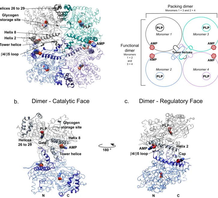
FIGURE 2. Overview of the structure of AMP-bound brain glycogen phosphorylase. Shown are schematic and ribbon representations of the C␣trace of the
tetramer and functional dimer of bGP. Key secondary structures described in this study are indicated. The catalytic site and the AMP-binding site are marked by the co-factor pyridoxal phosphate and allosteric effector AMP (surface representation), respectively. a, the tetrameric organization of bGP. Two bGP monomers associate by the horizontal axis to form the functional dimer (monomers 1 and 2). The tetramer is composed of two functional dimers that interact through their catalytic faces, by the vertical axis. The association of two monomers through the catalytic site forms the packing dimer (monomers 1 and 3). A schematic representation of the tetramer is shown (right panel). The functional dimer corresponds to the association of monomers 1 and 2 (gray and blue) and monomers 3 and 4 (turquoise and purple). The packing dimer corresponds to the association of monomers 1 and 3 (gray and turquoise) and monomers 2 and 4 (blue and purple). b, the catalytic face of bGP. The residues implied in the activation of GP and the subunit interfaces are indicated. The gate loop (residues 279 –289) is not stabilized allowing the glycogen to access the catalytic site. The tower helix interacts with its counterparts from the other subunits. The crossover angle of the tower helices in the functional dimer avoids the stabilization of the gate, promoting the opening of the catalytic site. c, the regulatory face of bGP contains key regions involved in the control of allosteric transition of the enzyme and is exposed to solvent in both the tetramer and the dimer. PLP, pyridoxal phosphate.
by guest on June 28, 2018
http://www.jbc.org/
The Subunit Interfaces—GPs are generally found in two major oligomeric states: dimer and tetramer. In the absence of AMP or phosphorylation, the dimer remains in an inactive con-formation. The subunit interfaces of the dimer are remodeled upon activation, allowing the transition from the inactive (T state) to the active (R state) conformations. The remodeling of the mGP interfaces allows this isozyme to form tetramers in which the catalytic sites are occluded (27, 33). The active form of lGP remains as a dimer because of its more rigid structure (29). To confirm the oligomeric state of inactive and active bGP in solution, we performed DLS experiments. We observed that bGP is found as an equilibrium between a monomeric and dimeric form, which is shift toward the dimeric state upon
acti-vation (supplemental Table S1). In crystals, bGP forms a
tetramer displaying three distinct interfaces, similar to the muscle isoform. Each monomer displays a series of contacts allowing it to interact with the three other monomers of the tetramer. We designate the three interfaces as: 1) the functional dimer, which corresponds to the active dimer (horizontal axis composed of monomers 1 and 2, as well as monomers 3 and 4); 2) the packing dimer, located at the interface between two dimers from the tetramer (vertical axis, composed of mono-mers 1 and 3, as well as monomono-mers 2 and 4); and 3) the core of the tower helices (Fig. 2a).
The Functional Dimer Interface of bGP—We found that the association of two GP monomers into the functional dimer
involves helix 2 (residues 49 –77), the 4/5 loop (residues
180 –198), the cap⬘ loop (residues 41–48), and the tower helices
(residues 259 –278), as observed for mGP and lGP (Fig. 5, a and
b). The helix 2-4/5 loop-cap⬘ loop interface includes the
binding site (Fig. 5a). As stated previously, the AMP-binding site in the PEG-bound bGP structure is occupied by a
PEG 400 molecule (supplemental Fig. S2and Fig. 4). The PEG
400 molecule was found to interact with several amino acids involved in the binding of AMP (see below). PEG 400 also acted with amino acids involved in the functional dimer inter-face (Fig. 4a). The presence of AMP in the AMP-bound bGP structure promoted the formation of specific contacts by con-certed movements of amino acid side chains and slight packing of the AMP-binding site around AMP relative to PEG-bound
bGP (Fig. 4, b and c). These movements facilitate interactions
between the two subunits by bringing the cap⬘ loop closer to
helix 2 and the4/5 loop and stabilize the enzyme in the active
conformation (Fig. 5a).
The tower helices consist of the anti-parallel association of helix 7 with its symmetric equivalent. The helices are connected to the gate loop (residues 279 –289). The gate loop adopts either an open or a closed configuration depending the activation state of the enzyme (27–29) (active or inactive). We found that the helices present a crossover angle of 85° in both the free and AMP-complexed forms (Fig. 5b), promoting the open confor-mation of the gate loop in a manner similar to that previously observed for the muscle isoform (74°) but not the liver isoform (45°) (27–29) (Fig. 5, c and d). The tower helices in all three isozymes are stabilized by non-polar interactions between
Val266, Leu267, and Asn270 and their counterparts from the
other subunit (Fig. 5, b– d). However, in contrast to mGP and lGP, the two tower helices from bGP also interact through two additional hydrogen bonds: one between the lateral side chains
of Tyr262and Asn270⬘ at one end of the helical interface and the
other between Asn270and Tyr262⬘ at the other end (Fig. 5b).
Moreover, whereas lGP has a rigid structure (27, 29) and little remodeling occurs during the activation process, mGP dis-plays a flexible functional dimer interface, which is remod-eled upon activation. In both bGP structures, the functional
dimer interface has a buried surface of 1400 Å2, whereas the
buried surface of the functional dimer interface of mGP is of
2240 Å2. This is consistent with a flexible functional dimer
interface and the similar regulatory features shared between muscle and brain GP.
The Packing Dimer Interface and the Tetramer Interface of
bGP—Interaction between the two functional bGP dimers
relies on contacts involving the tower helices, the glycogen
stor-age site (residues 426 – 434), and the bundle of helices␣26 to 29
(residues 721–769), as previously described for the muscle isozyme (33). No tetrameric structure has been observed for lGP, and thus, such an interface does not exist (29) (Fig. 6a). The bGP and AMP-bound bGP structures display a similar packing dimer interface, although there are a few more contacts involving the glycogen storage site in the bGP tetramer without AMP (data not shown).
The tower helices in bGP constitute a substantial part of the interactions established at the packing dimer interface, further stabilizing this interaction, contrary to the muscle isoform (33). Indeed, the N-terminal extremity of the tower helices in bGP
allow the formation of hydrophobic contacts between Val259,
Tyr262, Ile263, and their relative symmetric equivalents. The
gate from each subunit is stabilized in the open conformation
through hydrophobic contacts between Phe286and Asn281, and
their symmetric equivalents (Fig. 6b). Nevertheless, the number of contacts slightly decreases in the presence of the allosteric effector, affecting mostly in the glycogen storage site. Indeed, in this site, half of the contacts are lost (data not shown).
The Core of Tower Helices—The tower helices of bGP consti-tute the core of the tetramer interface as they do in mGP (33). The tower helices of AMP-bound bGP are longer by two turns than in AMP-bound mGP (28), allowing each tower helix to interact with the helices from the three other subunits.
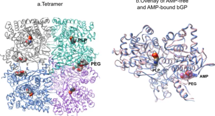
FIGURE 3. The overall structure of AMP-free bGP. a, AMP-free bGP forms a tetramer, obeying a 222 symmetry, similarly to AMP-bound bGP. PEG400, used as precipitant for crystallization, is found in the AMP binding site. b, structural alignment of AMP bound and AMP free monomers. View of the catalytic face of both structures. The catalytic site is marked by the pyridoxal phosphate (PLP; surface representation). AMP (blue) or PEG 400 (purple) is found in the AMP binding site and shown by surface representation.
by guest on June 28, 2018
http://www.jbc.org/
Opposite subunits from the tetramer (for example monomers 1 and 4) are thus able to interact through the tower helices via the
establishment of non-polar interactions between Leu271 and
Gly260⬘, as well as Leu268and Ile263⬘, on one side, and their
relative counterparts Leu271⬘, Gly260, Leu267⬘, and Ile263, on the
other (Fig. 6c). Although the tower helices contribute to less than 10% of the total interactions in the active muscle tetramer, they contribute to a fourth of the total contacts found in the whole tetramer in the PEG-bound bGP structure. In AMP-bound bGP, more than half of these contacts are lost. This leads to a reduction of the packing of the core formed by the tower helices, helix 8, and the gate, thus relaxing the tetramer inter-face (data not shown).
The AMP-binding Site—The AMP-binding site of bGP is
delimited by the cap⬘ loop from one subunit and helix 2, helix 8,
and the4/5 loop from the other subunit, because they are
in the muscle and liver isoforms (27–29). Helix 2 extends across the width of the protein and ensures the connection between the two AMP-binding sites of the functional dimer (Figs. 2c and 7a). Side chains of the residues involved in the binding of AMP move to improve their contacts with AMP in the AMP-bound bGP structure (Fig. 4c). The AMP-binding site can be divided into three subsites: one each for the phosphate, sugar, and base moiety of AMP (1, 27, 28). The phosphate group of AMP inter-acts with the AMP-binding site in the AMP-bound structure of bGP by forming several hydrogen bonds involving the side
FIGURE 4. Presence of PEG 400 in AMP binding site. a, PEG 400 is localized to the AMP binding. The C␣ trace of the main regions implicated in PEG 400 stabilization is represented as a ribbon. The amino acids interacting with PEG 400 are shown and labeled. b, plot diagram of the interactions of PEG400 with the residues from the bGP AMP-binding site. Hydrogen bonds are shown as dashed lines. Additional van der Waals contacts are represented as black curved lines. Most of the residues implicated in the stabilization of PEG 400 are also involved in the binding of AMP. c, overlay of the AMP-binding sites of the two bGP structures: bGP in complex with PEG 400 in pink; bGP in complex with AMP in blue. Residues that undergo reorientation following the binding of AMP are represented.
FIGURE 5. The functional dimer interface. Ribbon representations of the regions involved in the functional dimer interface of bGP are shown. The regions described are indicated, and the residues making contacts at the interface are represented and labeled. A schematic representation of the tetramer is shown (left panel), highlighting the interface we describe below. a, the functional dimer interface of bGP involves the helix 2-cap⬘ loop-4/5 loop region, comprising the AMP-binding site, and the tower helices. b, first panel, the tower helix of bGP from one monomer (blue) interacts with its counterpart (gray) forming hydrogen bonds and hydrophobic contacts. Second panel, interactions at the interface promote the arrangement of the tower helices by a crossover angle of 85°. c, overlay of the crystal structure of the tower helices interface of lGP (1 FA9; dark and light pink) and bGP (blue and gray). Active conformation (R state) of lGP is characterized by a 45° crossover angle of the tower helices and hydrophobic contacts between Val266, Leu267, and Asn270and their equivalents on the
other subunit. d, overlay of the crystal structures of the tower helices interface of the active R state of mGP (1PYG; green and beige) and bGP (blue and gray). The mGP tower helices interact solely through hydrophobic contacts involving Val266, Leu267, and Asn270and their equivalents on the other subunit, similar to lGP.
These interactions stabilize the tower helices in the active conformation, displaying a crossover angle of 74°.
by guest on June 28, 2018
http://www.jbc.org/
chains of Arg309and Arg310from helix 8 and the side chain of
Tyr196from the4/5 loop (Figs. 7a and 8a). The ribose of
AMP interacts with the cap⬘ loop, forming a hydrogen bond
with Asp42⬘ and van der Waals contacts with Asn44⬘ and Val45⬘.
The nucleotide moiety of AMP interacts weakly with helix 2, forming van der Waals interactions with the side chains of
Gln71and Gln72, and co-planar stacking with the side chain of
Tyr75(Figs. 7b and 8a). All these interactions may contribute to
a 24° rotation of the nucleotide moiety of AMP relative to AMP-bound mGP (Fig. 7b). The activation of mGP by AMP and its cooperative binding are partly attributed to the formation of several hydrogen bonds with the AMP-binding site and its fur-ther stabilization in the AMP-binding site by the adenine loop (residues 312–325) (27–29) (Fig. 7b and 8a). In contrast, the liver isoform interacts poorly with AMP, consistent with its weak activation by AMP (29) (Fig. 8a). Surprisingly, we did not observe electron density for the adenine loop in both structures (the PEG-bound and AMP-bound bGP structures) (Fig. 8b), despite the high sensitivity of bGP to AMP-dependent activa-tion and the sequence conservaactiva-tion between the muscle and
brain isoforms. The side chain of Tyr75adopts a conformation
that precludes the interaction of the adenine loop (312–325)
with AMP, as for lGP (29) (Fig. 7b). The conformation of Tyr75
in bGP is stabilized by the formation of a hydrogen bond
between the phosphate group of AMP and Tyr196, a residue
only found in the brain isozyme (corresponding to Phe196in
muscle and liver GP). This unique interaction thus contributes to the geometry of AMP in bGP and by favoring the co-planar
stacking between Tyr75 and the nucleotide part of AMP
(Fig. 7c). Discussion
Brain glycogen provides fuel for neuronal and astrocyte func-tions. It is critical for high cognitive processes including learn-ing and memory consolidation (10 –17). In humans, glycogen phosphorylase, the key enzyme for glycogen mobilization, con-sists of three isozymes, including one specific for brain. Whereas the structures of mGP and lGP have been known for decades, the structure of bGP is still lacking despite its critical role in brain glycogen metabolism (25, 26, 29).
We report the first crystal structure of human bGP. We found the overall crystal structure of bGP is similar to lGP and mGP, consistent with the sequence conservation of GP isozymes (83 and 80% sequence identity of bGP with the muscle and liver GP, respectively). GP isozymes differ in their responses to allosteric activation, in particular to AMP, despite their structural similarity. The liver isozyme is mostly con-trolled by phosphorylation, whereas the muscle and brain isozymes are strongly activated by AMP activation (1, 23, 24). These observations are consistent with our findings; the struc-ture of the AMP-bound bGP (active form) is more closely related to active mGP than active lGP, especially concerning the tower helices interface and the overall relationship between the two monomers of the functional dimers. These features suggest that the activation process of bGP relies on the remodeling of the functional dimer interface, as for mGP (27, 28). In con-trast, lGP has a more rigid structure, and no such remodeling occurs upon activation (29). This is further supported by the buried surface of the functional dimer interface of lGP (3350
Å2) relative to the two other GP isozymes (2240 Å2for mGP and
1400 Å2for bGP). However, bGP and mGP also demonstrate
distinct regulatory features (23, 24).
Although the phosphorylation peptide (1–21) is stabilized and seen in structures of AMP-bound mGP, no electron density was obtained for this region in AMP-bound bGP (Fig. 2, a and
c). It is possible that differences in flexibility/dynamics of this
region (which is not fully conserved between the three iso-forms) may explain the lower sensitivity of bGP to activation by phosphorylation (23).
Whereas mGP responds cooperatively to activation by AMP, bGP is activated noncooperatively by AMP (23). The AMP-bound bGP structure shows that AMP forms several hydrogen bonds within the AMP binding site (Figs. 7 and 8) as observed for AMP-bound mGP but not AMP-bound lGP. However, as observed for lGP, we found that helix 2 of bGP interacts poorly
with AMP (no hydrogen bond) and that Tyr75adopts a
confor-mation that may preclude the stabilization of the adenine loop (which is not observed in our structure because of a lack of electron density) (Fig. 7). These features have also been put
FIGURE 6. The tetramer interfaces. Ribbon representations of the regions involved in the tetramer interfaces in AMP-bound bGP are shown. The described regions are indicated, and the residues involved in the interactions are represented and labeled. a, the packing dimers interact through contacts involving the bundle of loops and helices 26 –29, as well as the glycogen storage site. b, the tower helices interactions at the packing dimer interface. Tower helices interact through Val259, Tyr262, and Ile263and stabilize the gate loop in the open configuration. c, interactions between the tower helices from two opposite subunits.
The tower helices from two opposite subunits from the bGP tetramer interact through hydrophobic contacts.
by guest on June 28, 2018
http://www.jbc.org/
forward to explain the absence of cooperative binding of AMP
to lGP (29). We also found that Tyr196in bGP, located at the
functional dimer interface (equivalent to Phe196in mGP and
lGP), makes a hydrogen bond with AMP that is specific for bGP (Figs. 7 and 8). Interestingly, this bGP specific substitution (F196Y) has been suggested to alter AMP cooperativity (23).
The conformation of the adenine loop is known to play a role in AMP binding and activation. The adenine loop is well defined in the AMP-bound mGP structures, whereas it is poorly ordered in AMP-bound lGP and not observed in AMP-bound bGP (no electron density) (28, 29). Altogether, these observa-tions may explain the non-cooperative binding of AMP to bGP upon AMP activation.
bGP is critical for the support of brain processes because it allows the mobilization of the brain glycogen store (10 –17). The importance of bGP is further supported by the fact that no mutations for this isozyme have been reported. Indeed, whereas deleterious mutations of the liver and muscle isozymes lead to the development of glycogen storage diseases (Hers dis-ease and McArdle disdis-ease, respectively), no deleterious muta-tions are known for bGP (1). This is likely because bGP is the only GP isozyme expressed during fetal development and is later replaced by mGP and lGP in their respective organs. It is likely that altered bGP activity is lethal during early embryonic development (1). The co-expression of the brain and muscle isoforms in astrocytes further underlines the functional impor-tance of the brain isozyme for neuronal processes (34). Indeed it
has been shown that whereas mGP rapidly responds to extra-cellular signals in brain such as noradrenaline, bGP is critical for local energy metabolism via changes in AMP concentration (23, 24). The distinct regulatory features of brain and muscle GP, in particular the cooperative and non-cooperative binding of AMP, allow astrocytes to tightly adapt their responses to their energy needs. Moreover, it is now accepted that the accu-mulation of glycogen in neurons leads to their rapid degenera-tion and is implicated in aging, as well as promoting several pathological conditions of the brain such as Lafora disease. The activity of bGP is critical for the controlled degradation of gly-cogen in neurons because they only express the brain isoform of GP (17). A reduction of bGP activity would lead to the uncon-trolled accumulation of glycogen in neurons and contribute to the pathogenesis of several brain diseases (18, 35, 36).
In conclusion, the crystal structures of human bGP reported in this study contribute to a better understanding of this spe-cific isozyme and a deeper knowledge of the differences in the activation properties of this important family of allosteric enzymes. Additionally, these new structural insights may enable isozyme-specific rational drug design for therapeutic development (37–39).
Experimental Procedures
Materials—Antibodies raised against brain glycogen phos-phorylase were obtained from Santa Cruz (SC-81751).
Anti-phosphoserine and anti-histidine tag antibodies,L-arabinose,
FIGURE 7. The AMP-binding site of human bGP. a, the binding mode of AMP in the bGP AMP-binding site. The AMP-binding site corresponds to a cleft formed by helix 2, helix 8,4/5, and the cap⬘ loop. The main regions implicated in AMP binding are represented as blue ribbons. Residues interacting with AMP are shown and labeled. No electron density was observed for the adenine loop, despite AMP binding. b, superposition of the crystal structure of AMP in the mGP (yellow) and bGP (orange) AMP-binding sites. The conformation of AMP in the AMP-binding site of bGP differs from that of mGP. c, overlay views of the AMP-binding site of bGP, mGP (1PYG), and lGP (1FA9). The main regions of bGP depicted in Fig. 6a (blue) are aligned with the corresponding regions in mGP (green) and lGP (pink). The AMP located in the AMP-binding site is not displayed for clarity. The replacement of Phe196of muscle and liver GP by a tyrosine
residue in bGP allows this amino acid to interact with the AMP. The Tyr75avoids the stabilization of the adenine loop in lGP and bGP.
by guest on June 28, 2018
http://www.jbc.org/
protease inhibitor mixture, nickel-nitrilotriacetic acid Super-flow resin, bovine serum albumin, imidazole, streptavidin-aga-rose resin, phosphoglucomutase, EDTA, glucose-1,6-diphos-phate, AMP, ATP, phosphorylase kinase from rabbit muscle, BSA, and glycogen were purchased from Sigma-Aldrich. Glu-cose-6-phosphate dehydrogenase was purchased from Roche. ECL Western blotting detection reagent was purchased from GE Heathcare. Biotinylated thrombin was purchased from Merck Millipore. NADP was purchased from Apollo Scientific. Cloning of bGP and Expression of Recombinant bGP—The human bGP cDNA (provided by OriGene Technologies, Inc.) was subcloned into the pET28a vector for further expression of
His6-tagged fusion recombinant proteins (6xHis-bGP). E. coli
C41(DE3)/pGro7 (encoding the GroEL-GroES chaperonin protein complex) strains were transformed with plasmid pET28a carrying 6xHis-bGP and used to express and purify the recombinant protein. Expression of the GroEL-GroES
chaper-onin protein complex was first induced by the addition of 1 mM
L-arabinose to cultures at an A600value of 0.2– 0.3. Expression
of recombinant bGP was then induced by the addition of 500
Mof isopropyl-1-thio--D-galactopyranoside to cultures at an
A600 value of 0.6 – 0.8. The bacteria were further cultured at
16 °C overnight. The bacteria were pelleted by centrifugation
(4,000⫻ g, 10 min), washed with cold PBS, and harvested by
centrifugation (4,000⫻ g, 10 min). The pellets were stored at
⫺80 °C until required.
Purification of Recombinant bGP from E. coli—Extraction and purification were repeatedly performed to obtain 20 mg of protein. The bacteria were resuspended in 35 ml of lysis buffer
(PBS, pH 8, 300 mM NaCl, 0.5% Triton X-100, 1 mg/ml
lysozyme, protease inhibitor mixture (Sigma-Aldrich)) and incubated for 1 h at 4 °C. The lysate was sonicated on ice (8-s
pulses for up to 7 min) and centrifuged (17,000⫻ g, 30 min,
4 °C). The supernatant was collected and incubated with 1 ml of nickel-nitrilotriacetic acid Superflow resin (Sigma-Aldrich) in
the presence of 10 mM imidazole (final concentration) for a
minimum of 2 h at 4 °C. The resin was then poured into a col-umn and washed successively with washing buffer (PBS, pH 8,
300 mMNaCl) containing 0.1% Triton X-100 and a stepwise
gradient of imidazole in washing buffer until a concentration of
20 mM imidazole (final concentration) was achieved.
His-tagged proteins were eluted with washing buffer containing 300
mMimidazole. Purified proteins were then incubated for 10
min with 10 mMDTT and protease inhibitor mixture. The
puri-fied protein was then exchanged against PBS, pH 7.1, using a PD10 (GE Healthcare) desalting column. The protein concen-tration was measured using the standard Bradford assay with BSA as standard and by absorbance measurement at 280 nm
using a theoretical⑀M: 115170 M⫺1 cm⫺1. The purity of the
protein was assessed by SDS-PAGE analysis.
His6Tag Cleavage by Thrombin Digestion—The His6tag was removed by thrombin digestion. Briefly, 20 mg of 6xHis-bGP
FIGURE 8. Residues involved in the binding of AMP. a, sequence alignment of the regions involved in the binding of AMP in GPs. Residues involved in the binding of AMP are in bold. Residues involved in hydrogen bonds with AMP are shown in red, and residues establishing van der Waals interactions with AMP are shown in black. Sequences are highly conserved between the different isoforms, except for the liver isoform adenine loop. b, interaction plot diagram of AMP with the AMP-binding site residues from brain, muscle, and liver GP. Hydrogen bonds are shown as dashed lines. Additional van der Waals contacts are represented as black curved lines. Whereas Tyr75and residues of the adenine loop forms hydrogen bonds with AMP in mGP, no such interactions are observed
for lGP and bGP. by guest on June 28, 2018
http://www.jbc.org/
Enzyme Assay—bGP activity was measured in the direction of glycogenolysis as described elsewhere (40). Briefly, the for-mation of glucose-1-phosphate was determined using a coupled assay system containing phosphoglucomutase, glucose-6-phosphate dehydrogenase, and NADP, by following
NADPH,H⫹formation at 340 nm. The phosphorylase activity
assay was carried out at 37 °C in PBS, pH 6.9. The mixture
consisted of bGP (final concentration, 0.1M) with or without 1
mMAMP, 0.25% glycogen, 2 mMEDTA, 0.8 mMNADP, 10 mM
magnesium acetate, 5Mglucose-1,6-diphosphate, 5 units of
glucose-6-phosphate dehydrogenase, and 5 units
phosphoglu-comutase, in a final volume of 250l. Each measurement was
performed in triplicate.
Biochemical Analysis of Recombinant bGP—The saturation curve by glycogen was obtained by measuring the bGP activity with various concentrations of glycogen (0 – 0.35%) in the
pres-ence of a saturating concentration of AMP (1 mM). The level of
activation of bGP by the allosteric activator AMP was obtained by measuring bGP activity in the presence of a saturating con-centration of glycogen (0.025%) and various concon-centrations of
AMP (0 –3 mM). Initial velocities were determined, and
coop-erativity of AMP binding was assessed by fitting the data to the Hill equation,
Log共Y/共1 ⫺ Y兲兲 ⫽ nlog关AMP兴 ⫺ logKA (Eq. 1)
where Y is the ratio Vi/Vmax, n is the Hill coefficient, [AMP] is
the corresponding concentration of AMP, and KAis the affinity
constant.
Phosphorylation of Ser14 and Activation by Phosphor-ylation—bGP was phosphorylated using phosphorylase kinase from rabbit muscle (Sigma-Aldrich) (1 unit/mg bGP) activated by preincubation for 1 h in phosphorylation buffer containing
20 mMTris-HCl buffer, pH 7.7, 0.22 mMATP, 3.3 mMMgCl2,
0.5 mM CaCl2, 0.5 mM NaF for activation.
Phosphor-ylation was performed by the addition of activated phosphor-ylase kinase to the bGP solution and in phosphorylation buffer and incubation for 2 h at room temperature. The resulting activity was assessed with or without AMP. Phosphorylation of rabbit mGP (Sigma-Aldrich) was used as a control. Phospho-rylated bGP was then subjected to buffer exchange against PBS buffer, pH 6.9, using a PD Minitrap G25 (GE Healthcare) desalt-ing column.
SDS-PAGE Electrophoresis and Western Blotting—Proteins were loaded onto 7.5% polyacrylamide gels and electrophoretic protein separation carried out at 110 V (constant voltage). For
mg/ml) using an Exactive Plus EMR mass spectrometer (Thermo Fisher Scientific), which allows analysis of proteins and complexes in native-like states (41). The proteins were desalted by buffer exchange prior to mass spectrometry analysis.
Dynamic Light Scattering Measurements—Dynamic light scattering measurements were performed with a DynaPro-MS800 molecular sizing instrument (Protein Solutions,
Lake-wood, NJ). Protein samples at 3.2 mg/ml in 20 mMTris-HCl, pH
7.0, in the presence or in the absence of 5 mMAMP were loaded
into a 45-l quartz cuvette. The hydrodynamic radius and
molec-ular mass were determined from 50 measurements at 18 °C. The data were analyzed using Dynamics 6.9.2.11 software.
Crystallization of bGP and Soaking with AMP—Initial screening of crystallization conditions was carried out by the
vapor diffusion method using a MosquitoTM
nanoliter-dispens-ing system (TTP Labtech) at 4 °C. Sittnanoliter-dispens-ing drops were set up using 400 nl of a 1:1 mixture of recombinant human bGP at 7 mg/ml and crystallization solutions (672 different
commer-cially available conditions) equilibrated against a 150-l
reser-voir in multiwell plates (Greiner Bio-One). The crystallization
plates were stored at 4 °C in a RockImager1000TM
(Formula-trix) automated imaging system to monitor crystal growth. Hits were improved by making handmade hanging drops in 24-well plates at 18 °C. The best crystals were obtained using a solution
of 28% (v/v) PEG 400, 0.2Mcalcium chloride, and 0.1MHepes,
pH 7.5. Crystals with dimensions of up to 0.05 mm ⫻ 0.05
mm⫻ 0.1 mm appeared within 2 weeks. The crystal of protein
complexed with AMP (AMP-bound bGP) was obtained by
soaking the apo-form crystal for 3 h in 50 mMof AMP dissolved
into the crystallization solution. For data collection, the crystals were flash-cooled in liquid nitrogen using the mother crystalli-zation solution as cryoprotectant. All x-ray diffraction data were collected on the PROXIMA-1 Beamline at the Soleil chrotron (St. Aubin, France) and the ID23-2 at European Syn-chrotron Radiation Facility (Grenoble, France). Diffraction images were integrated using the XDS program (42).
Structure Determination, Refinement, and Analysis—For the resolution of the structure, molecular replacement phases were obtained using Phaser (43) implemented in the CCP4 program suite (Collaborative Computational Project, Number 4 (44)). The model for molecular replacement was built by homology modeling using SWISS-MODEL (45) based on the template structure of human muscle glycogen phosphorylase (PDB code 1Z8D). The initial models were completed and adjusted using the COOT program (46). Refinement was performed using REFMAC as implemented in CCP4 (47). The crystal structure of the
by guest on June 28, 2018
http://www.jbc.org/
bound GPb at 2.5 Å resolution was refined to R and Rfree crystal-lographic factors of 18 and 24%, respectively (see statistics in Table 1). The crystal structure of the AMP-bound bGP at 3.4 Å
resolu-tion was refined to R and Rfreecrystallographic factors of 21 and
29%, respectively (see statistics in Table 1). Atomic coordinates and structure factors were deposited in the Protein Data Bank under accession codes 5IKO and 5IKP for the PEG-bound GPb and the AMP-bound bGP, respectively.
Molecular Graphics and Analyses—Molecular graphics and analyses were performed using the UCSF Chimera package (1.10.2). Chimera was developed by the Resource for Biocom-puting, Visualization, and Informatics at the University of Cal-ifornia, San Francisco (UCSF Chimera-a visualization system for exploratory research and analysis (48)).
Author Contributions—C. M. and F. R.-L. designed experiments, managed the project, and wrote the manuscript. C. M., I. L. S.-G., R. D., X. X., A. C., T. L., G. W., J.-M. C., C. E., A. H., J.-M. D., and F. R.-L. performed experiments and/or analyzed the data. All authors discussed the results and commented on the manuscript.
Acknowledgments—We acknowledge SOLEIL and ESRF for provision of synchrotron radiation facilities, and we thank the staff for assis-tance in using beamline PROXIMA1 and ID23–1, respectively.
References
1. Newgard, C. B., Hwang, P. K., and Fletterick, R. J. (1989) The family of glycogen phosphorylases: structure and function. Crit. Rev. Biochem. Mol.
Biol. 24,69 –99
2. Berg, J. M., Tymoczko, J. L., Stryer, L., and Clarke, N. D. (2002)
Biochem-istry, 5th ed., pp 831– 863, W. H. Freeman, New York
3. O¨ z, G., DiNuzzo, M., Kumar, A., Moheet, A., and Seaquist, E. R. (2015) Revisiting glycogen content in the human brain. Neurochem. Res. 40, 2473–2481
4. Obel, L. F., Mu¨ller, M. S., Walls, A. B., Sickmann, H. M., Bak, L. K., Waagepetersen, H. S., and Schousboe, A. (2012) Brain glycogen-new per-spectives on its metabolic function and regulation at the subcellular level.
Front. Neuroenergetics. 4,3
5. Brown, A. M. (2004) Brain glycogen re-awakened. J. Neurochem. 89, 537–552
6. Hossain, M. I., Roulston, C. L., and Stapleton, D. I. (2014) Molecular basis of impaired glycogen metabolism during ischemic stroke and hypoxia.
PLoS One 9,e97570
7. Choi, I.-Y., Seaquist, E. R., and Gruetter, R. (2003) Effect of hypoglycemia on brain glycogen metabolism in vivo. J. Neurosci. Res. 72, 25–32 8. Suh, S. W., Bergher, J. P., Anderson, C. M., Treadway, J. L., Fosgerau, K.,
and Swanson, R. A. (2007) Astrocyte glycogen sustains neuronal activity during hypoglycemia: studies with the glycogen phosphorylase inhibitor CP-316,819 ([R-R*,S*]-5-chloro-N-[2-hydroxy-3-(methoxymethyl-amino)-3-oxo-1-(phenylmethyl)propyl]-1H-indole-2-carboxamide).
J. Pharmacol. Exp. Ther. 321,45–50
9. Swanson, R. A., Sagar, S. M., and Sharp, F. R. (1989) Regional brain glyco-gen stores and metabolism during complete global ischaemia. Neurol. Res.
11,24 –28
10. Suzuki, A., Stern, S. A., Bozdagi, O., Huntley, G. W., Walker, R. H., Mag-istretti, P. J., and Alberini, C. M. (2011) Astrocyte-neuron lactate transport is required for long-term memory formation. Cell 144, 810 – 823 11. Newman, L. A., Korol, D. L., and Gold, P. E. (2011) Lactate produced by
glycogenolysis in astrocytes regulates memory processing. PLoS One 6, e28427
12. Pellerin, L., and Magistretti, P. J. (2012) Sweet sixteen for ANLS. J. Cereb.
Blood Flow Metab. 32,1152–1166
13. Gibbs, M. E., Anderson, D. G., and Hertz, L. (2006) Inhibition of
glycogen-olysis in astrocytes interrupts memory consolidation in young chickens.
Glia 54,214 –222
14. Gibbs, M. E., and Hutchinson, D. S. (2012) Rapid turnover of glycogen in memory formation. Neurochem. Res. 37, 2456 –2463
15. Duran, J., Saez, I., Gruart, A., Guinovart, J. J., and Delgado-García, J. M. (2013) Impairment in long-term memory formation and learning-depen-dent synaptic plasticity in mice lacking glycogen synthase in the brain.
J. Cereb. Blood Flow Metab. 33,550 –556
16. Duran, J., and Guinovart, J. J. (2015) Brain glycogen in health and disease.
Mol. Aspects Med. 46,70 –77
17. Saez, I., Duran, J., Sinadinos, C., Beltran, A., Yanes, O., Tevy, M. F., Mar-tínez-Pons, C., Mila´n, M., and Guinovart, J. J. (2014) Neurons have an active glycogen metabolism that contributes to tolerance to hypoxia.
J. Cereb. Blood Flow Metab. 34,945–955
18. Vilchez, D., Ros, S., Cifuentes, D., Pujadas, L., Valle`s, J., García-Fojeda, B., Criado-García, O., Ferna´ndez-Sa´nchez, E., Medran˜o-Ferna´ndez, I., Domínguez, J., García-Rocha, M., Soriano, E., Rodríguez de Co´rdoba, S., and Guinovart, J. J. (2007) Mechanism suppressing glycogen synthesis in neurons and its demise in progressive myoclonus epilepsy. Nat. Neurosci.
10,1407–1413
19. Duran, J., Tevy, M. F., Garcia-Rocha, M., Calbo´, J., Mila´n, M., and Guino-vart, J. J. (2012) Deleterious effects of neuronal accumulation of glycogen in flies and mice. EMBO Mol. Med. 4, 719 –729
20. Lebo, R. V., Gorin, F., Fletterick, R. J., Kao, F. T., Cheung, M. C., Bruce, B. D., and Kan, Y. W. (1984) High-resolution chromosome sorting and DNA spot-blot analysis assign McArdle’s syndrome to chromosome 11.
Science 225,57–59
21. Newgard, C. B., Fletterick, R. J., Anderson, L. A., and Lebo, R. V. (1987) The polymorphic locus for glycogen storage disease VI (liver glycogen phosphorylase) maps to chromosome 14. Am. J. Hum. Genet. 40, 351–364 22. Newgard, C. B., Littman, D. R., van Genderen, C., Smith, M., and Fletter-ick, R. J. (1988) Human brain glycogen phosphorylase. Cloning, sequence analysis, chromosomal mapping, tissue expression, and comparison with the human liver and muscle isozymes. J. Biol. Chem. 263, 3850 –3857 23. Crerar, M. M., Karlsson, O., Fletterick, R. J., and Hwang, P. K. (1995)
Chimeric muscle and brain glycogen phosphorylases define protein do-mains governing isozyme-specific responses to allosteric activation.
J. Biol. Chem. 270,13748 –13756
24. Mu¨ller, M. S., Pedersen, S. E., Walls, A. B., Waagepetersen, H. S., and Bak, L. K. (2015) Isoform-selective regulation of glycogen phosphorylase by energy deprivation and phosphorylation in astrocytes. Glia 63, 154 –162 25. Fletterick, R. J., Sygusch, J., Semple, H., and Madsen, N. B. (1976) Structure of glycogen phosphorylase a at 3.0 A resolution and its ligand binding sites at 6 A. J. Biol. Chem. 251, 6142– 6146
26. Barford, D., Hu, S. H., and Johnson, L. N. (1991) Structural mechanism for glycogen phosphorylase control by phosphorylation and AMP. J. Mol.
Biol. 218,233–260
27. Barford, D., and Johnson, L. N. (1989) The allosteric transition of glycogen phosphorylase. Nature 340, 609 – 616
28. Sprang, S. R., Withers, S. G., Goldsmith, E. J., Fletterick, R. J., and Madsen, N. B. (1991) Structural basis for the activation of glycogen phosphorylase b by adenosine monophosphate. Science 254, 1367–1371
29. Rath, V. L., Ammirati, M., LeMotte, P. K., Fennell, K. F., Mansour, M. N., Danley, D. E., Hynes, T. R., Schulte, G. K., Wasilko, D. J., and Pandit, J. (2000) Activation of human liver glycogen phosphorylase by alteration of the secondary structure and packing of the catalytic core. Mol. Cell 6, 139 –148
30. Gaboriaud-Kolar, N., and Skaltsounis, A.-L. (2013) Glycogen phosphoryl-ase inhibitors: a patent review (2008 –2012). Expert Opin. Ther. Pat. 23, 1017–1032
31. Vaidya, H. B., Ahmed, A. A., Goyal, R. K., and Cheema, S. K. (2013) Gly-cogen phosphorylase-a is a common target for anti-diabetic effect of iri-doid and secoiriiri-doid glycosides. J. Pharm. Pharm. Sci. 16, 530 –540 32. Agius, L. (2010) Physiological control of liver glycogen metabolism:
les-sons from novel glycogen phosphorylase inhibitors. Mini Rev. Med. Chem.
10,1175–1187
33. Barford, D., and Johnson, L. N. (1992) The molecular mechanism for the
by guest on June 28, 2018
http://www.jbc.org/
38. Cloix, J.-F., and He´vor, T. (2011) Glycogen as a putative target for diagno-sis and therapy in brain pathologies. ISRN Pathol. 2011, 1–17
39. Xu, L., and Sun, H. (2010) Pharmacological manipulation of brain glyco-genolysis as a therapeutic approach to cerebral ischemia. Mini-Rev. Med.
Chem. 10,1188 –1193
40. Maddaiah, V. T., and Madsen, N. B. (1966) Kinetics of purified liver phos-phorylase. J. Biol. Chem. 241, 3873–3881
41. Rose, R. J., Damoc, E., Denisov, E., Makarov, A., and Heck, A. J. (2012) High-sensitivity Orbitrap mass analysis of intact macromolecular assem-blies. Nat. Methods 9, 1084 –1086
46. Emsley, P., Lohkamp, B., Scott, W. G., and Cowtan, K. (2010) Features and development of Coot. Acta Crystallogr. D Biol. Crystallogr. 66, 486 –501 47. Murshudov, G. N., Skuba´k, P., Lebedev, A. A., Pannu, N. S., Steiner, R. A.,
Nicholls, R. A., Winn, M. D., Long, F., and Vagin, A. A. (2011) REFMAC5 for the refinement of macromolecular crystal structures. Acta Crystallogr.
D Biol. Crystallogr. 67,355–367
48. Pettersen, E. F., Goddard, T. D., Huang, C. C., Couch, G. S., Greenblatt, D. M., Meng, E. C., and Ferrin, T. E. (2004) UCSF Chimera: a visualization system for exploratory research and analysis. J. Comput. Chem. 25, 1605–1612
by guest on June 28, 2018
http://www.jbc.org/
Ahmed Haouz, Jean-Marie Dupret and Fernando Rodrigues-Lima
Cocaign, Thibaut Léger, Gary Woffendin, Jean-Michel Camadro, Catherine Etchebest,
Cécile Mathieu, Ines Li de la Sierra-Gallay, Romain Duval, Ximing Xu, Angélique
BRAIN GLYCOGEN PHOSPHORYLASE
doi: 10.1074/jbc.M116.738898 originally published online July 8, 2016 2016, 291:18072-18083.
J. Biol. Chem.
10.1074/jbc.M116.738898
Access the most updated version of this article at doi: Alerts:
When a correction for this article is posted
•
When this article is cited
•
to choose from all of JBC's e-mail alerts
Click here
Supplemental material:
http://www.jbc.org/content/suppl/2016/07/08/M116.738898.DC1 http://www.jbc.org/content/291/35/18072.full.html#ref-list-1This article cites 47 references, 8 of which can be accessed free at
by guest on June 28, 2018
http://www.jbc.org/