HAL Id: hal-00374522
https://hal.archives-ouvertes.fr/hal-00374522
Submitted on 8 Apr 2009
HAL is a multi-disciplinary open access
archive for the deposit and dissemination of sci-entific research documents, whether they are pub-lished or not. The documents may come from teaching and research institutions in France or abroad, or from public or private research centers.
L’archive ouverte pluridisciplinaire HAL, est destinée au dépôt et à la diffusion de documents scientifiques de niveau recherche, publiés ou non, émanant des établissements d’enseignement et de recherche français ou étrangers, des laboratoires publics ou privés.
Quinolinate synthase, an iron-sulfur enzyme in NAD
biosynthesis
Sandrine Ollagnier de Choudens, Laurent Loiseau, Yiannis Sanakis, Frédéric
Barras, Marc Fontecave
To cite this version:
Sandrine Ollagnier de Choudens, Laurent Loiseau, Yiannis Sanakis, Frédéric Barras, Marc Fontecave. Quinolinate synthase, an iron-sulfur enzyme in NAD biosynthesis. FEBS Letters, Wiley, 2005, 579, pp.3737-3743. �10.1016/j.febslet.2005.05.065�. �hal-00374522�
Quinolinate Synthetase, an iron-sulfur enzyme in NAD
biosynthesis
Sandrine Ollagnier-de Choudens1*, Laurent Loiseau2, Yiannis Sanakis3, Frédéric Barras2, and Marc Fontecave1*
1
Laboratoire de Chimie et Biochimie des Centres Rédox Biologiques, DBMS-CB, CEA/CNRS/Université Joseph Fourier, UMR 5047, 17 Avenue des Martyrs, 38054 Grenoble Cedex 09, France.
2
Laboratoire de Chimie Bactérienne, UPR-CNRS 9043, IBSM, 31 Chemin Joseph Aiguier, 13402 Marseille Cedex 20, France.
3
NCSR, Demokritos, Institute of Materials Science, 15310 Ag. Paraskevi, Attiki, Greece. Dept of Biological Applications And Technologies, Univ. of Ioannina, 45110 Ioannina.
*
To whom correspondence should be addressed.
Tel: 33 4 38 78 91 03; Fax:33 4 38 78 91 24; email: [email protected]
Abstract
Nicotinamide adenine dinucleotide (NAD) plays a crucial role as a cofactor in numerous essential redox biological reactions. NAD derives from quinolinic acid which is synthesized in E. coli from L-aspartate and dihydroxyacetone phosphate (DHAP) as the result of the concerted action of two enzymes, L-aspartate oxidase (NadB) and quinolinate synthetase (NadA). We report here the characterization of NadA protein from E. coli. When anaerobically purified, the isolated soluble protein contains 3-3.5 iron and 3-3.5 sulfide/ polypeptide chain. Mössbauer spectra of the
57
Fe-protein revealed that the majority of the iron is in the form of a (4Fe-4S)2+
cluster. An enzymatic assay for quinolinate synthetase activity was set up and allowed to demonstrate that the cluster is absolutely required for NadA activity. Exposure to air leads to degradation of the cluster and inactivate enzyme.
Introduction
Nicotinamide adenine dinucleotide (NAD) plays a crucial role as a cofactor in numerous essential redox biological reactions. In fact, in all living organisms, NAD derives from quinolinic acid, the biosynthetic pathway of which differs among organisms [1-5]. In most eukaryotes, quinolinic acid is produced via the degradation of tryptophan, whereas in E. coli or S. typhimurium, it is synthesized from L-aspartate and dihydroxyacetone phosphate (DHAP) as the result of the concerted action of two enzymes, L-aspartate oxidase, a flavin adenine dinucleotide (FAD)-dependent flavoenzyme encoded by the nadB gene, and quinolinate synthetase, encoded by the
nadA gene (Figure 1).
Surprisingly, very little is known on the NadA protein and the chemical mechanism of the corresponding catalyzed reaction. Previously, the E. coli 38 240 Da NadA protein was obtained from inclusion bodies and did not show any evidence for the presence of a prosthetic group [6]. On the other hand, several observations were used to suggest that the NadA protein was an iron-sulfur enzyme. First, E. coli NadA amino-acid sequence contains three cysteines within the CRSCAHCP motif, which is a common iron-sulfur chelating peptide sequence (Figure 2) [7-9]. Second, quinolinate synthetase in E. coli extracts is inactivated by oxygen and reactivation after removal of oxygen is strongly inhibited by ferrous chelators such as phenanthroline [10], a behaviour shared by a number of iron-sulfur enzymes such as dehydratases or members of the “Radical-SAM” (S-adenosylmethionine) enzyme superfamily [11]. Third, an iscS E. coli mutant strain, lacking one of the cysteine desulfurases involved in the assembly of iron-sulfur clusters, absolutely requires nicotinic acid for growth in
minimal media [12]. Such a phenotype might be related to the involvement of an iron-sulfur enzyme in the NAD biosynthetic pathway with NadA being the most obvious candidate. Presumably, nicotinic acid (NA) could suppress NAD auxotrophy by entering the biosynthesis pathway prior to the NadD catalysed step.
In this paper, we demonstrate that i) E. coli NadA is indeed an iron-sulfur protein in the form of a (4Fe-4S)2+
cluster which is sensitive to oxygen and ii) this cluster is absolutely required for NadA activity.
Abbreviations :
DTT : dithiothreitol , ( FeS) : iron-sulfur cluster, PCR : Polymerase Chain Reaction; IPTG, isopropyl -D-thiogalactoside; ISC, Iron Sulfur Cluster ; ara, arabinose; IA: Imino-Aspartate; DHAP: DiHydroxyAcetone Phosphate; QA: Quinolinic Acid; NadB: L-aspartate oxidase; NadA: Quinolinate synthetase; SAM: S-AdenosylMethionine.
Material and Methods
Plasmid and strain construction. The nadA and nadB inserts were PCR-amplified
using MG1655 chromosomal DNA as a template and primers Ns/Nas (5'-CCGCATATGAGCGTAATGTTTGATCCA-3’/5'-GTTCTCGAGTCCACGTAGTGTAGCC -3’), Bs/Bas (5'-CCGCATATGAATACTCTCCCTGAACATTCATG-3'/5'-GTTCTCGAGTCTGTTTATGTAATGATTGCCGGGGGAAAGG-3') respectively. The PCR products were digested with NdeI and XhoI and then ligated into a pET22b+ vector (Novagen) digested by the same enzymes, yielding plasmid pET-NadA and pET-NadB. An
aphA-3 cassette was subsequently inserted in the pETNadA plasmid at position 181nt in the nadA gene. Strain BW25113, carrying the pKD46 plasmid [13], was transformed by
electroporation with the NdeI-XhoI fragment containing the nadA::aphA-3 sequence and KmR
colonies were selected. Phage P1 was used to transduce the mutation into MG1655 strain, yielding MG1655 nadA strain. The nadA insert (using Nas stop 5'-GTTCTCGAGTCCACGTAGTGTAGCCCTCGAGTTA-3’ primer) was also cloned into pBAD vector digested by EcoRI /SalI enzymes yielding pBADNadA. PLBi-S, a pBAD vector carrying the iscS gene was described previously [14].
Expression and purification of NadA and NadB proteins. E. coli competent
BL21(DE3) strains were transformed with pET-NadA or pET-NadB vectors. Cells were grown at 37°C in LB medium containing 50 µg/ml ampicillin to an OD600 of 0.5 and
expression was induced with 0.5 mM of Isopropyl-ß-D-thiogalactopyranoside (Eurogentec) for 3 hours at 37°C. The bacterial pellet (3 g/1 L culture) was resuspended in 50 ml buffer A (100 mM Tris-HCl pH 7.5, 50 mM NaCl) and sonicated (10 sec x 10 times) for disruption. The cell lysate was centrifuged (30 min, at 4°C) and the supernatant (800 mg soluble proteins) loaded onto a 10 ml Hi-trap column (Amersham Pharmacia Biotch) charged with Nickel and
equilibrated with buffer A. Pure protein (50 mg) was eluted with buffer B (buffer A + 160 mM Imidazole for NadA and 210 mM imidazole for NadB) and was washed twice with 10 ml buffer A onto a BIOMAX-5K device (Millipore) in order to remove imidazole.
Aggregation state analysis. FPLC gel filtration with an analytical Superdex-200
(Pharmacia Amersham Biotech) at a flow rate of 0.5 ml/min equilibrated with buffer C (100 mM Tris-HCl pH 7.5, 100 mM NaCl, 5 mM DTT) was used for size determination of NadA. A gel filtration calibration kit (calibration protein II, Boehringer Inc) was used as molecular weight standards.
Anaerobic purification of NadA. Cell pellets obtained from an aerobic culture were
resuspended into a glove box (Jacomex B553 (NMT) in degassed buffer A containing 0.6 mg/ml lysozyme and 1 mM PMSF, then transferred into ultracentrifuge tubes. The solution was frozen quickly (outside the glove box) and thawed (inside the glove box). This procedure was repeated 3 times and followed by an ultracentrifugation (4°C, 45000 rpm, 1h30). After anaerobically streptomycine treatment (2%), the clear supernatant solution was loaded anaerobically into a Ni-NTA column (10 ml) equilibrated with buffer A. After an extensive washing (1 liter of buffer A) NadA was eluted with buffer B. Pure fractions were concentrated and stored at –80°C.
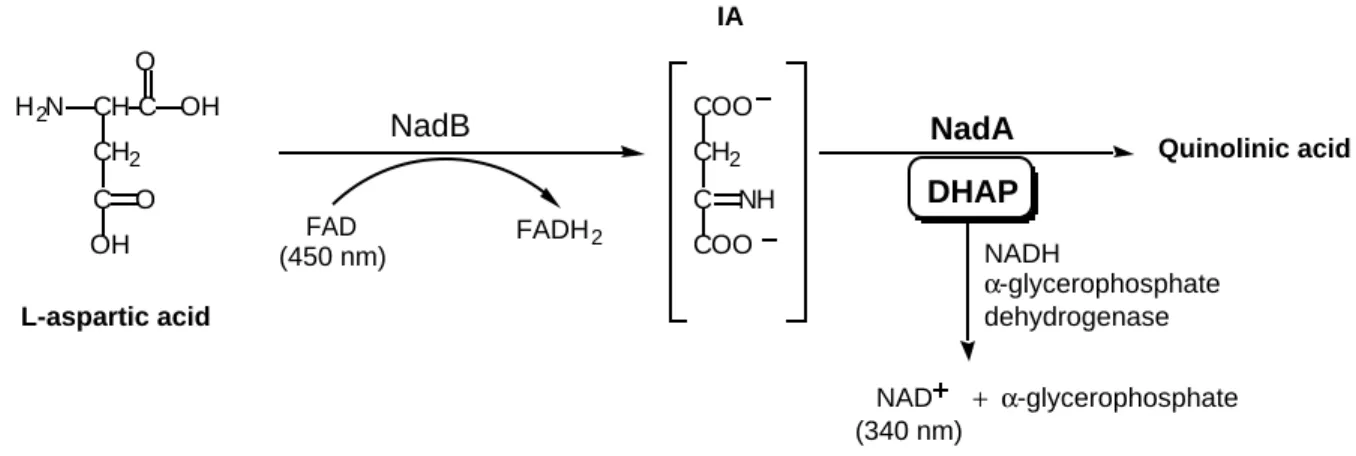
NadA enzymatic activity. NadA catalyzes the quinolinic acid biosynthesis from
iminoaspartate (IA) and DHAP. The whole two-step procedure was performed under anaerobic conditions, inside the glove box at 18°C. In a first step, iminoaspartate was synthetized from 60 µM aspartate oxidation by 60 µM FAD-NadB enzyme. Production of iminoaspartate was checked spectrophotometrically following the decrease of the absorption at 450 nm due to FAD reduction. To that solution, 50 µM DHAP (from Sigma) was then added with 0.7 µM of either apoNadA or iron-sulfur-containing NadA protein. The solution was incubated for 20 min. under anaerobic conditions to prevent oxidation and destruction of
the cluster. The reaction was monitored following the amount of DHAP remaining in solution. For that purpose, in a second step, 250 µM NADH and 0.5 µM α-glycerophosphate dehydrogenase enzyme (260 units/mg) were added to the solution. This enzyme catalyzes the oxidation of DHAP by NADH into α-glycerophosphate and NAD+
. The total amount of NAD+
formed, determined from the decrease of light absorption at 340 nm, is identical to that of remaining DHAP.
Complementation experiments.
MG1655nadA mutant strain, lacking an active NadA, was transformed with pLBi-S or pBAD-NadA allowing the overexpression of IscS and NadA respectively. These transformed strains were isolated on M9 medium supplemented with 2 mM MgSO4, 0.1 mM CaCl2, 0.4%
glycerol, 0.02% arabinose and in some experiments 12.5 µg/ml nicotinic acid (NA). Growth was controlled at 37°C after 12-36 hours.
Analysis. Protein concentration (by monomer) was determined by the method of
Bradford [15]. Protein-bound iron was determined under reducing conditions with bathophenanthroline disulfonate after acid denaturation of the protein [16] and labile sulfide by the method of Beinert [17].
UV-visible Spectroscopy. UV-visible spectra were recorded with a Cary 1 Bio
(Varian) spectrophotometer.
Mössbauer spectroscopy. BL21(DE3)pET-NadA cells were grown aerobically in
minimal M9 57
Fe-enriched medium (35 µM) supplemented with 2 mM MgSO4, 0.4% glucose, 2 µg/ml thiamine, 1 mM CaCl2 and the protein expressed by addition of 0.5 mM IPTG.
Anaerobic purification of NadA was performed as described above under aerobic conditions except that each was made inside the glove box. 10 mg of pure protein were obtained from 4 liters of culture. 57
Fe-Mössbauer spectra were recorded using 400 µl cuvettes containing 170-560 µM protein. Spectra were recorded on a spectrometer operating in constant acceleration
mode using an Oxford cryostat that allowed temperatures from 1.5 to 300 K and a 57
Co source in rhodium. Isomer shifts are reported relative to metallic iron at room temperature.
EPR spectroscopy. Spectra were recorded on a Bruker EMX (9.5 GHz) or ER200D
EPR spectrometers equipped with an ESR 900 helium flow cryostat (Oxford Instruments). Double integrals of the EPR signals and spin concentration were obtained through the Win-EPR software using the spectrum of a 200 µM Cu(EDTA) standard recorded under non saturating conditions.
RESULTS
Purification of the NadA protein.
pET-NadA which carries the nadA gene under the control of the T7 promoter was used to transform the E. coli BL21(DE3) strain and produce NadA, with a histidine tag at the C-terminus. After induction of NadA expression with IPTG, soluble cell-free extracts obtained were prepared and loaded onto an affinity chromatography column (Ni-NTA). Elution with a buffer containing imidazole resulted in a fraction containing 95% pure protein as judged by the SDS-gel electrophoresis (Inset Figure 3). Analysis by denaturing gel electrophoresis (SDS-PAGE) and size exclusion chromatography showed that the enzyme was obtained as an homogeneous protein with an apparent molecular weight of 40 kDa as expected from the amino-acid sequence (molecular weight: 39 305 Da). A gel filtration step using Superdex-200 was used to determine the oligomerization state of NadA. During that step, NadA eluted in a major peak corresponding to a dimeric form (80 kDa), but a minor amount of monomeric form (39.3 kDa) could also be observed. Combining the two Superdex fractions containing NadA, 50 mg of pure NadA could be obtained, from 800 mg of extracts, in a more than 95% pure form as judged by SDS electrophoresis analysis (data not shown). After affinity chromatography the protein was slightly pink, suggesting the presence of an iron-sulfur metal center. Accordingly it was found to contain comparable amounts of iron and sulfide as judged from the specific iron and sulfide quantitation assays. However the amount of iron and sulfide (0.15-0.2 iron and sulfide /monomer) was substoichiometric with regard to the protein, showing that the as-isolated protein was mainly in the apoprotein form.
Spectroscopic characterization of the NadA protein
Reasoning that the low cluster content was due to the aerobic manipulation of the protein and the oxygen-dependent degradation of the cluster, NadA was purified as described above but under strictly anaerobic conditions inside a glove box. The UV-visible spectrum of the anaerobic preparation is shown in Figure 3. It contains only one absorption band at 420 nm more characteristic for a (4Fe-4S) 2+
cluster. As expected, iron and sulfide determination revealed the presence of 3.1 iron and 3.0 sulfide atoms/monomer. Upon exposure to air, the UV-visible spectrum changed with a decrease of the 420 nm absorption band (t1/2=30 min) (data not shown). Reduction with dithionite under anaerobic conditions led to a rapid bleaching of the solution with disappearance of the 420 nm band (Figure 3). During this reaction the initial EPR-silent protein was converted to a S=1/2 species, characterized by an axial EPR signal with g values at g= 2.047 and 1.93 (Figure 4). Temperature dependence and microwave power saturation properties of the signal were in agreement with a (4Fe-4S)+1
center (data not shown). The signal integrated to 78% of total iron. Upon exposure of the tube to air for 10 minutes, the EPR signal totally disappeared.
We used Mössbauer spectroscopy to further characterize the iron sites in the anaerobically purified protein. Figure 5 shows the 4.2K Mössbauer spectrum of a 560 µM NadA preparation obtained after expression in 57
Fe-enriched M9 medium and anaerobic purification using a Ni-NTA column. This preparation contained 3.2 iron and 3.3 sulfide/polypeptide chain. The 4.2K spectrum (Figure 5A) consists of one doublet. For simulation of the spectrum we considered two nested doublets to account for the relatively large linewidths at roughly equal amounts [18]. The simulation yields the following parameters: δ(1)= 0.43(±0.01) mm/s, ∆EQ(1) = 0.94(±0.03); δ(2) = 0.44(±0.01) mm/s, ∆EQ(2)
= 1.27(±0.03) mm/s. The average values of these parameters (δ = 0.43-0.44 mm/s, ∆EQ = 1.11
Spectra 5B and 5C were recorded after thawing the sample and exposing it to air for 20 min and 80 min respectively. Significant changes were observed confirming that the (4Fe-4S)2+(S=0) cluster is air sensitive. An EPR spectrum recorded from the sample of spectrum 6C at liquid helium temperatures revealed the presence of an absorption peak at g~9.3 and a strong derivative signal at g~4.3 (data not shown). Such signals are indicative of S=5/2 species in rhombic environment (E/D~0.33). Although we have not attempted a more thorough characterization by Mössbauer spectroscopy, such species at 4.2K would give rise to a magnetic spectrum in a broad spectral region with small contribution in the –1 to +2 mm/s region. A ~50% reduction of the spectral area in the –1 to +2 mm/s region was observed in the spectra of 5B and 5C relative to the 5A spectrum suggesting that such magnetic species represent 50% of the total iron.
Although the deconvolution of the spectra 5B and 5C is not unique we attempted a qualitative interpretation of these spectra assuming that they accounted for ~50% of the iron relative to the 5A spectrum. We observed that the absorption area in the ~+1.0 mm/s region gradually decreased indicating a degradation of the (4Fe-4S)2+
(S=0) clusters; in the simulation of the spectra 5B and 5C we have included a doublet with parameters taken from the analysis of spectrum 5A thus corresponding to intact residual (4Fe-4S)2+(S=0) centers. We estimate that the iron in (4Fe-4S)2+(S=0) form is 20-25 % in 5B and <10 % in 5C. The decrease of the (4Fe-4S)2+(S=0) doublet is accompanied by the appearance of new species with one of the absorption lines at 0.5 - 0.6 mm/s. We have simulated these new features assuming two nested doublets A and B with parameters δ(A) = 0.29(±0.01) mm/s, ∆EQ(A) = 0.45(±0.03) mm/s
δ(B) = 0.30(±0.02) mm/s, ∆EQ(B) = 0.80(±0.07). The values of the parameters of doublets A
and B, and more specifically the isomer shift, strongly suggested tetrahedral high spin ferric ions, most probably in a diamagnetic environment. Combination of the above information
suggests that doublets A and B arise from (2Fe-2S)2+
(S=0) clusters (25-30% of the iron in 5B and ~40% in 5C).
In summary, Mössbauer spectroscopy indicates that the anaerobically isolated protein contains one (4Fe-4S)2+(S=0) per polypeptide chain. Exposure to air leads to a gradual degradation of this cluster to yield new species most probably in the (2Fe-2S)2+(S=0) form and S=5/2 species of presently unknown characteristics.
The (FeS) cluster of NadA is absolutely required for activity.
Quinolinic acid is supposed to be formed by a condensation reaction between iminoaspartate (IA) and DHAP (Figure 1) catalyzed by the quinolinate synthetase NadA protein. A NadA in vitro enzymatic assay was set up, based on the consumption of DHAP (Figure 6). It was carried out anaerobically in order to avoid degradation of the cluster. NadA under either its apo or (FeS)-containing form (3.4 iron/monomer) was mixed with iminoaspartate (formed during substoichiometric oxidation of L-aspartate by the FAD cofactor of NadB) and DHAP as described in the Materials and Methods section. Its activity i.e its ability to convert DHAP and IA into quinolinic acid was determined measuring the amount of DHAP remaining after a 20 min. reaction. This amount was determined in a second step using α-glycerophosphate dehydrogenase enzyme, an enzyme catalyzing the reaction between DHAP and NADH giving α-glycerophosphate and NAD+
. Thus the total amount of NAD+
formed during the reaction and determined from the decrease of absorption at 340 nm is equal to the amount of residual DHAP (Figure 6). The initial concentration of DHAP (50 µM) was chosen so that α-glycerophosphate dehydrogenase is not saturated. The results clearly demonstrated that consumption of DHAP is NadA-dependent and more interestingly that it absolutely requires the iron-sulfur cluster of NadA. Indeed, 100% of the initial DHAP was recovered after 20 min. reaction when apoNadA was used as an enzyme whereas only
44% was present in the assay mixture with the cluster-containing form of NadA after the same reaction time. After 60 min. reaction almost all DHAP was consumed (6% remaining) (data not shown). Consumption of DHAP was a function of NadA concentration (data not shown). Finally the DHAP assay was used to demonstrate that a preparation of active NadA was totally inactivated upon exposure to air for 60 min. Note that no activity was found when iminoaspartate (IA) was omitted in the reaction. All these results unambiguously show that the (4Fe-4S)2+
cluster is absolutely required for NadA activity.
NadA is required in vivo for NAD biosynthesis
An iscS mutant, lacking the functional FeS biogenesis ISC system, exhibits NAD auxotrophy. Bringing in the iscS mutant plasmidic copies of nadA failed to restore NAD prototrophy whereas multicopy of iscS did. A nadA mutant also exhibits NAD auxotrophy. Bringing in the nadA mutant plasmidic copies of iscS failed to restore NAD biosynthesis whereas multicopies of nadA did. Taken together this ascertains the structural role of nadA gene in NAD biosynthetic pathway and supports the view that NAD auxotrophy of the iscS mutant is due to its inability to synthesize a (4Fe-4S) holoform of NadA.
Discussion
In the present work, we report for the first time on the isolation and characterization of NadA, the quinolinate synthetase, and demonstrate that it absolutely requires a (4Fe-4S) cluster for activity. NadA has been shown to be essential for the growth of E. coli. Since this enzyme is not present in eucaryotes and furthermore is not essential in others microorganisms such as B. subtilis, this protein might be a potential target for selective antibacterial agents.
The anaerobically purified NadA dimeric protein indeed displays iron/sulfide content and spectroscopic properties which unambiguously show that it can assemble one (4Fe-4S)2+
cluster on each polypeptide chain. With a broad absorption band at 420 nm, a quadrupole Mössbauer doublet with δ: 0.43 mm/s and ∆Eq: 1.11 mm/s, the cluster in NadA is comparable to most (4Fe-4S)2+
clusters reported so far [19]. Similarly the g values of the features of the EPR spectrum of the reduced cluster are characteristic for S=1/2 (4Fe-4S)1+
clusters [19]. Several previous studies had led to the suggestion that NadA was an (FeS) protein but did not succeed in showing direct evidence for it [7-10]. This was probably due to the high oxygen-sensitivity of the cluster as demonstrated by the present study: when NadA is exposed to air, a decay of the visible band at 420 nm and the transformation of the (4Fe-4S)2+ cluster into a (2Fe-2S)2+
forms and S=5/2 species of presently unknown identity were observed by Mössbauer and EPR spectroscopy. This is in line with previous in vivo observations that de novo NAD biosynthesis is a pathway sensitive to hyperbaric oxygen and that NadA is specifically the oxygen-sensitive site [10].
O2 sensitivity has also been observed in the case of (4Fe-4S) clusters from other
dehydratases such as aconitase or in “Radical-SAM” enzymes [11, 20]. In these enzymes, (4Fe-4S)2+ clusters are not stable to oxygen and this sensitivity leads to partial or total degradation of the clusters with transient (3Fe-4S)+1
and/or (2Fe-2S)2+
loss of enzymatic activity [11, 20, 21]. All of these proteins have in common a (4Fe-4S) cluster ligated by only three cysteines whereas the fourth iron is coordinated by a non-protein ligand and this specific arrangement is likely to be at the origin of the instability of the cluster [20, 22]. It is thus tempting to suggest that NadA also belongs to this class of (4Fe-4S) proteins and contains a cluster only ligated by three cysteines. However so far there is no direct evidence for this hypothesis and the cluster could be chelated by four cysteines as well. There is indeed a precedent for a (4Fe-4S) cluster ligated by four cysteines with a significant reactivity towards oxygen resulting in the formation of (2Fe-2S) clusters. This is the case for the Transcription factor FNR, which is proposed to utilize this sensitivity to act as an O2 sensor for controlling the expression of specific enzymes under anaerobiosis [23]. NadA contains, in addition to the three conserved cysteine residues of the CXXCXXCP motif, 6 other cysteines among which two strictly conserved (cysteines 113 and 200 from E. coli). They are localized within the ECSL and GA(E)CI(V) motifs (Figure 2) and thus are also candidates as iron ligands. Futher mutagenesis, spectroscopic and structural studies are required to solve this issue.
A free coordination site, which would allow binding and activation of one or both substrates to the unique Fe site, would perfectly fit with a catalytic function for the iron sulfur center. This would be reminiscent of aconitase, in which the (4Fe-4S) cluster binds citrate and uses the Lewis acidity of the iron site to increase the electrophilicity of the substrate thus facilitating the reaction [20]. The reaction catalyzed by NadA is a complex one since it involves condensation of IA with DHAP with elimination of inorganic phosphate, ring closure and dehydration [24]. Several of these steps might be accelerated as a consequence of binding of substrates or intermediates to the cluster of NadA.
The mechanism of the reaction is currently under investigation to understand the chemical role of the cluster at the molecular level.
Acknowledgments
Thanks are due to members of the FB group for fruitful discussion. This work was supported by grants from the CNRS, the Université de la Méditerranée, the Toxicologie Nucleaire program (CEA) and the ACI Biologies Cellulaire Moléculaire et Structurale from the Ministère de l’Education Nationale. We give thanks also to J. Valton (Laboratory DRDC/CB) for valuable help for the enzymatic assay.
References
[1] Foster, J. W. and Moat, A. G. (1980) Regulation of NAD Metabolism in
Salmonella typhimurium: molecular Sequence Analysis of the Bifunctional nadR
Regulator and the nadA-pnuC Operon. Microbiol. Rev. 44, 83-105.
[2] Tritz, G. J. (1987) In S. typhimurium : Cellular and Molecular Biology. Neidhardt, F. C., Ingraham, J. L., Low, K. B., Magasanik, B., Schaechter, M., and Umbarger, H. E; Eds.), pp. 557-563. Am. Soc. Microbiol., Washington, DC.
[3] Begley, T. P., Kinsland, C., Mehl, R. A., Osterman, A. and Dorrestein, P. (2001) The biosynthesis of nicotinamide adenine dinucleotides in bacteria. Vitam Horm 61, 103-119.
[4] Rizzi, M. and Schindelin, H. (2002) Structural biology of enzymes involved in NAD and molybdenum cofactor biosynthesis. Curr. Opin. Struct. Biol. 12, 709-720. [5] Berger, F., Ramirez-Hernandez, M. H. and Ziegler, M. (2004) The new life of a centenarian: signalling functions of NAD(P). Trends. Biochem. Sci. 26: 111-118. [6] Ceciliani, F., Caramori, T., Ronchi, S., Tedeschi, G., Mortarino, M. and Galizzi, A. (2000) Cloning, Overexpression, and Purification of Escherichia coli Quinolinate Synthetase. Protein Expression and Purification 18, 64-70.
[7] Sun, D. and Stelow, P. (1993) Cloning, Nucleotide Sequence, and Regulation of the Bacillus subtilis nadB Gene and a nifS-like Gene, Both of Which are Essential for NAD Biosynthesis. J. Bacteriol. 175, 1423-1432.
[8] Suzuki, N., Carlson, J., Griffith, G. and Gholson, R. K. (1973) Studies on the de
novo biosynthesis of NAD in Escherichia coli. Biochem. Biophys. Acta. 304, 309-315.
[9] Flachmann, R., Kunz, N., Seifert, J., Gütlich, M., Wientjes, F. J., Laufer, A. and Gassen H. G. (1988) Molecular biology of pyridine nucleotide biosynthesis in
Escherichia coli. Cloning and characterization of quinolinate synthesis nadA and nadB. Eur. J. Biochem. 175, 221-228.
[10] Gardner, P. R. and Fridovich, I. (1991) Quinolinate Synthetase: The Oxygen-Sensitive Site of de novo NAD(P)+ Biosynthesis. Arch. Biochem. Biophys. 284, 106-111.
[11] Layer, G., Heinz, D. W., Jahn, D. and Schubert, W. D. (2004) Structure and function of radical SAM enzymes. Cur. Opin. Chem. Biol. 8, 1-9.
[12] Lauhon, C. T. and Kambampati, R. (2000) The iscS gene in Escherichia coli is required for the biosynthesis of 4-thiouridine, thiamin, and NAD. J. Biol. Chem. 275, 20096-20103.
[13] Datsenko, K. A. and Wanner, B. L. (2000) One-step inactivation of chromosomal genes in Escherichia coli K-12 using PCR products. Proc. Natl. Acad. Sci. USA. 97, 6640-6645.
[14] Loiseau, L., Ollagnier-de choudens S, Lascoux, D., Forest, E., Fontecave, M. and Barras, F. (2005) Analysis of the heterodimeric CsdA-CsdE cysteine desulfurase, assisting Fe-S cluster biogenesis in Escherichia coli. J. Biol. Chem. In press.
[15] Bradford, M. M. (1976) A rapid and sensitive method for the quantitation of microgram quantities of protein utilizing the principle of protein-dye binding. Anal.
Biochem. 72, 248-254.
[16] Fish, W. W. (1988) Rapid colorimetric micromethod for the quantitation of complexed iron in biological samples. Methods Enzymol 158, 357-364.
[17] Beinert, H. (1983) Semi-micro Methods for Analysis of labile Sulfide and of labile Sulfide plus Sulfane Sulfur in Unusually Stable Iron-Sulfur Proteins. Anal
[18] Cicchillo, R. M., Lee, K. H., Baleanuu-Cogonea, C., Nesbitt, N. M., Krebs, C. and Booker, S. J. (2004) Escherichia coli lipoyl synthase binds two distinct [4Fe-4S] clusters per polypeptide. Biochemistry 43, 11770-11781
[19] Cammack, R. (1992) Advances in Inorganic chemistry, Vol 38. Series Editor A. G. Sykes, Academic Press, INC. New York.
[20] Beinert, H., Kennedy, M. C. and Stout, C. D. (1996) Aconitase as an iron-Sulfur protein: enzyme and regulatory. Chem. Rev. 96, 2335-2373.
[21] Flint, D. H., Emptage, M. H., Finnegan, M. G., Fu, W. and Jonhson, M. K. (1993) The role and properties of the iron-sulfur cluster in Escherichia coli dihydroxy-acid dehydratase. J. Biol. Chem. 268, 14732-14742.
[22] Walsby, C. J., Hong, W., Broderick, W. E., Cheek, J., Ortillo, D., Broderick, J. B. and Hoffman, B. M. (2002) Electron-nuclear double resonance spectroscopic evidence that S-adenosylmethionine binds in contact with the catalytically active [4Fe-4S](+) cluster of pyruvate formate-lyase activating enzyme. J. Am. Chem. Soc. 124, 3143-3151.
[23] Khoroshilova, N., Popescu, C., Münck, E., Beinert, H. and Kiley, P. (1997) Iron-sulfur cluster disassembly in the FNR protein of Escherichia coli by O2: [4Fe-4S] to [2Fe-2S] conversion with loss of biological activity. Proc. Natl. Acad. Sci. USA 94, 6087-6092.
[24] Nasu, S., Wicks, F. D. and Gholson, R. K. (1982) L-Aspartate Oxidase, a Newly Discovered Enzyme of Escherichia coli, Is the B Protein of Quinolinate Synthetase. J.
Legends to Figures
Figure 1: Pathway for quinolinic acid biosynthesis.
Figure 2: Sequence alignment of NadA proteins from different organisms. Ecoli:
Escherichia coli; Shifl: Shigella flexneri; Salty: Salmonella typhimurium; Yerpe: Yersinia pestis; Vibch: Vibrio cholera. Astericks correspond to conserved aminoacid
residues and the box focussed on conserved cysteine residues.
Figure 3: UV-visible spectrum of anaerobically as-isolated NadA protein (15 µM)
before (solid line) and after reduction with 2 mM dithionite (dashed lines). Inset: SDS-polyacrylamide gel (15%) of NadA purification. Lane M, molecular mass markers (14.4; 20.1; 30; 45; 66 and 97 kDa from the bottom to the top), Lane 1, an aliquot of BL21(DE3)pETNadA crude extract; lane 2, an aliquot of the eluate from the HiTrap Ni2+chelating column.
Figure 4: EPR spectrum of anaerobically as isolated NadA (200 µM) reduced with 2
mM dithionite. T= 5K; P=0.1 mW; gain= 2.105; amplitude modulation: 10 mT.
Figure 5: Zero field Mössbauer spectra of anaerobically purified NadA protein (560
µM, 3.2 iron/monomer) at 4.2K before (A), and after exposure to air during 20 min (B) and 80 min. (C). Solid and dashed lines represent theoretical simulations using the parameters quoted in the text.
Figure 6: Scheme for the enzymatic assay of quinolinic acid biosynthesis. IA is
formed enzymatically from oxidation of L-aspartic acid by NadB. Then DHAP is added together with NadA. The amount of residual DHAP after a given reaction time is equal to the amount of NAD+
formed (determined by the decrease of the absorption at 340 nm) in a second step during incubation with NADH in the presence of α-glycerophosphate dehydrogenase.