HAL Id: hal-03176265
https://hal.sorbonne-universite.fr/hal-03176265
Submitted on 22 Mar 2021
HAL is a multi-disciplinary open access
archive for the deposit and dissemination of
sci-entific research documents, whether they are
pub-lished or not. The documents may come from
teaching and research institutions in France or
abroad, or from public or private research centers.
L’archive ouverte pluridisciplinaire HAL, est
destinée au dépôt et à la diffusion de documents
scientifiques de niveau recherche, publiés ou non,
émanant des établissements d’enseignement et de
recherche français ou étrangers, des laboratoires
publics ou privés.
Generation of two isogenic induced pluripotent stem cell
lines from a 4-month-old severe nemaline myopathy
patient with a heterozygous dominant c.553C > A
(p.Arg183Ser) variant in the ACTA1 gene
Joshua Clayton, Carolin Scriba, Norma Romero, Edoardo Malfatti, Safaa
Saker, Thierry Larmonier, Kristen Nowak, Gianina Ravenscroft, Nigel Laing,
Rhonda Taylor
To cite this version:
Joshua Clayton, Carolin Scriba, Norma Romero, Edoardo Malfatti, Safaa Saker, et al.. Generation of
two isogenic induced pluripotent stem cell lines from a 4-month-old severe nemaline myopathy patient
with a heterozygous dominant c.553C > A (p.Arg183Ser) variant in the ACTA1 gene. Stem cell
research, Elsevier, 2021, 53, pp.102273. �10.1016/j.scr.2021.102273�. �hal-03176265�
Stem Cell Research 53 (2021) 102273
Lab Resource: Multiple Cell Lines
Generation of two isogenic induced pluripotent stem cell lines from a
4-month-old severe nemaline myopathy patient with a heterozygous
dominant c.553C > A (p.Arg183Ser) variant in the ACTA1 gene
Joshua S. Clayton
a,b,*, Carolin K. Scriba
a,b,c, Norma B. Romero
d,e, Edoardo Malfatti
f,
Safaa Saker
g, Thierry Larmonier
g, Kristen J. Nowak
b,h,i, Gianina Ravenscroft
a,b,
Nigel G. Laing
a,b, Rhonda L. Taylor
a,baHarry Perkins Institute of Medical Research, QEII Medical Centre, Nedlands, WA, Australia
bCentre for Medical Research, University of Western Australia, QEII Medical Centre, Nedlands, WA, Australia cNeurogenetics Laboratory, Department of Diagnostic Genomics, PP Block, QEII Medical Centre, Nedlands, WA, Australia
dSorbonne Universit´e, Myology Institute, Neuromuscular Morphology Unit, Center for Research in Myology, GH Piti´e-Salpˆetri`ere, Paris, France eCentre de R´ef´erence de Pathologie Neuromusculaire Paris-Est, GHU Piti´e-Salpˆetri`ere, Assistance Publique-Hˆopitaux de Paris, Paris, France fReference center for Neuromuscular disorders, Henri Mondor teaching hospital, University of Versailles-Paris Saclay, France
gGenethon, DNA and Cell bank, 91000 Evry, France
hOffice of Population Health Genomics, Public and Aboriginal Health Division, Western Australian Department of Health, East Perth, WA, Australia iFaculty of Health and Medical Sciences, School of Biomedical Sciences, University of Western Australia, QEII Medical Centre, Nedlands, WA, Australia
A B S T R A C T
Nemaline myopathy (NM) is a congenital myopathy typically characterized by skeletal muscle weakness and the presence of abnormal thread- or rod-like structures (nemaline bodies) in myofibres. Pathogenic variants in the skeletal muscle alpha actin gene, ACTA1, cause approximately 25% of all NM cases. We generated two induced pluripotent stem cell lines from lymphoblastoid cells of a 4-month-old female with severe NM harbouring a dominant variant in ACTA1 (c.553C > A). The isogenic lines displayed characteristic iPSC morphology, expressed pluripotency markers, differentiated into cells of all three germ layers, and possessed normal karyotypes. These lines could be useful models of human ACTA1 disease.
1. Resource Table:
Unique stem cell lines
identifier 1. HPIi001-A 2. HPIi001-B
Alternative names of stem
cell lines 1. iPS-6303-C6B3 2. iPS-6303-R12
Institution Harry Perkins Institute of Medical Research
Contact information of
distributor Dr. Joshua Clayton joshua.clayton@perkins.org.au
Type of cell lines iPSC
Origin Human
Cell Source EBV-immortalized lymphoblastoid cell line (LCL)
Clonality Clonal
Method of
reprogramming Sendai virus
Multiline rationale Isogenic clones
Gene modification Yes
Type of modification Spontaneous variant
Associated disease
(continued on next column)
(continued)
Unique stem cell lines
identifier 1. HPIi001-A 2. HPIi001-B
Nemaline myopathy 3; NEM3 (OMIM#161800), severe form
Gene/locus Actin Alpha 1, Skeletal Muscle (ACTA1), NM_001100:
c.553C > A
Method of modification N/A
Name of transgene or
resistance N/A
Inducible/constitutive
system N/A
Date archived/stock date September 2020
Cell line repository/bank 1. https://hpscreg.eu/cell-line/HPIi001-A
2. https://hpscreg.eu/cell-line/HPIi001-B
Ethical approval Ethics approval was obtained from the Comit´e de
Protection des Personnes (Est IV DC-2012-1693), and national consent forms for genetic testing, banking and research were signed by the patients or their legal
(continued on next page)
Contents lists available at ScienceDirect
Stem Cell Research
Stem Cell Research 53 (2021) 102273
2
(continued)
Unique stem cell lines
identifier 1. HPIi001-A 2. HPIi001-B
guardian. The patient’s LCLs were banked by Genethon; activity authorization No. AC-2018-3156, import/ export authorization No. IE-2018-994. The study was approved by the University of Western Australia’s Human Research Ethics Committee (approval number: RA/4/20/1008).
2. Resource utility
There are currently few tractable cell or animal models that can be used to model disease pathobiology and/or test treatments for ACTA1 congenital myopathies. The patient-derived iPSC lines described here will complement existing Acta1 mouse and zebrafish models and enable evaluation of genomic therapies for nemaline myopathy.
3. Resource details
Nemaline myopathy (NM) is a form of congenital myopathy typically characterized by muscle weakness and the presence of abnormal thread- or rod-like structures in myofibres on histologic examination (Sewry et al., 2019). Dominant variants in the skeletal muscle alpha-actin gene (ACTA1) are one of the most common causes of NM (Nowak et al., 1999; Sewry et al., 2019). Several recessive ACTA1 variants are also known to cause NM (Sewry et al., 2019). Variants in ACTA1 typically cause severe NM, which presents at birth with hypotonia and profound muscle weakness. Patients have few spontaneous movements with difficulties swallowing and sucking (Sanoudou and Beggs, 2001). Affected infants typically die from respiratory insufficiency or pneumonia within the first months of life (Sanoudou and Beggs, 2001). As such, patient samples for cell reprogramming can be difficult to obtain. The iPSC lines presented here were generated from a lymphoblastoid cell line (LCL) from a 4- month-old female with severe NM (Table 1). The patient showed neonatal disease onset, including hypotonia and hydramnios, and required ventilatory support. Genetic testing revealed the patient was heterozygous for a pathogenic variant in ACTA1; c.553C > A, p. Arg183Ser (Sparrow et al., 2003).
LCLs were reprogrammed into iPSCs using the CytoTune™-iPS 2.0 Sendaivirus reprogramming system. Clones were selected and expanded in mTESR™ 1 culture medium and characterized at passage 10. Both iPSC clones (HPIi001-A, HPIi001-B) had normal morphology; colonies were tightly packed and had defined edges with little to no spontaneous differentiation (Fig. 1A). Pluripotency was confirmed by qRT-PCR (Fig. 1B) and immunocytochemistry (ICC) (Fig. 1C, D). Specifically, iPSCs were enriched for OCT4, SOX2, NANOG and CRIPTO by qPCR compared to parental LCLs (Fig. 1B), and stained positively for OCT4, SOX2, SSEA4 and TRA-1-60 by ICC (Fig. 1C, D). Trilineage differentia-tion potential was assessed by directed differentiadifferentia-tion followed by qRT-
PCR (Fig. 1E) and ICC (Fig. S1A, B). Differentiated mesoderm cultures were enriched for TBXT and BMP4 by qPCR, and Brachyury (TBXT) by ICC. Ectoderm cultures were enriched for OTX2 and PAX6 by qPCR, and OTX2 by ICC. Endoderm cultures were enriched for GATA4 and SOX17 by qPCR, and GATA4 by ICC. Parental (undifferentiated) iPSC cultures showed no or negligible expression of each germ layer marker (Fig. 1E). Both iPSC clones were confirmed to be EBV- and SeV-free at passage 10 by PCR and RT-PCR, respectively (Fig. 1F and 1G). Pre-screening for the 8 most common karyotypic abnormalities in human iPSCs by hPSC Genetic Analysis (qPCR) indicated normal copy number (2.0 ± 0.3) at all tested loci (Fig. S1C). Karyostat analyses further verified both clones possess a normal female karyotype (46, XX) with no aneuploidies (Fig. 1H). Short tandem repeat (STR) typing was used to verify culture identity and purity; both iPSC clones matched the original parental lymphoblastoid cell line (data archived with journal). The original ACTA1 mutation (c.553C > A) was confirmed to be present and het-erozygous in both clones by PCR and Sanger sequencing (Fig. 1I). The iPSC lines were free of mycoplasma by a universal PCR test and agarose gel electrophoresis (Fig. S1C). Characterization of the iPSC clones is summarized in Table 2.
4. Materials and methods
4.1. Generation and maintenance of iPSC lines
Patient lymphoblastoid cell lines (LCLs) were cultured in RPMI1640 medium supplemented with 10% fetal bovine serum and 1% L-glutamine
(R10 medium) at 37 ◦C and 5% CO
2. LCLs were reprogrammed using the
CytoTune™-iPS 2.0 Sendai Reprogramming Kit (ThermoFisher). Briefly, 3 × 10⁵ cells were transduced at recommended multiplicity of infection
and plated on growth factor-reduced (GFR) Matrigel® (1:100 in DMEM/ F-12; ThermoFisher) in R10 medium. Cells were gradually adapted to mTESR™1 medium (StemCell) and individual clones picked for expan-sion and validation. iPSCs were cultured on GFR Matrigel and passaged every 3–5 days (at ~ 80% confluency) using 1X Versene (Thermo-Fisher). Cells were cryopreserved in 90% KnockOut™ Serum Replace-ment (ThermoFisher) with 10% DMSO. Control iPSCs were a gift from Prof. Rhonda Bassel-Duby and were maintained as above.
4.2. Immunocytochemistry – pluripotency marker expression and trilineage differentiation potential
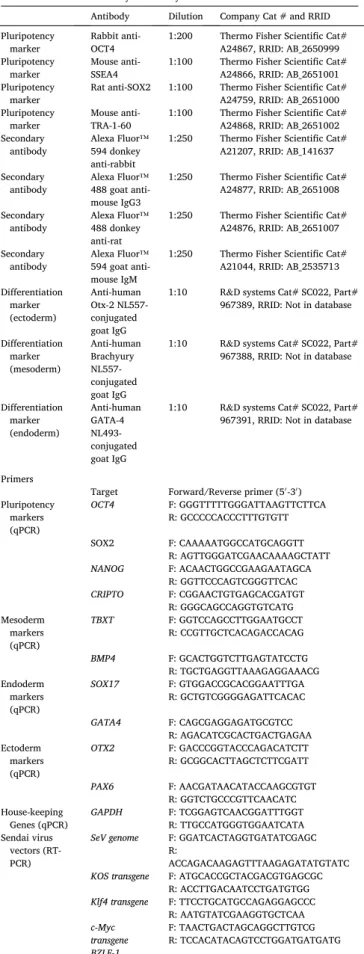
For qualitative pluripotency analysis, iPSCs were plated on Matrigel- coated 96-well Nunc polymer optical bottom plates and stained for OCT4, SSEA4, SOX2, and TRA-1-60 (Table 2). Trilineage differentiation potential was assessed using the STEMdiff™ Trilineage Differentiation Kit (StemCell) using StemPro™ Accutase™ (Gibco) for cell dissociation. Immunocytochemistry was performed as described in the PSC 4-marker Immunocytochemistry Kit (ThermoFisher), except that cells were incu-bated with primary antibodies (Table 3) overnight at 4 ◦C in 3% BSA.
Nuclei were stained using NucBlue™ Fixed Cell stain (ThermoFisher).
Table 1
Summary of lines.
iPSC line names Abbreviation in figures Gender Age Ethnicity Genotype of locus Disease
HPIi001-A (iPS-6303-C6B3) HPIi001-A Female 4 months Turkish C/A Nemaline myopathy 3; NEM3 (OMIM#161800), severe form
HPIi001-B (iPS-6303-R12) HPIi001-B Female 4 months Turkish C/A Nemaline myopathy 3; NEM3 (OMIM#161800), severe form
Stem Cell Research 53 (2021) 102273
4
Cells were imaged on an Olympus IX71 microscope with a DP74 camera and CellSens software.
4.3. DNA and RNA extraction
Genomic DNA was extracted using the QIAamp DNA Mini Kit (QIAGEN). RNA was extracted using the RNeasy Mini Kit (QIAGEN). 4.4. Quantitative reverse transcriptase polymerase chain reaction (qRT- PCR)
Total RNA was reverse transcribed into cDNA using the Super-Script™ III First-Strand Synthesis System (ThermoFisher). qRT-PCR was performed using the Rotor-Gene SYBR Green RT-PCR Kit on a Rotor- Gene Q thermocycler. Cycling conditions were as follows: 95 ◦C for 5
min, 45 cycles of 95 ◦C for 10 sec and 60 ◦C for 15 sec (acquiring),
followed by melt curve analysis (60 ◦C to 95 ◦C, 1◦/step). GAPDH was
used for normalization, using the ΔCT method. Primers are listed in
Table 3.
4.5. Polymerase chain reaction (PCR)
EBV genes (OriP, EBNA1, LMP1 and BLZF1) and a reference gene (GAPDH) were detected by PCR using GoTaq® G2 Hot Start Master Mix (Promega) with primers from Barrett et al., 2014. Presence of Sendai virus genome and transgenes were assessed by RT-PCR at passage 10 as per the CytoTune™ manufacturer’s protocol. Transduced LCLs (day 3) were used as a positive control. Primers are listed in Table 3.
4.6. KaryoStat analysis
Karyostat analysis was performed by the Ramaciotti Centre for Ge-nomics (Sydney, NSW, Australia). Data were analyzed using Chromo-some Analysis Suite 4.2 (ThermoFisher).
4.7. Confirmation of pathogenic ACTA1 variant
ACTA1 exon 4 was amplified by PCR using GoTaq® G2 Hot Start Master Mix (Promega). Sanger sequencing was performed by the Australian Genome Research Facility (Perth, WA, Australia). Chro-matograms were analyzed using Benchling (benchling.com). Primers are listed in Table 3.
4.8. Mycoplasma testing
Lines were screened for mycoplasma using the ATCC Universal My-coplasma PCR test kit.
4.9. STR typing
STR typing was performed by PathWest Diagnostic Genomics (Perth, WA, Australia) using the QSTR Plus assay (Elucigene).
Declaration of Competing Interest
Nigel G Laing reports financial support was provided by A Founda-tion Building Strength (AFBS). Kristen J Nowak reports financial support was provided by The French Muscular Dystrophy Association (AFM- Telethon).
Table 2
Characterization and validation.
Classification Test Result Data
Morphology Photography (light microscopy) Normal Fig. 1, panel A
Phenotype Qualitative analysis
(Immunocytochemistry) Positive for OCT4, SOX2, SSEA4, TRA-1–60 Fig. 1, panels C/D
Quantitative analysis (qRT-PCR) Expression of OCT4, SOX2, NANOG, CRIPTO Fig. 1, panel B
Genotype KaryoStat™ assay (CytoScan
Optima) 46, XX. No chromosomal aberrations were found in either line. Minimum resolution = 1 MB for losses, 2 MB for gains, 5 MB for LOH/AOH Fig. 1, panel H
Identity Microsatellite PCR (mPCR) OR STR
analysis Microsatellite PCR not performed Matched to parental LCL line at 22/22 STR loci Archived with journal
Mutation analysis (IF
APPLICABLE) Sanger sequencing Southern Blot OR WGS Heterozygous ACTA1 mutation; NM_001100:c.553C > A Not performed Fig. 1, panel I
Microbiology and virology Mycoplasma Negative by PCR Supplementary Fig. 1,
panel D
Differentiation potential Directed differentiation, qPCR Enrichment of TBXT and BMP4 (mesoderm), GATA4 and SOX17 (ectoderm), and
OTX2 and PAX6 (endoderm) Fig. 1, panel E
Directed differentiation,
immunocytochemistry Positive for OTX2 (ectoderm), Brachyury (mesoderm), GATA4 (endoderm) Supplementary Fig. 1, panel A/B
Donor screening
(OPTIONAL) HIV 1 + 2 Hepatitis B, Hepatitis C Not performed
Genotype additional info
(OPTIONAL) Blood group genotyping HLA tissue typing Not performed Not performed
Acknowledgements
This work was supported by funding from an AFM Telethon Tram-poline Grant (REF-21816) to Kristen Nowak, and A Foundation Building Strength Research Grant (ID-A3TR22, PI Nigel Laing). We also gratefully acknowledge funding from the Australian National Health and Medical Research Council, including a Principal Research Fellowship (APP1117510) to Nigel Laing and a Career Development Fellowship (APP1122952) to Gianina Ravenscroft.
Appendix A. Supplementary data
Supplementary data to this article can be found online at https://doi. org/10.1016/j.scr.2021.102273.
References
Barrett, R., Ornelas, L., Yeager, N., Mandefro, B., Sahabian, A., Lenaeus, L., Targan, S.R., Svendsen, C.N., Sareen, D., 2014. Reliable generation of induced pluripotent stem cells from human lymphoblastoid cell lines. Stem Cells Transl. Med. 3, 1429–1434.
Nowak, K.J., Wattanasirichaigoon, D., Goebel, H.H., Wilce, M., Pelin, K., Donner, K., Jacob, R.L., Hübner, C., Oexle, K., Anderson, J.R., Verity, C.M., North, K.N., Iannaccone, S.T., Müller, C.R., Nürnberg, P., Muntoni, F., Sewry, C., Hughes, I., Sutphen, R., Lacson, A.G., Swoboda, K.J., Vigneron, J., Wallgren-Pettersson, C., Beggs, A.H., Laing, N.G., 1999. Mutations in the skeletal muscle α-actin gene in patients with actin myopathy and nemaline myopathy. Nat. Genet. 23 (2), 208–212.
Sanoudou, D., Beggs, A.H., 2001. Clinical and genetic heterogeneity in nemaline myopathy – a disease of skeletal muscle thin filaments. Trends Mol. Med. 7 (8), 362–368.
Sewry, C.A., Laitila, J.M., Wallgren-Pettersson, C., 2019. Nemaline myopathies: a current view. J. Muscle Res. Cell Motil. 40 (2), 111–126.
Sparrow, J.C., Nowak, K.J., Durling, H.J., Beggs, A.H., Wallgren-Pettersson, C., Romero, N., Nonaka, I., Laing, N.G., 2003. Muscle disease caused by mutations in the skeletal muscle alpha-actin gene (ACTA1). Neuromuscul. Disord. 13 (7-8), 519–531.
Table 3
Reagents details.
Antibodies used for immunocytochemistry
Antibody Dilution Company Cat # and RRID
Pluripotency
marker Rabbit anti- OCT4 1:200 Thermo Fisher Scientific Cat# A24867, RRID: AB_2650999 Pluripotency
marker Mouse anti- SSEA4 1:100 Thermo Fisher Scientific Cat# A24866, RRID: AB_2651001 Pluripotency
marker Rat anti-SOX2 1:100 Thermo Fisher Scientific Cat# A24759, RRID: AB_2651000 Pluripotency
marker Mouse anti- TRA-1-60 1:100 Thermo Fisher Scientific Cat# A24868, RRID: AB_2651002 Secondary
antibody Alexa Fluor™ 594 donkey anti-rabbit
1:250 Thermo Fisher Scientific Cat#
A21207, RRID: AB_141637 Secondary
antibody Alexa Fluor™ 488 goat anti- mouse IgG3
1:250 Thermo Fisher Scientific Cat#
A24877, RRID: AB_2651008 Secondary
antibody Alexa Fluor™ 488 donkey anti-rat
1:250 Thermo Fisher Scientific Cat#
A24876, RRID: AB_2651007 Secondary
antibody Alexa Fluor™ 594 goat anti- mouse IgM
1:250 Thermo Fisher Scientific Cat#
A21044, RRID: AB_2535713 Differentiation marker (ectoderm) Anti-human Otx-2 NL557- conjugated goat IgG
1:10 R&D systems Cat# SC022, Part#
967389, RRID: Not in database Differentiation marker (mesoderm) Anti-human Brachyury NL557- conjugated goat IgG
1:10 R&D systems Cat# SC022, Part#
967388, RRID: Not in database
Differentiation marker (endoderm) Anti-human GATA-4 NL493- conjugated goat IgG
1:10 R&D systems Cat# SC022, Part#
967391, RRID: Not in database
Primers
Target Forward/Reverse primer (5′-3′)
Pluripotency markers (qPCR) OCT4 F: GGGTTTTTGGGATTAAGTTCTTCA R: GCCCCCACCCTTTGTGTT SOX2 F: CAAAAATGGCCATGCAGGTT R: AGTTGGGATCGAACAAAAGCTATT NANOG F: ACAACTGGCCGAAGAATAGCA R: GGTTCCCAGTCGGGTTCAC CRIPTO F: CGGAACTGTGAGCACGATGT R: GGGCAGCCAGGTGTCATG Mesoderm markers (qPCR) TBXT F: GGTCCAGCCTTGGAATGCCT R: CCGTTGCTCACAGACCACAG BMP4 F: GCACTGGTCTTGAGTATCCTG R: TGCTGAGGTTAAAGAGGAAACG Endoderm markers (qPCR) SOX17 F: GTGGACCGCACGGAATTTGA R: GCTGTCGGGGAGATTCACAC GATA4 F: CAGCGAGGAGATGCGTCC R: AGACATCGCACTGACTGAGAA Ectoderm markers (qPCR) OTX2 F: GACCCGGTACCCAGACATCTT R: GCGGCACTTAGCTCTTCGATT PAX6 F: AACGATAACATACCAAGCGTGT R: GGTCTGCCCGTTCAACATC House-keeping
Genes (qPCR) GAPDH F: TCGGAGTCAACGGATTTGGT R: TTGCCATGGGTGGAATCATA Sendai virus
vectors (RT- PCR)
SeV genome F: GGATCACTAGGTGATATCGAGC R:
ACCAGACAAGAGTTTAAGAGATATGTATC
Table 3 (continued)
Antibodies used for immunocytochemistry
Antibody Dilution Company Cat # and RRID
EBNA testing (PCR) F: CACCTCAACCTGGAGACAAT R: TGAAGCAGGCGTGGTTTCAA LMP1 F: ATGGAACACGACCTTGAGA R: TGAGCAGGATGAGGTCTAGG EBNA1 F: ATCAGGGCCAAGACATAGAGA R: GCCAATGCAACTTGGACGTT OriP F: TCGGGGGTGTTAGAGACAAC R: TTCCACGAGGGTAGTGAACC House-keeping
Genes (PCR) GAPDH F: ACCACAGTCCATGCCATCAC R: TCCACCACCCTGTTGCTGTA Targeted mutation analysis (PCR/ sequencing) ACTA1 exon 4 (F primer used for sequencing) F: TAGCGCTGAGAGCCTAGCC R: CTGTGGTCACGAAGGAGTAGC