HAL Id: tel-01164978
https://tel.archives-ouvertes.fr/tel-01164978
Submitted on 18 Jun 2015HAL is a multi-disciplinary open access
archive for the deposit and dissemination of sci-entific research documents, whether they are pub-lished or not. The documents may come from teaching and research institutions in France or abroad, or from public or private research centers.
L’archive ouverte pluridisciplinaire HAL, est destinée au dépôt et à la diffusion de documents scientifiques de niveau recherche, publiés ou non, émanant des établissements d’enseignement et de recherche français ou étrangers, des laboratoires publics ou privés.
Denise Galzerano
To cite this version:
Denise Galzerano. Electronic properties of carotenoids in natural and artificial photosynthesis. Vegetal Biology. Université Pierre et Marie Curie - Paris VI, 2014. English. �NNT : 2014PA066153�. �tel-01164978�
i
Université Pierre et Marie Curie
Ecole doctorale iViv- ED 387
Étude des propriétés électroniques des caroténoïdes dans
la photosynthèse naturelle et artificielle
Par Denise Galzerano
Thèse de doctorat de Biologie
Dirigée par Dr. Bruno ROBERT
Présentée et soutenue publiquement le 12/05/2014Devant un jury composé de :
ZITO Francesca Rapporteur
OUCHANE Soufian Rapporteur
RAPPAPORT Fabrice Examinateur
VAN GRONDELLE Rienk Examinateur
ROBERT Bruno Directeur de thèse
iii
Sommaire
La photosynthèse est un processus à plusieurs étapes qui commence par la capture de la lumière par des structures moléculaires spécialisées incluses dans les organismes photosynthétiques. Les pigments présents dans les protéines de l'appareil photosynthétique jouent un rôle essentiel dans les premiers événements.
Ces pigments, des chlorophylles et des caroténoïdes, peuvent absorber la lumière et transférer l'énergie résultante en excitation aux molécules voisines, garantissant le respect de la succession des étapes photosynthétiques. En plus de l'absorption de la lumière, les caroténoïdes protègent l'appareil photosynthétique du stress photo-oxydatif survenant en condition de lumière intense. De cette manière, ce processus garantit l’équilibre entre l’absorption de l’énergie lumineuse, son utilisation et la protection à une exposition excessive. Les caroténoïdes possèdent une structure moléculaire composée d’une chaine linéaire de polyene conjuguée. Cette structure confère à ces pigments des propriétés électroniques uniques grâce auxquelles ils réalisent leurs fonctions. La connaissance de ces propriétés est essentielle pour comprendre leurs mécanismes d’actions.
Malgré la simplicité apparente de leur structure, les calculs précis de leurs propriétés électroniques et vibrationnels ainsi que la prédiction de leur comportement selon l’environnement dans lequel le caroténoïde se trouve, s’effectuent difficilement et exigent des approches complexes. Ces travaux de recherche illustrent différentes approches pour être au plus près des mécanismes d'interactions entre les pigments et la lumière, avec un accent mis sur le rôle photo-protecteur joué par les caroténoïdes. Une série d'échantillons, à différents niveaux d'organisations structurelles des protéines collectrices de lumière contenants ces pigments, sera décrite et analysée.
Après une introduction générale, nous analysons deux cas où des molécules de caroténoïdes cycliques identiques sont attachés à la même protéine, mais exposent néanmoins des transitions d’absorption différentes. Le centre de réaction du photosystème II (PSII) contient deux -carotènes, une qui absorbe à 489 nm et l'autre à 507 nm. Par contre, le complexe majeur du photosystème II (LHCII) contient deux molécules de lutéine, une qui absorbe à 495 nm et l'autre à 510 nm. Dans chaque cas, on remarque que l'espèce moléculaire identique dans la même protéine expose des propriétés électroniques différentes.
iv Nous démontrons que l'environnement du site de liaison à la protéine influence de façon différente les propriétés de ces caroténoïdes possédant un cycle à leur extrémité. Le carotène du PSII qui absorbe à 487 nm et la lutéine du LHCII qui absorbe à 495 nm sont principalement influencés par la polarisabilité locale de leur site de liaison. Inversement, le carotène qui absorbe à 507 nm et la lutéine qui absorbe à 510 nm sont influencés par la
structure de leur site de liaison : des encombrements stériques locaux force les cycles à se
retrouver sur le même plan que la chaine polyene.
Dans le chapitre 3, nous analysons des mutants d’Arabidopsis thaliana qui présentent une modification de la voie de biosynthèse des caroténoïdes. Ces mutants expriment la phytoène désaturase bactérienne (CRTI) en plus de l’enzyme endogène impliqué dans les réactions de désaturation du phytoène (PDS). En conséquence ils présentent un changement de la composition des caroténoïdes, montrant une diminution de la quantité de lutéine et une augmentation des xanthophylles dérivées du -carotène. Les mutants présentent une sensibilité majeure à la lumière intense. La cause de ce changement est l’altération de la chaîne de transport électronique photosynthétique plutôt que les changements de la composition de caroténoïde dans les protéines collectrice de lumière. Il s'avère que dans les lignes qui expriment CRTI, le niveau de protéine de l'oxydase terminale plastid (PTOX) augmente, tandis que le flux électronique cyclique est supprimé.
D’après les résultats, PTOX rivalise efficacement avec le flux électronique cyclique au niveau du plastoquinol dans les mutants et joue un rôle crucial dans le contrôle de l'état de réduction du plastoquinone « pool ». Ceci confirme l'hypothèse que PTOX est capable de moduler l'équilibre entre le flux d’électron linéaire et le flux cyclique autour du photosystème I (PSI).
Dans le chapitre 4, des antennes photosynthétiques artificielles (dyades), imitant l'interaction entre les chlorophylles et les caroténoïdes, sont analysées. Grâce à une ingénierie précise des interactions entre leurs composants (une molécule tétrapyrrolique et une molécule similaire au caroténoïde), ces molécules synthétiques peuvent imiter un certain nombre de caractéristiques et fonctions de leurs équivalentes naturelles. Ici nous nous concentrons sur le mécanisme de transfert d'énergie parmis les états excités de triplet (transfert T-T) entre le tétrapyrrole et le caroténoïde de deux différents dyades artificielles. Nous étudions les caractéristiques spectroscopiques de leurs états de triplet et nous les comparons aux systèmes naturels, où le même mécanisme à lieu.
Les analyses spectroscopiques exécutées indiquent que dans la dyade carotenophthalocyanine (dyade 1) la structure électronique de l’état de triplet est partagée
v entre le caroténoïde et le tétrapyrrole. Ce couplage permet un transfert d'énergie T-T extrêmement rapide. Des résultats similaires ont été trouvés dans des complexes de protéines collectrices de lumière des organismes qui réalisent une photosynthèse oxygénique.
Nous suggérons qu'une structure électronique partagée est essentielle pour la protection de la production de l’état singlet de l’oxygène dans des membranes photosynthétiques de ces organismes. Dans le carotenopurpurpine (dyade 2) le lien tétrapyrrole-caroténoïde fournit un couplage électronique plus faible. En conséquence, le transfert d'énergie T-T est plus lent et montre moins de preuves spectroscopiques d'un état de triplet délocalisé. Cette dyade imite le comportement des pigments dans les complexes LH2 de bactéries photosynthétiques anaérobies dans lesquelles l'exposition à l'oxygène est intermittente et beaucoup plus basse.
Les deux cas analysés suggèrent qu’un transfert d'énergie T-T rapide, tant dans les protéines photosynthétiques naturelles que dans les dyades artificielles, exige un changement de la structure d'état de triplet du caroténoïde, probablement à travers le partage de triplet avec le tétrapyrrole. Les systèmes artificiels de cette étude décrivent l'avantage évolutif des organismes aérobies d’utiliser des chromophores bien couplés pour un transfert d’énergie rapide T-T. Cela permet de dissiper efficacement le triplet des chlorophylles pour empêcher la production de l’état singlet de l’oxygène. Au contraire, les organismes anaérobies ont une version plus faible et probablement plus archaïque de ce mécanisme de protection. Ce travail démontre que le mécanisme naturel de protection peut être pourvu dans des constructions photosynthétiques artificielles.
Dans le chapitre 5, des résultats préliminaires concernant l'effet des conditions de solubilisation sur LHCII, sont présentés. Nous décrivons le caractère dynamique de l’LHCII et de ses pigments en fonction des propriétés du détergent.
En conclusion, l'étude des fonctions des molécules de caroténoïde, impliquant l'interaction avec la lumière, exige un approche pluridisciplinaire et à plusieurs niveaux.
Cette thèse illustre qu’une combinaison de techniques spectroscopiques et biochimiques contribue à mettre en évidence et décrire les propriétés physico-chimiques des caroténoïdes leur conférant un rôle vaste et essentiel dans la photosynthèse.
vii
This work was carried out at the
Laboratoire bioénergétique membranaire et stress (LBMS)
Address
iBiTec-S, Bâtiment 532
CEA, UMR 8221 CNRS,
ix
Acknowledgments
I would like to thank, first of all, my supervisor Prof. Bruno Robert for giving me the opportunity to work in his lab and for always encouraging and supporting me. When I started the PhD I had a limited background and no practical experience in the photosynthetic field, but Bruno trusted me and I will always be grateful for it.
I thank Cristian who introduced me to the world of spectroscopy and who was always around me to give good advices and to discuss about the experimental results. Many thanks to my colleagues Andrew, Andy, Maria, Luc and Ana for their constant help and thanks also to Dr. Ghada Ajlani, who was always “next door” to answer to any kind of questions. A special thanks to my colleague and good friend Liz; without her these years at the Cea wouldn’t have been the same.
To Dr. Anja Krieger-Lizskay and Dr. Winfried Leibl, thanks for the fruitful collaborations we had and for giving me the opportunity of learning new experimental techniques.
I also express my gratitude to my collaborators Tom and Ana Moore and Katie Wong-Carter form Arizona State University and to people from the Free University of Amsterdam for sharing their advices on part of my work.
To all the people from the iBiTeC-S, thank you for your support all over the time course of my thesis.
Special thanks go to people from the Marie Curie ITN HARVEST Network: being part of this international, multidisciplinary and wonderful group was one of the most exciting experiences of my life from both the professional and human point of view. Huge thanks for the happy moments I had with the students and post-doc I met during the network meeting and courses all around Europe. Thanks to Luca and Julien: the best memories of these last three years are from the time I spent with you.
To Liz, Sané, Denise, Qian, Kathleen, Margaux, Mehdi, Stéphanie, Michal, Amin, Eiri, Hassina, Eduardo and Manolis, thanks for all the good moments we spent together inside and outside the Cea.
Finally special thanks to my family, my childhood friends and my dear Hugues for always encouraging me, especially during the bad moments and for being so understanding and patience.
xi
List of abbreviations
4-POBN= 4-pyridyl-1-oxide-N-tert-butylnitrone; Chl a= chlorophyll a Chl b= chlorophyll b Chl= chlorophyllCRTI= phytoene desaturase;
DCMU= 3-(3,4-Dichlorophenyl)-1,1-dimethylurea;
DNP-INT= 2’-iodo-6-isopropyl-3-methyl-2’, 4-4’-trinitrodiphenylether; EADS= evolution associated difference spectra
EET= electronic energy transfer FWHM=full width at half maximum ISC= intersystem crossing
kDa= kilodalton
LHCII= light harvesting complex II; LHCs= light harvesting complexes LT= low temperature
Lut= lutein
NDH= plastid NAD(P)H dehydrogenase; Neo= neoxanthin
NPQ= non-photochemical quenching OG= octyl-gallate;
P700= chlorophyll a molecule in association with photosystem I; PQ= plastoquinone;
PSII= photosystem II;
PTOX= plastid terminal oxidase;
qE= rapidly relaxing , energy dependent quenching component of NPQ ; qI= very slowly relaxing, photoinibitory quenching component of NPQ; qP= photochemical quenching
qT= slowly relaxing quenching component of NPQ cause by state transitions RC= reaction centre;
ROS= reactive oxygen species; RR= resonance Raman spectroscopy
xii RT= room temperature
TEMPD= 2,2,6,6-tetramethyl-4-piperidone hydrochloride THF= tetrahydrofuran Vio= violaxanthin XC= xanthophyll cycle Zea= zeaxanthin -DDM= n-dodecyl--D-maltoside -DDM= n-dodecyl--D-maltoside -car = -carotene
xiii
Table of contents
Title page i Summary iii Acknowledgments ix List of abbreviations xi Contents xiii List of figures xiv List of tables xviiCHAPTER 1 Introduction
... 11.1 General introduction ... 3
1.2 Carotenoids ... 4
1.3 Chlorophylls ... 11
1.4 The proteins of the photosynthetic apparatus from higher plants ... 12
1.4.1 Peripheral Photosystem II Antenna Complexes ... 12
1.4.2 Photosystem II supercomplexes... 15
1.4.3 Photosystem II ... 16
1.4.4 Cytochrome b6f ... 18
1.4.5 Photosystem I ... 18
1.4.6 ATP-synthase complex ... 20
1.4.7 Linear electron transport ... 20
1.4.8 Cyclic electron transport ... 20
1.5 Photoprotective mechanisms in higher plants ... 21
1.5.1 Photoprotective role of carotenoids ... 22
1.5.2 Non-photochemical quenching (NPQ) ... 23
1.6 Photoinhibition ... 28
1.7 Realizing artificial photosynthesis ... 29
1.8 Experimental approach ... 31
1.8.1 Resonance Raman spectroscopy ... 32
1.8.2 Carotenoid molecules ... 34
1.8.3 Chlorophylls and derivatives ... 35
1.8.4 Step-scan Fourier transform infrared spectroscopy ... 35
1.9 Project outline ... 36
CHAPTER 2 Mechanisms underlying carotenoid absorption in oxygenic
photosynthetic proteins
………...512.1 Introduction ... 54
2.2 Experimental procedure ... 56
2.3 Results and discussion ... 57
2.3.1 Isolated β-Carotene and Lutein ... 57
xiv
2.3.3 Lutein Molecules in LHCII ... 62
2.3.4 Mechanisms tuning carotenoid absorption in PSII-RC and LHCII ... 65
CHAPTER 3 Effect of constitutive expression of bacterial phytoene
desaturase CRTI on photosynthetic electron transport in Arabidopsis
thaliana
... 753.1 Introduction ... 78
3.2 Materials and Methods ... 80
3.3 Results ... 83
3.3.1 Light sensitivity of CRTI expressing lines ... 83
3.3.2 Generation of H2O2-derived hydroxyl radicals in the CRTI-lines ... 87
3.3.3 Reduction state of the plastoquinone pool in CRTI-lines ... 91
3.3.4 Cyclic electron flow in the CRTI-lines ... 92
3.4 Discussion ... 94
CHAPTER 4
Carotenotetrapyrrole Dyads Mimic Photosynthetic
Triplet-Triplet Energy Transfer ... 105
4.1 Introduction ... 109
4.2 Methods ... 110
4.3 Results ... 114
4.4 Discussion ... 121
CHAPTER 5Effect of the isomeric forms of dodecyl-maltoside detergent on
LHCII spectroscopic properties
... 1315.1 Introduction ... 134
5.2 Material and methods ... 136
5.3 Results ... 137
5.4 Discussion ... 143
CHAPTER 6 General discussion and future perspective
... 1496.1 Introduction ... 151
6.2 Tuning of carotenoid absorption properties ... 151
6.3 Regulation of the photosynthetic electron flow in Arabidopsis thaliana ... 153
6.4 Reengineering photosynthesis: artificial antenna system ... 156
6.5 Dynamic and flexibility of LHCII ... 157
6.6 Conclusions ... 159
xv
List of figures
Figure 1.1 Zeaxanthin molecular structure ... 6
Figure 1.2 Carotenoid biosynthesis pathway (Diretto et al., 2006). ... 7
Figure 1.3 General energy level scheme of carotenoids. ... 10
Figure 1.4 Absorption spectrum of zeaxanthin inTHF. ... 10
Figure 1.5 Molecular structure of chlorophylls derivatives. ... 11
Figure 1.6 Pigment and absorption maxima of the most common chlorophylls . ... 12
Figure 1.7 Crystal structure of LHCII ... 13
Figure 1.8 CP29 crystal structure ... 14
Figure 1.9 Model of the PSII supercomplex C2S2M2 from higher plants. ... 16
Figure 1.10 Overall structure of PSII dimer.. ... 18
Figure 1.11 Overall structure of plant PSI, represented in surface and schematic ... 19
Figure 1.12 Schematic representation or the behavior of photosynthetic organisms in presence of different light intensities. ... 21
Figure 1.13 Mechanisms of photoprotection played by carotenoids in photosynthesis ... 23
Figure 1.14 Structural model of an LHCII monomer showing the key pigments involved in the establishment of qE. ... 27
Figure 1.15 Schematic representation of an artificial photosynthetic system for water splitting. ... 29
Figure 1.16 Biosynthetic scheme of a carotenoid ether dyad ... 31
Figure 1.17 The Raman effect designates an exchange of energy between a molecule and a photon during scattering………32
Figure 1.18 Resonance Raman spectrum of zeaxanthin in THF at 488nm excitation wavelength………34
Figure 1.19 Resonance Raman spectrum of chlorophyll a in THF at 413 nm excitation wavelengt………..35
Figure 2.1 Molecular structures of -carotene and lutein ... 54
Figure 2.2 Correlation between the S0→S2 electronic transition and the frequency of the ν1 Raman band for linear carotenoids with different conjugation length ... 58
Figure 2.3 Absorption spectra of PSII-RC particles ... 59
xvi
Figure 2.5 Correlation between the S0→S2 electronic transition and the frequency of the ν1
Raman band for the two -carotene molecules in PSII-RC ... 62
Figure 2.6 Absorption spectra of LHCII trimers ... 63 Figure 2.7 RR spectra of LHCII trimers recorded with 496.5- and 514.5-nm excitation ... 64 Figure 2.8 Correlation between the S0→S2 electronic transition and the frequency of the ν1
Raman band for the two lutein molecules in LHCII trimers. ... 65
Figure 2.9 Structural details of the carotenoid end rings in PSII-RC and LHCII ... 69 Figure 3.1 Phenotypes of Arabidopsis thaliana wild type (wt), CRTI-lines 11 and 14 after 7
weeks grown on soil. ... 84
Figure 3.2 Chlorophyll and carotenoid content of mature leaves from wt and CRTI-lines
11and 14. ... 85
Figure 3.3 Non-photochemical (NPQ) and photochemical (qP) quenching in wt, CRTI-lines
11 and 14 ... 86
Figure 3.4 Generation of 1O2 in isolated thylakoids from wild-type and CRTI-lines 11 and 14
... 87
Figure 3.5 Light-induced hydroxyl radical formation in thylakoids from wt and the
CRTI-lines 11 and 14. ... 88
Figure 3.6 Light-induced hydroxyl radical formation in thylakoids from wt and line 14 in the
presence and absence of 10 M octylgallate (OG). ... 89
Figure 3.7 PTOX content in wt and the CRTI-lines 11 and 14. ... 90 Figure 3.8 F0′ rise after illumination with actinic light in leaves from wt and the CRTI-lines
11and 14………91
Figure 3.9 Thermoluminescence measurements of dark-adapted leaves……….92 Figure 3.10 Dependence of PSI oxidation on the intensity of actinic light.. ... 94 Figure 3.11 PSI oxidation probed by far-red illumination in wt and CRTI-lines 11 and 14... 94 Figure S1 Protein complexes of PSII-enriched membrane fragments from wt, 11 and 14..97 Figure S2 77K fluorescence emission spectra of thylakoids membranes from wt, and the CrtI
insertion lines 11 and 14………...………98
Figure S3 Pigment analysis from 7 days old Arabidopsis plantlets grown in the absence
(control) or in the presence (NF) of 1M norflurazon………98
Figure 4.1 Molecular structure of carotenophthalocyanine dyad and model phthalocyanine,
carotenopurpurin dyad and model purpurin ... 113
Figure 4.2 Evolution-associated-difference spectra (EADS) for the carotenophthalocyanine,
xvii
Figure 4.3 A: Resonance Raman spectra of the carotenophthalocyanine, dyad 1………..116 Figure 4.4 Time-resolved FTIR absorption measurements performed on the phthalocyanine
model compared to the carotenophthalocyanine, dyad 1 ... 118
Figure 4.5 Evolution-associated-difference spectra (EADS) for the carotenopurpurin, dyad 2...………..119 Figure 4.6 Resonance Raman spectra of carotenopurpurin, dyad 2. ... 120 Figure 4.7 Time-resolved FTIR absorption measurements in THF performed on the purpurin
model, compared to carotenopurpurin, dyad 2. ... 120
Figure 4.8 Orbital diagrams of the carotenophthalocyanine, dyad 1, and the carotenopurpurin,
dyad 2 calculated using DFT (B3LYP/6-31G(d)). ... 125
Figure 5.1 Structural formula of n-dodecyl--D-maltoside (-DM) and n-dodecyl- -D-maltoside (-DM) and chemical properties of the two isomers…………..….……….135
Figure 5.2 Absorption spectra of LHCII trimers in -DM or DM and difference between the two samples………...137
Figure 5.3 Resonance Raman spectra of LHCII trimers at 488 excitation
wavelength………..………..139
Figure 5.4 4 band Raman band spectra of LHCII trimers in -DM, -DM (from figure 5.3B)
and of trimers in -DM with the addition of b-DM……….………140
Figure 5.5 Resonance Raman spectra of LHCII trimers in -DM from wild type (wt) and npq2 mutants (npq2) at 488 nm excitation wavelength………141
Figure 5.6 Comparison of 4 band between LHCII trimers and aggregates in -DM at 488 nm
excitation wavelength………..…….142
Figure 5.7 Resonance Raman spectra of LHCII trimers in -DM or -DM with the addition
of -DM……….………143
Figure 5.8 CD spectra of LHCII trimers in -DM compared to the trimers in -DM with the
addition of -DM………..144
Figure 6.1 Phytoene desaturation reactions. ... 155 List of tables
Table 3.1 Photoinhibition of PSII in leaves from wild-type and the CRTI-lines 11 and14..85 Table 3.2 Hydroxyl radical production in isolated thylakoids from wild-type, line 11 and line
1
3
1.1 General introduction
Photosynthesis is a multistep process occurring in green plants, algae and some species of bacteria. Its function is to convert sunlight into biologically useful energy such as electrochemical potential or proton motive force and store it in the form of chemical energy - (carbohydrate synthesis).
For higher plants, photosynthesis can be basically summarized by the following formula: 6H2O+6CO2+light energy C6H12O6+6O2
where light energy is used to oxidise water and reduce carbon dioxide in complex sugars, releasing O2 as a by-product.
In plants and algae, photosynthetic reactions occur in special organelles called chloroplasts. These organelles are surrounded by a chloroplast envelope, made up of a double membrane with two bilayers separated by an intermembrane space. The region inside the inner chloroplast envelope membrane is called stroma and it contains the enzymes necessary for the “dark” reactions, i.e. the synthesis of sugars. The inner membrane system forms the thylakoids, which accommodate all the light harvesting proteins and the electron transport system that carries out the first photosynthetic steps, also called ‘light’ reactions, which comprise all the necessary steps to transduce the energy of the light into chemical potential energy.
The first (and only endergonic) process in photosynthesis process is the absorption of solar photons by specialized molecules, most often carotenoids or chlorophyll molecules. This occurs in specialized proteins termed LHCs for light-harvesting complexes. After absorption, pigments are excited to a higher singlet state and the excitation energy is transferred, from pigment to pigment to the reaction centres of the two photosystems: photosystem I (PSI) and photosystem II (PSII). When the excitation reaches a special chlorophyll structure, a charge separation occurs, triggering a transmembrane cascade of electron transfers. The released electrons pass through a series of electron carriers till the reduction of NADP+ (nicotinamide adenine dinucleotide phosphate) to NADPH. At the same time, protons are transferred across the thylakoid membrane from the stroma to the lumen and used to drive the synthesis of ATP (adenosine triphosphate). NADPH and ATP are then used to assimilate CO2 into
4 carbohydrates through light-independent reactions (“dark” reactions). In the following, we will focus on the ‘light’ reaction of photosynthesis.
An essential role in the first photosynthetic events is played by the pigments embedded into the proteins of the photosynthetic apparatus. These pigments, namely chlorophylls and carotenoids, are able to absorb light, and transfer the resulting excitation energy to neighboring molecules, guaranteeing the correct sequence of the photosynthetic steps.
Many studies have been performed in order to highlight how this whole process works with a quantum efficiency close to unity, how it is regulated and eventually reproducing it in reengineered photosynthesis. Different advanced spectroscopic techniques can be used to follow the physics and the dynamic of the cascade of energy exchanges.
This research work exploits a series of approaches to investigate the precise mechanisms beyond the interaction between pigments, with a special focus on the photoprotective role played by carotenoids. A series of samples which represents different degree of organization and structuration of pigment-binding proteins will be described and analyzed.
1.2 Carotenoids
A large part of this work has been devoted to specific photosynthetic pigments, carotenoid molecules. Carotenoid molecules are natural organic molecules built from the assembly of isoprenoid units (figure 1.1). I will here first address the role and properties of these molecules.
In the last decades the study of carotenoids underwent a significant increase in basically all the research fields. The main interest in this class of pigments is basically due to their ubiquity and versatility. They are indeed present all over the kingdoms of life, from prokaryotes like photosynthetic bacteria to humans, where they assume numerous physiological and biological functions which can involve or not the interaction with light (Britton et al., 2008). For instance the variety of colours, mainly red, orange or yellow, we find in fruits, vegetables marine organisms and birds is often due to the presence of one or a combination of carotenoid molecules, unique for a specific living organism. Highlighting the way they are arranged and combined and the significance and role of a specific colour in different species represents a wide area of research.
Some carotenoids such as - and -carotene and -cryptoxanthin are provitamin A molecules, i.e. they are converted into vitamin A, an essential component of the human diet.
5 Humans absorb carotenoids from their food at the level of the intestine. Human serum contains carotene, -carotene,cryptoxanthin, lycopene, and lutein as major components, with smaller concentrations of zeaxanthin, other xanthophylls, and polyenes such as phytofluene and phytoene (see Rao and Rao 2007 for a review). Epidemiological studies revealed a positive association between higher dietary intake and tissue concentrations of carotenoids and lower risk of human diseases including cardiovascular diseases, cancer and other chronic diseases. Even though a causal relation has not been established yet, the antioxidant properties of carotenoids have been suggested as being the main mechanism by which they afford their beneficial effects, together with their ability of enhancing animal and human immune system.
In animal and human photoactive tissues, like the retina in the eye, carotenoids also involved in different ways in the visual process (Snodderly, 1995). It has been recently discovered, though not yet fully understood, that the carotenoids present in the human retina, namely zeaxanthin and lutein, provide protection against the age-related macular degeneration (AMD), a multifactorial degenerative disease of the retina. Results showed a significantly lower risk of developing the eye disease in people with high amounts of lutein+zeaxanthin in their blood (Bone et al., 2000).The first major function of carotenoids in photosynthesis is to act as accessory pigments, absorbing light in region of the electromagnetic spectrum where the chlorophylls do not absorb or poorly absorb (Cogdell et al., 1994). Additionally they are able to protect the photosynthetic apparatus from photo-oxidative stress and thus to balance between the beneficial uses of light energy and protection against energy damage (Foote, 1976).
In photosynthesis, both carotenoids and chlorophylls are generally bound in a non-covalent way to peptides, to form pigment-protein complexes in the thylakoid membrane. The close proximity and the specific orientations between the pigments guaranteed by the binding to proteins maximize the interactions between them, facilitating the chlorophyll-chlorophyll and the carotenoid-chlorophyll fast energy exchange, a prerequisite of a highly efficient photosynthetic process. At the same time the binding of carotenoids to the photosynthetic proteins is essential for their correct folding, assembly and stabilization (Humbeck et al., 1989; Paulsen, 1997) underlying that the role of carotenoids is also structural and not only functional.
6
1.2.1 Molecular structure of carotenoids
Carotenoids are lipophilic pigment molecules with a structure consisting of conjoined units of the hydrocarbon isoprene, with alternating single and double bonds that form a conjugated -electron system. All carotenoids are tetraterpenoids, meaning that they are produced from 8 isoprene molecules and contain 40 carbon atoms. The resulting assembly is a linear polyene hydrocarbon chain which is sometimes terminated by rings at one or both extremities (as e.g. in -carotene) and which may contain several functional groups which may or may not be conjugated with the isoprenoid chain, such as carbonyl groups in the case of fucoxanthin and spheroidenone. Other possible modifications involve the degree of unsaturation, cis-trans isomerization, double-bond rearrangements including allenic and actylenic units, and gliycosylation/acylation of the substituent groups.
So far over then 800 different carotenoids have been identified and divided into two main classes: xanthophylls, which contain oxygen, and carotenes, which are purely hydrocarbons and contain no oxygen.
Figure 1.1 Zeaxanthin molecular structure
1.2.2 Synthetic pathway
Animals and humans cannot synthetize carotenoids and they can only assume them by dietary intake, while plants and some bacteria and algae synthetize and accumulate them in plastids.
The core carotenoid pathway is conserved in most plant species although some plants accumulate special and rare carotenoids via unique biosynthetic routes.
The first committed step in carotenoid biosynthesis is the condensation of two molecules of geranyl-geranyl diphosphate (GGPP) by phytoene synthase (PSY) to form phytoene, the first C40 compound (figure 1.2). GGPP is also the precursor for several other groups of
metabolites, including chlorophylls, ubiquinones and tocopherols. Phytoene then undergoes four sequential reactions to form lycopene.
7 In bacteria, only one phytoene desaturase, crtI, catalyzes the conversion of phytoene to lycopene; while in plants, at least four enzymes are required for this step. These enzymes are phytoene desaturase (PDS) and zeta-carotene desaturase (ZDS) which produce respective poly-cis-compounds, which are then isomerized to trans form by zeta carotene isomerase (ZISO) and carotenoid isomerase (CRTISO) to produce lycopene.
Figure 1.2 Carotenoid biosynthesis pathway (Diretto et al., 2006).
In higher plants, the cyclization of lycopene by lycopene ε- and β-cyclases is a critical branch-point in carotenoid biosynthesis (figure 1.2) (Cazzonelli and Pogson, 2010). In one branch, a single enzyme, lycopene β-cyclase (β-CYC), introduces a β-ring at both ends of lycopene to form β-carotene in a sequential two-step reaction. The first dedicated reaction in the other branch, leading to lutein, requires both ε-CYC and lycopene β-cyclase (β-CYC) to introduce one β- and one ε-ring into lycopene to form α-carotene (Cunningham et al., 1996).
8 hydroxylated by an ε-ring hydroxylase to produce lutein, the most abundant carotenoid in green plant tissues.
β-carotene can be hydroxylated in a two-step reaction to zeaxanthin, with β-cryptoxanthin as
an intermediate product. In green tissues, zeaxanthin can be epoxidized to violaxanthin, and a set of light- and dark-controlled reactions known as the xanthophyll cycle rapidly optimize the concentration of violaxanthin and zeaxanthin in the cell (Demmig-Adams and Adams, 1996).
The knowledge of the key enzymes involved in carotenoid biosynthesis and regulation (Cunningham and Gantt, 1998; Farré et al., 2010; Walter and Strack, 2011)has led to a series of attempts at metabolic engineering of carotenoids in economically important crops. Considering the importance of carotenoids for industry, human health and plant development, emphasis has been given to targeted manipulation of carotenoid biosynthesis to modify their production and accumulation in vivo. In recent years, the carotenoid biosynthetic pathway in higher plants including Arabidopsis, maize, rice, potato, tomato and canola has successfully been engineered through transgenic approaches. An important example is the Golden Rice. In this project two genes have been inserted into the rice genome by genetic engineering, to restart the carotenoid biosynthetic pathway leading to the production and accumulation of β-carotene in the grains (the edible part), where normally it’s not present (Beyer et al., 2002).
1.2.3 Photophysics properties
Carotenoids achieve all their roles through their electronic properties, which arise from their linear conjugated polyene chain, and more precisely through the energies, structures and dynamics of their low-energy excited electronic states. Despite of the apparent simplicity of their structure, the electronic states of carotenoid molecules is quite complex to predict through modern molecular physics. Although considerable progress has been achieved in this field, precise calculations of their electronic and vibrational properties are still difficult to perform (Wirtz et al., 2007).
Carotenoids absorb in the visible range of light from 450-550 nm, a spectral range in which the sun irradiation is maximal. Figure 1.3 shows a general carotenoid excited state diagram (Papagiannakis et al., 2002; Wang et al., 2005).
The transition from the ground state to the second excited singlet state, the S0-S2 transition, is
the lowest-energy allowed (Tavan and Schulten, 1987) and is responsible for the intense yellow-orange coloration characteristic of all polyenes and carotenoids. Its energy decreases with the molecular conjugation length (Dale, 1954; Hemley and Kohler, 1977; Christensen et
9
al., 2004) and it also depends on the refractive index and, consequently, on the polarizability
of the solvent (Lerosen and Reid, 1952; Hirayama, 1955; Andersson et al., 1991; Kuki et al., 1994; Chen et al., 2006; Renge and Sild, 2011). The presence of additional conjugated chemical group in the carotenoid molecule influences the position of this absorption transition, as well as distorsions and isomerisations of the linear conjugated chain. Usually, this transition displays a clear vibrational sub-structure (see for instance figure 1.4), however, the presence of conjugated carbonyl groups tends to smear this substructure (Frank et al., 2000).
The S2 excited state has a very short lifetime (200 fs; Truscott, 1990) and because of
that the quantum yield fluorescence emission for carotenoids from this state is very low (<10
-4). However this state plays an important role in transferring excitation energy to chlorophyll
molecules in photosynthesis (Andersson et al., 1996; Ricci et al., 1996).
At lower energies than the S2 level, there are a number of excited states termed as dark, since
they are not visible in conventional absorption spectroscopy. The lowest excited singlet state S1 has the same symmetry as the ground state. Consequently the transition from the ground
state to the lowest excited singlet state of carotenoids, S1, is optically forbidden, it is
populated by internal conversion of S2 and decays almost totally via internal conversion. Its
lifetime is in the order of 10-40 ps (Wasielewski and Kispert, 1986) making the fluorescence from S1 very weak. It has been shown that S1 excited state is involved in energy transfer in
light-harvesting systems (Gradinaru et al., 2000; Zhang et al. 2000; Polivka and Sundström 2004, Berera et al. 2007) and in protecting the photosynthetic apparatus in condition of high light stress (Berera et al., 2006; Ruban et al., 2007). The energy level of S1 also depends on
the length of the conjugated chain as S2.
Besides S1, a number of low energy lying state have been postulated, generally termed
S*, although it is not always clear that this denomination always describes the same states. The clearest of these S* states was shown to act as intermediate in the S2 to S1 internal
conversion (Berera et al., 2007) and it also plays a role in generation of triplet states in bacterial light harvesting complexes or in the energy transfer to bacteriochlorophyll (Papagiannakis et al., 2002).
Underneath the S1 state lays the lowest triplet state of carotenoids, T1 (Foote et al.,
1970). The production of the lowest energy triplet state of a carotenoid by direct absorption of light into that state (S0-T1) is forbidden and therefore quite difficult to populate directly.
10 triplet chlorophyll or singlet oxygen. The capacity of carotenoids to deactivate singlet oxygen confers them their (photo)protective functions (see paragraph 1.5.1). Carotenoid triplet states have absorption maxima between 500 and 560 nm and a lifetime between 5-10 s. Their precise energy is not known, but it is below that of singlet oxygen at around 1.0 eV, allowing energy transfer from the latter. The T1-S0 transition is radiationless and thus provides a safe
way to dissipate excitation energy as heat.
S
nS
2S*
S
1S
02
1A
-g1
1B
-u1
1A
-gFigure 1.3 General energy level scheme of carotenoids. S0 (11A-g) ground state, S1 (21A-g), S2 (11B+u) and Sn excited
electronic states; S*(11B
-u) intermediate excited state; T1 (13B0) triplet state.
350 400 450 500 550 600 0,0 0,2 0,4 0,6 0,8 OD Wavelength (nm)
Figure 1.4 Absorption spectrum of zeaxanthin inTHF.
T
11
3B
01
1B
+u11
1.3 Chlorophylls
Apart from carotenoids, photosynthetic organisms possess two other classes of pigments: (bacterio)chlorophylls and phycobilins. The latter are light-harvesting pigments present only in cyanobacteria and some groups of algae, and they will not be further described in this chapter. Chlorophylls are molecule central to photosynthesis, as they perform not only light absorption in photosynthetic organisms, but are also responsible of the conversion of the absorbed light into chemical potential energy.
Chlorophylls are cyclic tetrapyrroles with a characteristic isocyclic five-membered ring, which are biosynthetically derived from a common precursor, protoporphyrin IX. They usually contain Mg as the central metal ligand. Physically, they are characterized by long-lived excited states and by intense absorption transitions in the blue (around 430 nm) and in the red (640-680 nm).
From a chemical point of view, the different kind of chlorophylls can be divided according to the degree of unsaturation of the macrocycle (figure 1.5):
1) the fully unsaturated porphyrin system present in the c-type chlorophylls of chromophyte algae and some prokaryotes;
2) the 17,18-dyhydroporphyrin system (chlorin) present in chlorophyll a, b and d of photoxygenic organisms and in the BChls c, d and e of green anoxygenic bacteria; 3) the 7,8,17,18-tetrahydroporphyrin system (bacteriochlorin) present in the
bacteriochlorophyll a, b and g of anoxygenic bacteria.
Figure 1.5 Molecular structure of chlorophylls derivatives.
The two absorption bands in the blue or near UV and in the red or near IR spectral region are called the B (or Soret) and Qy bands respectively. They arise from the –*
transitions, involving the electrons in the conjugated system of the chlorin macrocycle (Weiss, 1978). Because of the variation in symmetry of the conjugated -system of macrocycle, the relative intensities and positions of these bands depends on the precise chemical structure of the the pigments, as shown in figure1.6.
12 Chl-a Chl-b BChl-a BChl-b BChl-c BChl-d BChl-e Chl-a Chl-b BChl-a BChl-b BChl-c BChl-d BChl BChl-ee B bands Q bands
Figure 1.6 Pigment and absorption maxima of the most common chlorophylls (with the exception of chlorophyll g). Adapted
from Frigaard et al., 1996.
The introduction of substituent groups or additional ring which alters this system can indeed strongly influence the spectroscopic properties of the pigment, including light absorption and redox potential, and nowadays tetrapyrroles are used in many industrial applications (Senge and Sergeeva, 2006). There has been an immense surge in the preparation of structural homologues, isomers and derivatives with different arrangements/distortion of the macrocycle atoms of these systems, different substituent groups and/or coordination with metals in order to improve their photophysical and biological activities. This surge was accompanied by significant progress in the synthetic methodology for their preparation as well as for the development of novel conformationally designed systems. Few examples are: 1) artificial porphyrins used as artificial light harvesting systems and in opto-electronic devices (Holten et al., 2002; Moore et al., 2007); 2) metallo-substituted chlorins for the structural analysis of chlorophylls and chlorin-containing proteins (Strachan et al., 2000, Taniguchi et al., 2010); 3) synthetic chlorins used as photosensitizers (PS) for Photodynamic Therapy (PTD) (Detty et al., 2004; Josefsen and Boyle, 2008).
1.4 The proteins of the photosynthetic apparatus from higher plants
1.4.1 Peripheral Photosystem II Antenna Complexes
PSII receives the light energy from an ensemble of peripheral light-harvesting complexes, which belong to the Lhc gene family (Jansson, 1994). The antennas surrounding PSII can be divided in two groups: the major antenna complex LHCIIb and the three minor antenna complexes CP29, CP26 and CP24, all binding chlorophyll a and b and xanthophylls.
13 The two groups share large homology sequence thus suggesting a similar structure, going from the transmembrane helices arrangement to most of the pigment-binding sites (Green and Kuhlbrandt, 1995).
1.4.1.1 LHCII
The major light-harvesting antenna, LHCII is the product of the gene Lhcb1-2-3 (Jansson, 1994) which assembles in a trimeric structure (figure.1.7). Recently, the structure of LHCII at 2.5-2.7 Å resolution has been obtained (Liu et al., 2004: Standfuss et al., 2005). Each monomer is composed of three transmembrane helices and two amphipathic helices and binds 14 chlorophyll molecules (8 Chl a and 6 Chl b) and 4 xanthophylls (1 neoxanthin, 1 violaxanthin, 2 luteins). The two lutein molecules are situated in two binding sites, L1 and L2, at the centre of the molecule forming a cross-brace, while neoxanthin and violaxanthin are located at the periphery of the trimer, in site N1 and V1, respectively.
LHCII, as the main protein constituent of the thylakoid membrane, has also a structural role in the thylakoid membrane (Garab and Mustárdy, 1999). Thanks to its flexibility it also plays an important function in regulatory processes such as non-photochemical quenching (Horton et
al., 1996) and the regulation of phosphorilation by light (Allen et al., 1981; Zer et al., 1999).
Figure 1.7 Crystal structure of LHCII (1RWT from PDB databank). In grey the polypeptide backbone; in green chlorophyll
a, olive green for chlorophyll b, yellow for luteins and orange and red for neoxanthin and violaxanthin, respectively. Adapted
14
1.4.1.2 CP29
CP29, encode by the Lhcb4 gene (Jansson, 1994), is the largest of the minor light-harvesting protein. It’s located between the outmost antenna LHCII and the inner antenna CP47 in the core complex (Caffarri et al., 2009) and it’s always present in monomeric form. The crystal structure of CP29 from Spinacia oleracia at 2.8 Å resolution has been recently obtained (Pan et al., 2011; figure. 1.8). The crystal revealed a different ratio between chlorophylls and carotenoids bound to the molecule compared to previous biochemical and specroscopic analysis performed on it (Bassi et al., 1999; Ruban et al., 1999). The crystallized protein contains 13 chlorophyll and 3 carotenoid molecules. The 13 chlorophyll-binding sites are assigned as eight chlorophyll a sites, four chlorophyll b sites and one putative mixed site occupied by both chlorophylls a and b. The carotenoids identified correspond to one molecule of neoxanthin, lutein and violaxanthin bound in site N1, L1 and V1 respectively.
Figure 1.8 CP29 crystal structure (3PL9 from PDB databank); blue: backbone; green: chlorophyll a; grey: chlorophyll b;
orange: neoxanthin; red: xanthophyll; yellow: lutein.
In addition to its role as light harvesting and transfer, CP29 is proposed to have a role in the non-photochemical quenching. In the work of Pan and coworkers (2011), two special clusters of pigment molecules, namely a615–a611–a612–Lutein and Vio(Zea)–a603–a609, have been identified and suggested to function as potential energy-quenching centres and as the exit or entrance in energy-transfer pathways. CP29 is also is necessary for PSII organization and a key component for the stability of the PSII–LHCII supercomplex (Van Oort. et al., 2010).
15
1.4.1.3 CP26
CP26 is encoded by Lhcb5 gene and it’s also present as a monomer. The structura data from this protein are not currently available and studies performed so far are quite contrasting between them concerning the precise number of chlorophylls (comprised between 8 and 9) and carotenoids (from 2 to 3 sites) binding this protein (Peter and Thornber, 1991, Sandona et
al., 1998; Ruban et al., 1999). 1.4.1.4 CP24
CP24 is encoded by Lhcb6 gene and is the smallest of the Lhcb proteins. Biochemical and spectroscopic studies showed that the molecule binds 5 chlorophylls b and 5 chlorophylls
a, together with two-three xanthophyll binding sites (Peter and Thornber, 1991; Pagano et al.,
1998). As for CP29, CP24 is also believed to have a structural role in the assembly of the PSII-LHCII supercomplex and its correct functioning (Kovács et al., 2006)
1.4.2 Photosystem II supercomplexes
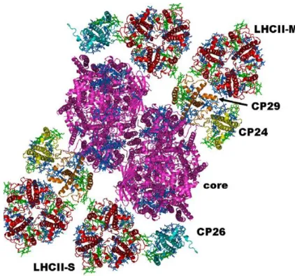
Heterogeneous preparations of PSII supercomplexes have been obtained directly from mildly solubilized thylakoid membranes or after a fast purification step, which allows enrichment of the PSII-enriched particles, BBYs (Boekema et al., 1999; Yakushevska et al., 2001). Cross-linking experiments first (Harrer et al., 1998) and electron microscopy (EM) and single particle analysis later, have been used in order to investigate how the different molecules were positioned and interconnected in these macrocomplexes (Yakushevska et al., 2003). The biggest complex identified so far is called C2S2M2 (Dekker and Boekema, 2005;
figure 1.9). It contains a dimeric core (C2), two LHCII trimers (trimer S) strongly bound to the complex on the side of CP43 and CP26, and two more trimers, moderately bound (trimer M) in contact with CP29 and CP24.
16
Figure 1.9 Model of the PSII supercomplex C2S2M2 from higher plants. From Croce and Van Hamerongen, 2011.
In the work of Caffarri and coworkers (2009) more homogeneous and stable preparations of the various types of PSII–LHCII supercomplexes with different antenna sizes have been obtained. The comparison of their protein composition to that one of mutants lacking some of the supercomplex components gives the possibility to relate the supercomplex organization to the protein content and thus to determine the role of the individual subunits in the overall organization.
An additional smaller complex it has also been isolated and characterized after mild detergent solubilisation of PSII membranes (Bassi and Dainese, 1992). It contains CP24, CP29 and LHCII. Phosphorylation of the membranes induces dissociation of the LHCII moiety from the CP24 moiety and changes in the aggregation state of LHCII components of the CP29-CP24-LHCII complex, showing that this complex is involved in the mechanism of regulation of excitation-energy distribution between the photosystems (Bassi and Dainese, 1992).
1.4.3 Photosystem II
Photosystem II (PSII) is a large supramolecular pigment-protein complex, which works in series with PSI during the first steps of photosynthesis. It collects light energy and uses it for the reduction of plastoquinone; it oxidizes water and contributes to the formation of a proton gradient across the thylakoid membrane. The most recent PSII structure (Umena et
17
al., 2011, figure 1.10) from the cyanobacterium T. vulcanus has been obtained at 1.9 Å
resolution allowing the detailed characterization of the water oxidation Mn4CaO5-cluster. Its
structure is supposed to be very similar to that of higher plants.
Each PSII monomer contains a core complex constituted by the reaction centre (RC) and the inner antenna proteins CP43 and CP47, the oxygen-evolving complex (OEC) and peripheral light-harvesting antenna.
The inner antenna proteins, CP43 and CP47, bound to D1 and D2 are involved in both light harvesting and energy transfer from the peripheral antennas to the RC (Ferreira et al., 2004).
Several other small subunits are included in the PSII dimer and others are located at its luminal side. They might mainly have a role in stabilizing and guiding the assembly of the macrocomplex, respectively (Zouni et al., 2001; Ferreira et al., 2004).The RC is made of the cytochrome b559 who is thought to have a role in the protection of RC against photodamage (Stewart and Brudvig, 1998), and of the two proteins D1 and D2 which carry out the charge separation and the electron transport. They bind the cofactors involved in these events, including the primary electron donor of PSII, known as P680 and two -carotene molecules. After light excitation, an electron is transferred from P680 to a pheophytin (Ph), resulting in a charge separation (P680+-Ph-). From pheophytin the electron goes to the quinone Q
A, then to
the quinone QB. P680+ is reduced by the water oxidation carried out by D1 protein and the
Mn4CaO5-cluster. After two charge separations and after proton uptake a reduced
18
Figure 1.10 Overall structure of PSII dimer. View from the direction perpendicular to the membrane normal. The protein
subunits are colored individually in the right-side monomer and in light gray in the left-side monomer, and the cofactors are colored in the left-side monomer and in light gray in the right-side monomer. Orange balls represent water molecules.
1.4.4 Cytochrome b6f
The cytochrome b6f complex (cyt b6f) provides the electronic connection between the
photosystem I and photosystem II reaction centres of oxygenic photosynthesis. It acts by oxidizing lipophilic plastoquinol and reducing plastocyanin and simultaneously, by translocating protons into the thylakoid lumen, generates a trans-membrane electrochemical proton gradient for ATP synthesis. Its crystal structure revealed 4 large (cytochrome b6,
cytochrome f, Rieske iron-sulfur, and subunit IV) and 4 smaller (PetG, PetM, PetL and PetN) subunits (Kurisu et al., 2003; for a review see Baniulis et al., 2008). The monomeric form contains four heme molecules, one chlorophyll a and one -carotene.
1.4.5 Photosystem I
Photosystem I is a large membrane protein complex which together with PSII catalyse the light-induced charge separation across the photosynthetic membrane. Solar energy absorbed by PSI antennas is transferred to reaction centre of the complex, which mediates the electron-transfer from plastocyanin at the luminal side to ferredoxin at the stromal side of the thylakoid membrane (Jensen et al., 2002).
19 A first crystal structure of PSI at 4.4 Å shows 12 core subunits, 4 different light-harvesting membrane proteins (the antenna complex LHCI) assembled into dimers in a half-moon shape on one side of the core, 45 transmembrane helices, 167 chlorophylls, 3 Fe–S clusters and 2 phylloquinones as highlighted by its crystal structure (Ben-Shem et al., 2003a). LHCI proteins are unique among the chlorophyll-a/b binding proteins in their red-shifted absorbance and in the formation of dimers (Croce et al., 2002).
Plant PSI is present in a monomeric structure both in vitro and in vivo (Ben-Shem et
al., 2003b). A more recent crystal structure (Amunts et al., 2010, figure 1.11) obtained at
3.3-Å resolution includes an additional protein subunit, and a total number of 173 chlorophylls and 15 β-carotenoids. The two large core complex subunits, PsaA and PsaB, form a symmetry-related dimer, which binds the majority of pigments, including the P700 special pair which forms the primary electron donor in the photosynthetic pathway.
PSI represents also the binding site for phosphorylated LHCII following the state 1 to state 2 transition phenomenon (Lunde et al., 2000).
Figure 1.11 Overall structure of plant PSI, represented in surface and schematic. View from the stroma. The 17 individual
20
1.4.6 ATP-synthase complex
In light reactions, the proton gradient formed by the photosynthetic process is ultimately converted into ATP by the plant ATP-synthase complex, a large multisubunit macromolecular enzyme of about 600 kDa. This complex is responsible for the generation of ATP, from adenosine diphosphate (ADP) and inorganic phosphate (Pi), utilising the proton gradient created by electron transport. In chloroplasts, ATP synthase is called the CF0CF1 complex (Groth and Pohl, 2001). The CF0 unit is a hydrophobic transmembrane multiprotein complex which contains a water-filled proton conducting channel. The CF1 unit is a hydrophilic peripheral membrane protein complex that protrudes into the stroma (McCarty et
al., 2000). It contains a reversible ATPase and a gate which controls proton movement
between CF0 and CF1, a key step of the ATP synthase mechanism (Noji et al., 1997). Entire CF0CF1 complexes are restricted to non-appressed portions of thylakoid membranes due to their bulky CF1 unit.
1.4.7 Linear electron transport
The electron transport generated by the photo-induced water oxidation catalyzed by PSII is defined as “linear” (LEF). The stoichiometry of the reactions is indicated for 4 photons absorbed by PSII and 4 photons absorbed by PSI. Electrons are transferred from PSII through the PQ pool to cytb6f, which acts as proton pump through the Q cycle. Electrons are then
transferred from cytb6f to the soluble electron carrier plastocyanin (PC) and then to PSI,
which acts as light-driven plastocyanin ferredoxin oxidoreductase. Ultimately FNR reduces NADP+ to NADPH at the expense of reduced ferredoxin.
1.4.8 Cyclic electron transport
In addition to LEF, cyclic electron transfer reactions (CEF) can also occur (Joliot and Johnson, 2011). CEF operates via two different routes, one involving a plastoquinone reductase which is homologous to the mitochondrial complex I, the so-called NDH complex, and the other via a putative ferredoxin-quinone reductase, feeding electrons directly into the cytochrome b6f complex, the so-called PGRL1/PGR5-dependent pathway (Hertle et al.,
2013). CEF involves PSI only and generates ATP without NADPH accumulation (for a review see Johnson, 2011).
When electrons from PS I pass through their primary electron acceptor (ferredoxin), they do not then proceed to form NADPH and O2. Rather, they cycle back to plastoquinone
21 and then to the cytochrome b6/f system, where the energy released by each electron causes two protons (H+) to be pumped into the interior of the thylakoid membrane. The protons produce a concentration gradient, or a flow of current, that is used to power chemiosmosis and the production of ATP. The process is cyclic because no outside source of electrons is required. Functioning of either CEF is thought to achieve the appropriate ATP/NADPH balance required for the biochemical needs of the plant, especially under specific environmental conditions. In addition to maintaining the proper balance of ATP and NADPH for the Calvin cycle, it may also serve as a photoprotective device in stress conditions (Rumeau et al., 2007). The regulation of CEF especially in regard to the possible competition with LEF is still under investigation.
1.5 Photoprotective mechanisms in higher plants
During the day photosynthetic organisms are exposed to different light intensities. The photosynthetic apparatus has evolved for both maximizing light capture in case of low light conditions and to guarantee protection during exposure to high light.
In low light conditions the amount of light energy absorbed matches the amount utilized in photosynthesis. In high light conditions, the rate of incoming photons is higher than the rate of electron transfer through the photosynthetic apparatus, and the reaction centers become progressively saturated (closed) (figure 1.12; Ruban et al., 2012).
22 Plants are able to respond to strong light (and thus to avoid photodamage) on different level of organization: at the whole organism level via leaf movements and leaf deposit, at the cellular level via chloroplast number and at the molecular level by the control of the number of pigments within the antenna (Bjorkman and Powles, 1987; Chow et al., 1988; Koller, 1990). Molecular adaptations can be long-term (acclimation) based on genetic regulation and short term (regulatory mechanisms).
1.5.1 Photoprotective role of carotenoids
The communication between carotenoids and (Bacterio)chlorophyll molecules at level of the light harvesting complexes is bidirectional. Carotenoids not only are able to capture the light and transfer the excitation energy to chlorophylls, but they can also interfere with excited singlet and triplet states of chlorophyll to avoid photodamage. This action takes place in the nanosecond/microsecond timescale thus guaranteeing an extremely fast response. In high light conditions, the energy reaching the photoynthetic apparatus can exceed the normal level used for photoynthesis. The excess of energy can then provocate the persistence of excited states of chlorophylls. Triplet-excited chlorophylls can react with molecular oxygen to produce singlet O2, which is a powerful oxidizing agent and rapidly kills those cells exposed
to it (Foote, 1976). Carotenoids are able to overcome this effect in one of two ways: 1) they can quench singlet oxygen directly through energy transfer or chemical reaction, or 2) they can quench the chlorophyll triplet itself via a rapid triplet-triplet energy transfer, preventing the production of singlet oxygen (Krinsky et al., 1971).
In vivo, the latter process is dominant. For this reaction to occur efficiently, the energy
level of the carotenoid’s triplet state must be lower than that of chlorophyll and lower than that of singlet oxygen i.e. < 1eV (1274 nm-7849 cm-1- or 94 kj/mole; Foote et al., 1970) in
order to prevent the triplet-excited carotenoid from reacting with molecular oxygen itself. In practice, this means that only those carotenoids with 9 or more conjugated double bonds (n≥9) have the ability to photoprotect (Foote et al., 1970). This property could thus be one of the reasons why nature has selected only carotenoids of this length in the photosynthetic proteins. Moreover, because the carotenoid triplet state is lower in energy than singlet oxygen, it returns harmlessly to the ground state with the liberation of heat (Cogdell and Frank, 1987). Carotenoids can also quenching the singlet excited state of chlorophyll, which could also represent a source of singlet oxygen, through a not yet clarified mechanism which plays an essential role in the non-photochemical quenching or NPQ (Truscott et al., 1973; Palozza and
23 Krinsky, 1992; Niyogi et al., 1997; Pascal et al., 2005; Ruban et al., 2007; Britton et al., 2008). This function will be better described in paragraph 1.5.2.
The different mechanisms of photoprotection guaranteed by carotenoids are schematized in figure 1.13.
1.5.2 Non-photochemical quenching (NPQ)
As already anticipated, photosynthetic organisms possess short-term regulatory mechanisms which serve to reduce the flow of electrons to the acceptor side of PSII, thereby preventing the production of reactive oxygen and thus the irreversible damage of the photosystem in conditions of strong light.
Non-photochemical quenching (NPQ) is based on the reduction of PSII antenna chlorophyll fluorescence yield in order to dissipate the excess energy before it reaches the PSII reaction centers (Muller et al., 2001; Ruban et al., 2012). NPQ is a heterogeneous process and can be divided in three components (Walters and Horton, 1991). qI (photoinhibitory) is the slowest forming and relaxing component. It’s partially related to the photodamage of PSII and partially to the antenna quenching associated with the photoprotective downregulation of PSII (Ruban and Horton, 1995; Horton et al., 1996). The second component, qT or state transition, is mainly observed in low light conditions (Walters and Horton, 1991). It forms and relaxes in tens of minutes and is related to the balance of excitation energy between PSII and PSI (Horton and Hague, 1988; Ruban and Johnson, 2009). qE, or energy dependent quenching, is the major component of NPQ in high light and it forms and relaxes within seconds to minutes (Horton and Hague, 1988). This component is dependent on the formation of an intrathylakoid proton gradient during the illumination (Briantais et al., 1979), the xanthophyll cycle (the reversible de-epoxidation of violaxanthin to neoxanthin through an intermediate called antheraxanthin; Yamamoto et al., 1962; 1999) and particularly on the amount of zeaxanthin present (Demmig et al., 1987, Demmig-Adams et
al., 1989) and on PsbS which is a PSII-related protein (Funk et al., 1995; Li et al., 2002).
3
Chl-a* + Car Chl-a + 3Car* chlorophyll triplet quenching (triplet-triplet energy transfer-TT)
1
Chl-a* + Car chlorophyll singlet quenching (NPQ) ??
24
1.5.2.1 Site of qE
Most of evidences indicate that the qE component of NPQ occurs within the antenna. Some of these evidences are as follow:
-quenching of excitation energy within the antenna complexes has been mainly studied by the analysis of PSII fluorescence at 77K (Ruban and Horton, 1995): following the induction of qE, the PSII emission spectrum of PSII resembled that of partially aggregated LHCII, with an enhancement band at 700 nm;
-the xanthophyll cycle carotenoids are associated with the light-harvesting antennae;
- in vitro quenching of isolated antenna complexes reproduces many features of in vivo qE, such as the kinetics of fluorescence change, the enhancement by zeaxanthin and the absorbance changes accompanying the in vivo quenching process (Ruban and Horton, 1992; Wentworth et al., 2000; 2001)
Nowadays is widely accepted the site of qE is within LHCII antenna (Horton and Ruban, 1992; Ruban and Horton, 1995; Wentworth et al., 2000) but there is no agreement on whether this is the only quenching site, and on which part or parts of the antenna is located the qE quencher(s). It seems that no individual LHCII complex acts as the sole site of qE and that potentially the quenching could happen in both the major and minor complexes (Horton et al., 1996; Horton et al., 2005; Kovacs et al., 2006). Important insights into this question were found by investigating various Lhc mutants (Yakushevska et al., 2003, Andersson et al., 2003; Ruban et al., 2003; Kovacs et al., 2006), which also demonstrated the importance of retaining a correct macro-structure organization of the LHCII antenna system in order to reach maxima levels of NPQ.
1.5.2.2 Mechanism(s) of qE
Most of the experiments on qE have been focused on the investigation of the precise role of zeaxanthin in this NPQ component. At first it was suggested a direct role of the xanthophyll cycle zeaxanthin in the quenching mechanism, in the so called “molecular gearshift” (Demmig-Adams, 1990). According to this model, as the energy of the S1 state of zeaxanthin
should lay below that of chlorophyll a, a possible energy transfer between the two pigments could happen, allowing to dissipation of the excess energy as heat. Anyway a weak point of this model is that the assignment of the energy levels mentioned above is not straightforward.
It is currently proposed that zeaxanthin acts as an allosteric modulator of the quenching rather than being the direct quencher (Horton et al., 2000). Horton et al. (1991; 2005) proposed the “LHCII aggregation model”, where 4 LHCII states are described,