HAL Id: hal-02448983
https://hal.archives-ouvertes.fr/hal-02448983
Submitted on 13 Nov 2020HAL is a multi-disciplinary open access archive for the deposit and dissemination of sci-entific research documents, whether they are pub-lished or not. The documents may come from teaching and research institutions in France or abroad, or from public or private research centers.
L’archive ouverte pluridisciplinaire HAL, est destinée au dépôt et à la diffusion de documents scientifiques de niveau recherche, publiés ou non, émanant des établissements d’enseignement et de recherche français ou étrangers, des laboratoires publics ou privés.
Céline Burel, Alexandre Teolis, Ahmed Alsayed, Christopher Murray,
Bertrand Donnio, Rémi Dreyfus
To cite this version:
Céline Burel, Alexandre Teolis, Ahmed Alsayed, Christopher Murray, Bertrand Donnio, et al.. Plas-monic Elastic Capsules as Colorimetric Reversible pH-Microsensors. Small, Wiley-VCH Verlag, 2020, 16 (6), pp.1903897. �10.1002/smll.201903897�. �hal-02448983�
1
Plasmonic elastic capsules as colorimetric reversible
pH-microsensors
Dr. Céline A. S. Burel1, Alexandre Teolis1, Dr. Ahmed Alsayed1, Pr Christopher B. Murray2,
Dr. Bertrand Donnio3, and Dr. Rémi Dreyfus1*
Dr. C. A. S. Burel, A. Teolis, Dr. A. Alsayed, Dr. R. Dreyfus
Complex Assemblies of Soft Matter Laboratory (COMPASS), UMI 3254, CNRS-Solvay-University of Pennsylvania, RIC, Bristol, PA 19007, United States
E-mail: [email protected] Dr. C.B. Murray
Dept of Chemistry and Materials Science, University of Pennsylvania 231 S. 34th Street, Philadelphia, PA 19104, USA
Dr. B. Donnio
Institut de Physique et Chimie des Matériaux de Strasbourg (IPCMS), UMR 7504, CNRS-Université de Strasbourg, 67034, Strasbourg, France
KEYWORDS: pH-sensors, plasmonics, microcapsules, nanoparticles, bacteria.
ABSTRACT: There is a crucial need for effective and easily dispersible colloidal
microsensors able to detect local pH changes before irreversible damages caused by
demineralization, corrosion, or biofilms occur. One class of such microsensors is based on
molecular dyes encapsulated or dispersed either in polymer matrices or in liquid systems
exhibiting different colors upon pH variations. They are efficient but often rely on
sophisticated and costly syntheses, and present significant risks of leakage and
photobleaching damages, which is detrimental for mainstream applications. Another approach
consists in exploiting the distance-dependent plasmonic properties of metallic nanoparticles.
Still, assembling nanoparticles into dispersible colloidal pH-sensitive sensors remains a
challenge. Here, we show how to combine optically active plasmonic gold nanoparticles and
2
shrink. Concomitantly, the distance between the gold nanoparticles embedded in the
polymeric matrix varies, resulting in an unambiguous color change. Billions of micron-size
sensors can thus be easily fabricated. They are non-intrusive, reusable, and sense local pH
changes. Each plasmocapsule is an independent reversible microsensor over a large pH range.
Finally, we demonstrate their potential use for the detection of bacterial growth, thus proving
that plasmocapsules are a new class of sensing materials.
1. Introduction
When corrosion occurs in a material, damages start from randomly located microscopic
sites, where pH has already decreased.[1-3] When biofilms develop, bacteria change
phenotypes and secrete substances that result in a local decrease in pH.[4-6] In the case of teeth,
the more acidic environment causes local demineralization and is responsible for painful
cavities.[7],[8] Although these three examples describe very different natural processes, they all
carry one common feature: they describe heterogeneous processes, which start with local
chemical changes at a microscopic level. Being able to track and detect these changes early
open perspectives to prevent these processes and their subsequent adverse effects develop.
Monitoring these local alterations requires the fabrication of smart microsensors that are
cost-effective, easy to disperse, sense minute local pH changes rapidly, and able to translate these
changes into a large, unambiguous signaling output. Optical signals have been used as output
for microsensors because they are easy to read and visible to the naked eye without the need
3
So far, the dominant optical pH sensors able to reach some of the microsensors goals relies
on the use of dyes such as fluorophores[9-15] or chromophores[11],[16],[17] which light absorption
or fluorescence emission depends on the pH. By matching the adsorption peak of dyes with
the emission wavelengths of nanoparticles (NPs) such as quantum dots, the fluorescence of
the pH indicators can be greatly enhanced.[18],[19] Organic indicator dye molecules can be
dispersed, adsorbed, entrapped, covalently bonded, or encapsulated in a support
matrix.[5],[11],[13],[14],[20],[21] Though this type of sensors has proved very efficient, it usually
requires careful monitoring of the dye self-aggregation, large concentrations, complex
chemistry for their synthesis or their chemical attachment onto the polymeric backbone or a
perfect control of the leaking for dyes encapsulation. [5],[11],[20],[21] Moreover, most dyes are
sensitive to photobleaching, which limits their long-term use.[11] Furthermore, depending on
the type of matrix and indicator, each pH sensor has a different measurable pH range, which
usually goes only over a couple of pH units. Metallic NPs such as gold (Au) and silver (Ag)
NPs absorb light in the visible due to their plasmon resonance.[22],[23] Additionally, plasmonic
NPs extinction is extremely sensitive depending on NP composition, size, shape, orientation,
local dielectric environment, and interparticle distance.[22-31] Furthermore, contrasting with
organic dyes, plasmonic nanoparticles do not photobleach, can be easily synthesized in one
single step process[32] and produced in large quantities. Based on their very interesting optical
properties, their resistance to light and their easy synthesis, plasmonic NPs constitute a very
good alternative to organic dyes. Therefore, a new generation of optical pH sensors made of
plasmonic NPs and actuating polymers has been developed in recent years.[33],[34] The working
principles of these sensors are based on the chemical and mechanical properties of the
4
and carboxyl group-terminated poly(2-vinylpyridine)[33] and, the optical properties of the NPs
used such as Au or Ag NPs. The combined assembly of optically active plasmonic NPs and
pH-responsive polymers leads to an alternative class of optical pH sensors able to transform
chemical potentials into a strong and easily detectable optical response. Although these
systems are efficient, they do not provide a dispersible micron size sensor and are not suitable
for real-life applications as their incorporation in different matrices strongly impact their
optical response.
Here, we present an original alternative for the fabrication of highly sensitive and
dispersible pH plasmonic microsensors. Briefly, these microsensors are obtained from
Pickering emulsions made of Au NPs closely packed at an oil/ water emulsion interface.[35],[36]
Then, the Au NPs are locked within a polyelectrolyte polymer shell at the interface giving the
final shape to the Au NPs-polymer microcapsules’ sensors, thereafter referred to as
plasmocapsules. We demonstrate how these microsensors show a significant optical response
to pH changes due to their shift in the plasmonic extinction maximum. As schematically
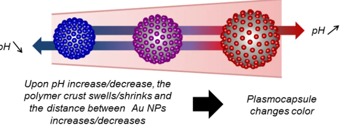
shown in Figure 1, as the polymeric matrix swells or shrinks upon pH change, the distance
between neighbored Au NPs, and thus their interparticular coupling, changes, resulting in a
different optical response.33 In our emulsion templated method, billions of microsensors are
fabricated simultaneously, each plasmocapsule acting as a single micron-size sensor. These
colloidal microsensors are easy to fabricate, dispersible, robust, non-intrusive, reusable,
reversibly sense minute local changes over a very large pH range (7 pH units), and able to
translate these changes rapidly into a large, unambiguous signaling output. As proof of
5
demonstrated. We anticipate that our approach enables the development of a large body of
similar hybrid microcapsule sensors for tracking and sensing other stimuli in spatially
homogeneous and heterogeneous systems.
2. Principle of the Synthesis
The precise synthetic protocol is described in details in the Methods Section and relies on
the following methodology: Au NPs functionalized with
poly(diallyldimethylammonium-nitrate-co-1-)vinylpyrrolidone (PVP DADMAN, full structural formula available in Figure S1
of the Supporting Information), are synthesized by reduction of Au salt with ascorbic
acid[32],[37]. The Au NPs obtained are polydispersed, the mean diameter d is d = 30 nm ± 11
nm and the PVP-DADMAN capping layer is about 1.5 nm (TEM images, histogram of size
distribution, and extinction spectrum of the Au NPs can be found in Figures S2 of the
Supplementary information). In this size range, the polydispersity does not affect the position
of the plasmonic peak but results in a small broadening of the peak.[22],[37] Then, a solution of
toluene containing a mixture of monomers of methyl methacrylate (MMA) and butyl acrylate
(BA) (50:50 w/w), is emulsified in a continuous aqueous phase of the previously synthesized
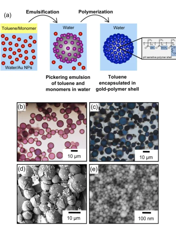
Au NPs. By setting the pH of the solution to 1.2, electrostatic barriers are removed, and the
NPs adsorb at the surface of the emulsion due to energy minimization, forming a so-called
Pickering emulsion of Au NPs[37] (Figure 2a). The red color of the emulsion at the beginning
of the process indicates Au NPs adsorption (Figure 2b). As synthesis proceeds, color change
from red to blue acts as a good visual indicator of the NPs close-packed arrangement at the
emulsion interface. Indeed, only a tight and dense packing results in the coupling of the
6
The polymerization of the encapsulated monomers occurs at the interface by raising the
temperature above 60°C, giving rise to blue capsules (Figure 2c) made of an elastic shell in
which gold nanoparticles are very closely embedded (Figures 2d). The plasmocapsules’
spherical shells are observed by SEM. A high magnification image of the shell shows the
dense packing of the nanoparticles (Figure 2e). The thickness of the shell is measured from
the SEM images to be approximately 100 nm +/- 20 nm (see Figure S3 in supplementary
information).
Mass spectroscopy is performed to quantify the amount of methyl methacrylate (MA), and
butyl acrylate (BA) polymerized. Results show that essentially all of the two monomers are
consumed (98.3% of MMA and 95.4% of BA, see Figure S4 in supplementary information).
Upon addition of sodium hydroxide, about 24 mol% of the acrylate and carboxylic acid
moieties present on the polymer shell are transformed into carboxylate ions (see the
Supplementary information section for more details on the characterization of the
plasmocapsules’ polymer shell). The pH sensitivity of the polymer[39],[40] in which the Au NPs
are embedded, makes the collective assembly of nanoparticles sensitive to any pH-changing
process. As pH is increased, the presence of numerous free carboxylic groups on the surface
confers charges to the shell, resulting in osmotic swelling of the shells. The whole construct
made of Au NPs embedded in a pH sensitive crust can then be used as a pH-microsensor,
7
3. Experimental Results and Discussion
The hypothesis that the synthesized microcapsules are de facto pH sensitive needed to be
demonstrated first. To do so, we performed microfluidics experiments. We first introduced an
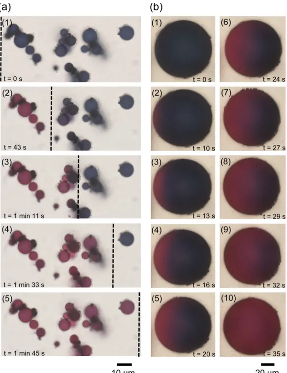
acidic solution (pH ~ 1.2) containing blue microcapsules in a capillary, and then, a small
droplet of a basic solution (pH ~ 13.8) was deposited gently at one end of the capillary. The
droplet locally changed the pH, generating a front of increasing pH that propagated in the
capillary. This front propagation could be easily followed by optical microscopy, the
microcapsules changed colors as the pH solution was increased (Figure 3a, images 1-5,
plasmocapsules in the capillary under different pH locally exhibit strong red and blue colors
in the regions of high and low pH, respectively. The propagation of the pH front can be seen
in movie 1 in the supplementary materials). At low pH, the polymeric backbone is uncharged,
the nanoparticles are densely packed in the crust, and thus strongly coupled, and the
microcapsules are blue. At higher pH, the polymer acquires charges (hydrolysis of acrylic
acid –m- and acrylate ester –n and p- groups into carboxylate groups –m’-, see Figure S8 in
supplementary information), the swelling crust increases the interparticle distance between the
Au NPs, the plasmons are no more coupled and the microcapsules appear red. We also
showed that this color change could even be monitored at the level of a single capsule (Figure
3b, images 1-10, and movie 2 in the supplementary materials).
To ensure that the change of color is solely due to the mechanical swelling of the polymeric
crust embedding the Au NPs, we measured and plotted the color and size of the microcapsule
8
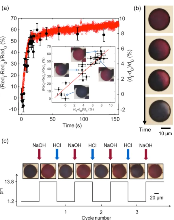
moves with time over a microcapsule (Figure 4a), both the red intensity and the capsule
diameter increase. When the red intensity was plotted as a function of capsule size (inset
Figure 4a), a clear correlation between size and color could be found: a relative diameter
change of about 10% is found sufficient to induce this color transition. To preclude the
possibility that leaking or desorption of the Au NPs from the polymeric matrix might be
responsible for the color change, we also check the reversibility of the color transition. We
show that by changing the pH inside the microfluidics chamber back to acidic, the color of the
microcapsules turns back to blue (Figure 4b). Moreover, the color of the capsules can be
cycled, at least three times, from blue to red to blue, and so on, attesting of their flexibility
and robustness (Figure 4c). This simple set of experiments evidences the pH-sensing potential
of the microcapsules and how the coupling between the mechanical properties of the
polymeric crust and the optical properties of the Au NPs allows us to obtain an easy read of
local pH changes.
For the calibration of the colloidal microsensors, we performed a series of experiments in
which the microcapsules were prepared and redispersed in different buffers of pH ranging
from 2 to 12 (see the materials and methods section). They were first synthesized following
the protocol described in the materials and methods section. Once they were formed, the
capsules were redispersed in several solutions of different pH. The pH was measured using a
microelectrode connected to a pH-meter. Direct observations of the microcapsules under the
microscope confirmed that the color of the capsules was dependent on pH. Typical images of
the plasmocapsules at five different pH values (i.e., pH = 2.4, 4.4, 7.4, 8.2, and 11.2) are
9
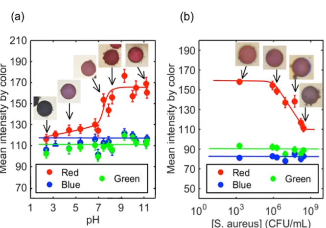
surrounding suspension. To be more quantitative, for each pH sample, about 50 images were
taken using a color camera, and the red, blue and green intensities were extracted from the
camera channels and plotted as a function of pH (Figure 5a). A large red color transition in a
narrow zone of pH, which extended from pH = 7.2 to pH = 8.2, was observed. The blue and
green intensities remained quasi-constant at all pH values. The transition and the distinction
between pH below 7.2 and pH above 8.2 are observable by the naked eye. In the pH range of
2 to 9, microcapsules color change due to pH increment as small as 0.5 pH unit were
distinguished by eye under the microscope. As seen on the images of Figure 5 (a), a large
range of colors is observed: blue, purple, pink, red, and strong red. The diffusion of
hydronium ions over a 100 nm polymeric shell can be estimated by taking 10-10 m2/s as typical
value for the diffusion coefficient.[41],[42] The time it takes for a thin 100 nm pH-responsive
polymer membrane to swell is then of the order of 0.1 ms: the membrane swelling of the
plasmocapsules is almost instantaneous. This is consistent with the immediate change of color
observed by eye under a microscope when a droplet of the base is added to an acidic droplet
of plasmocapsules. This shows that the capsules can be indeed used to detect or monitor
processes, which alter the pH of surrounding solutions in a large pH-range.
Finally, to demonstrate the applicability of these pH-sensitive plasmocapsules as efficient
optical sensors, we used them to detect a natural process and replaced usual microelectrode
measurements by simple in-situ straightforward optical measurements. As proof of concept,
we used them to monitor pH change when a bacterial growth process occurred in a solution.
When certain bacteria strains grow in a solution with nutrients, they can metabolize
10
a decrease in the pH of the medium. Measuring a pH change is thus one way to diagnose the
presence of a living and growing strain in a contaminated solution. A strain of Staphylococcus
aureus was grown in a nutrient broth containing 1 wt% D-(+)-glucose. The bacteria were
incubated for two days at 35°C. The broth alone (no bacteria) was also incubated and the pH
was controlled during the entire duration of the experiment. S. aureus colony forming units
(CFU)/mL was determined at different times by serial dilution, and plating. Simultaneously,
the pH was measured both in the broth alone (without bacteria) and in the broth containing
bacteria (see Figure S9). Over time, the S. aureus grew from 103 CFU/mL to 108 CFU/mL, the
exponential growth phase, and the stationary phase respectively occurring in about 10 and 28
h after incubation. Concomitantly, due to the fermentation of glucose by S. aureus, the pH
was found to decrease from 8 to 4.5. The larger pH drops occurred during the exponential
phase when bacterial growth was the most important, the pH stabilized at 4.5 once the
bacteria reached the stationary phase. As a reference, the pH of the broth alone containing no
bacteria remained constant with time, showing that the observed pH decrease is only due to
the fermentation of glucose by S. aureus. The plasmocapsules were then dispersed in the
culture media at a different time of the incubation, and the different colors were analyzed. As
the bacteria grew in solution and the pH subsequently decreased, the microcapsules changed
color from red at pH 8 to purple at pH 4.5. The Red, Blue and Green intensities of the
microcapsules were plotted versus the concentration of S. aureus (see Figure 5b). We
observed that as the population of the bacteria increased, the red intensity decreased, and the
blue and green intensities remained constant. A color shift in the red intensity could be
11
the plasmocapsules can be used to detect the presence of acid-producing bacteria by sensing
pH variations.
4. Conclusion
We have demonstrated the synthesis of pH-sensitive plasmonic microcapsules. They
represent a new type of dispersible optical sensors made of a combination of a pH-sensitive
polymeric shell, which swells or shrinks under pH changes, and embedded plasmonic
nanoparticles, the optical response of which depends on the interparticle distance. Using
regular colorimetric analysis, we have shown a direct correlation between the capsules color d
the size of the plasmocapsules. We have calibrated the relation between the outside pH and
color of the plasmocapsules. We have demonstrated that the optical response of the
plasmocapsules is contrasted enough to detect different pHs, even though we use a path of
synthesis that yields polydisperse nanoparticles. The plasmocapsules sensor allows for
detection of pH in a relatively large range of 2 to 9. The plasmocapsules microsensors
response time is of the order of 0.1ms, which is much smaller than competing organic dye
technologies whose typical response time range from several tenths of seconds to a minute.[11]
In the case where the microsensor would be a 10 microns particle of polyacrylic acid with Au
NPs on its surface, the response time would be increased to about 1s. This validates the use of
a thin shell of pH-responsive polymer instead of a large particle. We did not observe leaching
of the NPs out of the polymeric shell, which was evidenced by the reversibility of the capsules
over at least three cycles. Finally, we have shown that the plasmocapsules can be used to
12
replicate. Because of all these characteristics, the pH-responsive plasmocapsules could be of
interest to detect and monitor bacterial contamination in health care and the food industry.
The microsensors showed here were specially developed for pH measurement, however we
believe that the effect and principle demonstrated here are general enough to be extended to
other types of detectors. Swapping a pH-sensitive polymeric shell with, for instance, a
fluoropolymer one, which, depending of the fluorination degree, would be able to selectively
dissolve a wide range of gases (H2, CO, CO2, NO2, CH4) and vapors’ solvents, would allow
the fabrication of very specific gas sensors.[43] Alternatively, a polymeric shell sensitive to e.g.
polynitroaromatic compounds would be of interest in the area of explosive sensing.[44] Using a
temperature sensitive polymeric shell, which would swell/shrink under small temperature
variations, could also be used as a temperature sensor in the cold chain to ensure fresh and
frozen aliment preservation, chemical hazards limitation, or drugs stabilities. Therefore, the
nature of the shell provides the type of detection to be done while the optical reading would
still lie on the plasmonic properties of the embedded nanoparticles. As such we anticipate that
these microcapsules will likely represent a new class of cheap non-intrusive microsensors able
to transform chemical potentials or external forces into a large optical absorption shift, and
easily detectable by the naked eye.
5. Experimental Section
Materials: Gold (III) chloride trihydrate (99.9%, Sigma-Aldrich), L-ascorbic acid (99%,
13
Plus, 99.9%, Sigma-Aldrich), poly(diallyldimethyl-ammoniumnitrate-co-1-vinylpyrrolidone
(PVP-DADMAN, Solvay®), Sodium Hydroxide (50/50 w/w Baker), methyl methacrylate
(99%, Sigma-Aldrich), butyl acrylate (99%, Sigma-Aldrich),
2,2′-Azobis(2-methyl-propionitrile) (AIBN) (98%, Sigma-Aldrich), citric acid (ACS reagent, EMD Chemicals),
sodium phosphate dibasic (ACS reagent, VWR), sodium carbonate dibasic (99.5%, ACS
reagent, Sigma-Aldrich), sodium bicarbonate (99.7%, ACS reagent, Sigma-Aldrich),
D-(+)-glucose (99.5%, Sigma Aldrich), sodium chloride BioXtra (99.5%, Sigma Aldrich), sodium
hydroxide pellets (98%, Alfa Aesar), Nutrient Broth powder (NEOGEN Corporation), 9 mL
Nutrient Broth tubes (General Laboratory Products), Tryptic Soy Agar plates (USP G60,
Hardy Diagnostics). The bacterium used in the studies was Staphylococcus aureus subsp.
aureus (ATCC 6538). Ultrapure water (type 1, 18.2 MΩ.cm) was used for all culture media
preparation and deionized water was used otherwise.
Gold nanoparticles synthesis: The Au NPs used in this procedure were synthesized by
reduction of HAuCl4 with ascorbic acid in presence of PVP-DADMAN. A solution of 5.7.10-6
M PVP-DADMAN and 2.510-3 M HAuCl4 was brought to boil. Then, 12.5 mL of ascorbic
acid (0.1 M) was added. The solution protected from light by aluminum foil was stirred for 1
h at 97°C. After synthesis, the Au NPs were left to rest for one day to remove the biggest
nanoparticles. The rest of the dispersion was centrifuged and concentrated into a few
milliliters’ solution. MilliQ water was used throughout the entire synthesis process.
Plasmocapsules synthesis: An aqueous phase containing the Au NPs ([Au0] = 0.02 M) and
HCl (20 µL) is mixed with a toluene phase (0.5 mL) of the dissolved acrylate monomers
(MMA 2.2 M and BA 1.7 M) and AIBN (0.12 M). The two phases are strongly agitated by
oil-in-14
water emulsion. The formation of the polymer shell by polymerization of the monomer at the
interface of the emulsion is carried out at 62°C for 2hrs.
Plasmocapsules in buffer solutions: buffer solutions were prepared by mixing the
following four solutions in different ratios: Citric acid (0.75 M), Na2HPO4 (1.5 M), Na2CO3
(0.75 M) and NaHCO3 (0.75 M). Buffer solutions at a specific pH were mixed with the
dispersion of microcapsules in a 50/50 v/v ratio, and the pH was controlled afterward. Each
time the final pH value was close (within 0.5 unit of pH) to the pH of the buffer solution. For
high (> 11) and low (< 3) pH, NaOH and HCl were respectively used to adjust the pH of the
plasmocapsules dispersion.
Electron microscopy: Scanning electron microscopy (SEM) was performed on JEOL 7500
HRSEM. The samples were prepared by air drying under ambient conditions by laying a drop
of microcapsules dispersion in water on an electron microscopy science carbon-coated copper
grid. Transmission electron microscopy (TEM) was carried out on a JEOL JEM-1400. The
accelerating voltage was set at 120 kV.
Optical characterizations: Optical microscopy images were acquired on an Olympus
microscope in bright field and transmission mode.
pH measurements: The pH of all solutions was measured with pH-meter (model 8015,
VWR International) with a micro pH combination electrode (Z113425-1EA, Millipore
Sigma).
Bacteria cell culture and plate counting: A fresh culture of S. aureus was prepared by
inoculating 9 mL Nutrient Broth with a single colony from a Tryptic Soy Agar plate streaked
15
conditions (250 rpm) in an incubating mini shaker (12620-942, VWR International). Bacteria
were harvested by centrifugation for 10 min at 2000×g, washed twice using the medium
studied, before resuspending in the medium studied. The concentration of S. aureus was
determined by serial dilution with subsequent plating on Tryptic Soy Agar plates and
measurement of colony forming units (CFUs) after 24 h at 35 °C.
pH-bacteria growth study in planktonic conditions: Two sterile culture media were
prepared: a Nutrient Broth with 1 wt% D-(+)-glucose, 3 wt% sodium chloride; and a Nutrient
Broth with just 3 wt% sodium chloride. Both solutions were adjusted to pH 8 with NaOH and
filtered using 0.2 µm PES sterile syringe filters (VWR International). Bacteria were washed
twice and resuspended in both solutions. Initial S. aureus concentration was adjusted by
dilution at around 103 CFU/mL. Then, bacteria were cultivated in closed vials on a
MaxQ4000 shaker (model 4342, Thermo Scientific) for 48 h at 35 °C with shaking (100 rpm).
S. aureus concentration was determined by plating and pH was measured. The microcapsules
were concentrated and washed several times with aliquot of the bacteria solution at a different
time of incubation until the pH of the final dispersion matches the pH of the bacteria
solutions. Then the microcapsules were imaged by optical microscopy.
Supporting Information
Structural formula of PVP-DADMAN; SEM image of a plasmocapsule polymer shell; HPLC
chromatograms obtained for plasmocapsules at pH 1.2 and at pH 13.8; characterizations of the
plasmocapsules polymer shell with FTIR spectra, GC and GC-MS chromatograms; schematic
16
and color of the plasmocapsules as a function of the concentration of S. aureus; additional
materials and methods. Supporting Information is available from the Wiley Online Library or
from the author.
6. Acknowledgments
The authors thank the ANRT, GIE AIFOR, CNRS, and Solvay for financial support. The
authors acknowledge the support of the French National Agency of Research (ANR) to the
project REACT through the grant ANR15-PIRE-0001-06. SEM imaging was performed in
facilities supported by the NSF MRSEC program under award No DMR-1720530. The
authors also thank D. Bendejacq, R. Giordanengo, L. Gage, D. Radtke, C. Anderson, J.
Hutchinson, and A.J. Yodh for fruitful discussions.
Received: ((will be filled in by the editorial staff)) Revised: ((will be filled in by the editorial staff)) Published online: ((will be filled in by the editorial staff))
REFERENCES
[1] D. E. Williams, J. Electrochem. Soc. 1985, 132, 1796.
[2] M. P. Ryan, D. E. Williams, R. J. Chater, B. M. Hutton, D. S. McPhail, Nature 2002,
415, 770.
[3] P. Guo, E. C. La Plante, B. Wang, X. Chen, M. Balonis, M. Bauchy, G. Sant, Sci. Rep.
17
[4] G. Hidalgo, A. Burns, E. Herz, A. G. Hay, P. L. Houston, U. Wiesner, L. W. Lion,
Appl. Environ. Microbiol. 2009, 75, 7426.
[5] S. Milo, F. B. Acosta, H. J. Hathaway, L. A. Wallace, N. T. Thet, A. T. A. Jenkins,
ACS Sensors 2018, 3, 612.
[6] L. C. Z. Chan, G. Khalili Moghaddam, Z. Wang, C. R. Lowe, ACS Sensors 2019, 4,
456.
[7] I. Dige, V. Baelum, B. Nyvad, S. Schlafer, J. Oral Microbiol. 2016, 8, 4.
[8] J. Xiao, A. T. Hara, D. Kim, D. T. Zero, H. Koo, G. Hwang, Int. J. Oral Sci. 2017, 9,
74.
[9] Y. Wang, T. Zhao, Y. Li, L. Su, Z. Wang, G. Huang, B. D. Sumer, J. Gao, J. Am. Chem.
Soc. 2014, 136, 11085.
[10] M. Tantama, Y. P. Hung, G. Yellen, J. Am. Chem. Soc. 2011, 133, 10034.
[11] D. Wencel, T. Abel, C. McDonagh, Anal. Chem. 2014, 86, 15.
[12] J. Han, K. Burgess, Chem. Rev. 2010, 110, 2709.
[13] S. Schreml, R. J. Meier, O. S. Wolfbeis, M. Landthaler, R.-M. Szeimies, P. Babilas,
Proc. Natl. Acad. Sci. 2011, 108, 2432.
[14] X. D. Wang, J. A. Stolwijk, T. Lang, M. Sperber, R. J. Meier, J. Wegener, O. S.
Wolfbeis, J. Am. Chem. Soc. 2012, 134, 17011.
[15] W. Shi, S. He, M. Wei, D. G. Evans, X. Duan, Adv. Funct. Mater. 2010, 20, 3856.
[16] W. F. Jager, T. S. Hammink, O. Den Van Berg, F. C. Grozema, J. Org. Chem. 2010, 75,
2169.
[17] J. P. O’Reilly, C. P. Butts, I. A. I’Anson, A. M. Shaw, J. Am. Chem. Soc. 2005, 127,
18
[18] Y. Hiruta, N. Yoshizawa, D. Citterio, K. Suzuki, Anal. Chem. 2012, 84, 10650.
[19] Z. Bai, R. Chen, P. Si, Y. Huang, H. Sun, Appl. Mater. Interfaces 2013, 5, 5856.
[20] Z. Jin, Y. Su, Y. Duan, Sensors Actuators B Chem. 2000, 71, 118.
[21] O. Kreft, A. M. Javier, G. B. Sukhorukov, W. J. Parak, J. Mater. Chem. 2007, 17, 4471.
[22] V. Myroshnychenko, J. Rodríguez-Fernández, I. Pastoriza-Santos, A. M. Funston, C.
Novo, P. Mulvaney, L. M. Liz-Marzán, F. J. García de Abajo, Chem. Soc. Rev. 2008,
37, 1792.
[23] L. M. Liz-Marzán, Langmuir 2006, 22, 32.
[24] P. K. Jain, W. Huang, M. A. El-sayed, Nano Lett. 2007, 7, 2080.
[25] H. Gehan, C. Mangeney, J. Aubard, G. Lévi, A. Hohenau, J. R. Krenn, E. Lacaze, N.
Félidj, J. Phys. Chem. Lett. 2011, 2, 926.
[26] H. Lange, B. H. Juárez, A. Carl, M. Richter, N. G. Bastús, H. Weller, C. Thomsen, R.
Von Klitzing, A. Knorr, Langmuir 2012, 28, 8862.
[27] S. K. Ghosh, T. Pal, Chem. Rev. 2007, 107, 4797.
[28] H. Chen, X. Kou, Z. Yang, W. Ni, J. Wang, Langmuir 2008, 24, 5233.
[29] M. A. El-Sayed, P. K. Jain, Nano Lett. 2008, 8, 4347.
[30] A. B. Taylor, P. Zijlstra, ACS Sensors 2017, 2, 1103.
[31] J. Lee, J. H. Huh, K. Kim, S. Lee, Adv. Funct. Mater. 2018, 28, 1.
[32] L. Malassis, R. Dreyfus, R. J. Murphy, L. A. Hough, B. Donnio, C. B. Murray, RSC
Adv. 2016, 6, 33092.
[33] I. Tokareva, S. Minko, J. H. Fendler, E. Hutter, J. Am. Chem. Soc. 2004, 126, 15950.
[34] Y. Shen, M. Kuang, Z. Shen, J. Nieberle, H. Duan, H. Frey, Angew. Chemie - Int. Ed.
19
[35] S. U. Pickering, J. Chem. Soc., Trans. 1907, 91, 2001.
[36] J. Wu, G.-H. Ma, Small 2016, 12, 4633.
[37] C. A. S. Burel, A. Alsayed, L. Malassis, C. B. Murray, B. Donnio, R. Dreyfus, Small
2017, 13, 1.
[38] K. Larson-smith, D. C. Pozzo, Langmuir 2012, 28, 11725.
[39] S. Bazban-Shotorbani, M. M. Hasani-Sadrabadi, A. Karkhaneh, V. Serpooshan, K. I.
Jacob, A. Moshaverinia, M. Mahmoudi, J. Control. Release 2017, 253, 46.
[40] A. D. Drozdov, J. Declaville Christiansen, Phys. Rev. E - Stat. Nonlinear, Soft Matter
Phys. 2015, 91, 1.
[41] G. Schuszter, T. Gehér-Herczegh, Á. Szűcs, Á. Tóth, D. Horváth, Phys. Chem. Chem.
Phys. 2017, 19, 12136.
[42] B. Penke, S. Kinsey, S. J. Gibbs, B. R. Locke, J. Magn. Reson. 1998, 132, 240.
[43] T. Graunke, K. Schmitt, S. Raible, J. Wöllenstein, Sensors 2016, 16, 1605.
20
Figure 1: Schematic representation of the plasmonic response of the designed plasmocapsule
microsensor as a function of pH change. As pH increases the plasmocapsule swells, and
21
Figure 2: (a) Schematic representation of the formation of plasmocapsules. (b) Optical
microscope images of typical emulsions right after sonication. (c) Optical microscope images
of typical plasmocapsules obtained after polymerization of the emulsion. (d) SEM images of
dried plasmocapsules. (e) SEM image of densely packed Au NPs embedded in the polymer
22
Figure 3: (a) Plasmocapsules swelling. Initially in acidic solution (pH=1). The pH is slowly
increased by flowing a solution of sodium hydroxide (pH=13.8, 10-1 M) from left to right
(1-5); the dashed black line represents the progression of the pH front. (b) Color change of an
23
Figure 4: (a) Plasmocapsules swelling and change of color over time (microcapsule diameter
is 12 µm). Insert: Plasmocapsule change of color as a function of its increase in diameter. (b)
Reversibility of swelling and color change upon pH decrease. (c) Plasmocapsule color change
24
Figure 5: (a) Change of color of the plasmocapsules as a function of (a) the pH and (b) the
25
ToC:
Plasmocapsules made of optically active plasmonic gold nanoparticles and pH-responsive polyacrylate are used as pH microsensors. Upon pH change, the polymer shell of the plasmocapsules swells or shrinks. Concomitantly, the distance between the gold nanoparticles embedded in the polymeric matrix varies, resulting in a color change. Each single plasmocapsule is a reversible independent microsensor over a large range of pH.
Keyword: Microsensors
C.A. S. Burel1, A.Teolis1, A. Alsayed1, B. Donnio1, 2, and R. Dreyfus1*
Dispersible Elastic Plasmocapsules as Reversible pH-microsensor Based on Color Change
![[PDF] Tutoriel Arduino pdf projet exemple | Cours Arduino](data:image/gif;base64,R0lGODlhAQABAIAAAP///wAAACH5BAEAAAAALAAAAAABAAEAAAICRAEAOw==)