HAL Id: hal-01274555
https://hal.univ-reunion.fr/hal-01274555
Submitted on 22 Mar 2018
HAL is a multi-disciplinary open access archive for the deposit and dissemination of sci-entific research documents, whether they are pub-lished or not. The documents may come from teaching and research institutions in France or abroad, or from public or private research centers.
L’archive ouverte pluridisciplinaire HAL, est destinée au dépôt et à la diffusion de documents scientifiques de niveau recherche, publiés ou non, émanant des établissements d’enseignement et de recherche français ou étrangers, des laboratoires publics ou privés.
Development of real-time RT-PCR for the detection of
low concentrations of Rift Valley fever virus
Marianne Maquart, Sarah Temmam, Jean-Michel Heraud, Isabelle
Leparc-Goffart, Catherine Cetre-Sossah, Koussay Dellagi, Eric Cardinale,
Hervé Pascalis
To cite this version:
Marianne Maquart, Sarah Temmam, Jean-Michel Heraud, Isabelle Leparc-Goffart, Catherine
Cetre-Sossah, et al.. Development of real-time RT-PCR for the detection of low
concentra-tions of Rift Valley fever virus. Journal of Virological Methods, Elsevier, 2014, 195, pp.92-99.
Development of real-time RT-PCR for the detection of low concentrations of Rift Valley
fever virus
Marianne Maquarta,b,c, Sarah Temmamc,d, Jean-Michel Héraude, Isabelle Leparc-Goffartf,
Catherine Cêtre-Sossaha,b,c, Koussay Dellagi c,g, Eric Cardinalea,b,c, Hervé Pascalisc,g
Author affiliations: a CIRAD, UMR CMAEE, F-97490 Sainte-Clotilde, La Réunion, France; b
INRA, UMR 1309 CMAEE, F-34398 Montpellier, France ; c CRVOI, F-97490 Sainte
Clotilde, La Réunion, France; d CNRS-Université Claude Bernard- UMR 5557, F-69000
Lyon, France ; e Institut Pasteur de Madagascar, Virology Unit, Antananarivo, Madagascar; f
IRBA antenne Marseille, Centre National de Référence (CNR) des Arbovirus, F-13007
Marseille, France; g IRD, F-97490 Sainte Clotilde, La Réunion, France
Corresponding author: Marianne Maquart, CIRAD, UMR 15 CMAEE
CRVOI
Plateforme de recherche CYROI, 2 rue Maxime Rivière, F-97490 Sainte Clotilde, La
Réunion, France
Tel.: +262 6 92 435588
E-mail address: mariannemaquart@yahoo.fr
Manuscript
Abstract (193 words)
In recent years, Madagascar and the Comoros archipelago have been affected by epidemics of Rift Valley fever (RVF), however detection of Rift Valley fever virus (RVFV) in zebu, sheep and goats during the post epidemic periods was frequently unsuccessful. Thus, a highly sensitive real-time RT-PCR assay was developed for the detection of RVFV at low viral loads. A new RVF SYBR Green RT-PCR targeting the M segment was tested on serum from different RVF seronegative ruminant species collected from May 2010 to August 2011 in Madagascar and the Comoros archipelago and compared with a RVF specific quantitative real time RT-PCR technique, which is considered as the reference technique. The specificity was tested on a wide range of arboviruses or other viruses giving RVF similar clinical signs. A total of 38 out of 2,756 serum samples tested positive with the new RT-PCR, whereas the reference technique only detected 5 out of the 2,756. The described RT-PCR is an efficient diagnostic tool for the investigation of enzootic circulation of the RVF virus. It allows the detection of low viral RNA loads adapted for the investigations of reservoirs or specific epidemiological situations such as inter-epizootic periods.
Keywords: Rift Valley fever virus; diagnostic tool; semi-nested RT-PCR; inter-epidemic period
1. Introduction
Rift Valley fever (RVF) is an arthropod-borne zoonotic disease affecting mainly domestic and
wild ruminants as well as humans (Olive et al., 2012). Rift Valley fever virus (RVFV) is an
enveloped RNA segmented virus belonging to the family Bunyaviridae, genus Phlebovirus,
transmitted by a wide range of mosquito species with a tri-segmented single-stranded RNA
genome of negative or ambisense polarity (S, M and L) (Suzich et al., 1990; Müller et al.,
1994; Nichol et al., 2001; Schmaljohn and Hopper, 2001; Bird et al., 2007a; Turrell et al.,
2008). Outbreaks have been reported in 19 countries from continental Africa, Indian Ocean
islands (Madagascar, Union of the Comoros, Mayotte) and the Arabian Peninsula (Balenghien
et al., 2013). Epidemiology of RVF is complex, associated with specific ecologic and climatic
conditions such as heavy rainfalls that favor the emergence of a large number of adult
mosquitoes maintaining RVF during interepidemic periods (IEP) through vertical
transmission in Aedes vectors for Southern and Eastern Africa (Linthicum et al., 1985;
Swanepoel and Coetzer, 2004). In addition, RVF isolations have been successful in
forest-sampled mosquitoes in the Afro-Malagasy area, implicating wild vertebrates as potential
reservoir hosts (Fontenille, 1989; Smithburn et al., 1948).
Whether the emergence of outbreaks in insular ecosystems is due to enzootic maintenance of
the virus within the island or to importation from continental landmasses is unknown in the
majority of cases (Tortosa et al., 2012). Recently, Bird et al. (2008) confirmed that RVFV
activity can be detected at low levels in animal reservoirs during IEPs. In 2007, human RVF
cases were noticed in 2 islands of the Comoros archipelago, Mayotte and Grande Comore
(Sissoko et al., 2009).
Even if no case has been reported in this area since, seroconversion in animals has been
observed in Madagascar and the Comoros archipelago including Mayotte, indicating
2012). Considering the proximity of these islands and the continuous animal trade between
them, an active surveillance system for RVF was implemented between May 2010 and
August 2011. The surveillance network targeting farm animal species was specifically
designed to pick up active virus circulation in the context of the current post-epidemic phase
in Madagascar and the Comoros archipelago. The implementation of a highly sensitive and
specific diagnostic tool was a prerequisite for this large survey.
Techniques for the diagnosis of RVF include virus isolation, detection of specific antibody
responses, and detection of the viral nucleic acids. Enzyme-linked immunosorbent assays,
based on antigens or the recombinant nucleocapsid protein of RVFV have been extensively
validated for the serodiagnosis of RVF (World Organisation for Animal Health, 2004;
Paweska et al., 2005, 2008). Reverse transcriptase PCR (RT-PCR) assays are currently the
most rapid and sensitive tests for the detection and quantification of RVFV. Conventional
RT-PCR (Sall et al., 2002) and real-time RT-RT-PCR (rtRT-RT-PCR) based on TaqMan probe
technology are the most used techniques during outbreaks (Garcia et al., 2001; Drosten et al.,
2002; Bird et al., 2007b; Weidmann et al., 2008). Several studies have highlighted the highest
analytical sensitivity obtained when using two-steps protocol RT-PCR detection (Sall et al.,
2002). In order to improve the detection of very low concentrations of viral nucleic acids in
samples taken during a RVF post-epidemic or inter-epizootic period, a highly sensitive,
RVFV specific, real-time semi-nested RT-PCR detection system was developed. Specificity
and sensitivity of the test was comparatively assessed with one of the currently available
techniques used as reference. The new RT-PCR proved to be more adapted to detect low-level
of RVF circulation in specific epidemiological conditions such as those in Comoros
archipelago, Mayotte and Madagascar where seroconversions occur in the absence of clinical
The main objective was to develop a highly sensitive real-time RT-PCR assay for the
detection of Rift Valley fever virus.
2. Material and Methods
2.1. Cells, Viruses and samples
Vero cells were grown at 37°C in modified medium (DMEM) (GIBCO, USA)
containing 10% fetal bovine serum (FBS), 2 mM L-glutamine, 1% non essential amino-acids,
1% fungizone, 1% penicilin-streptomycin.
The following RVFV strains were used: the Smithburn vaccine strain obtained from the OBP
(Ondersterpoort biological products, Onderstepoort, South Africa), four distinct Mauritanian
RVF strains isolated during the 2010 outbreak (MT24010, MT25010, MT26010, MT28010)
by the LNERV/ISRA laboratories respectively in Mauritania and Senegal and three RVF
strains from Madagascar isolated during the 2008-2009 outbreak (776-08, 1464-08, 1585-08)
by the Institut Pasteur, Madagascar.
The animal cohort (204 zebu in Madagascar, 104 zebu, sheep and goats in Mayotte and 182
zebu, sheep and goats in the Union of Comoros) was chosen because of their RVF
seronegative status tested with a commercial ELISA kit (Comtet et al., 2010). The animals
were bled monthly as part of an epidemiological survey held in a post-epidemic period from
May 2010 to August 2011, giving a total of 2,756 serum samples. Serum samples were stored
at -80°C.
2.2. RNA extraction
Viral RNA was extracted from 10 µl of either Smithburn strain infected Vero cell supernatant
titrating to 106.3 TCID50/ml or from 200 µl of a pool of 5 serum samples and spiked with an
v2.0 (Qiagen, Hilden, Germany) and the EZ1 robot, according to .
Positive pools were analyzed individually with the aim of identifying the positive individual.
2.3. Primer design
The complete M RNA segments of 52 RVF virus isolates retrieved from GenBank were
aligned via Muscle (Edgar, 2004) using the Geneious pro software package (v5.5)
(Drummond et al., 2011). A highly conserved 110 bp genomic region within the RVF
Gc-glycoprotein gene was identified matching 100% of the selected RVF isolates (nucleotides
2,656 to 2,766). This region was used for the design of the forward and reverse primers.
2.4. cDNA synthesis and real-time PCR conditions
cDNA templates were produced from a 20 l RNA aliquot with 0.4 µg of random hexamers
(Promega, Madison, USA) by reverse transcription using the GoScript kit (Promega,
Madison, USA) in a final volume of 40 l, as described by the manufacturer. The 2,756
samples collected during the epidemiological serosurvey were also analyzed without the step
of reverse transcription to verify the absence of residual DNA.
The following primers listed in figure 1 were determined to be optimal: RVFV-M-2,656F,
RVFV-M-2,840R (first round PCR) and RVFV-M-2,766R (second round semi-nested PCR).
Several experiments highlighted that the highest analytical sensitivity could be obtained using
a two-step RT-PCR protocol detection. To avoid cross-contamination, experiments were
performed within an imposed one-way laboratory environment where amplified PCR products
were never manipulated in the same area as the original samples. Additionally, individual
tubes were used and multiple negative controls and no positive control were included in each
PCR protocol. Amplification reaction mixtures were prepared in a final volume of 25 l
consisting of 12.5 µl GoTaq qPCR SYBR Green mastermix 2X (Promega, Madison, USA)
and 5 µl of the nucleic acid matrix. For the first round PCR, 0.1 M of each primer
RVFV-M-2,766R was used for the second round PCR. Thermal cycling for both amplification
steps was performed in a Mx3005P real-time thermocycler (Stratagene, USA) as follows:
95°C for 5 min; 45 cycles of [95°C for 15 s, 60°C for 30 s and 72°C for 45 s]. A dissociation
analysis was carried out following PCR to identify the right product by its specific melting
temperature. The dissociation cycle was 95°C for 1 min, 60°C for 30 s and heating to 95°C at
0.1°C/s with continuous measurement of fluorescence. The expected sizes of these PCR
products are 185 bp and 110 bp for the first and second round PCR, respectively.
2.5. Comparative efficiency
The performance of the new RT-PCR was compared with that of a Taqman RT-PCR
considered as the current reference detection system (Bird et al., 2007b; Escadafal et al.,
2013). The comparison was made by determining the detection limit of each assay using
10-fold serial dilutions (10-2 to 10-9 prepared from the supernatant of Vero cells that had
previously been infected by the Smithburn strain. The supernatant titrated to 106.3 TCID50/ml.
2.6. Analytical specificity of the new RT-PCR
RNAs from other vector-borne viruses were tested, as well as other viruses giving clinical
signs similar to RVF, such as abortions or fever. These viruses are listed in Table 1.
2.7. Analytical sensitivity based on field samples
The sensitivity of the new RT-PCR was assessed on a bank of 2,756 serums collected from
490 zebu, sheep and goats that were serially bled in Madagascar and the Comoros archipelago
and compared to the same Taqman RT-PCR (Bird et al., 2007b; Escadafal et al., 2013) and
calibrated using a standard control based on the dilution range described above. Samples
tested positive with the new RT-PCR were considered as true positives only after sequencing
of the amplified material.
The serological status of the 490 animals was assessed monthly with a commercial ELISA kit
(Comtet et al., 2010).
2.9. Sequence analysis
PCR products sequencing was performed , UK. Blast
alignments were generated using MUSCLE through the Geneious pro software package (v5.5)
(Biomatters Ltd, UK) (Edgar, 2004; Drummond et al., 2011).
2.10. Statistical analysis
Data were recorded and analyzed statistically with the R software package (version 3.0.1) (R
Development Core team, 2009). Results with a two-sided
p-significant.
3. Results
3.1. Primer design
From the analysis of multiple sequence alignments of the M segment of 52 biologically and
geographically diverse RVF virus strains, a highly conserved region was identified at
positions 2511 to 2880 as illustrated in figure 1. Three distinct primers spanning this region
were designed.
The nucleotide alignment and the position of the three primers are presented in Figure 1: two
of the primers (RVFV-M-2656F, RVFV-M-2766R) gave a 100% match with all strains and
the remaining primer named RVFV-M-2840R included 2 degenerated nucleotides (Fig. 1).
3.2. Comparative efficiency
Based on ten-fold serial dilutions of the Smithburn vaccine strain, the reference Taqman
RT-PCR test (Bird et al., 2007b; Escadafal et al., 2013)was able to detect RVFV to a dilution of
10-3 whereas the new RT-PCR was able to detect to a dilution of 10-5 (Fig 2).
No false positive or false negative results were observed: All viruses belonging to different
tested viral families (listed in Table 1) were negative using the new RT-PCR, and all PCR
products obtained during screening gave the expected RVFV sequence in sequence analysis.
3.4. Analytical sensitivity based on field samples
All serum samples from the 2,756 serially bled zebu, sheep and goats were tested by the new
RT-PCR and by the reference technique. A total of five samples tested positive by the
reference RT-PCR, whereas 38 samples tested positive using the new RT-PCR, including all
five detected using the reference technique. The new RT-PCR system, thus, demonstrated at
least a 7.7-fold (p=2.313e-7) higher sensitivity than the reference system (Table 2). Integration
of RNA viruses into host genomes has previously been demonstrated (Katzourakis and
Gifford, 2010), however, no PCR products were amplified in the absence of reverse
transcription, thus excluding this as a possibility for tested RVFV PCR-positive animals.
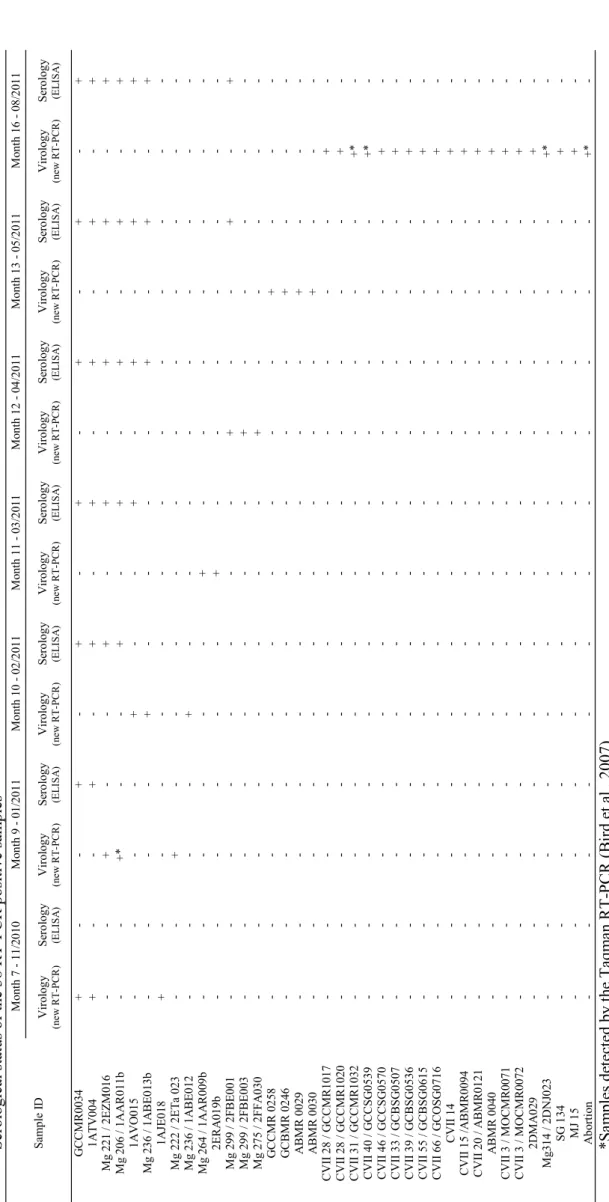
3.5. Serological status of the RVF RT-PCR samples
Serum samples were available for consecutive months of sampling for 18 of the 38 RT-PCR
positive animals. Seven of these animals seroconverted one month after sampling, whereas 11
animals did not (Table 3).
4. Discussion
Highly sensitive molecular assays have already been developed for the detection of RVFV
based either on conventional PCR or on real-time PCR. These techniques have been shown to
diagnose RVFV infection during epidemics. The specifically low viral loads associated with
post-epidemic or inter-epidemic periods require extreme sensitivity and robustness from
detection systems for the successful study of RVF circulation in potential reservoir animals
The presented RT-PCR proved to be both specific and sensitive and could detect 2 log
dilutions further than the reference Taqman system (Bird et al., 2007b; Escadafal et al., 2013).
Using 2,756 serum samples collected in the two countries as a test group, the reference
technique detected only 5 positive samples. In contrast, the new RT-PCR allowed detection of
38 positive samples, corresponding to at least a 7.7 times higher sensitivity than the currently
available gold-standard.
The new RT-PCR detection system performed well, even in the presence of mutations. On the
forward-sense primer RVFV-M-2656F, 100 % of homology was observed among all strains
and on the antisense primer RVFV-M-2766R, only one mutation was observed on the first
nucleotide for 6 strains. Genomic diversity was assessed and showed the presence of 7
mutations in 2 sequences (CVII 14 and CVII 15) and 4 mutations for 2 sequences (ABMR
030 and ABMR 040), all isolated from Anjouan (Figure 3). The remaining sequences only
presented erratic mutations. These results confirmed the strength of the primer designed in 2
conserved areas.
In 2007, the first cases of RVF were reported in the Comoros archipelago with evidence of
animal seroconversions. During the epidemiological survey conducted between May 2010
and August 2011, the incidence of infection assessed by animal seroconversion was estimated
at 14.4 % in the Union of Comoros in goats, sheep and zebu and 4.94 % in Madagascar in
zebu. Out of the 18 positive animals detected by the new RT-PCR, 7 animals seroconverted
with a specific RVF IgG antibody response, 11 did not seroconvert. The fact that a large
proportion of animals that tested PCR positive did not show evidence of seroconversion to
RVFV as assessed by Elisa is interesting and deserves comments. Firstly, the serology-based
detection methods employed have inherent limits to the level of antibody that they can detect.
More generally, the scale of the host immune response mounted after an infection is
immuno competence of the host. The pathogen infecting dose and its potential to actively
replicate in dendritic cells are also likely to correlate with the magnitude of the invoked
immune response, and therefore low-level responses may be expected during inter-epidemic
periods, when viral loads are (on the whole) low. During epizootics, occasional animals may
prove positive to the pathogen by molecular techniques but fail to show evidence of
seroconversion or have delayed seroconversion. In a serosurvey of naturally acquired caprine
arthritis encephalitis virus infection in goats, almost 20% of seronegative animals and
apparently healthy animals had positive PCR test results and only half of them seroconverted
by 8 months later (Rimstad et al., 1993). All these factors are likely operational in the field
conditions characteristic of Madagascar and Comoros and may have implications to the
epidemiology of RVFV; particularly, whether animals detected PCR positive but failed to
seroconvert are contagious and participate to the low level transmission of RVF in the post
epidemic period is presently unknown. The new RT-PCR allowed the detection of RVFV in
38 samples collected in a global epidemiological network in the Comoros archipelago and
Madagascar considered as insular ecosystems (Tortosa et al., 2012). Both serological and
molecular findings strongly suggest that the RVF virus is circulating in this area without
inducing any clinical signs in infected animals.
During IEPs, the epidemiology of RVF remains unclear and involves a possible cryptic
enzootic transmission cycle (Linthicum et al., 1985; Bird et al., 2008). Up to now, the
detection of the RVF virus in RVF potential species reservoirs such as bats (Roussetus
obliviosus, Chaerephon pusillus, Miniopterus griveaudi) in Comoros (n=96) and Maki catta
(Lemur catta) in Mayotte (n=52) was unsuccessful (unpublished data), as well as in
Madagascar (Olive et al.; 2013). The question of a possible inside virus propagation via an
use of sensitive detection tools will be a great help in the understanding of the RVF
inter-epidemic circulation.
Acknowledgments
The authors would like to thank the CRVOI team, La Réunion (Drs Roger and Girard, C.
Foray) for their implication in the epidemiological survey. They also thank Dr D.A.
Wilkinson for his comments and careful reading, all the field team in Madagascar (J.
Rasolofoniaina, N. Andriamanampy, M.-M. Olive) the veterinarians (Drs Evenar, Rabenja,
Remoro) and veterinarian assistants of Tulear I, Tulear II, Mampikony districts, the
FOFIFA-DRZV (Drs J. Ravaomanana and R. Rakotondravao), Dr L. Bibias Armand and the Comorian
partners (Mohamed Halifa) for their assistance during the collection campaign and P. Gil, for
her help in lab experiments at CIRAD, Montpellier.
References
Balenghien, T., Cardinale, E., Elissa, N., Failloux, A.-B., Nipomichene, T.N.J.J., Nicolas, G.,
Rakotoharinome, V.M., Roger, M., Zumbo, B., 2013. Towards a better understanding of Rift
Valley fever epidemiology in the south-west of the Indian Ocean. Vet. Res. 44, 78.
Bird, B.H., Khristova, M.L., Rollin, P.E., Ksiazek, T.G., Nichol, S.T., 2007a. Complete
genome analysis of 33 ecologically and biologically diverse Rift Valley fever virus strains
reveals widespread virus movement and low genetic diversity due to recent common ancestry.
J. Virol. 81, 2805-2016.
Bird, B.H., Bawiec, D.A., Ksiazek, T.G., Shoemaker, T.R., Nichol, S.T., 2007b. Highly
sensitive and broadly reactive quantitative reverse transcription-PCR assay for
Bird, B.H., Githinji, J.W., Macharia, J.M., Kasiiti, J.L., Muriithi, R.M., Gacheru, S.G.,
Musaa, J.O., Towner, J.S., Reeder, S.A., Oliver, J.B., Stevens, T.L., Erickson, B.R., Morgan,
L.T., Khristova, M.L., Hartman, A.L., Comer, J.A., Rollin, P.E., Ksiazek, T.G., Nichol, S.T.,
2008. Multiple virus lineages sharing recent common ancestry were associated with a Large
Rift Valley fever outbreak among livestock in Kenya during 2006-2007. J. Virol. 82,
11152-1166.
Cêtre-Sossah, C., Pédarrieu, A., Guis, H., Defernez, C., Bouloy, M., Favre, J., Girard, S.,
Cardinale, E., Albina, E., 2012. Prevalence of Rift Valley Fever among Ruminants, Mayotte.
Emerg. Infect. Dis. 18, 972-975.
Comtet, L., Pourquier, P., Marié, J.L., Davoust, B., Cêtre-Sossah, C., 2010. Preliminary
validation of the ID Screen® Rift Valley Fever Competition Multi-species ELISA. Poster
presented at the 2010 EAVLD meeting, Lelystad, The Netherlands.
Drosten, C., Göttig, S., Schilling, S., Asper, M., Panning, M., Schmitz, H. Günther, S., 2002.
Rapid detection and quantification of RNA of Ebola and Marburg viruses, Lassa virus,
Crimean-Congo hemorrhagic fever virus, Rift Valley fever virus, dengue virus, and yellow
fever virus by real-time reverse transcription-PCR. J. Clin. Microbiol. 40, 2323-2330.
Drummond, A.J., Ashton, B., Buxton, S., Cheung, M., Cooper, A., Duran, C., Field, M.,
Heled, J., Kearse, M., Markowitz, S., Moir, R., Stones-Havas, S., Sturrock, S., Thierer, T.,
Wilson, A., 2011. Geneious v5.4. Available from http://www.geneious.com/
Edgar, R.C., 2004. MUSCLE: multiple sequence alignment with high accuracy and high
throughput. Nucleic Acids Res. 32, 1792-1797.
Escadafal, C., Paweska, J.T., Grobbelaar, A., le Roux, C., Bouloy, M., Patel, P., Teichmann,
A., Donoso-Mantke, O., Niedrig, M., 2013. International External Quality Assessment of
Fontenille, D., 1989. Transmission cycles of arboviruses in Madagascar. Arch. Inst. Pasteur
Madagascar. 55, 7-317.
Garcia, S., Crance, J.M., Billecocq, A., Peinnequin, A., Jouan, A., Bouloy, M., Garin, D.,
2001. Quantitative real-time PCR detection of Rift Valley fever virus and its application to
evaluation of antiviral compounds. J. Clin. Microbiol. 39, 4456-4461.
Katzourakis, A. and Gifford, R.J., 2010. Endogenous viral elements in animal genomes. PLoS
genetics. 6, e1001191.
Linthicum, K.J., Davies, F.G., Kairo, A., Bailey, C.L., 1985. Rift Valley fever virus (family
Bunyaviridae, genus Phlebovirus). Isolations from Diptera collected during an inter-epizootic
period in Kenya. J. Hyg. (Lond). 95, 197-209.
Müller, R., Poch, O., Delarue, M., Bishop, D.H., Bouloy, M., 1994. Rift Valley fever virus L
segment: correction of the sequence and possible functional role of newly identified regions
conserved in RNA-dependent polymerases. J. Gen. Virol. 75, 1345-1352.
Nichol, S.T., 2001. Bunyaviruses, In: Knipe, D.M., Howley, P.M., Griffin, D.E., Lamb, R.A.,
Martin, M.A., Roizman, B., Straus S.E. (Eds.), Fields virology, fourth ed. Lippincott,
Williams & Wilkins, Philadelphia, pp. 1603-1633.
Ninove L , Nougairede A , Gazin C , Thirion L , Delogu I , Zandotti C Charrel, R.N., De
Lamballerie, X., 2011. RNA and DNA bacteriophages as molecular diagnosis controls in
clinical virology: a comprehensive study of more than 45,000 routine PCR tests. PLoS One 6
e16142.
Olive, M.M., Goodman, S.M., Reynes, J.M., 2012. The role of wild mammals in the
Olive, M.M., Razafindralambo, N., Andrianaivo Barivelo, T., Rafisandratantsoa, J.T.,
Soarimalala, V., Goodman, S.M., Rollin, P.E., Heraud, J.M., Reynes, J.M., 2013. Absence of
Rift Valley fever virus in wild small mammals, Madagascar. Emerg. Infect. Dis. 19, 6.
Paweska, J.T., Mortimer, E., Leman, P.A., Swanepoel, R., 2005. An inhibition enzyme-linked
immunosorbent assay for the detection of antibody to Rift Valley fever virus in humans,
domestic and wild ruminants. J. Virol. Methods. 127, 10-18.
Paweska, J.T., van Vuren, P.J., Kemp, A., Buss, P., Bengis, R.G., Gakuya, F., Breiman, R.F.,
Njenga, M.K., Swanepoel, R., 2008. Recombinant nucleocapsid-based ELISA for detection of
IgG antibody to Rift Valley fever virus in African buffalo. Vet. Microbiol. 127, 21-28.
R Development Core team, 2009. R: A language and environment for statistical computing.
In: R foundation for statistical computing. ISBN 3-900051-07-0, Vienna, Austria.
Rimstad, E., East, N.E., Torten, M., Higgins, J., DeRock, E., Pedersen, N.C., 1993. Delayed
seroconversion following naturally acquired caprine arthitis-encepahilitis virus infection in
goats. Am. J. Vet. Res. 54, 1858-1862.
Roger, M., Girard, S., Faharoudine, A., Halifa, M., Bouloy, M., Cetre-Sossah, C., Cardinale,
E., 2011. Rift valley fever in ruminants, Republic of Comoros, 2009. Emerg. Infect. Dis. 17,
1319-1320.
Sall, A.A., Macondo, E.A., Sène, O.K., Diagne, M., Sylla, R., Mondo, M., Girault, L.,
Marrama, L., Spiegel, A., Diallo, M., Bouloy, M., Mathiot, C., 2002. Use of reverse
transcriptase PCR in early diagnosis of Rift Valley fever. Clin. Diag. Lab. Immunol. 9,
713-715.
Schmaljohn, C., Hooper, J.W., 2001. Bunyaviridae: the viruses and their replication, In:
Knipe, D.M., Howley, P.M., Griffin, D.E., Lamb, R.A., Martin, M.A., Roizman, B., Straus
S.E. (Eds.), Fields virology, fourth ed. Lippincott, Williams & Wilkins, Philadelphia, pp.
Sissoko, D., Giry, C., Gabrie, P., Tarantola, A., Pettinelli, F., Collet, L., 2009. Rift Valley
Fever, Mayotte, 2007-2008. Emerg. Infect. Dis. 15, 568-570.
Smithburn, K.C., Haddow, A.J., Gillett, J.D., 1948. Rift Valley fever; isolation of the virus
from wild mosquitoes. Br. J. Exp. Pathol. 29, 107-121.
Suzich, J.A., Kakach, L.T., Collett, M.S., 1990. Expression strategy of a phlebovirus:
biogenesis of proteins from the Rift Valley fever virus M segment. J. Virol. 64, 1549-1555.
Swanepoel, R., Coetzer, J.A.W., 2004. Rift Valley fever. In: Coetzer, J.A.W., Thomson, G.R.,
Tustin, R.C. (Eds.), Infectious diseases of livestock. Oxford University Press, Oxford, pp.
1037-1070.
Tortosa, P., Pascalis, H., Guernier, V., Cardinale, E., Le Corre, M., Goodman, S.M. Dellagi,
K., 2012. Deciphering arboviral emergence within insular ecosystems. Infect. Genet. Evol.
12, 1333-1339.
Turell, M.J., Linthicum, K.J., Patrican, L.A., Davies, F.G., Kairo, A., Bailey, C.L., 2008.
Vector competence of selected African mosquito (Diptera:Culicidae) species for Rift Valley
fever virus. J. Med. Entomol. 45, 102-108.
Weidmann, M., Sanchez-Seco, M.P., Sall, A.A., Ly, P.O., Thiongane, Y., Lô, M.M. Schley,
H., Hufert, F.T., 2008. Rapid detection of important human pathogenic Phleboviruses. J. Clin.
Virol. 41, 138-142.
World Organisation for Animal Health, 2004. Manual of diagnostic tests and vaccines for
terrestrial animals (mammals, birds and bees), fifth ed. World Organisation for Animal
Figures captions
Figure 1
(A) Alignment of nucleotides 2653 to 2850 of M segment from 52 strains from different
origins. Identical nucleotides for all strains are represented by a dot. Strains are identified by
their country of origin and date of isolation. Primers were represented on the consensus
sequence of the alignment and designed from the most conserved parts of the alignment. Only
the RVFV-M-2840R primer was degenerated for 2 nucleotides. (B) Nucleotide sequences,
position on the M segment and annealing temperatures of the primers (Tm calculator
http://www6.appliedbiosystems.com/support/techtools/calc/).
Figure 2
Comparative efficiency of the Taqman RT-PCR and the new RT-PCR of ten-fold serial
dilutions of tissue culture infected supernatant starting from a Smithburn strain stock titrating
to 106.3 TCID50/ml. Dilutions detected with (A) the Taqman RT-PCR (Bird et al., 2007) (B)
the new RT-PCR .
Figure 3
Alignment of nucleotides 2653 to 2766 of M segment from 52 strains of different origin and
from 30 strains detected with the new RT-PCR. Identical nucleotides for all strains are
represented by a dot. Strains are identified by their country of origin and date of isolation.
G en us Sp ec ie s V ec to r-bo rn e vi ru s R V F s yn dr om e lik e as so ci at ed M ai n ho st P hl eb ov ir us B el te rr a vi ru s C ul ic id ae - H um an Ic oa ra ci v ir us C ul ic id ae - H um an P un ta T or o vi ru s C ul ic id ae - H um an T os ca na v ir us P hl eb ot om in ae Fe ve r H um an Sa nd fl y N ap le s vi ru s P hl eb ot om in ae - H um an Sa nd fl y S ic il ia v ir us P hl eb ot om in ae - H um an Fl av iv ir us W es t N ile v ir us C ul ic id ae - H um an /A ni m al D en gu e ty pe 1 v ir us C ul ic id ae Fe ve r H um an D en gu e ty pe 2 v ir us C ul ic id ae Fe ve r H um an D en gu e ty pe 3 v ir us C ul ic id ae Fe ve r H um an D en gu e ty pe 4 v ir us C ul ic id ae Fe ve r H um an Y el lo w f ev er v ir us C ul ic id ae Fe ve r H um an Ja pa ne se e nc ep ha lit is v ir us C ul ic id ae Fe ve r H um an A po ï v ir us N A - H um an A lp ha vi ru s C hi ku ng un ya v ir us C ul ic id ae Fe ve r H um an O 'N yo ng n yo ng v ir us C ul ic id ae Fe ve r H um an O rt ho bu ny av ir us Sc hm al le nb er g vi ru s C er at op og on id ae A bo rt io n A ni m al R eo vi ru s B lu et on gu e vi ru s se ro ty pe 2 C er at op og on id ae A bo rt io n A ni m al M or bi lli vi ru s P es te d es p et it s ru m in an ts v ir us N A A bo rt io n, D ia rr he a A ni m al In fl ue nz a vi ru s H 5N 1 vi ru s A N A Fe ve r H um an /A ni m al N A : N ot A pp li ca bl e T ab le 1 L is t o f vi ru se s te st ed w it h th e ne w ly d ev el op ed R T -P C R d et ec ti on s ys te m T a b le (s )
T ab le 2 C om pa ri so n of t w o R T -P C R d et ec ti on s ys te m s on a c ol le ct io n of 2 ,7 56 s er um s am pl es f ro m M ad ag as ca r an d th e C om or os a rc hi pe la go ( M ay 20 10 to A ug us t 2 01 1) . C ol le ct io n ar ea N um be r of an im al s co ll ec te d du ri ng th e se ro su rv ey N ew R T -P C R , b as ed o n th e M se gm en t* ( % o f po si ti ve a ni m al s) T aq m an R T -P C R ( B ir d et a l., 20 07 ), b as ed o n th e L s eg m en t (% o f po si ti ve a ni m al s) In ci de nc e le ve ls o f se ro co nv er si on s (% ) M ay ot te 10 4 3/ 12 4 (0 .2 9) 0/ 12 4 (0 ) N D U ni on o f C om or os 18 2 20 /1 06 3 (1 0. 9) 3/ 10 63 ( 1. 6) 14 .4 M ad ag as ca r 20 4 15 /1 56 9 (7 .3 5) 2/ 15 69 ( 0. 9) 4. 94 *E ac h of th e po si ti ve P C R s am pl e de te ct ed w ith th e ne w R T - P C R o n M s eg m en t w as c on fi rm ed b y se qu en ce a na ly se s. N D : N ot d et er m in ed T a b le (s )
T ab le 3 Se ro lo gi ca l s ta tu s of th e 38 R T -P C R p os it iv e sa m pl es Sa m pl e ID M on th 7 1 1/ 20 10 M on th 9 0 1/ 20 11 M on th 1 0 - 02 /2 01 1 M on th 1 1 - 03 /2 01 1 M on th 1 2 - 04 /2 01 1 M on th 1 3 - 05 /2 01 1 M on th 1 6 -08 /2 01 1 V ir ol og y (n ew R T -P C R ) Se ro lo gy (E L IS A ) V ir ol og y (n ew R T -P C R ) Se ro lo gy (E L IS A ) V ir ol og y (n ew R T -P C R ) Se ro lo gy (E L IS A ) V ir ol og y (n ew R T -P C R ) Se ro lo gy (E L IS A ) V ir ol og y (n ew R T -P C R ) Se ro lo gy (E L IS A ) V ir ol og y (n ew R T -P C R ) Se ro lo gy (E L IS A ) V ir ol og y (n ew R T -P C R ) Se ro lo gy (E L IS A ) G C C M R 00 34 + - - + - + - + - + - + - + 1A T V 00 4 + - - + - + - + - + - + - + M g 22 1 / 2 E Z M 01 6 - - + - - + - + - + - + - + M g 20 6 / 1 A A R 01 1b - - + * - - + - + - + - + - + 1A V O 01 5 - - - - + - - + - + - + - + M g 23 6 / 1 A B E 01 3b - - - - + - - - - + - + - + 1A JE 01 8 + - - - - - - - - - - - - -M g 22 2 / 2 E T a 02 3 - - + - - - - - - - - - - -M g 23 6 / 1 A B E 01 2 - - - - + - - - - - - - - -M g 26 4 / 1 A A R 00 9b - - - - - - + - - - - - - -2E R A 01 9b - - - - - - + - - - - - - -M g 29 9 / 2 FB E 00 1 - - - - - - - - + - - + - + M g 29 9 / 2 FB E 00 3 - - - - - - - - + - - - - -M g 27 5 / 2 F FA 03 0 - - - - - - - - + - - - - -G C C M R 0 25 8 - - - - - - - - - - + - - -G C B M R 0 24 6 - - - - - - - - - - + - - -A B M R 0 02 9 - - - - - - - - - - + - - -A B M R 0 03 0 - - - - - - - - - - + - - -C V II 2 8 / G C C M R 10 17 - - - - - - - - - - - - + -C V II 2 8 / G C C M R 10 20 - - - - - - - - - - - - + -C V II 3 1 / G C C M R 10 32 - - - - - - - - - - - - + * -C V II 4 0 / G C C S G 05 39 - - - - - - - - - - - - + * -C V II 4 6 / G C C S G 05 70 - - - - - - - - - - - - + -C V II 3 3 / G C B S G 05 07 - - - - - - - - - - - - + -C V II 3 9 / G C B S G 05 36 - - - - - - - - - - - - + -C V II 5 5 / G C B S G 06 15 - - - - - - - - - - - - + -C V II 6 6 / G C O SG 07 16 - - - - - - - - - - - - + -C V II 1 4 - - - - - - - - - - - - + -C V II 1 5 /A B M R 00 94 - - - - - - - - - - - - + -C V II 2 0 / A B M R 01 21 - - - - - - - - - - - - + -A B M R 0 04 0 - - - - - - - - - - - - + -C V II 3 / M O C M R 00 71 - - - - - - - - - - - - + -C V II 3 / M O C M R 00 72 - - - - - - - - - - - - + -2D M A 02 9 - - - - - - - - - - - - + -M g3 14 / 2D N J0 23 - - - - - - - - - - - - + * -SG 1 34 - - - - - - - - - - - - + -M J 15 - - - - - - - - - - - - + -A bo rt io n - - - - - - - - - - - - + * -*S am pl es d et ec te d by th e T aq m an R T -P C R ( B ir d et a l., 2 00 7) T a b le (s )
Pr im er Se qu en ce N uc le ot id e po si ti on o n th e M s eg m en t A nn ea lin g T R V FV -M -2 65 6F -C T A G C C G T T T C A C A A A C T G G G -2, 65 6 - 2, 67 6 60 R V FV -M -2 84 0R -G A C T G A R G A Y T C T G A A T T G C A C C -2, 81 7 - 2, 84 0 55 R V FV -M -2 76 6R -C A A T T G C A T A C C C T T T G C C T G G G C -2, 74 2 - 2, 76 6 70 Fi gu re 1 F ig u re (s )
B
.
A
.
di lu ti on 1 0 -3 di lu ti on 1 0 -4 di lu ti on 1 0 -5 di lu ti on 1 0 -2 di lu ti on 1 0 -3Fi
gu
re
2
F ig u re (s )Fig.3. Figure(s)
Highlights
A highly sensitive real-time PCR assay for the detection of Rift Valley Fever virus Sensibility tested on 2,756 serum from Madagascar and the Comoros archipelago Comparison with a RVF specific quantitative real time RT-PCR reference technique System specific and 7.7 times more sensitive than the reference technique
Diagnostic tool for the detection of low viral loads in inter-epizootic periods