HAL Id: tel-01340856
https://tel.archives-ouvertes.fr/tel-01340856
Submitted on 2 Jul 2016HAL is a multi-disciplinary open access archive for the deposit and dissemination of sci-entific research documents, whether they are pub-lished or not. The documents may come from teaching and research institutions in France or abroad, or from public or private research centers.
L’archive ouverte pluridisciplinaire HAL, est destinée au dépôt et à la diffusion de documents scientifiques de niveau recherche, publiés ou non, émanant des établissements d’enseignement et de recherche français ou étrangers, des laboratoires publics ou privés.
The role of shed GP in Ebola virus pathogenicity
Beatriz Escudero Pérez
To cite this version:
Beatriz Escudero Pérez. The role of shed GP in Ebola virus pathogenicity. Virology. Ecole normale supérieure de lyon - ENS LYON, 2014. English. �NNT : 2014ENSL0933�. �tel-01340856�
THÈSE
en vue de l'obtention du grade de
Docteur de l’Université de Lyon, délivré par l’École Normale Supérieure de Lyon
Discipline : Sciences de la vie
Laboratoire Bases Moléculaires de la Pathogénicité Virale École Doctorale de Biologie Moléculaire, Intégrative et Cellulaire
présentée et soutenue publiquement le 3 Octobre 2014
par Madame Beatriz Eugenia ESCUDERO PÉREZ
_____________________________________________
The role of shed GP in Ebola virus pathogenicity
______________________________________________
Directeur de thèse : M. Viktor VOLCHKOV
Devant la commission d'examen formée de :
M. Renaud MAHIEUX, Centre International de Recherche en Infectiologie, Président M. Christophe PEYREFITTE, IRBA-ERL, Examinateur
M. Daniel PINSCHEWER, University of Basel, Rapporteur
M. Viktor VOLCHKOV, Centre International de Recherche en Infectiologie, Directeur de thèse M. Thierry WALZER, Centre International de Recherche en Infectiologie, Examinateur
Acknowledgments
1
Acknowledgments
I would first like to thank Professor Viktor Volchkov for having welcomed me to his laboratory and for making my dream come true: since I was eight years old I have been dreaming of working with the Ebola virus. I would specially like to thank him for his trust, which has encouraged me to be more independent within the research framework, and for his bright ability to present and organise concepts.
J’aimerais aussi remercier les membres de mon jury pour m’avoir fait l’honneur d’évaluer mon travail et pour l’échange qui aura lieu le jour de ma défense: M. Peyrefitte, qui a très aimablement accepté de juger mon travail en tant que représentant de la DGA (Délégation Générale pour l’Armement), M. Walzer et M. Pinschewer et M.Weissenhorn qui ont accepté d’être les rapporteurs de ma thèse ainsi que le Professeur Mahieux, qui a accepté d’être le président de mon jury.
Je tiens à remercier spécialement à Thierry Walzer pour sa totale et inconditionnelle disponibilité pour m’aider avec des doutes en immunologie et pour ses conseils professionnels. Je l´admire profondément pour son intelligence, son honnêteté, et pour sa capacité à motiver et inspirer les autres. J’aimerais aussi exprimer ma profonde gratitude aux membres de son équipe pour leur gentillesse, leur sympathie, leur disponibilité, leur soutien, leur intérêt et leur aide constante avec tout ce dont j’ai eu besoin. Je remercie en spécial à Antoine, Sophie, Fabrice et Sébastien. Un grand merci également à Armelle, avec qui j’ai passée des très bons moments ensemble et qui a réussi à m’arracher du laboratoire pour jouer au foot.
Je remercie aussi à Thierry Defrance, Denis Gerlier et Mathias Faure pour leur aide désintéressée. Je les admire énormément non seulement parce qu´ils sont des grands scientifiques mais aussi parce qu´ils sont des personnes honnêtes, intelligentes et intègres.
Je voudrais aussi exprimer ma plus sincère gratitude à Marc Jamin et de nouveau à Thierry Walzer pour m’avoir fait l’honneur de faire partie de mon comité de thèse il y a deux ans. Je les remercie fortement pour m’avoir aidé à
Acknowledgments
2
prendre du recul sur mon projet de thèse et pour les enrichissantes discussions qui m’ont permis d’avoir un regard plus critique sur mes résultats et d´avancer dans mon projet.
J’aimerais bien remercier aussi à Veronique Santyi pour sa gentillesse, sa disponibilité et pour m´avoir initiée au Biacore et l’ AKTA, machines que sans son aide, je n’aurais pas réussi à maîtriser.
Je remercie également le plateau technique de cytometrie, en particulier à Sébastian et Thibault pour leur aide. Je ne peux pas non plus oublier de remercier à Renaud Colisson qui a toujours essayé de m’aider, de me faire faire des pauses pendant les intenses journées de travail et qui m’a appris patientent (même les week-ends) les bases de la cytometrie. Un grand merci aussi à tout l’équipe P4, en spécial à Audrey, pour son amabilité et son professionnalisme, à Delphine, pour sa sympathie et les bons moments passés pendant la formation, à Stephanie et à Stephan ainsi qu´au poste central de biosécurité pour m’avoir permis de réaliser toutes mes expériences dans d’excellentes conditions.
Je suis très reconnaissante a la Délégation Générale pour l’Armement (DGA), La Caixa en Espagne et l’INSERM pour m´avoir permis de réaliser ce travail. Je remercie aussi à ma petite étudiante, Camille Gibourt, avec qui, malgré le fait que le temps passé ensemble fut bref, j’ai beaucoup profité de cette échange qui m´a permis de ratifier ma passion pour l’enseignement et la recherche.
Un grand merci à Mathieu Mateo, qui à été mon premier maître de stage et qui m’a surtout appris dès le début l´engagement au quotidien envers la recherche. J’aimerais aussi le remercier de m´avoir soutenu et accompagné tout au long de mon stage: ses conseils, sa patience et sa facilité à expliquer des concepts pas évidents pour moi à l’époque on rendu ma première expérience au laboratoire très agréable et enrichissante. J’aimerais remercier aussi à Audrey Page, pour la confiance dont elle m´a toujours témoignée, pour s´être toujours intéressée à moi et pour avoir su garder le contact même si je ne suis pas très souvent disponible. Je remercie également à Olivier Reynard qui nous a permis
Acknowledgments
3
de profiter de son large expérience dans beaucoup de domaines. Je présente aussi mes remerciements à Valentina pour avoir été ma deuxième maître de stage et m’avoir introduit au intéressant monde des GPs du virus Ebola; je la remercie également pour sa gentillesse et sa douceur qui imprègnent l´esprit du laboratoire. Même s’il n’est plus là, je tiens à remercier spécialement à Kirill Nemirov, qui est arrivé au laboratoire au même temps que moi et qui a été un énorme soutien et un très bon ami: il est une des meilleures personnes que je n’ai jamais connues. Je remercie aussi à Nicolas pour les très bons moments passés ensemble, pour sa disponibilité, ses encouragements et sa précieuse connaissance du mystérieux monde de la statistique. J’aimerais remercier à Audrey Baule, qui m’a beaucoup appris sur l’immunologie et qui a été une amie et un appui en tout moment. Je la remercie aussi pour toujours me faire confiance, pour sa compréhension et pour me pousser à élargir mes ambitions professionnelles. Sa présence au laboratoire m’a beaucoup manqué. Finalement, le plus grand remerciement est pour Philip Lawrence, qui a été mon phare dans le laboratoire depuis son arrivée. Il m`a poussé à mettre en valeur mes résultats, il m’a corrigé, écouté, recorrigé, il a toujours respecté mes opinions, il m’a défendu et il a cru en moi. Au delà des remerciements pour avoir été ma bouée professionnelle, je le remercie surtout pour avoir été un vrai ami. Je ne trouve pas assez des mots pour exprimer ma gratitude envers lui. J’adresse également mes remerciements, pour leur sympathie et leurs conseils à tous les anciens membres de l’équipe Filovirus (Sébastian, Mikel, St Patrik, Jennifer, Cidney, Ilona, Nolwen, Ludo, Amandine) ainsi qu’à nos voisins, l’équipe Pasteur. Je voudrais aussi remercier à toutes les personnes de la TOUR que j’ai croisé à un moment où à un autre et avec qui nous avons partagé des bons moments (Ludovic, Joel, Chloé…).
Un grand merci aussi à mon copain Joévin, pour son inestimable soutien (je me demande encore comment il fait pour me supporter tout le temps surtout en fin de thèse), je le remercie aussi pour notre passion commune des voyages qui me rappelle qu’il y a un monde parallèle à / en dehors de la thèse que je hâte d’explorer. Merci de me rappeler de temps en temps que le travail ce n’est pas tout dans la vie et mercie de t’être toujours adapté à moi et à mes ambitions professionnelles.
Acknowledgments
4
A les meves amigues Cèlia i Irene, amb qui he pogut mantenir una veritable amistat al llarg dels anys tot i la llarga distància que ens separa. Els hi agreixo molt el seu suport, la seva forma de ser i el fet que em fassin sentir que malgrat el temps que no ens veiem, encara podem comptar les unes amb les altres i compartir moments genials. També ocupades per la seva tesis, estic contenta d’haver pogut compartir aquesta difícil experiència amb elles i espero que l’acabem aviat i ens poguem veure més sovint.
A Laura, por su apoyo, su alegría, su sonrisa, los viajes rélampago y su amistad.
A Richard Preston, por haberme seducido con su libro Zona Caliente y haber despertado mi interés por Ebola a la temprana edad de 8 años.
A toda mi familia, en especial a mis padres y a mi hermana. A mi hermana, Raquel Escudero Pérez, por haberme hecho sentir que no estaba sola, que siempre podría contar con ella y, a pesar de ser una de las personas mas inteligentes y trabajadoras que conozco, considerarme capaz de sacarme la tesis.
Finalmente, a mis padres, a quien les debo todo. Quisiera agradeceros por vuestra eterna paciencia conmigo, por haber sido los únicos que me habeis escuchado, entendido y luchado a mi lado por mis sueños y mi persona. Os agradezco por haberme dado los valores que tengo, porque nunca han sido los conceptos que he aprendido a lo largo de mis estudios los que me han permitido salir adelante sino vuestros principios, vuestra fuerza y vuestro carácter que ahora también son los míos. No sé si la tesis me definirá como una buena científica, pero sí estoy segura que esta tesis me ha puesto a prueba como persona y que sin vosotros, no podría haberla hecho. Vosotros habeis sido los que habeis marcado la diferencia.
Merci à tous,
Acknowledgments
5
La verdadera ciencia enseña, por encima de todo, a dudar y a ser
ignorante.
Miguel de Unamuno (1864-1936)
“Don't let the noise of other's opinions drown out your own inner
voice. And most important, have the courage to follow your heart
and intuition. They somehow already know what you truly want to
become. Everything else is secondary.”
Acknowledgments
Abstract
7
Abstract
Study of the role of Ebola virus shed GP
During Ebola virus (EBOV) infection several soluble glycoproteins are released in high amounts from infected cells but as yet still no clear role has been identified for these viral proteins. We hypothesized that the impairment of coagulation and vascular systems observed during EBOV infection could be, at least in part, due to these soluble glycoproteins.
Here, for the first time we identify the cellular targets of EBOV soluble protein shed GP and provide evidence that through its glycosylation, shed GP can activate non-infected dendritic cells and macrophages, inducing, through TLR4, the secretion of pro-inflammatory cytokines. We also demonstrate that shed GP activity is negated upon addition of Mannose-Binding sera Lectin (MBL), a molecule known to interact with sugar arrays present on the surface of different microorganisms. We further demonstrate that shed GP binds MBL and activates the MBL-associated proteases MASP-1 and MASP-2, implicated in the coagulation pathway. Moreover, shed GP is capable of competing with mannose for MBL binding. Importantly, we show that shed causes monocytes, macrophages and HUVECs to increase their expression of tissue factor (TF), which has been shown to be the primary molecule in endothelial cells that results in alterations in both permeability and procoagulant activity. Treatment of HUVECs with shed GP resulted in increased surface expression of the attachment factors ICAM and VCAM but also the release of soluble ICAM, s-VCAM, s-E-selectine s-TF and s-thrombomodulin. We have also revealed that shed GP activates permeability of HUVECs both directly and indirectly through cytokine release.
Overall, this study suggests that shed GP may be one of the principal factors responsible for the early stimulation of immune cells that then produce high amounts of proinflammatory cytokines. Moreover shed GP also activates MBL and therefore may play a critical role in the dysregulation of coagulation and permeability responses that, combined with massive virus replication and virus-induced cell damage, can lead to a septic shock-like syndrome and high mortality.
Keywords 8
Keywords
Ebola virus Soluble glycoproteins Shed GP Immune response Coagulopathy Permeability PathogenesisList of figures
9
List of figures
Figure 1. Geographic distribution of filovirus outbreaks ...27
Figure 2. Discovery of an ebolavirus-like filovirus in europe …...28
Figure 3. Possible Ebola virus reservoirs ...30
Figure 4. Phases of EBOV hemorrhagic fever (EHF) ...37
Figure 5. Ebola virus morphology and organisation ...51
Figure 6. Ebola virus glycoprotein synthesis and conformation ...55
Figure 7. Schematic representation of Ebola virus GP ...56
Figure 8. TACE cleavage site selectivity ...62
Figure 9. Illustration of Ebola virus infection cycle ...68
Figure 10. Ebola virus infection and spreading ...70
Figure 11. IFN pathway ...74
Figure 12. Type I IFN induction, signaling and action ...75
Figure 13. Function of Ebola virus-encoded IFN-antagonist proteins ...78
Figure 14. Tissue Factor coagulation pathway...84
Figure 15. Expression of Tissue Factor can lead to Disseminated Intravascular Coagulation syndrome ...85
Figure 16. Disseminated Intravascular Coagulation ...87
Figure 17. Schematic representation of pathogenetic pathways in disseminated intravascular coagulation ...88
Figure 18. Cross talk between the inflammation and coagulation systems ...95
Figure 19. Effects of physiological anticoagulant systems and fibrinolysis on coagulation and inflammation ...98
List of figures
10 Figure 20. MBL structure ...100 Figure 21. MBL binding to certain patterns activates MASPs and subsequently
activates the lectin complement pathway...102
Figure 22. Role of MBL during infection ...103 Figure 23. Induction of cytopathic effects by Ebola virus glycoproteins ……..105 Figure 24. EBOV GP masks cellular surface proteins ...106 Figure R1. Production of recombinant EBOV shed GP and analysis of shed GP
release in the blood of infected guinea pigs ……….…118
Figure R2. Shed GP production and characterisation ...120 Figure R3. DCs and MØ are primary targets of shed GP ...123 Figure R4. Shed GP induces transcriptional activation of cytokines in DCs and
MØ ...126
Figure R5. Shed GP induces secretion of pro- and anti-inflammatory cytokines
from DCs and MØ and induces their phenotypic maturation ...129
Figure R6. Deglycosylation of shed GP affects activation of DCs and MØ ....131 Figure R7. Ebola virus shed GP binds to human monocytes ...135 Figure R8. Ebola virus shed GP does not induce mRNA synthesis and release
of cytokines from human monocytes ...136
Figure R9. Ebola virus replication in human monocytes after shed GP treatment
...138
Figure R10. EBOV glycoproteins interact with rhMBL
...142
Figure R11. Shed GP induces VPR-AMC cleavage through MASP-1 ...146 Figure R12. C4 fixation and cleavage by shed GP through MBL ...149
List of figures
11 Figure R13. Expression of tissue factor in monocytes, DC, macrophages and
HUVEC cells ...151
Figure R14. Surface expression and release of coagulation and permeability
markers by HUVEC ...153
Figure R15. EBOV Shed GP increases cell permeability of HUVEC in vitro
………..156
Figure R16. Shed GP effect on virus binding and replication ...162 Figure 25. Possible role of shed GP in inducing lymphocyte apoptosis …...169 Figure 26. Potential effects of shed GP on the immune system during EBOV
infection ...172
Figure 27. Role of shed GP in the impairment of coagulation ...178 Figure 28. Role of shed GP in inducing endothelial cell permeability
...183
Figure 29. Role of shed GP in virus spreading ...185 Figure 30. Role of shed GP during EBOV pathogenesis ...189
List of tables
12
List of tables
Table 1. Ebola virus outbreaks in human beings ...26 Table 2. Techniques to analyse and detect Ebola virus infection ...41 Table 3. Comparison of pathological features of different animal models of
Ebola virus infection ...43
Table 4. Efficacy of post-exposure treatment in animal models of Ebola virus
infection ...46
Table 5. Overview of Ebola virus vaccines ...49
Table 6
.
Correlative differences between survivors and non survivors of Ebolavirus infection ………...82
List of abbreviations 13
List of abbreviations
α Anti- / Alpha Δ Delta Ab Antibody AT AntiThrombinATP Adenosine TriPhosphate
AP-1 Activator Protein-1
APC Antigen Presenting Cell BEBOV Bundibugyo EBOla Virus BSL-4 BioSecurity Level 4
DNA DeoxyRibonucleic Acid ºC degrees Celsius
cDC conventional Dendritic Cells
CIEBOV Côte d’Ivoire EBOla Virus (=Taï Forest)
CK CytoKines
CMV CytoMegaloVirus
CRD Carbohydrate-Recognition Domain CTL CytoToxic Lymphocytes
DC Dendritic cell
DC-SIGN Dendritic Cell-Specific Intercellular adhesion molecule-3-Grabbing
Non-integrin
List of abbreviations
14 DIC Dissemeinated Intravascular Coagulation
DMEM Dulbecco modified Eagle’s medium DRC Democratic Republic of Congo EBOV EBOla Virus
EC Endothelial Cells
EHF Ebola Haemorrhagic Fever
ELISA Enzyme Linked ImmunoSorbent Assay
EPCR Endothelial Protein C Receptor
FA Fluoresence Assay
FBS Fetal Bovine Serum
FDP Fibrinogen Degradation Products
GFP Green Fluorescence Protein GP Glycoprotein
HA HemAgglutinin
HIV Human Immunodeficiency Virus HR1/HR2 Heptated Repeat 1/2
HS High Shedding
*HS deglycosylated High Shedding
HUVEC Human umbilical vein endothelial cells ICAM -1 Intercellular Adhesion Molecule-1 (=CD54)
IFA ImmunoFluorescence Assay
IgG Immunoglobulin G IgM Immunoglobulin M
List of abbreviations
15
IFL Internal Fusion Loop
IFN InterFeroN IL InterLeukine
IRADs Innate Response Antagonist Domains IRF-3/IRF-9 Interferon Regulatory Factor 3/9
ISG Interferon Stimulated Genes
ISRE Interferon Stimulated Response Elements kDa kilo Dalton
LBP Lypopolysaccharide Binding Protein
LLOV LLOviu Virus
LPS Lipopolysaccharide
LS Low Shedding
L-SECtin Liver/Lymph node Sinusoidal Endothelial cell C-type lectin
L-SIGN Liver/Lymph node-Specific Intercellular adhesion molecule-3-
Grabbing Nonintegrin
MØ Macrophages
MAL Membrane Attack Complex
MASP MBL Associated Serin Proteases
MARV MARburg Virus
MBL Mannose Binding Lectin
MDA-5 Melanoma Differentiation-Associated protein 5
MGL Macrophage Galactose Lectin
List of abbreviations
16
MLD Mucin Like Domain
MOI Multiplicity Of Infection mRNA messenger RiboNucleic Acid
NF-κB Nuclear Factor-kappa B
NHP Non-Human Primates NK Natural Killer cell
NO Nitric Oxide
NP NucleoProtein
NPC1 Niemann-Pick C1
PA Plasminogen Activator
PAI-1 Plasminogen Activator Inhibitor-1
PAF Platelet-Activating Factor
PAR Protein-Activated Receptors
PCA Protein C Activated
PCR Polymerase Chain Reaction
pDC plasmacytoid Dendritic Cells
PFA ParaFormAlehyde
PKR Protein Kinase R
qPCR quantitative Polymerase Chain Reaction
RBD Receptor-Binding Domain
REBOV Reston EBOla Virus
rNAPC2 recombinant Nematode Anticoagulant Protein C2
List of abbreviations
17 RNA RiboNucleic Acid
RNP RiboNucleoProtein complex
RPMI Roswell Park Memorial Institute medium
RT-PCR Reverse Transcriptase Polymerase Chain Reaction
SARS Severe Acute Respiatory Syndrome
SEBOV Sudan EBOla Virus
sGP secreted GlycoProtein
ssGP small secreted GlycoProtein
SNP Single Nucleotide Polymorphism
STAT-1 Signal Transducers and Activators of Transcription- 1
TACE TNF-α converting enzyme
TAFI Thrombin-Activatable Fibrinolysis Inhibitor
TAM Tyro-3, Axl, and Mer families
TIM-1 T-cell Immunoglobulin Mucin Domain 1
TF Tissue Factor
TFPI Tissue Factor Pathway Inhibitor
TLR Toll Like Receptor
TM TransMembrane
ThM THromboModulin
TNF Tumor Necrosis Factor
TRAIL TNF Related Apoptosis-Inducing Ligand
PAMP Pathogen Associated Molecular Patterns
List of abbreviations
18 PBMC Peripheral Blood Mononucleated Cells
PC Protein C
PECAM Platelet Endothelial Cell Adhesion Molecule (=CD31)
pi post infection
PRR Pattern Recognition Receptor
t-PA tissue-type Plasminogen Activator
u-PA urokinase-type Plasminogen Activator
VCAM-1 Vascular Cell Adhesion Molecule- 1 VLP Virus Like Particles
VP Viral Protein
VSV Vesicular Stomatitis Virus
WT Wild Type
Index 19
INDEX
Acknowledgments ………...1 Abstract ……….7 Keywords ………..8 List of figures ………...9 List of tables ………..12 List of abbreviations ………13 Introduction ………231. Ebola virus hemorrhagic fever ………25
1.1. Epidemiology ………...…25
1.2. Ebola virus natural cycle ………..………29
1.2.1. Ebola virus reservoir ………29
1.2.1.1. Bats as a possible reservoir ………...…31
1.2.2. Ebola virus transmission ………33
1.3. Clinical aspects of Ebola virus hemorrhagic fever ………35
1.4. Diagnostic techniques to identify Ebola virus ………39
1.5. Ebola virus animal models ………..…41
1.6. The fight against Ebola virus ………..…44
1.6.1. Treatments ………..…44
1.6.2. Vaccines ………..…47
2. Molecular biology of Ebola virus ..………..…50
2.1. Morphology and genome organisation ……….…50
2.2. EBOV proteins and their role during pathogenesis …………..……52
2.2.1. Nucleoprotein (NP) ………52
2.2.2. VP35 ………..53
Index
20 2.2.4. GP ………..…54 2.2.5. Ebola virus soluble glycoproteins ………60 2.2.5.1. Secreted GP ………...…60 2.2.5.2. Shed GP ………61 2.2.5.3. Small secreted GP ……… 63 2.2.6. VP30 ………..…63 2.2.7. VP24 ………..………64 2.2.8. L polymerase ………..………65 2.3. Ebola virus replication cycle ………...………65 3. Ebola virus pathogenesis ……….………69 3.1. Host immune response to Ebola virus infection ………69 3.1.1. Innate immune response ……….69 3.1.1.1. Ebola virus increases the cytokine response …………69 3.1.1.2. Ebola virus inhibits the interferon response .…………73 3.1.2. Adaptative immune respone ………..………79 3.1.2.1. Cellular immune response ………..……79 3.1.2.2. Humoral immune response ……….…80 3.1.3. Coagulation impairment ………..……82 3.1.3.1. Disseminated Intravascular Coagulation (DIC) ….……86 3.1.3.2. DIC pathogenesis ………..………88 3.1.3.2.1. Tissue factor expression ……….…………89 3.1.3.2.2. Impairment of anticoagulant mechanisms .……91 3.1.3.2.3. Impairment of fibrin removal ………..…93 3.1.3.3. Inflammatory response and DIC ………95 3.1.3.4. Cross-talk between inflammation and coagulation …..96 3.1.3.5. DIC diagnosis ……….………99 3.1.4. Another player in the coagulation pathway: MBL ………99
Index
21 3.2. Direct cytotoxicity caused by the virus itself and GP protein ………104 3.3. Overview of Ebola virus pathogenesis ……….………107 Objectives ……….…109 Results ………...…113 Results I: Role of shed GP in innate immune response ………..…115 Results II: Ebola virus shed GP allows monocyte infection by EBOV ………..133 Results III: Role of shed GP in coagulation and vascular permeability ………139 Results IV: Impact of shed GP on EBOV entry ………...………159 Discussion ………163 Shed GP targets the immune system ………165 Shed GP impairs coagulation and permeability pathways ……..…177 Shed GP as a key factor in EBOV pathogenicity ………185 Material and methods ………193 Publications and other communications ……….…209 References ………213 Annexes ……….……251
Glycosylation in EBOV ………..………253
Publications ………..………261
o Shed GP of Ebola Virus Triggers Systemic Immune Activation and Increased Vascular Permeability
………..………263
o Surface glycoproteins of the recently identified African Henipavirus promote viral entry and cell fusion in a range of human, simian and bat cell lines
………..………317
o Dérégulation de l’hémostase dans l’infection à filovirus
Index
Introduction
23
Introduction
Introduction
24 1. Ebola virus hemorrhagic fever
Introduction
25
Ebola virus is the etiologic agent of a highly lethal hemorrhagic fever in humans and nonhuman primates (NHPs) with mortality rates of up to 90% in humans (Zampieri, Sullivan et al. 2007). Due to its high mortality rate, its potential for human-to-human transmission and the lack of an approved vaccine or therapy, the virus is classified as a category A pathogen requiring biosafety level 4 (BSL-4) containment.
Ebola virus belongs to the family Filoviridae, in the order Mononegavirales which includes Rhabdoviridae and Paramyxoviridae. The family consists of three genera: Marburgvirus, Ebolavirus and Cuevavirus (Kuhn, Becker et al. 2010).
Ebola haemorrhagic fever (EHF) is caused by five genetically distinct members of the Filoviridae family: Zaire ebolavirus (ZEBOV), Sudan ebolavirus (SEBOV),
Côte d’Ivoire ebolavirus (CIEBOV), Bundibugyo ebolavirus (BEBOV) and Reston ebolavirus (REBOV). The species vary in their pathogenicity in humans
with ZEBOV being the most pathogenic (up to 90% case fatality rate). For Sudan and Bundibugyo Ebolavirus the mortality rate is lower, 53% and 34% respectively. The species Côte d’Ivoire is lethal for NHPs (Formenty, Boesch et
al. 1999). Ebolavirus Reston is not pathogenic for humans (4 asymptomatic cases have been described), however, it is lethal for NHPs and pigs. All species are endemic in Africa except Ebolavirus Reston, which is endemic in the Philippines (Miranda, Ksiazek et al. 1999, Barrette, Metwally et al. 2009). A number of sporadic outbreaks have been reported since the initial discovery of EBOV in 1976 (Table 1) (Arthur 2002, Mahanty and Bray 2004, Baize,
Introduction
26
Date EBOV
strain Source of infection human casesNumber of Fatality rate Location
1976 ZEBOV Unknown 318 88% DRC
1976 SEBOV Unknown 284 53% Sudan
1977 ZEBOV Unknown 1 100% DRC (Tandala)
1979 SEBOV Unknown 34 65% Sudan
1989 REBOV Imported macaques - - USA (Virginia)
1990 REBOV Imported macaques - - USA (Pennsylvania)
1992 REBOV Imported macaques - - Italy
1994 CIEBOV Dead chimp 1 0% Côte d’Ivoire
1994-1995 ZEBOV Unknown 49 59% Gabon
1995 ZEBOV Unknown 317 77% DRC (Kiwit)
1996 REBOV Imported macaques - - USA (Texas)
1996 ZEBOV Dead chimp 35 68% Gabon (Mayibout)
1996 ZEBOV Unknown 60 75% Gabon (Booué)
2000-2001 SEBOV Contact with NHP 425 53% Ouganda (Gulu)
2001 ZEBOV Contact with NHP 123 79% Gabon/Congo
2002-2003 ZEBOV Unknown 143 90% Congo (Mbomo, Kéllé)
2003 ZEBOV Unknown 35 83% Congo(Mbomo)
2004 SEBOV Unknown 17 42% Sudan (Yambio)
2005 ZEBOV Contact with bats 12 75% Congo (Etoumbi,Mbomo)
2007 ZEBOV Unknown 249 74% DRC (Kasaï)
2007 BEBOV Unknown 56 40% Ouganda (Bundibugyo)
2008-2009 ZEBOV Unknown 32 47% DRC (Kasaï)
2011 SEBOV Unknown 1 100% Ouganda (Luwero)
2012 SEBOV Unknown 24 71% Ouganda (Kibaale)
2012 BEBOV Unknown 52 48% DRC (Haut-Uélé)
2014 ZEBOV Unknown 604 63% Guinea, Liberia, Sierra Leone
Table 1.- Ebola virus outbreaks in human beings. Adapted from (Arthur
2002, Mahanty and Bray 2004, Baize, Pannetier et al. 2014).
The geographical distribution of the different epidemics of filovirus hemorragic fevers corresponds to the equatorial zone of the globe (Figure 1). Ebola haemorrhagic fever remains a plague for the population of equatorial Africa, with an increase in the numbers of outbreaks and cases since 2000. Almost all human cases are due to the emergence or reemergence of Zaire Ebola virus in regions of Gabon, Republic of the Congo, DRC and very recently in west Africa, and of Sudan Ebola virus in Sudan and Uganda. Subsequently, Reston Ebola virus has been found in the Philippines on several occasions, including reports documenting infections in pigs (Barrette, Metwally et al. 2009). The emergence of Reston Ebola virus in pigs raises important concerns for public health,
Introduction
27
agriculture, and food safety in the Philippines and could represent a serious issue for parts of Asia (Feldmann and Geisbert 2011).
Figure 1.- Geographic distribution of filovirus outbreaks. (Prior to 2014)
Adapted from (Feldmann and Geisbert 2011).
A novel filovirus, provisionally named Lloviu virus (LLOV), was detected during the investigation of Miniopterus schreibersii die-offs in Cueva del Lloviu in southern Europe. LLOV is genetically distinct from other marburgviruses and ebolaviruses and is the first filovirus detected in Europe that was not imported from an endemic area in Africa (Negredo, Palacios et al. 2011). Phylogenetic analysis of LLOV indicates a common ancestor of all filoviruses, dating to at least 150,000 years ago (Figure 2). The discovery of LLOV in M. schreibersii is
consistent with filovirus tropism for bats. However, unlike MARV and EBOV, where asymptomatic circulation is posited to be consistent with evolution to avirulence in this long-term host-parasite relationship, several observations suggest that in the case of LLOV, filovirus infection may be pathogenic; LLOV was found in the affected bat population but not in other healthy bats. In addition, RNA sequences were found in lung and spleen of the affected bat population. The discovery of a novel filovirus in western Europe, and the wide geographical distribution of the associated insectivorous bat is a significant concern. While the virus was detected in the north of Spain, simultaneous bat die-offs were also been observed in Portugal and France (Negredo, Palacios et al. 2011). Filoviruses had been suggested to show a geographically related
Introduction
28
phylogeographic structure (Peterson, Bauer et al. 2004). Viruses from particular geographic areas cluster together phylogenetically, suggesting a stable host-parasite relationship wherein viruses are maintained in permanent local pools. The discovery of a novel filovirus in a distinct geographical niche suggests that the diversity and distribution of filoviruses should be studied further.
Figure 2.- Discovery of an ebolavirus-like filovirus in europe.Adapted from
(Negredo, Palacios et al. 2011).
Recently, a study has shown that additional bat species not previously associated with EBOV such as Epomorphorus gambianus, Nanyecteris veldkampii and Eiodolon helvum contain antibodies against ZEBOV and REBOV (Hayman, Yu et al. 2012). This reinforces the hypothesis of bats being the possible natural reservoir of filoviruses.
Serological evidence of ebolavirus infection in several bat populations in China has also recently been reported (Yuan, Zhang et al. 2012). This, together with the Lloviu finding, suggests that filoviruses have a wider host range and geographical distribution than previously thought.
Introduction
29 1.2. Ebola virus natural cycle
1.2.1. Ebola virus reservoir
Despite the discovery of this virus family almost 40 years ago, the natural reservoir of these lethal pathogens remains an enigma.
Several studies have been undertaken in order to try and identify the natural reservoir of the filoviruses. Since 1976 these studies have been based on the capture and analysis of plants and wild animals (vertebrates and invertebrates) suspected to be potential natural hosts of filoviruses.
In outbreaks where information is available, the human index cases have invariably had direct contact with gorillas, chimpanzees, antelope or bats. The search for a reservoir of EBOV has been very aggressive. Although great apes are generally involved in EHF outbreaks, NHPs are not thought to be natural reservoirs but, rather, susceptible hosts, based on the sudden sharp decline in populations of the great apes in Gabon and the Republic of Congo which coincided with EBOV outbreaks in humans (Pourrut, Kumulungui et al. 2005). In one study, short EBOV-specific genetic sequences were amplified from organs of six mice (Mus setulosus and Praomys sp) and a shrew (Sylvisorex
ollula). However no firm conclusions as to the EBOV reservoir status of these
animals can be drawn, given the lack of specific serologic responses, the lack of nucleotide specificities in the amplified viral sequences, the failure of virus isolation attempts and the non-reproducible nature of the results. Also several attempts were made to infect various plant and animal species, including rodents (mice, shrews, rats, guinea pigs, etc.), birds (mainly pigeons), reptiles (tortoises, snakes, geckos, frogs), molluscs (mainly snails), arthropods (spiders, cockroaches, ants, centipedes, mosquitoes and butterflies) and more than 33 plant varieties belonging to 24 species (tomatoes, cucumbers, wheat, cotton, lupin, corn, tobacco, etc.). All attempts to inoculate these species failed to yield evidence of virus replication (Pourrut, Kumulungui et al. 2005).
In contrast, EBOV infection of vertebrates and invertebrates gave the first evidence that both insectivorous and frugivorous bats can support the replication and circulation of EBOV (Swanepoel, Leman et al. 1996). This
Introduction
30
evidence, along with reports of bat exposures for some of the Ebola index cases, directed research towards bats as potential reservoirs. Indeed, an ecological survey revealed the presence of ZEBOV-specific antibodies in six bat species caught in the field (Epomops franqueti, Hypsignathus monstrosus,
Myonycteris torquata, Micropteropus pusillus, Mops condylurus and Rousettus aegyptiacus) (Pourrut, Kumulungui et al. 2005). Since 2000, trapping
expeditions have been undertaken in areas close to outbreaks in order to identify the viral reservoir. In total, in one study 1,030 animals were captured and were tested for evidence of infection by Ebola virus. Immunoglobulin G (IgG) specific for Ebola virus was detected in serum from three different bat species: Hypsignathus monstrosus, Epomops franqueti and Myonycteris
torquata (Figure 3). Viral nucleotide sequences, some of them clustering
phylogenetically within the Zaire clade, were detected in liver and spleen in other bats from the same populations. Surprisingly, none of the IgG-positive animals was PCR-positive, and none of the PCR-positive animals was IgG-positive. Moreover it was not possible to isolate the virus itself (Leroy, Kumulungui et al. 2005).
Hypsignathus monstrosus Epomops franqueti Myonycteris torquata Figure 3.- Possible Ebola virus reservoirs
The unsuccessful identification of ebolavirus-related genes in the samples is likely attributable to the often low-level of virus replication, the similarly transient
Introduction
31
nature of the infection in bats or the sequence mis-match of the PCR primers used and the possibly divergent target sequence of the potential unknown ebolavirus genomes.
There are several possibilities to account for the failure to detect neutralizing antibodies. In general, bats seem to produce lower level of neutralizing antibodies in response to viral infection, possibly due to the lower affinity of the bat antibodies (Baker, Tachedjian et al. 2010). Alternatively, it is possible that one or more as-yet-unknown ebolaviruses are circulating among the bat populations sampled.
In addition, investigations are usually implemented retrospectively, several weeks or months after the index case has been infected from a putative reservoir. It is possible that by that time the putative reservoirs may have moved to another site. A surveillance system capable of early detection of Ebola cases could allow animal reservoir studies in ‘real time’, which is not always easy in remote places in African forests.
1.2.1.1. Bats as possible reservoir
Numerous publications in the past few years have reinforced the observation that bats carry a wide range of novel RNA and DNA viruses. One might hypothesize that they could play an important role in facilitating the dispersal of these viruses to different geographical locations and different hosts. The recent surge of interest in bats as a reservoir of viruses was driven by two factors. First, in the last 20 years, several high profile viral pathogens have been proven or hypothesized to have a bat origin (Hendra virus (Halpin, Young et al. 2000), Nipah virus (Yadav, Raut et al. 2012), Severe acute respiratory syndrome (SARS)(Field 2009),(Balboni, Battilani et al. 2012)) and MERS (Kupferschmidt 2013) .
The second driver for the recent surge in bat virus research has come from advances in modern molecular techniques which have presented opportunities for discovery of novel viruses, that were considered impossible or nonpractical just a decade ago. Using pan-virus-specific primers and next-generation
Introduction
32
sequencing, it is now possible to detect and characterize novel viral sequences without the need for virus isolation by cell culture or the identification of virions by electron microscopy.
Bats are one of the longest living mammalian orders, and probably constitute the order with the most diversity between species. They originated early, around 50 million years ago, and the different species have changed relatively little over time compared to mammals of other taxa. The wide range of bat species could provide a large ‘breeding ground’ for viruses. Bat viruses may therefore have co-evolved with or adapted to bats over many millions of years. Besides, bats are the only mammalian species that can fly and some bat species can migrate hundreds of miles to their overwintering or hibernation sites. Thus, bats have more opportunities than terrestrial mammals to have direct or indirect contact with other animal species at different geographical locations, thereby enhancing the opportunity for interspecies virus transmission (Calisher, Childs et al. 2006). Persistence in the absence of pathology or disease appears to be a common characteristic of bat viruses in their natural host population and this is also indicative of a highly evolved relationship (Taylor, Leach et al. 2010). It could be argued, therefore, that the most significant characteristic of viral infections in bats may not be the effectiveness of a highly evolved host immune response, but rather the absence of pathology as the result of an ancient and highly evolved viral survival strategy. For many RNA viruses such as those commonly infecting bats, accessory proteins and evolved secondary functions of other viral proteins play a key role in infection by blocking host innate immune defences, modulating cellular signaling pathways and re-directing normal cellular functions (Frieman and Baric 2008). Highly adapted viruses persistently infecting bat populations might also serve to protect bats at the species or population level from predators, in a sense acting as defensive ‘biological weapons’. The best defensive weapons are those that do no harm to the host species and are released only when there is an imminent threat of danger and the emerging bat viruses satisfy these requirements. Henipaviruses are believed to persist in bat populations at a very low viral load and are totally harmless to their natural host. However, under stress, the viral load increases, facilitating transmission to other
Introduction
33
animals. Such a mechanism might not be able to protect every individual animal in a population, but it would be an effective way to preserve the species.
In principle, such a symbiotic relationship with viruses would benefit any animal species and there is evidence that such relationships do exist in very different hosts including species ranging from humans to mice to fungi and bacteria. It is perhaps the long period of co-evolution and some unique selective pressures that have driven viral emergence and dominance in bats. Bats have a relatively low reproductive rate compared to other animals such as rodents and bats tend to live in very large and dense populations. These biological and behavioral characteristics may demand far more robust mechanisms to fight infection and predation in order to avoid extinction.
1.2.2. Ebola virus transmission
Filovirus transmission among humans occurs through mucosal surfaces, breaks and abrasions in the skin, or by parenteral introduction. Most human infections in outbreaks seem to occur by direct contact with infected patients (particularly in the late stages of infection) or cadavers, when viral loads are highest. Transmission can also happen through contaminated needle reuse following a 4 to 16 day incubation period (the period between infection and onset of symptoms).
Infectious virus particles or viral RNA have been detected in semen, genital secretions (Yu, Liao et al. 2006), and in skin of infected patients (Zaki, Shieh et al. 1999); they have also been isolated from body fluids, and nasal secretions of experimentally infected non-human primates (Geisbert, Young et al. 2003). The natural reservoir of infection remains unknown, but the virus clearly has a zoonotic origin. As discussed above, infection cases of people having visited caves containing bats suggest a possible transmission of filoviruses from bats to humans. Despite the fact that this type of transmission has not been demonstrated, transmission between monkeys and humans has clearly been established. In most outbreaks, Ebola virus is introduced into human populations via the handling of infected animal carcasses. In these cases, the
Introduction
34
first source of transmission is an animal found dead or hunted in the forest, followed by person-to-person transmission from the index case to family members or health-care staff. Animal-to-human transmission occurs when people come into contact with tissues and bodily fluids of infected animals, especially with infected nonhuman primates (Leroy, Rouquet et al. 2004). Virus transmission is also amplified during funerary rituals typical of some regions of Africa. Although proper cooking of foods should inactivate infectious Ebola virus, ingestion of contaminated food cannot wholly be ruled out as a possible route of exposure in natural infections. Notably, handling and consumption of freshly killed bats was associated with an outbreak of Zaire Ebola virus in Democratic Republic of Congo (Leroy, Epelboin et al. 2009).
Organ infectivity titres in non-human primates infected with Ebola virus are frequently in the range of 107 to 108 pfu/g; thus, exposure through the oral route
could be associated with very high infectious doses. Transmission of Zaire Ebola virus to uninfected macaques housed in the same room as experimentally infected macaques has been reported; however, the study did not exclude the possibility that exposure had occurred from excreted virus that was aerosolized during cleaning of the cages rather than “true” primate-to-primate transmission (Jaax, Jahrling et al. 1995). In fact, studies have shown that filoviruses are relatively stable as aerosols, retain virulence after lyophilisation and can persist for long periods on contaminated surfaces (Belanov, Muntianov et al. 1996). Another study shown that Zaire Ebola virus is highly lethal when given orally to Rhesus monkeys causing a pathology close to the one observed through intraperitoneal infection and also leads to death of the animal (Johnson, Jaax et al. 1995). The possibility to generate aerosol containing filoviruses is a possible bioterrorism threat. This form of transmission has been observed with REVOB. In 1990 all monkeys from Reston animal facility were contaminated, including those who were separated from infected macaques, which could be explained by an aerosol inter-simian transmission. However, there is no evidence that primate-to-primate transmission by aerosol actually occurred (Jahrling, Geisbert et al. 1990).
Introduction
35
Even though aerosolised filoviruses are highly infectious for non-human primates in the laboratory, transmission patterns during epidemics indicate that the virus does not spread naturally among human beings by the respiratory route, which suggests that it is not efficiently aerosolised by humans. Thus, aerogenic transmission of filovirus has still not been demonstrated in humans. In human beings, the route of infection seems to affect the disease course and outcome. The mean incubation period for cases of Zaire Ebola virus infection known to be due to injection is 3-6 days, versus 5-9 days for contact exposures. Moreover, the case-fatality rate in the 1976 outbreak of Zaire Ebola virus was 100% (85 of 85) in cases associated with injection compared with about 80% (119 of 149) in cases of known contact exposure. For nonhuman primates infected with Zaire Ebola virus the disease course seems to progress faster in animals exposed by intramuscular or intraperitoneal injection than in animals exposed by aerosol droplets (Geisbert, Daddario-Dicaprio et al. 2008).
Currently there is no treatment or vaccine approved to use in humans against Ebola virus.
1.3. Clinical aspects of Ebola virus haemorrhagic fever
Ebola viruses (EBOV) are among the most pathogenic viruses known to humanswith a high case fatality rate of up to 90% that is rarely observed in viral diseases. EBOV hemorrhagic fever (EHF) is often characterized by the sudden onset of fever, weakness, muscle pain, headache, and a sore throat, which are followed by vomiting, diarrhea, rash, kidney and liver dysfunction, and often internal and external bleeding (Sadek, Khan et al. 1999).
After an incubation period of 1–21 days, patients become abruptly ill. The signs and symptoms evolve in three successive phases, which sometimes overlap
(Figure 4). The first phase is characterized by nonspecific symptoms; the
second by multiple organ involvement; and the third, terminal phase, by recovery or death, depending on both host and viral factors.
Introduction
36 Phase I
Symptoms typically begin with a flu-like syndrome with abrupt-onset fever (39– 40ºC), chills, violent headaches and generalized fatigue with myalgia. Sometimes, myalgia with concomitant neck muscle tension occurs, mimicking meningitis.
Phase II
The second phase begins with severe visceral disorders on day 2–4 after symptom onset, and lasts for 7–10 days. During this phase several sympthoms occur: gastrointestinal problems (stomach pain, vomiting, watery diarrhoea), respiratory disorders (throat and chest pain, cough) and neurological manifestations (prostration, confusion, delirium), indicating systemic viral dissemination and multi-organ failure. Haemorrhagic manifestations vary in severity and location, and often include skin rashes, nosebleeds, melaena, haematemesis and bleeding at venepuncture sites. There is also a loss of appetite leading to weight loss and marked dehydration.
Patients have ‘‘a ghostly appearance’’ with deep-set eyes, an expressionless face, and extreme lethargy. Respiratory symptoms appear at the same time, with violent sore throat accompanied by chest pain and dry cough, resulting from pharyngeal damage. Swallowing is hindered, further aggravating nutritional status. A non-itchy rash appears between the second and seventh day, followed by fine scaling. Bleeding is frequent, with petechiae at various sites, an ocular burning sensation, reddening, melena, hematemesis, hematuria, epistaxis, hemoptysis, and bleeding at puncture sites. Ultimately, the nervous system can be involved, leading to behavioural disorders (aggressiveness, confusion, delirium), paresthesia, hyperesthesia, and seizures.
Phase III
The terminal phase results in recovery or death. Tachypnea may occur with hiccups, followed by anuria. Death occurs after 2–3 days due to plasmatic shock with capillary extravasation and multiorgan failure. Survivors experience a lengthy period (one month or more) of painful convalescence with intense
Introduction
37
fatigue, loss of appetite, profound prostration, weight loss, and migratory arthralgia.
INFECTION DECLARATION DEATH
Convulsions Delires Coma Difuse coagulopathy Respiratory problems Metabolic disorders Arthralgy Muscle pain Hepatitis Ocular affections Neurologic desorders Psycosis
Incubation Phase I Phase III
Abdominal pain Nausea Diarrhea Cough Rash Ocular burning 2 to 21 days Until 6 months From 5 to 10 days REMISSION Phase II Fever Chills Myalgia Headache Fatigue
Figure 4.- Phases of EBOV hemorrhagic fever (EHF)
Filovirus infection triggers the development of coagulopathy. Haemorrhaging can be severe but is only present in fewer than half of patients. In those individuals who survive the illness, signs of coagulopathy generally remain limited to conjunctival haemorrhages, easy bruising, and failure of venepuncture
Introduction
38
sites to clot. By contrast, many fatally infected individuals have blood in the urine and faeces, and some bleed massively from the gastrointestinal tract during the terminal phase of illness, consistent with disseminated intravascular coagulation (DIC). The condition of these patients continues to decline, and the persistence of severe nausea and vomiting and the development of prostration, tachypnoea, anuria, delirium, or coma signal the onset of irreversible shock (Kortepeter, Bausch et al. 2011).
Biological parameters include mild and early leucopenia, associated with lymphopenia, thrombocytopenia (< 100 000 platelets/mm3), hyperproteinaemia,
and marked transaminase elevation. The prothrombin time is always prolonged and fibrin split products are often detected, indicating disseminated intravascular coagulation (Leroy, Gonzalez et al. 2011).
Fatal illness is associated with high and increasing amounts of virus in the bloodstream. By contrast, patients who survive infection begin to show a decrease in amounts of circulating virus and clinical improvement around day 6–11. In most cases, this improvement coincides with the appearance of ebolavirus-specific antibodies. Patients with non-fatal or asymptomatic disease increase specific IgM and IgG responses that seem to be associated with a temporary, early and strong inflammatory response, including interleukin β, interleukin 6, and tumour necrosis factor α (TNFα). However, whether this is the mechanism for protection from fatal disease remains to be proven (Feldmann and Geisbert 2011). The humoral response can result in the formation of antigen-antibody complexes, and some recovering patients develop acute arthralgia and other symptoms consistent with immune complex disease. Although virus disappears quickly from the bloodstream of survivors, it may persist in particular immunologically privileged sites and has been recovered from the semen of a few patients weeks after the acute illness. Some survivors are unable to recall the most severe period of illness. Thus, convalescence is extended and often associated with sequelae which may include orchitis, recurrent hepatitis, transverse myelitis, and uveitis. Psychiatric sequelae include confusion, anxiety, and restless and aggressive behaviour (Nkoghe, Leroy et al. 2012).
Introduction
39 1.4.Diagnostic techniques to identify Ebola virus
Ebola haemorrhagic fever can be suspected in acute febrile patients with a history of travel to an endemic area, if they present with fever and constitutional symptoms. Identification might be difficult because severe and acute febrile diseases can have a wide range of causes in areas endemic for Ebola virus, with the most prominent being malaria and typhoid fever followed by others such as shigellosis, menigococcal septicaemia, plague, leptospirosis, anthrax, relapsing fever, typhus, murine typhus, yellow fever, Chikungunya fever, and fulminant viral hepatitis. Laboratory diagnosis for viral haemorrhagic fevers is generally done in national and international reference centres, which are generally contacted immediately on suspicion for advice on sampling, sample preparation and sample transport. Laboratory diagnosis of Ebola virus is achieved in two ways: measurement of host-specific immune responses to infection and detection of viral particles, or particle components in infected individuals.
The development of sensitive diagnostic assays capable of detecting EBOV in human and nonhuman primates is important for identifying outbreaks and supporting ensuing epidemiologic investigations.
Several diagnostic assays for Ebola infection are currently used (Table 2). Virus
isolation, the most definitive but not necessarily the most sensitive method, requires high level biocontainment, and regardless is too slow for outbreak detection and management (Leroy, Baize et al. 2000). Similarly electron microscopy and histological techniques are sensitive methods particularly reliable for post-mortem diagnosis but require sophisticated equipment and reagents (Zaki, Shieh et al. 1999).
Several serological techniques have been developed to diagnose EBOV infection. Nowadays, RT-PCR and antigen detection ELISA are the primary assays to diagnose an acute infection. Viral antigen and nucleic acid can be detected in blood from day 3 up to 7–16 days after onset of symptoms. For antibody detection the most generally used assays are direct IgG and IgM ELISAs and IgM capture ELISA. IgM antibodies can appear as early as 2 days
Introduction
40
post onset of symptoms and disappear between 30 and 168 days after infection. IgG-specific antibodies develop between day 6 and 18 after onset and persist for many years. An IgM or rising IgG titre constitutes a strong presumptive diagnosis. Decreasing IgM, or increasing IgG titres (four-fold), or both, in successive paired serum samples are highly suggestive of a recent infection. ELISAs show a good measure of sensitivity and specificity and are able to detect all of the EBOV species. Antigen-capture diagnostic assays together with nested reverse-transcription (RT)–polymerase chain reaction (PCR) have been developed and used in previous outbreaks. ELISA is the best test for identifying patients because of its rapidity and robustness; it is particularly likely to be positive in the more severely ill patients. RT-PCR is usually more sensitive but also more subject to artifacts and contamination. Both methods have proved to be very effective as field diagnostic tools for the detection of EBOV antigen and nucleic acids in patient serum, plasma, and whole blood (Onyango, Opoka et al. 2007).
Moreover these assays can only be done on materials that have been rendered non-infectious. An efficient way to inactivate the virus for antigen and antibody detection is the use of gamma irradiation or heat inactivation. Similarly, nucleic acid can be amplified by purification of the virus, such as RNA from materials treated with guanidinium isothiocyanate - a chemical chaotrope that denatures the proteins of the virus and renders the sample non-infectious.
Repeated Ebola virus outbreaks in several countries of equatorial Africa have occurred in recent years. Often these outbreaks occur in remote sites where advanced medical support systems are scarce and timely diagnostic services are very difficult to provide. Provision of basic on-site diagnostics (including confounding differential diagnosis, truly portable real-time thermocyclers and simple serological assays appropriate for field use) could help with the management of patients specifically and with the outbreak in general.
Introduction
41
Test Element
detected Samples Advantages Disadvantages
RT-PCR
Viral ARN Blood, serum, tissues
• Fast, sensitive ,specific
• Use of inactivated material •False positives and negatives•Requires special equipment
Antibody capture by
ELISA Viral antigens
Blood, serum, tissues
• Fast, sensitive
•Use of inactivated material •Requires special equipment
ELISA Specific
antibodies (IgM or IgG)
Serum •sensitive ,specific •Longer than IFA •No antibodies in fatal cases
Immuno-histochemistry Viral antigens Tissues (skin, liver)
•Specific
•Use of inactivated and fixed material
•Long
•Requires special equipment
Fluorescent assay
(FA) Viral antigens Tissues (liver) •Fast •Subjective interpretation Indirect
immunofuorescence (IFA)
Specific
antibodies Serum •Easy
•Low sensitivity •False positives •Subjective interpretation
Electronic
microscopy Viral particles Blood, tissues
•Unique morphology of Filovirus
•Low sensitivity •Requires special equipment
Immunoblot
Viral proteins Serum •Specific •Sometimes difficult interpretation
Virus isolation
Viral particles Blood, tissues •Virus available for subsequent studies
•Requires special equipment and BSL-4 conditions
Table 2.-Techniques to analyse and detect Ebola virus infection.
1.5.Ebola virus animal models
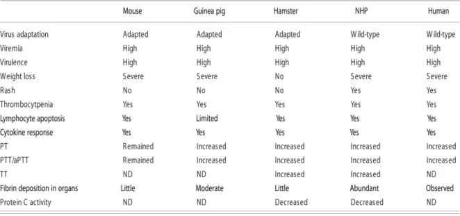
Development of animal models is critical to understanding Ebola virus pathogenesis. Several animal models have been developed in order to study pathophysiology, vaccines and therapeutics of Ebola hemorrhagic fever including mice, guinea pigs, hamsters and NHPs (Connolly, Steele et al. 1999, Bente, Gren et al. 2009, Bradfute, Warfield et al. 2012). However, they do not all show the same pathological features when infected by Ebola virus (Table 3).
-Mouse model: development of mice, in contrast to guinea pigs and
NHP models, has been unsuccessful due to the fact that serial passages of wild-type EBOV in mice are needed for adaptation. The adapted virus resulted in 8 amino acid changes in both coding and non-coding regions of the virus genome compared to the original wild-type virus; nucleotide substitutions leading to amino acid changes were found in VP35, VP24, NP and L polymerase (Ebihara, Takada et al. 2006). Moreover, the route of viral inoculation is determinant to observe or not infection: intraperitoneal inoculation causes lethal infection whereas subcutaneous inoculation does not cause a
Introduction
42
symptomatic illness (Gibb, Bray et al. 2001). This phenomenon is not observed in NHP models, which are susceptible to wild-type infection. Infected mice become acutely ill with symptoms of ruffled fur, reduced activity and loss of weight. However, mice infected with mouse-adapted filovirus show differences when compared to infection in human disease or NHP models in terms of a lack of severe coagulation disorder and fibrin deposition; in addition, the cytokine profile observed during infection is not the same as in humans and NHP. Nevertheless, mouse models can be useful tools for studying basic aspects of replication, pathogenesis, and immune responses and also serve to evaluate candidate vaccines and therapeutic agents (Nakayama and Saijo 2013).
-Guinea pig model: Infection of guinea pigs with wild-type EBOV causes
only a transient febrile illness; Ebola virus needs to be serially passaged in guinea pigs to acquire the ability to cause lethal infection in this model (Ryabchikova, Strelets et al. 1996). The lack of virulence of wild-type virus in guinea pigs is associated with an inability of the virus to replicate/or be released from macrophages and hepatocytes. Guinea pigs inoculated with guinea pig-adapted virus showed similar symptoms to those reported in humans and NHP: fever, anorexia, dehydratation, lymphopenia, fibrin deposition and a decrease in platelet count during infection (Nakayama and Saijo 2013). Guinea pig adapted virus resulted in 8 nucleotide changes that lead to 5 amino acids changes found in NP and L (single mutations) and VP24 (with three substitutions) (Volchkov, Chepurnov et al. 2000). Guinea pig models are useful tools for studying basic aspects of replication, pathogenesis and coagulation disorders. The guinea pig provides a more restringent evaluation of vaccine efficacy and is more predictive of success in NHP model than the mouse model. Unfortunately, significantly fewer tools are available for immunological research using this animal model.
-Syrian Golden hamster model: this model is based on infection of
adult hamsters with mouse-adapted Ebola virus and shows an uniformly lethal outcome. In contrast to other rodent models, hamsters accurately reproduce the severe coagulation abnormalities associated with Ebola virus infection, including fibrin deposition, thrombocytopenia and reduction of Protein C levels.
Introduction
43
Importantly, this model also shows cytokine dysregulation and suppression of IFN type I responses, which are important for EBOV pathogenesis (Ebihara, Zivcec et al. 2013). Differently from guinea pigs, in the hamster model prolonged thrombin time (TT) and severe lymphopenia is observed. However, as with guinea pigs, immunological tools are limited.
-Non-human primate (NHP) model: African green monkeys,
cynomolgus macaques and hamadryas baboons have been employed as models for EBOV infection (Baskerville, Bowen et al. 1978, Fisher-Hoch, Platt et al. 1985, Jaax, Davis et al. 1996). The NHP model has been proven to be valuable in providing new information regarding filoviral pathogenesis; the differences in the disease pathology observed in other models (Table 3) means
that the NHP model remains the most useful and closest to haemorrhagic fever observed in humans despite the practical, and especially ethical, considerations that lead to the restriction of experiments (Geisbert, Young et al. 2003). NHP is the best model for vaccine and drug testing, since it shows both the characteristic clinical and pathological features seen in human EBOV infection. It should be taken into account that genetic differences, even among the same animal species, and the origin of the species may influence disease presentation and progression.
Table 3.- Comparison of pathological features of different animal models of Ebola virus infection. Adapted from (Nakayama and Saijo 2013).


