HAL Id: hal-02969477
https://hal.archives-ouvertes.fr/hal-02969477
Submitted on 19 Oct 2020
HAL is a multi-disciplinary open access
archive for the deposit and dissemination of
sci-entific research documents, whether they are
pub-lished or not. The documents may come from
teaching and research institutions in France or
abroad, or from public or private research centers.
L’archive ouverte pluridisciplinaire HAL, est
destinée au dépôt et à la diffusion de documents
scientifiques de niveau recherche, publiés ou non,
émanant des établissements d’enseignement et de
recherche français ou étrangers, des laboratoires
publics ou privés.
mouse models of Parkinson’s disease
Nora Bengoa-Vergniory, Emilie Faggiani, Paula Ramos-Gonzalez, Ecem
Kirkiz, Natalie Connor-Robson, Liam Brown, Ibrar Siddique, Zizheng Li, Siv
Vingill, Milena Cioroch, et al.
To cite this version:
Nora Bengoa-Vergniory, Emilie Faggiani, Paula Ramos-Gonzalez, Ecem Kirkiz, Natalie
Connor-Robson, et al.. CLR01 protects dopaminergic neurons in vitro and in mouse models of Parkinson’s
disease. Nature Communications, Nature Publishing Group, 2020, 11 (1),
�10.1038/s41467-020-18689-x�. �hal-02969477�
CLR01 protects dopaminergic neurons in vitro and
in mouse models of Parkinson
’s disease
Nora Bengoa-Vergniory
1
, Emilie Faggiani
2,3
, Paula Ramos-Gonzalez
4
, Ecem Kirkiz
1
,
Natalie Connor-Robson
1
, Liam V. Brown
5
, Ibrar Siddique
6
, Zizheng Li
6
, Siv Vingill
1
, Milena Cioroch
1
,
Fabio Cavaliere
4
, Sarah Threlfell
1
, Bradley Roberts
1
, Thomas Schrader
7
, Frank-Gerrit Klärner
7
,
Stephanie Cragg
1
, Benjamin Dehay
2,3
, Gal Bitan
6
, Carlos Matute
4
, Erwan Bezard
2,3
&
Richard Wade-Martins
1
✉
Parkinson’s disease (PD) affects millions of patients worldwide and is characterized by
alpha-synuclein aggregation in dopamine neurons. Molecular tweezers have shown high potential
as anti-aggregation agents targeting positively charged residues of proteins undergoing
amyloidogenic processes. Here we report that the molecular tweezer CLR01 decreased
aggregation and toxicity in induced pluripotent stem cell-derived dopaminergic cultures
treated with PD brain protein extracts. In micro
fluidic devices CLR01 reduced alpha-synuclein
aggregation in cell somas when axonal terminals were exposed to alpha-synuclein oligomers.
We then tested CLR01 in vivo in a humanized alpha-synuclein overexpressing mouse model;
mice treated at 12 months of age when motor defects are mild exhibited an improvement in
motor defects and a decreased oligomeric alpha-synuclein burden. Finally, CLR01 reduced
alpha-synuclein-associated pathology in mice injected with alpha-synuclein aggregates into
the striatum or substantia nigra. Taken together, these results highlight CLR01 as a
disease-modifying therapy for PD and support further clinical investigation.
https://doi.org/10.1038/s41467-020-18689-x
OPEN
1Oxford Parkinson’s Disease Center (OPDC) and Department of Physiology, Anatomy and Genetics, Oxford University, South Parks Road, Oxford OX1 3QX, UK.2Institut des Maladies Neurodégénératives, UMR 5293, Univ. de Bordeaux, F-33000 Bordeaux, France.3CNRS, Institut des Maladies
Neurodégénératives, UMR 5293, F-33000 Bordeaux, France.4Departamento de Neurociencias, Achucarro Basque Center for Neuroscience, and Centro de Investigación Biomédica en Red en Enfermedades Neurodegenerativas (CIBERNED), Universidad del País Vasco (UPV/EHU), S-48940 Leioa, Spain. 5Mathematical Institute, Oxford University, Oxford OX2 6GG, UK.6Department of Neurology, Brain Research Institute and Molecular Biology Institute, University of California, Los Angeles, 635 Charles E Young Drive South, Gordon 451, Los Angeles, CA 90095, USA.7Institute of Organic Chemistry, University of Duisburg-Essen, Essen 45117, Germany. ✉email:richard.wade-martins@dpag.ox.ac.uk
123456789
P
arkinson’s disease (PD) is the second most common
neu-rodegenerative disorder affecting millions of people
worldwide presenting with resting tremor, altered gait,
bradykinesia, rigidity, hyposmia, constipation, and rapid eye
movement sleep behavior disorder
1. Classic neuropathological
examination of PD patient post-mortem tissue shows a loss of
dopaminergic neurons in the substantia nigra (SN) pars compacta
(SNc) of the midbrain, as well as an accumulation of
α-synuclein
(α-syn) in inclusions known as Lewy bodies (LBs) and Lewy
neurites
2. It was previously thought that
fibrils were the only
pathogenic
α-syn species within LBs in the midbrain of PD
patients, but several lines of research have shown that
α-syn
oligomers are also important in PD pathogenesis
3. Molecular
tweezers have shown potential for tackling pathological protein
aggregation
4,5. These compounds inhibit key interactions in the
self-assembly of amyloidogenic protein by binding to positively
charged amino acid residues and disrupting both hydrophobic
and electrostatic interactions. Specifically, the molecular tweezer
CLR01 binds to lysine residues crucial for
α-syn oligomerization
and aggregation
6. CLR01 has been shown to inhibit
α-syn
aggregation in cell lines and zebra
fish
7, and, interestingly, to be
neuroprotective in a triple transgenic Alzheimer’s disease mouse
model
8. There is still a lack of thorough translational studies of
neurodegenerative therapeutic approaches combining
state-of-the-art models, such as induced pluripotent stem cell
(iPSC)-derived dopaminergic cultures, humanized transgenic animal
models, and animals inoculated with PD brain extracts.
iPSC-derived dopaminergic cultures have been shown to
recapitulate midbrain development, and neurons survive in vivo
and integrate into Parkinsonian cellular networks
9. Using
iPSC-derived dopaminergic cultures, we have detected cellular disease
phenotypes that recapitulate
findings from post-mortem PD
cases
10,11. We have also shown previously that expressing the
complete human SNCA locus as a transgene in mice on a Snca
−/−background recapitulates the spatial and temporal expression of
the endogenous human
α-syn
12. Transgenic animals exhibit
age-dependent motor behavioral phenotypes, dopaminergic cell loss,
as well as deficits in dopamine neurotransmission. We have also
previously demonstrated that LB-containing fractions of
Parkin-sonian brains can be isolated, and that injection of this purified
material into the SN of wild-type (WT) mice recapitulates
Par-kinsonian degeneration of dopaminergic neurons after 4 months
with
α-syn aggregation spreading into connected areas of the
brain
13.
In this study, we show that CLR01 reduces both the
aggrega-tion of recombinant
α-syn and dissociates pre-aggregated α-syn.
We show that CLR01 reduces
α-syn aggregation in LB extracts
from PD patients in vitro. iPSC-derived dopaminergic cultures
insulted with LB extracts show shortening and blebbing of their
processes, which is rescued by CLR01 treatment. In microfluidic
chambers, CLR01 also reduces
α-syn aggregation and transport.
Our in vivo results show that CLR01 rescues motor behavior
defects and reduces
α-syn aggregates and accumulation of α-syn
oligomers in the SN of SNCA transgenic animals dosed at
12 months, but not at 6 months, and only partially at 18 months.
These data indicate that the timing of treatment within a
ther-apeutic time window is critical when considering CLR01 for use
in the clinic. Finally, CLR01 reduces pathology in two different
α-syn injection models, namely an LB-injection model and a
pre-formed
fibril injection model, demonstrating broad therapeutic
potential.
Results
CLR01 impairs
α-syn aggregation in vitro. CLR01 binds
spe-cifically with high on–off rate to lysines and, therefore, its
specificity is not to a particular protein but to the process of
abnormal protein self-assembly itself. CLR01 has been shown to
be suitable for a high therapeutic dosing window in rodent
models that tolerate high doses of this compound well
14. These
data highlight the need to investigate the anti-aggregation and
neuroprotective effects of this molecule further, to determine
whether it is promising as a disease-modifying treatment for PD.
To test whether CLR01 was able to have an impact on
recom-binant
α-syn aggregation in vitro, we performed an electron
microscopy (EM) time-course of recombinant
α-syn aggregation.
Recombinant
α-syn was shaken at 250 r.p.m. and 37 °C for 1, 2, 3,
4, 5, and 10 days in the presence or absence of CLR01. In the
absence of CLR01, recombinant
α-syn aggregated, forming both
fibrils and amorphous structures representative of oligomers,
whereas in the presence of CLR01 aggregation was severely
impaired (Supplementary Fig. 1A). We then evaluated whether
CLR01 could reduce the aggregation of recombinant
α-syn
pre-formed oligomers and
fibrils. Recombinant α-syn species were
incubated in the presence or absence of CLR01 for a further
10 days and then evaluated by EM. CLR01 reduced the presence
of oligomers and
fibrils in these preparations (Supplementary
Fig. 1B), which is in agreement with the effects of CLR01 on
recombinant
α-syn species previously shown by Prabhudesai
et al.
7. We then tested whether CLR01 could reduce the presence
of aggregates in LB extracts from PD patients, which were
pro-duced as previously described
13,15,16. LB extracts and their
cor-responding non-LB (noLB) control extracts were subjected to the
α-syn proximity ligation assay (AS-PLA) and EM to evaluate the
presence of oligomers
17,18and the morphology of the aggregates
(Supplementary Fig. 2), respectively. We have previously shown
that we can detect
α-syn oligomers with AS-PLA, which α-syn
oligomers accumulate in the human where LB pathology is barely
detectable
17, and that oligomeric deposition can be modulated
18.
Here we found that
α-syn oligomers could only be detected in
untreated LB extracts but were no longer detected when treated
with CLR01 for 10 days (Supplementary Fig. 2A–C). We could
also only
find structures resembling LBs in LB extracts by EM
(Supplementary Fig. 2D), which were greatly reduced upon
incubation with CLR01. In summary, CLR01 inhibited
α-syn
aggregation and caused dissociation of both oligomeric and
fibrillar α-syn.
α-Syn aggregation and toxicity are reduced by CLR01 in
human dopaminergic cultures. We initially explored the effects
of CLR01 and CLR03 (an inactive analog) in cell lines. We
transfected neuroblastoma SH-SY5Y cells with a plasmid
over-expressing
α-syn and measured the resulting AS-PLA signal
(Supplementary Fig. 3A). Cells expressing the plasmid
accumu-lated high amounts of intracellular
α-syn oligomers and were
treated by the addition of CLR01 to evaluate its ability to
pene-trate cells and to determine an EC50
for the compound. We found
that CLR01 was able to efficiently dissociate oligomers and/or
prevent their aggregation with an EC50
of 85.4 nM
(Supplemen-tary Fig. 3B, C) but that CLR03 did not have a significant effect on
the aggregation status of
α-syn. To further explore the therapeutic
potential of CLR01, we next tested its ability to reduce
α-syn
aggregation and toxicity in iPSC-derived dopaminergic neuron
cultures. Treatment with LB or noLB extracts produced different
surface textures upon microscopic inspection. Whereas we could
observe clear neuronal cell cultures in control and noLB samples,
LB-treated cells exhibited an abnormal texture indicating
deposition of putative proteinaceous material on the cell surface
(Fig.
1
a and Supplementary Fig. 4A). However, when pretreated
with CLR01, this material or texture disappeared. The number of
dead cells labeled by To-pro was unchanged after incubation of
brain extract with CLR01 (Fig.
1
a and Supplementary Fig. 4A, G,
I). As a next step, we evaluated the levels of
α-syn oligomers in
the culture and the number of tyrosine hydroxylase (TH)+ cells
(Fig.
1
b–c and Supplementary Fig. 4B–C, H, J). The number of
TH+ cells was unchanged but we detected an upregulation of
AS-PLA puncta in LB-treated cells, which was reduced by 10 days of
10 µM CLR01 pre-treatment. Finally, as axonal degeneration has
been shown to precede cell death in PD
19, we analyzed processes
labeled by the Tuj1 antibody to
βIII-tubulin (Fig.
1
d–f and
Supplementary Fig. 4D–F). NoLB extracts did not affect process
length or blebbing, whereas LB extract treatment shortened
neuronal processes and increased blebbing, which was reduced by
CLR01 pre-treatment back to control levels. This indicated that
LB extract treatment caused axonal degeneration of iPSC-derived
dopaminergic cultures, whereas CLR01 rescued the neurons
from these toxic effects. Finally, we exposed iPSC-derived
dopa-minergic cultures to LB extracts pretreated with increasing
concentrations of CLR01 or the inactive analog CLR03.
CTRLa
b
c
d
e
g
i
j
h
f
CTRL TO-PRO BF TUJ1 TH AS-PLA LB LB LB + CLR01 LB + CLR01 CTRL LB LB + CLR01 CTRL LB LB + CLR01 CTRL LB LB + CLR01 CTRL LB LB + CLR01 2.0 1.5 1.0Norm AS-PLA puncta
Norm process length
Norm blebbing % Apoptotic cells % Apoptotic cells 0.5 0.0 1.5 1.0 0.5 0.0 1.5 1.0 0.5 0.0 50 40 30 20 10 0 50 40 30 20 10 0 CTRL LB LB + CLR01 CTRL LB LB + CLR01 CTRL Neurons Astrocytes LB LB + CLR01 CTRL LB LB + CLR01
Fig. 1 CLR01 reducesα-syn aggregation and toxicity in iPSC-derived dopaminergic and primary rat cortical cultures. a, c, e Representative brightfield and immunofluorescent images of live and dead cells (labeled by To-pro), dopaminergic cells (TH-positive), AS-PLA (red puncta), and neuronal processes of four independent control cell lines analyzed. Cells were treated with LB extracts, which had been pretreated with CLR01 or PBS as a negative control. LB treatment caused an abnormal deposition of material in treated wells, accompanied by increased AS-PLA signal process degeneration, which were reduced by CLR01 pre-treatment. Scale bars= 100, 25, 25 µm, respectively. b, d, f Quantification of indicated parameters (from c and e) of four independent control cell lines analyzed using a one-way ANOVA (Sidak’s). b F(2, 9) = 5.154, p = 0.0213. d F(2, 9) = 7.309, p = 0.0112, p = 0.0356. f F(2, 9) = 10.67, p = 0.0038,p = 0.0126. g, i Representative images of neuronal (top) and astrocytic (bottom) primary rat cultures stained with DAPI (n = 3 independent cultures and treatments). Scale bars 20µm. h, j Quantification of apoptotic cells (expressed as % from total) analyzed using a one-way ANOVA (Sidak’s, n = 3 independent cultures and treatments). k F(2, 6) = 35.09, p = 0.0005, p = 0.0012. *p < 0.05, **p < 0.01, ***p < 0.001. For all appropriate panels, data are presented as mean values ± SEM. BF brightfield, Norm normalized.
CLR01 showed a dose-dependent reduction of
α-syn oligomers in
dopaminergic cultures whereas CLR03 had no effect
(Supple-mentary Fig. 5A, B).
To test whether CLR01 could reduce
α-syn aggregation and
toxicity in other neural populations, we treated primary rat
neuronal and astrocytic cultures with LB extracts in the presence
or absence of CLR01 (Fig.
1
g–j). LB treatment induced cell death
in the neuronal cultures, but not in astrocytes, in agreement with
our previous
findings
20, and these effects were prevented by
CLR01 treatment. Altogether, these data suggest that CLR01 is
able to reduce
α-syn aggregation and toxicity in human
iPSC-derived dopaminergic cultures and primary rat cultures.
CLR01 prevents
α-syn aggregation in microfluidic chambers.
To investigate how
α-syn is taken up and transported, we used
microfluidic chambers that allow axonal separation of two
dis-tinct cellular populations. As depicted in Fig.
2
a in our
first
experiment, we plated cells on both sides, the home and the insult
chamber, and kept the medium
flow at all times towards the
insult chamber. This
flow system prevents any passive diffusion
occurring from the insult chamber to the home chamber,
there-fore allowing us to determine that, if any insult is detected in the
home chamber, it would have been transported along an axon
(either by anterograde or retrograde transport). AS-PLA showed
that when we insult iPSC-derived dopaminergic cultures with
recombinant
α-syn oligomers in the insult chamber, we can detect
this insult in the home chamber, indicating that oligomers are
actively transported along the axons of these cells (Fig.
2
b). In the
presence of CLR01, the level of AS-PLA signal is greatly reduced
in the home chamber, indicating that CLR01 treatment protects
neurons from an oligomeric insult. To test whether there was a
specific involvement of retrograde transport in this process, we
examined the same paradigm without seeding cells in the insult
chamber (Fig.
2
c). If an insult is detected in the home chamber in
the absence of cell bodies in the insult chamber under the same
flow conditions, the insult must have been transported by axonal
retrograde transport. Upon recombinant
α-syn oligomer insult at
the terminals, we detected an increase in AS-PLA in the
dopa-minergic neuron cell bodies in our cultures (Fig.
2
d), which
indicated that oligomers are moved by retrograde transport.
Application of CLR01 reduced the levels of AS-PLA seen after
α-syn oligomer insult.
We therefore decided to study whether
α-syn interacted with
retrograde and anterograde transport proteins (dynein and
kinesin, respectively) through PLA, and whether these
interac-tions could be inhibited by CLR01.
α-syn-dynein-PLA revealed
that dynein does interact with
α-syn, and that this interaction can
be increased by insulting dopaminergic cultures with
α-syn
monomers, which is then reduced by CLR01 treatment
(Supplementary Fig. 6A, B). When we examined the interactions
between
α-syn and kinesin through α-syn-kinesin-PLA we could
detect a clear upregulation of the signal upon
α-syn monomer
insult, which was depleted with CLR01 treatment. This indicates
that CLR01 is able to reduce this interaction and therefore
influence α-syn transport (Supplementary Fig. 6C, D).
In summary, these data show that recombinant
α-syn
oligomers are actively transported along axons. Importantly,
CLR01 reduces
α-syn oligomeric pathology following active
transport efficiently in iPSC-derived dopaminergic neurons and it
is specifically able to reduce interaction of α-syn with axonal
transport proteins.
CLR01 reduces
α-syn aggregation and its detrimental effects
in vivo. Our laboratory has previously described a human
α-syn-overexpressing mouse model (SNCA-OVX), which showed
accurate spatial and temporal expression of human
α-syn in a
mouse Snca-null background. Overexpression of
α-syn in this
humanized model led to age-dependent neuronal loss, impaired
dopamine release, altered vesicle clustering, and motor defects
12.
Here we conducted further analysis and found that, although
SNCA-OVX mice do not show pronounced whole-brain gliosis,
the SN has increased levels of GFAP (glial
fibrillary acidic
pro-tein, astrocytes) and a shift in morphology for microglia
(Iba1-positive cells) (Fig.
3
a–d and Supplementary Fig. 7). Microglial
cells in the SN are preferentially ramified (homeostatic) in WT
animals, yet microglia of SNCA-OVX animals adopt a more
ameboid morphology, which is suggestive of an activated state.
We have reported previously that these animals do not exhibit
LB-like aggregates; however, here, by using our recently
devel-oped AS-PLA assay
17,18, we detected
α-syn oligomers (Fig.
3
e–f).
We detected a significant age-dependent accumulation of α-syn
oligomers, which likely is linked to the motor defects detected.
We therefore decided to use the SNCA-OVX model to test
whether CLR01 treatment could affect these phenotypes in vivo.
Mathematical in silico models have been previously used to
predict and model pharmacological and biological processes
21–24.
We
first administered radiolabeled CLR01 systemically to
12-month-old animals to measure the blood–brain penetration of the
compound to inform an in silico model that would allow us to
determine whether a monthly dose of 40 µg/kg/day, which was
previously used
8, would provide favorable pharmacokinetic
profile in the brain. The proportion of CLR01 in the brain was
determined as the percentage of radioactivity in the perfused
brain, relative to that of the blood, at three different doses and
three time points in 12-month-old animals. Our results indicate
that 10–35% of the drug concentration available in the blood is
present in the brain, and that this is a time- and dose-dependent
process (Supplementary Fig. 8A). Based on these data and
previously published blood kinetics
14, we
first predicted the
extra-and intracellular accumulation of the compound in the brain
(Supplementary Fig. 8B), then simulated a 2-month treatment
spread across 16 subcutaneous injections at 40 µg/kg/day
(Supplementary Fig. 8C), to obtain pharmacokinetic parameters
(Supplementary Table 1). We found that the intracellular time
averaged area under the curve was 240 ng/ml, with a predicted
maximum concentration of 510 ng/ml, which resulted in cells
being 93% of the experimental time above the EC50
(85.4 nM).
The model predicted a rapid intracellular accumulation of CLR01,
which lead to lower concentrations and time over EC50
in the
extracellular space, which were still above EC50
levels and
generally high (extracellular time averaged area under the curve
was 100 ng/ml, maximum concentration was 230 ng/ml, and time
above the EC50
was 81%). This simulation showed that CLR01
would be available at therapeutically relevant concentrations in
the extracellular space and the intracellular compartment of
the brain.
We then treated transgenic animals with CLR01 using three
different paradigms (Supplementary Fig. 9) as follows:
first,
4-month-old mice were injected for 2 months subcutaneously, then
12-month-old mice were subcutaneously injected for 2 months,
and,
finally, 18-month-old animals were treated with a
sub-cutaneous implant of an osmotic mini-pump, to minimize
manipulation and stress of aged animals. As an end-point for
our 12- and 18-month cohorts, we assessed the animals for both
motor behavior and neuropathology, whereas the 6-month-old
cohort was evaluated for dopamine release and microglial
morphology. The 6-month cohort would be too young to show
a neuronal-loss-associated motor phenotype, considering our
previous
findings
12.
We treated 6-month-old animals to assess whether treatment
at an early timepoint was able to change early phenotypes, which
may be linked to
α-syn overexpression rather than α-syn
aggregation. At 6 months, dopamine release evoked by discrete
electrical stimuli in the striatum in acute slices as measured by
fast-scan cyclic voltammetry (FCV) was unchanged
(Supplemen-tary Fig. 10A, B). Likewise, we could not detect a difference in
α-syn oligomers in dopaminergic cells or microglial morphology
(Supplementary Fig. 10C–F), precluding testing treatment effect
in these young mice.
These results prompted us to treat older mice to assess whether
they would show the expected deficits and allow measurement of
neuroprotection, leading to a rescue of motor function. The
12-month-old SNCA-OVX mice showed motor deficits in the latency
to fall on a rotarod and speed in catwalk analysis, which was
rescued by a twice-weekly subcutaneous injection of CLR01
(Fig.
4
a, c), which was accompanied by restorative trends for
several other motor phenotypes (Fig.
4
b, d and Supplementary
Fig. 11). Stool analysis, locomotor activity (LMA), and muscle
strength and coordination were unaffected by the treatment
(Supplementary Fig. 11). To test whether these effects were
specific to SNCA-OVX, we dosed a further small cohort of Wt
Insult chamber Insult chamber Home chamber Home chamber Neurons NeuronsNeurons Neurons Neurons
Neurons Neurons Neurons Oligomer CLR01 CLR01 Flow Oligomer
a
b
c
d
Oligomer Oligomer Oligomer CTRL CTRL CTRL CTRL CLR01 CTRL CTRL CLR01 Oligo Oligo CTRL AS-PLA TH TH AS-PLA CTRL CLR01 10 UM CTRL CTRL CLR01 10 UM Oligomer Neurons Home chamber Home chamber Insult chamber Flow 6 4 2 0 1.0 0.8 Norm puntca/field Norm puntca/field Norm puntca/field 0.6 0.4 250 200 150 100 50 0 0.2 0.0 Home chamber Insult chamber Insult chamber Home chamberFig. 2 CLR01 preventsα-syn aggregation in microfluidic chambers. a, c Schematic representation of the experimental design for the microfluidic-chamber experiments.b, d Representative immunofluorescence images of dopaminergic neurons grown in the microfluidic devices insulted with oligomers and/or treated with CLR01 for 5 days (25µg/ml and/or 10 µM, respectively). Images were taken at least one field of view away from the microgrooves to avoid artifacts due to contact with the silicone. Scale bars= 25 µm. Quantification of normalized puncta per field of three independent control cell lines analyzed using a one-way ANOVA (Tukey). Oligo: oligomer.b F(2, 6)= 24.31, p = 0.0009 and F(2, 6) = 39.09, p = 0.0005, p = 0.0006. d F(2, 6) = 71.45. *p < 0.05, **p < 0.01, ***p < 0.001. For all appropriate panels, data are presented as mean values ± SEM.
and SNCA-OVX animals with the same dose and paradigm as
above and subjected them to rotarod and catwalk analysis. Wt
animals showed no changes in their behavior after CLR01
treatment as measured by both rotarod and catwalk
(Supple-mentary Fig. 12), whereas a trend for improvement (which did
not reach significance probably due to smaller sample size) was
seen in the SNCA-OVX animals, suggesting that CLR01 may
require the presence of aggregates to elicit its effects. We also
observed a significant reduction in α-syn oligomers in
dopami-nergic SNc cells, and a reduction in GFAP staining (Fig.
4
e–h).
Interestingly, the morphology of microglia shifted further towards
ameboid rather than ramified (Fig.
4
i, j), which could be due to a
shift towards a phagocytic phenotype to clear the debris generated
by CLR01 treatment. Overall, we found that twice-weekly CLR01
injection is able to restore motor behavior and downregulate
oligomeric deposition as well as astrogliosis.
Finally, we decided to test the effects of CLR01 treatment in
18-month-old animals. We could not detect any changes in the
behavior or stool analysis of 18-month-old animals carrying a
surgically implanted mini-pump releasing CLR01 at a constant
rate of 0.11 µl/h (Supplementary Fig. 13). However, we did detect
a significant reduction in the number of α-syn oligomers per
dopaminergic neuron in the SNc as shown in Fig.
5
a, c. This
prompted us to investigate whether the treatment had impacted
the astrocytic and microglial populations of the SN. We could not
detect any changes in the GFAP-positive area or the morphology
of Iba1-positive cells in the SN of CLR01-treated animals
(Supplementary Fig. 14A–C). These results were unsurprising,
as by 18 months SNCA-OVX mice have lost ~30% of their SNc
dopaminergic neurons and, therefore, a rescue in motor behavior
or astrogliosis would be difficult to achieve after neurons are lost.
However, CLR01 was able to reduce the
α-syn oligomer burden in
SN astrocytes and microglia (Fig.
5
b, e), and to significantly
elevate the levels of PGC1α and Lamp2A in the midbrain of these
animals (Fig.
5
f, g), but not those of Lamp1 (Supplementary
Fig. 14D). PGC1α is a neuroprotective mitochondrial biogenesis
3 months WTa
c
e
f
d
b
4 3 2Norm GFAP stained area
Norm AS-PLA per TH+
cell
Norm ramified morphology
Norm ameboid morphology 1 0 100 80 60 40 20 0 100 80 60 40 20 0 10 8 6 4 2 0 3 Months 6 9 12 3 Months 6 9 12 3 6 Months 9 12 OVX WT WT OVX WT OVX WT OVX OVX WT OVX
6 months 9 months 12 months
3 months 6 months 9 months 12 months
3 months
AS-PLA TH GFAP TH
IBA1
6 months 9 months 12 months
Fig. 3 Glial activation andα-syn aggregation in SNCA-OVX mice. a, c, e Representative images of immunofluorescence of GFAP (astrocytes), IBA1 (microglia), and AS-PLA in dopaminergic neurons, respectively. Scale bars= 200, 25, and 25 µm, respectively. b, d, f Quantification of normalized stained area, morphology (expressed as a % from total), and puncta per TH+ cell analyzed using a two-way ANOVA (Dunnett’s for age and Sidak’s for genotype, for age all values compared to 3 months),n = 3–4 animals per group. b F(3, 20) = 1.211, p = 0.0177 for age and F(1, 20) = 0.1572, p = 0.0106 for genotype. d F(1, 22)= 2.216, p = 0.0287 for genotype and F(3, 22) = 1.764, p = 0.0209, p = 0.0104, p = 0.0192 for age. f F(3, 21) = 4.528 for age, p = 0.0058, p = 0.0016,p = 0.0024 and F(1, 21) = 33.41, p = 0.0043, p = 0.0015, p = 0.0061 for genotype. *p < 0.05. For all appropriate panels, data are presented as mean values ± SEM. OVX:SNCA-OVX.
master regulator that plays a role in mitophagy and reactive
oxygen species (ROS) reduction, often perturbed in PD
25;
Lamp2a is involved in chaperone-mediated autophagy (CMA)
and has been shown to be reduced in post-mortem PD brains
26. It
is interesting to hypothesize that CLR01 might be able to
unburden TH+ cells from oligomeric α-syn, thereby indirectly
enabling mitochondrial turnover and ROS reduction, and
restoring CMA function, which are decreased with aging and
disease
25,27,28. Finally, we confirmed the impact of CLR01
treatment on
α-syn aggregates by evaluating midbrain levels of
Triton X-100 and SDS-soluble and -insoluble fractions of
α-syn.
SNCA-OVX showed no signs of SDS-insoluble aggregates (which
agrees with the detection of oligomeric pathology), with most of
their
α-syn content detectable in the Triton X-100 fraction
(Fig.
5
h, i and Supplementary Fig. 14E). However, a small fraction
of
α-syn was present in the SDS-soluble fraction in the midbrains
of SNCA-OVX animals, which was reduced by CLR01 treatment.
In conclusion, CLR01 treatment downregulated SN
α-syn
oligomers and upregulated neuroprotective markers at 18 months
of age in SNCA-OVX mice.
400 Rotarod
a
e
g
i
j
h
f
b
c
d
WT WT THAS-PLA GFAPTH IBA1 OVX OVX+CLR01 WT OVX OVX+CLR01 WT OVX OVX+CLR01 OVX OVX + CLR01 WT OVX OVX + CLR01 WT OVX OVX + CLR01 WT OVX OVX + CLR01 WT OVX OVX + CLR01 WT OVX OVX + CLR01 WT % Ameboid % Ramified OVX OVX + CLR01 Forefootswing speed Speed Cadence
AS-PLA
Astrocytes
65 30 16
2.5 2.0
Relative puncta per TH+ neuron
Relative GFAP stained area
% Morphology 1.5 1.0 0.5 0.0 4 3 2 1 0 150 100 50 0 14 12 10 8 25 20 15 10 60 55 50 45 40 35 300
Time to fall (s) Speed (cm/s) Speed (cm/s)
Steps/s
200 100 0
Fig. 4 CLR01 restores motor behavior and reduces early pathology in vivo at 12 months of age. a–d Behavior analysis of rotarod (a) and catwalk gait analysis (b–d) after 2 months of subcutaneous administration of 40 µg/kg/day drug or PBS analyzed using a one-way ANOVA (Sidak), n = 11–14 animals per group.e, f Representative images and quantification of AS-PLA puncta per TH+ cell. Scale bars = 25 µm. g–j Representative images and quantification of GFAP-stained area perfield and microglial morphology (expressed as a % from total). Scale bars = 100 and 50 µm, respectively. a–j were analyzed using one-way ANOVA (Holm–Sidak), e–j n = 8/8. CLR: CLR01 *p < 0.05, **p < 0.01. a F(2, 35) = 4.138, p = 0.0376, p = 0.0205. b F(2, 37) = 4.000, p = 0.0058.c F(2, 38)= 4.884, p = 0.0078, p = 0.0451. d F(2, 38) = 3.982, p = 0.0151. f F(2, 19) = 6.057, p = 0.0072, p = 0.0338. h F(2, 21) = 8.797, p = 0.0091,p = 0.0012. j F(2, 23) = 4.750, p = 0.0106. For all appropriate panels, data are presented as mean values ± SEM.
Taken together, these results highlight that CLR01 effects are
age-dependent and likely linked to
α-syn oligomerization and
aggregation, which increase with age, rather than to
α-syn
overexpression itself.
CLR01 reduces
α-syn aggregation after intracranial aggregate
injection. We tested whether CLR01 could reduce protein
pathology using two different intracranial injection models
(Supplementary Fig. 9), representing more late-stage pathology
than that of SNCA-OVX mice, which are a model for early
oli-gomeric accumulation of
α-syn. First, we used a recombinant
mouse
α-syn pre-formed fibril (mPFF) injection model, which
was previously described by Luk et al.
29. We injected
3-month-old C57Bl/6 WT mice with mPFF preparations in the dorsal
striatum and then followed pathology in the SNc of these mice.
Upon inspection of the SNc, lesions labeled by phospho-α-syn
were found to be more prevalent in the SNc than in the ventral
tegmental area, as was expected following dorsal striatum
injec-tion (Supplementary Fig. 15A–D). Subcutaneous injecinjec-tions of
CLR01 twice-weekly reduced the number of lesions in the SNc
compared to animals treated for a month with
phosphate-buffered saline (PBS) (Supplementary Fig. 15A, B).
We next tested whether CLR01 could reduce pathology
induced by the injection of LB extracts as described by Recasens
et al.
13. LB extracts were injected into the SN of 4-month-old
C57Bl/6 WT mice, which were aged for a further 3 months before
CLR01 treatment. As these extracts are toxic and can lead to
neurodegeneration, we decided to implant an osmotic
mini-pump to deliver either PBS or CLR01 for the last month to
reduce manipulation and stress for the mice. Stereology and
CTRL THAS-PLA 15 1.0 0.8 0.6 0.4 0.4 0.3 0.2 0.1 0.0 0.2 0.0 5 6 4 2 0 4 3 2 1 0 TH+ Neuronsa
c
f
h
i
g
d
e
b
Astrocytes MicrogliaAv puncta per cell
PGC1a levels
A-SYN levels A-SYN levels
Lamp2a levels
10
5
0
15
Av puncta per cell
10
5
0
15
Av puncta per cell
10 5 0 CTRL PGC1αα LAMP2A ACT PBS CLR01 Triron-X100 α-SYN SDS α-SYN PBS CLR01 PBS CLR01 PBS CLR01 ACT 120 kDa 720 kDa 480 kDa 242 kDa 720 kDa 480 kDa 242 kDa 40 kDa 100 kDa 40 kDa CLR01 CTRL CLR01 CTRL CLR01 CTRL CLR01 CTRL CLR01 CTRL CLR01 CTRL CLR01
GFAPAS-PLA IBA
CLR01 CTRL CLR01
Fig. 5 CLR01 reducesα-syn aggregation in vivo at 18 months of age. a–e Representative images and quantification of AS-PLA puncta per TH+, GFAP+, and Iba1+ cells after osmotic mini-pump implantation of 40 µg/kg/days CLR01 or PBS, analyzed using a two-tailed t-test, n = 3/4 independent animals. Scale bars= 50 µm. c p = 0.0117, d p = 0.0319, e p = 0.0179. f, g Western blottings of PGC1α and Lamp2a, analyzed using a one-tailed Mann–Whitney U-test, n = 3–4 independent animals. f p = 0.0286, g p = 0.0286. h, i Native western blottings of Triton X-100 and SDS-soluble α-syn, analyzed using a one-tailed Mann–Whitney U-test, n = 3–4 independent animals. i p = 0.0286. *p < 0.05. AV average, CLR CLR01. For all appropriate panels, data are presented as mean values ± SEM.
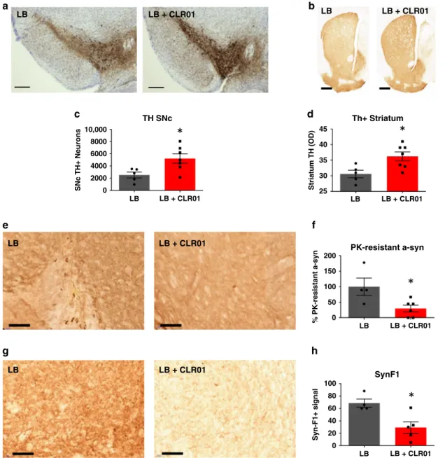
densitometry analysis showed that CLR01 treatment was able to
rescue TH+ immunoreactivity in the SN and striatum,
respectively (Fig.
6
a–d). Nissl+ cell counts in the SN and DAT
in the striatum were only partially restored (Supplementary
Fig. 15E, F). Further analysis of the midbrains of these mice
showed that there was a reduction in
α-syn PK-resistant puncta
and a reduction of staining by the Syn-F1 antibody, which are
indicators of a reduction of aggregated
α-syn (Fig.
6
e, h).
Discussion
Neuroprotective therapies targeting
α-syn in PD have a high
potential for translation to patients in the clinic. Working across a
range of human in vitro and rodent in vivo
α-syn-based models of
PD, we have shown that CLR01 reduces
α-syn aggregation and
alleviates disease-related phenotypes. CLR01 dismantles synthetic
oligomeric and
fibrillar, and patient LB-derived α-syn, alleviating
their toxic effects in vitro, reducing neurodegeneration-associated
α-syn accumulation in vivo, and correcting behavioral
pheno-types in a transgenic mouse model of PD.
Applying patient LB extracts treated with CLR01 to the highly
physiological human iPSC-derived dopaminergic cultures in vitro
gives a fully humanized model of disease. The LB extracts show a
higher
α-syn oligomeric content than noLB extracts. CLR01
reduced the
α-syn oligomeric content and with it the toxic effect
of the patient LB extracts. However, LB extracts likely contain
many other proteins
30–32, which could be sensitive or resistant to
CLR01 treatment. When the extracts were applied to
iPSC-derived dopaminergic cultures, we observed that CLR01 potently
LBa
c
e
g
h
f
d
b
LB + CLR01 LB LB + CLR01 LB LB + CLR01 LB LB + CLR01 LB LB + CLR01 LB LB + CLR01 TH SNc Th+ Striatum PK-resistant a-syn SynF1 % PK-resistant a-syn Syn-F1+ signal LB LB + CLR01 10,000 45 40 35 30 25 200 150 100 100 80 60 40 20 0 50 0 8000 6000 4000SNc TH+ Neurons Striatum TH (OD) 2000
0
LB LB + CLR01
Fig. 6 CLR01 protects dopaminergic neurons from LB-induced cell death through reduction ofα-syn aggregation in mice. a, b Representative photomicrographs of TH-immunostained SNc and striatum (respectively) in LB-inoculated mice after osmotic mini-pump implantation for CLR01 (or PBS) delivery (40µg/kg/h). c Stereological cell counts of SNc TH-immunoreactive neurons (a) in LB-inoculated mice, at 3 months post-LB inoculation. p = 0.0177.d Optical densitometry of striatal TH immunoreactivity in LB-inoculated mice (b), at 3 months post-LB inoculation.p = 0.0354. e, g Representative photomicrographs ofα-syn and PK-resistant α-syn immunostained SN from LB-inoculated mice, and corresponding quantification. f, h Representative images of Syn-F1 immunostaining in the SN in LB-inoculated mice and corresponding quantification of the intensity levels of Syn-F1 staining. f p = 0.0333, hp = 0.0556. a–h n = 4–5 for LB-inoculated mice, n = 5–7 for LB + CLR01-independently treated animals, analyzed with a two-tailed Mann–Whitney U-test. *p < 0.05. Scale bars = 0.1 mm (SN) and 1 mm (Str). For all appropriate panels, data are presented as mean values ± SEM.
inhibits the toxic effects of LB extracts in both iPSC-derived and
primary neuron culture systems, as previously shown in the case
of Aβ-induced LTP defects in primary mouse hippocampal and
cortical neurons, and in an AD mouse model
8.
Microfluidic chambers facilitate the isolation of axonal
term-inals from cell bodies in culture modelling pathology along the
neuron from synapse to soma. Several studies have shown that
degeneration may start in the striatal axons before affecting the
cell soma of dopaminergic neurons
33. In support of this theory,
neuroimaging studies have shown that human PD presents early
with a larger loss of transporters and other DA axon markers in
the striatum than in the SNc
34,35. As a result several groups have
studied this early degeneration in
α-syn overexpression mouse
models and shown early striatal synaptic and dopamine release
defects prior to neurodegeneration
12,36. It is therefore important
to investigate in vitro and in vivo whether the toxic effects of
α-syn on axon terminals can be alleviated with CLR01
adminis-tration. Using microfluidic chambers in vitro, we observed that
CLR01 reduced the accumulation of
α-syn oligomers in the cell
soma after treatment of axonal terminals, although it is possible
that this was in part due to an inhibition of retrograde transport
of
α-syn, or to the minimal flow of CLR01-containing medium to
the insult chamber. In parallel studies in vivo, CLR01
adminis-tration reduced lesions in dopaminergic cell somas in the SNc
after injection of mPFFs to the dorsal striatum, confirming a
protective effect of CLR01 against
α-syn pathology in both axons
and cell bodies.
The age-dependent microglial and astrocytic activation in the
SNCA-OVX mouse model overexpressing
α-syn supports a role for
inflammation in PD and other neurodegenerative disorders
37–39.
The observation that microglial activation occurs in a
time-dependent manner almost simultaneously to oligomer deposition,
and that astrocyte activation is delayed in comparison, is consistent
with studies showing that astrocytic activation is induced by
microglial activation and is mediated at least in part by Il1α, TNF,
and C1q
40. In SNCA-OVX mice at 6 months, the overexpression of
α-syn causes only mild oligomer accumulation and disease-related
motor phenotypes were not observed. The window for treatment
with a molecular tweezer is optimal at twelve months, when early
disease processes are underway and neuronal dysfunction has
begun, but before extensive and widespread dopaminergic neuron
degeneration. By 18 months, when dopaminergic neurons have
already been lost and neuroinflammation has been manifest for the
previous 6–12 months, inhibition of α-syn self-assembly by CLR01
is unable to reverse cellular and behavioral phenotypes. This
highlights the importance of studying the disease in a late-onset
age-dependent model and of treating the disease before it has
progressed beyond a putative point-of-no-return. Our work also
demonstrates that continuous administration, as used previously in
the rapidly-progressing Thy-1
α-syn overexpression model of
PD
41, is unnecessary, further supporting the clinical development
of CLR01 in PD patients. We have also found that CLR01 is able to
elicit its function in the nanomolar range, which makes it a potent
compound that can be effective even at low doses, such as the ones
we use in vivo. These low doses showed promising predicted
pharmacokinetic properties in silico, which support previous
findings
8,14and our in vivo results.
Finally, to extend the application of CLR01 beyond transgenic
strains and to models of protein aggregate burden, we
demon-strated a positive therapeutic effect of CLR01 against
α-syn-induced pathology after intracranial injection of LB extract into
the SN. CLR01 treatment exhibited a neuroprotective effect
demonstrated by reduced
α-syn pathology and a rescue of
dopaminergic neurons in the SNc and increased striatal TH+
innervation in the striatum. The positive effect of CLR01 in vivo
in both late-onset, age-dependent models, and in rapid models of
accelerated protein pathology demonstrates the robust
applica-tion of the molecule across diverse disease models.
We have investigated the broad utility of CLR01 against
dif-ferent forms of aggregated
α-syn which complements previous
work on other amyloid assembly inhibitors. For example,
epigallocatechin-3-gallate (EGCG) has been shown to bind
α-syn
inhibiting its aggregation
42,43. It has also been shown to be
pro-tective in vitro
44and has shown promise by restoring
MPTP-induced neuronal loss
45and reducing peripheral immune
response after MPTP treatment
46in mouse models. The relatively
small size of CLR01 allows it to be internalized into cells as has
been shown in our study and by others
47, providing an advantage
over antibody based therapies, which generally target external
epitopes, and are therefore unable to fully tackle toxic
intracel-lular
α-syn species. Taken together, these data in diverse in vitro
and in vivo preclinical models suggest that molecular tweezers are
highly promising candidates for the treatment of PD.
Methods
Study design. Sample size was determined according to the experimental para-digm. Sixteen animals were bread to account for loss of animals during ageing and avoid n < 10 at the end of the study where behavioral differences of ageing cohorts were investigated. Any animals showing any welfare issues were taken off the study, otherwise animals remained in the sample. For neuropathological examination four tofive animals were bred per condition in order to avoid n < 3. For iPSC culture, three to four independent control lines were used in order to ensure n≥ 3. For biochemical analysis of recombinant or biological material, three independent experiments were performed on three independent samples.
Treatments were administered blindly by coding of the different groups into numbers. Samples were blinded by taping over identifiers, to avoid bias when sampling. For EM imaging, four independentfields were imaged and a representative image was shown. For cell imaging, three to nine independentfields of view were analyzed per well and one to two wells were analyzed per condition and control line. For tissue analysis, two to four independentfields (i.e., at least 100 µm apart to avoid re-sampling) of anterior SN were analyzed, and for puncta counts associated with TH+ cells, 25 neurons were sampled across the SNc. For blood–brain barrier experiments, one radioactivity value was obtained per brain and animal. For behavior and stereology, sampling is described in their particular methods section.
Electron microscopy. Ten microliters of 0.2 mg/ml sample were applied to glow-discharged, carbon-coated EM grids (TAAB). Samples were incubated for 2 min on the grids at room temperature (RT), followed by 2% uranyl acetate staining for 10 s. Samples were rinsed with water, dried, and stored at RT for analysis. Images were acquired with a FEI Tecnai 12 transmission EM (120 kV) with a Gatan US1000 camera.
α-Syn aggregation. α-Syn (2 mg/ml; Peptide) were shaken at 37 °C and 250 r.p.m. for as many days as described in each case, to induce aggregation ofα-syn. To produce oligomers,α-syn was shaken at 1 mg/ml and for 3 days (Supplementary Fig. 16A).
PLA, immunofluorescence, and immunohistochemistry. AS-PLA experiments were performed using the Duolink kits (Sigma) according to the manufacturer’s instructions17,18. Anα-syn antibody (mouse monoclonal anti-α-syn4D6, ab1903, Abcam, 1:2000), was used to prepare conjugates using Duolink Probemaker Plus and Minus kits. Forα-syn-dynein and α-syn-kinesin interaction experiments, we used the sameα-syn antibody with monoclonal antibodies targeting dynein and kinesin (mouse monoclonal anti-dynein MAB1618 and anti-kinesin MAB1614 from Millipore, respectively, both 1:100). Forfluorescent AS-PLA of brain extracts and cell samples, samples werefixed in 4% paraformaldehyde (PFA). All samples were incubated with Duolink block solution at 37 °C for 1 h and then with α-syn conjugates diluted in Duolink PLA diluent overnight (ON) at 4 °C. Samples were washed with tris buffered saline (TBS) containing 0.05% Tween-20 (TBS-T) and incubated with Duolink ligation reagents for 1 h at 37 °C, washed four times with TBS-T, and then incubated with Duolink amplification reagents for 2.5 h at 37 °C. Samples were washed and then mounted in FluorSave (Calbiochem).
Paraffin-embedded tissue was dewaxed by 2 min consecutive incubations in Xylene, Histoclear, 100% ethanol, 95% ethanol, 70% ethanol, and H2O. After rehydration, samples were incubated in 10% H2O2in PBS, to reduce background and heated in a microwave in citrate buffer pH 6.0 (Abcam) for antigen retrieval. After antigen retrieval, samples were subjected to immunofluorescence or AS-PLA as necessary.
For AS-PLA co-immunofluorescence, immunofluorescence was performed after antigen retrieval and before PLA block. This process consisted of 1 h RT incubation in 10% Serum TBS-T block, 1 h RT incubation in primary antibody (GFAP Z0334 Sigma 1:1000, Iba1 019-19741 Wako 1:1000, TH ab152 Millipore 1:1000, TH ab76442 Abcam, 1:100, Tuj1 ab107216 Abcam, 1:100), after which slides were washed with TBS-T. Slides then were incubated for 1 h at RT with secondary antibodies (Alexa488, Alexa594, or Alexa680, Life Technologies), after which they were washed again with TBS-T. If AS-PLA analysis was required, samples went on to AS-PLA block, if not samples were mounted with FluorSave.
For recombinant protein analysis, 10 µl of 2 mg/ml samples were spotted on poly-L-lysine coated cover slips, left at RT for 30 min, then PFA-fixed for 10 min, andfinally treated as above for fluorescent AS-PLA analysis.
Histopathological analysis of cryoprotected tissue. Extent of lesion: To assess the integrity of the nigrostriatal pathway, TH immunohistochemistry was per-formed on 50 µm free-floating SNc and striatal sections. Briefly, sections from three representative levels of the striatum (anterior, medial and posterior) and serial sections (1/6) corresponding to the whole SNc were incubated with a rabbit monoclonal antibody raised against TH (Abcam, EP1532Y, 1:5000) for one night at RT and developed with an anti-rabbit peroxidase EnVisionTM system (DAKO, K400311) followed by 3,3′-diaminobenzidine (DAB) visualization. Free-floating SNc sections were mounted on gelatinized slides, counterstained with 0.1% cresyl violet solution, dehydrated, and cover-slipped, whereas striatal sections were mounted on gelatinized slides and cover-slipped. The extent of the lesion in the striatum was quantified by optical density (OD). Sections were scanned in an Epson expression 10000XL high-resolution scanner and images were used in ImageJ open source software to compare the grey level in the putamen. TH-positive SNc cells were counted by stereology, blind with regard to the experimental con-dition, using a Leica DM6000B motorized microscope coupled with the Mercator software (ExploraNova).
α-Syn pathology: Synucleinopathy was assessed using a primary antibody that recognizes both mouse and humanα-syn (mouse anti-α-syn monoclonal antibody, BD Transduction Laboratories, clone 42, #610787; BD Biosciences, SYN1, 1:1000) and aggregatedα-syn antibody (mouse anti-α-syn monoclonal antibody, BioLegend, Syn-F1, 1:2000). In addition, SN sections werefirst incubated with or without proteinase K (PK) at 1μg/mL (Sigma-Aldrich) in PBS for 10 min at room temperature13,48. PK-treated and nontreated adjacent sections then were incubated ON at room temperature with SYN1 antibody, 1:1000. The following day, they were incubated with a secondary antibody conjugated to a peroxidase EnVision system (DAKO) DAB incubation. Sections then were mounted on gelatinized slides, dehydrated, counterstained if necessary and cover-slipped until further analysis. Images were used in ImageJ to count PK-resistantα-syn-positive dots in SN (SYN1) or to measure OD in SN (Syn-F1). For striatal injections, tissue was paraffin embedded and processed as specified above. The whole SN was sectioned at 5 µm and every tenth section was stained for p-α-syn (EP1536Y, Abcam) and TH (sc-25269, Santa Cruz). The sections with most p-α-syn+/TH+lesions were blindly selected, and the resulting number of lesions was averaged for independent animals.
Imaging. Samples were imaged with the Opera Phenix confocal microscope and Harmony software (Perkin-Elmer) or the EVOSflAUTO (Thermo). For standard cell culture, images were automatically captured with the Opera Phenix and ana-lyzed with ImageJ or Cell Profiler for the detection of puncta (AS-PLA signal) or cell nucleus/body (TO-PRO3, DAPI, TH counts), respectively. To analyze process morphology, average length and number of puncta were analyzed. For microfluidic and tissue samples, images were taken blinded to treatment on the EVOSflAUTO. We used automatic measurement with ImageJ for puncta counts (AS-PLA signal) or stained areas (GFAP signal). To count PLA puncta inside cells of interest, puncta were counted blindly by eye in 25 cells per animal. In order to determine microglial morphology, all microglial cells in the SN were clasified by a blinded operator by eye as ramified or ameboid.
iPSC-derived dopaminergic cultures. iPSCs were cultured according to the methods described in Supplementary Information and previous reports9,11,49.
NoLB and LB brain extracts were purified as described below and treated for 10 days ±10μM CLR01 (Supplementary Fig. 16B) with continuous shaking at 37 °C and 250 rpm. Cells were treated with 600 pg/ml noLB/LB extract ±10μM CLR01 as indicated on DIV36 and DIV38, and then harvested and measured on DIV41. To culture cells in microfluidic chambers, dual axonal separation microfluidic devices were purchased from Millipore and adapted for dopaminergic cell survival. Briefly, the cell channel was carefully opened with a scalpel without disturbing the microgrooves for axonal separation. The device was tested ON by leaving the chambersfilled with unequal volumes; if the volumes remained unequal after ON incubation the chambers were used for experiments. For microfluidics assays, CLR01 was used at 10μM and oligomers were applied at 25 μg/ml.
For live-dead staining, TO-PRO-3 was added to culture medium 1:1000 (Thermo).
Neuroblastoma cell cultures. Neuroblastoma SH-SY5Y cell cultures were cul-tured in Dulbecco’s modified Eagle’s medium supplemented with 10% fetal bovine serum (FBS), 1% penicillin/streptomycin, and 1%L-glutamine (Life Technologies) at 37 °C and 5% CO2. On DIV0 cells were plated at 10,000 cells/well on half-area 96-well plates (Greiner). On DIV1 cells were transfected with a pcDNA3.1 vector containing humanα-syn (Addgene) with Lipofectamine 2000 and Plus reagent (Life Technologies) in OptiMem (Life Technologies). On the next day, the medium was changed to CLR03-/CLR01-containing medium. Plates werefixed on DIV4 and then processed for AS-PLA analysis.
Purification of LBs from human PD brains. The human brain samples were obtained from brains collected in a Brain Donation Program of the Brain Bank “GIE NeuroCEB” run by a consortium of Patients Associations: ARSEP (Asso-ciation for Research on Multiple Sclerosis), CSC (cerebellar ataxias), France Alz-heimer, and France Parkinson. The consents were signed by the patients themselves or their next of kin in their name, in accordance with the French Bioethical Laws. The Brain Bank GIE NeuroCEB (Bioresource Research Impact Factor number BB-0033-00011) has been declared at the Ministry of Higher Education and Research and has received approval to distribute samples (agree-ment AC-2013-1887). Human SNc was dissected from fresh frozen post-mortem striatal samples. Tissue was homogenized in 9 vol (wt/vol) of an ice-cold MSE buffer (10 mM MOPS/KOH pH 7.4, 1 M sucrose, 1 mM ethylene glycol tetraacetic acid, and 1 mM ethylenediaminetetraacetic acid) with protease inhibitor cocktail (Complete Mini) with 12 strokes of a motor-driven glass/Teflon homogenizer. For LB purification, a sucrose step gradient was prepared by overlaying 2.2 M with 1.4 M andfinally with 1.2 M sucrose in volume ratios of 3.5:8:8 (vol/vol)50. The homogenate was layered onto the gradient and centrifuged at 160,000 × g for 3 h using a SW32.1 rotor (Beckman Coulter). Twenty-six fractions (500 µL each) were collected from each gradient from top (fraction 1) to bottom (fraction 26) and were analyzed for the presence ofα-syn aggregates by a filter retardation assay as pre-viously described13,51. LB-containing fractions from PD patients were those between fractions 21 and 23. NoLB-containing fractions (i.e., fraction 3, at the beginning of the 1.2 M interface) derived from the same PD patients (which contain soluble orfinely granular α-syn) but lack large LB-linked α-syn aggregates were obtained from the same sucrose gradient purification. The amount of α-syn in the LB fractions was quantified using a human α-syn ELISA kit (#KHB0061). Further characterization of LB fractions was performed by immunofluorescence analysis. Briefly, LB fractions from the sucrose gradient were spread over slides coated with poly-D lysine andfixed with 4% PFA in PBS for 30 min. Fixed slides were stained with 0.05% thioflavin S for 8 min and then washed three times with 80% EtOH for 5 min, followed by two washes in PBS for 5 min. Finally, all samples were washed 3 times with PBS and blocked with 2% casein and 2% normal goat serum for 30 min. For immunofluorescence analyses, samples were incubated with a humanα-syn-specific antibody (clone syn211, Thermo Scientific, 1:1000) for 30 min, washed three times with PBS, incubated with a goat anti-mouse TRITC-conjugated antibody (Jackson, 1:500), before being cover-slipped for microscopic visualization usingfluorescence mounting medium.
Primary rodent cultures. Cortical neurons were obtained from the cortical lobes of E18 Sprague–Dawley rat embryos: selected cortical tissue was digested with 0.25% trypsin and 0.004% deoxyribonuclease in Hank’s balanced salt solution (HBSS; Sigma-Aldrich) for 5 min at 37 °C. The reaction was stopped by adding Neurobasal medium (Invitrogen) supplemented with 10% FBS, B27 (Invitrogen), 2 mM glutamine, and antibiotic-antimycotic mixture, centrifuged at 1000 r.p.m. for 5 min, and the cell pellet was resuspended in 1 ml of the same solution. Mechanical dissociation was performed by using 23-, 25-, and 27G-gauge needles, and the resulting cell suspension wasfiltered through a 40 µm nylon mesh (Millipore)52. Neurons were seeded onto poly-L-ornithine-coated glass cover slips (12 mm) at 10,000 cells/cm2. After 24 h, the medium was replaced with serum-free, B27-supplemented Neurobasal medium and maintained at 37 °C and 5% CO2. The cultures were essentially free of macroglia and microglia.
Primary cultures of cerebral cortical astrocytes were prepared from newborn (P0–P2) Sprague–Dawley rats: cortical lobes were extracted and enzymatically digested with 400 µl of 2.5% trypsin and 40 µl of 0.5% deoxyribonuclease in HBSS for 15 min at 37 °C. The reaction was stopped by adding Iscoveʼs Modified Dulbeccoʼs Medium (IMDM) supplemented with 10% FBS (Gibco) and centrifuged at 1200 r.p.m. for 6 min. The cell pellet was resuspended in 1 ml of the same solution and mechanical dissociation was performed by using 21- and 23G-gauge needles53. After 2 weeks, cells were trypsinized and astrocytes were plated (15,000 cells/cm2) onto poly-lysine-coated glass cover slips (12 mm).
Apoptosis assays. Rat astrocytes and neurons were plated respectively on poly-lysine and poly-ornithine, and then treated with LB (120 pg/ml), CLR01 (10 µM), or with the combination of LB and CLR01. Where indicated, LB was preincubated for 10 days with 10 µM CLR01 in agitation before treating the cells. After 5 days of incubation of the cells with the treatment, cells werefixed with 4% PFA and stained with DAPI (Molecular probes). Images were taken with the Zeiss AxioVision microscope. Apoptosis was calculated as a percentage of nuclei with condensed chromatin vs. total nuclei.