HAL Id: hal-01496180
https://hal.archives-ouvertes.fr/hal-01496180
Submitted on 7 May 2018
HAL is a multi-disciplinary open access
archive for the deposit and dissemination of
sci-entific research documents, whether they are
pub-lished or not. The documents may come from
teaching and research institutions in France or
abroad, or from public or private research centers.
L’archive ouverte pluridisciplinaire HAL, est
destinée au dépôt et à la diffusion de documents
scientifiques de niveau recherche, publiés ou non,
émanant des établissements d’enseignement et de
recherche français ou étrangers, des laboratoires
publics ou privés.
Mycobacterium ulcerans Suggests Potential
Environmental Reservoirs
Dezemon Zingue, Amar Bouam, Muriel Militello, Michel Drancourt
To cite this version:
Dezemon Zingue, Amar Bouam, Muriel Militello, Michel Drancourt. High-Throughput Carbon
Sub-strate Profiling of Mycobacterium ulcerans Suggests Potential Environmental Reservoirs. PLoS
Ne-glected Tropical Diseases, Public Library of Science, 2017, 11 (1), �10.1371/journal.pntd.0005303�.
�hal-01496180�
High-Throughput Carbon Substrate Profiling
of Mycobacterium ulcerans Suggests Potential
Environmental Reservoirs
Dezemon Zingue
☯, Amar Bouam
☯, Muriel Militello, Michel Drancourt
*
Aix Marseille Univ, INSERM, CNRS, IRD, URMITE, Marseille, France
☯These authors contributed equally to this work.
*Michel.drancourt@univ-amu.fr
Abstract
Background
Mycobacterium ulcerans is a close derivative of Mycobacterium marinum and the agent of
Buruli ulcer in some tropical countries. Epidemiological and environmental studies pointed
towards stagnant water ecosystems as potential sources of M. ulcerans, yet the ultimate
reservoirs remain elusive. We hypothesized that carbon substrate determination may help
elucidating the spectrum of potential reservoirs.
Methodology/Principal findings
In a first step, high-throughput phenotype microarray Biolog was used to profile carbon
strates in one M. marinum and five M. ulcerans strains. A total of 131/190 (69%) carbon
sub-strates were metabolized by at least one M. ulcerans strain, including 28/190 (15%) carbon
substrates metabolized by all five M. ulcerans strains of which 21 substrates were also
metabolized by M. marinum. In a second step, 131 carbon substrates were investigated,
through a bibliographical search, for their known environmental sources including plants,
fruits and vegetables, bacteria, algae, fungi, nematodes, mollusks, mammals, insects and
the inanimate environment. This analysis yielded significant association of M. ulcerans with
bacteria (p = 0.000), fungi (p = 0.001), algae (p = 0.003) and mollusks (p = 0.007). In a third
step, the Medline database was cross-searched for bacteria, fungi, mollusks and algae as
potential sources of carbon substrates metabolized by all tested M. ulcerans; it indicated
that 57% of M. ulcerans substrates were associated with bacteria, 18% with alga, 11% with
mollusks and 7% with fungi.
Conclusions
This first report of high-throughput carbon substrate utilization by M. ulcerans would help
designing media to isolate and grow this pathogen. Furthermore, the presented data
sug-gest that potential M. ulcerans environmental reservoirs might be related to micro-habitats
where bacteria, fungi, algae and mollusks are abundant. This should be followed by targeted
investigations in Buruli ulcer endemic regions.
a1111111111
a1111111111
a1111111111
a1111111111
a1111111111
OPEN ACCESSCitation: Zingue D, Bouam A, Militello M,
Drancourt M (2017) High-Throughput Carbon Substrate Profiling of Mycobacterium ulcerans Suggests Potential Environmental Reservoirs. PLoS Negl Trop Dis 11(1): e0005303. doi:10.1371/ journal.pntd.0005303
Editor: Pamela L. C. Small, University of
Tennessee, UNITED STATES
Received: October 24, 2016 Accepted: January 4, 2017 Published: January 17, 2017
Copyright:© 2017 Zingue et al. This is an open access article distributed under the terms of the
Creative Commons Attribution License, which permits unrestricted use, distribution, and reproduction in any medium, provided the original author and source are credited.
Data Availability Statement: All relevant data are
within the paper and its Supporting Information files.
Funding: The authors received no specific funding
for this work.
Competing Interests: The authors have declared
Author Summary
Buruli ulcer is a neglected tropical disease which has been reported in over 33 countries,
mainly located in tropical and subtropical regions. It is caused by
Mycobacterium ulcerans,
an environmental pathogen associated to slow-moving water. The sources and reservoirs
of
M. ulcerans remain elusive and are still to be discovered. In a first attempt to address
this issue we used high-throughput carbon substrate profiling of
M. ulcerans. The reported
results show that some nutrients, naturally available in organisms present in
M. ulcerans’
environment, are metabolized by this microorganism. This carbon substrate
determina-tion should help improve the culture of
M. ulcerans as well as suggest potential
environ-mental reservoirs in Buruli ulcer endemic regions.
Introduction
Mycobacterium ulcerans is the etiologic agent of Buruli ulcer, a disabling infection of the
cuta-neous and subcutacuta-neous tissues [
1
–
3
].
M. ulcerans has been discovered in Bairnsdale,
Austra-lia, where Buruli ulcer was initially described [
4
,
5
]. Buruli ulcer is a World Health
Organization notifiable infection and has been reported at least once by 33 countries located
in the rural tropical regions of Africa and South America, in addition to Australia and Japan
[
6
,
7
]. Over the past ten years, 83.6% (80.89–86.30) of cases were declared by eight West African
countries [
8
]. In these highly endemic regions, the exact reservoirs of
M. ulcerans remain
elu-sive [
6
,
9
–
11
]. However, epidemiological studies conducted in West African countries all
indi-cated a significant association between the prevalence of Buruli ulcer and the contact of
populations with stagnant water sources [
12
–
17
] through routine activities such as washing,
swimming, fishing and farming [
18
,
19
]. A significant progress was recently made by
narrow-ing the possible sources down to contacts with rice fields in Coˆte d’Ivoire which are sources of
stagnant water [
16
,
18
,
20
,
21
]. Parallel environmental investigations of stagnant water [
20
,
22
],
water insects [
23
–
25
], fishes [
26
,
27
] and aquatic mammals [
12
] showed the presence of
PCR-amplified
M. ulcerans insertion sequences (IS) IS2404, IS2606 and KR-B gene. Furthermore,
M. ulcerans partial DNA coding sequences were also recovered from the soil in the vicinity of
stagnant water [
20
,
22
,
26
,
28
,
29
]. This finding was strengthened by an experimental study
con-firming a four-month survival of
M. ulcerans in soil [
30
].
M. ulcerans DNA has been also
detected in water plants [
28
,
31
] and in
Thryonhuomys swinderianus (agouti), a small mammal
causing damages to rice fields and in close contacts with rural populations in West Africa [
20
].
Moreover, this compelling amount of information concerning the presence of
M. ulcerans
DNA-related sequences found in the environment has been strengthened by the isolation of
five wild strains from those sources [
3
,
32
,
33
].
Here, we propose that a characterization of the metabolic profile of
M. ulcerans may give
clues to better define its natural environment including its environmental reservoirs. In this
perspective, we used the Biolog Phenotype MicroArray (Biolog Inc., Hayward, CA) for
high-throughput carbon substrate profiling of
M. ulcerans. Indeed, Biolog Phenotype MicroArray
was previously used to classify and characterize heterotrophic microbial communities from
different natural habitats according to their sole-carbon-source utilization profiles [
34
].
Accordingly, this approach previously unraveled the phenotypic patterns of some
Mycobacte-rium tuberculosis complex mycobacteria [
35
] and
Mycobacterium avium subsp.
paratuberculo-sis [
36
]. It is used here in the context of unique carbon metabolisms such as chitinase exhibited
by
M. ulcerans [
37
].
Materials and Methods
M. ulcerans strains
This experimental study investigated
M. ulcerans strain CU001 (a gift from Pr V. Jarlier, Paris,
France), a clinical isolate representative of the West African epidemic,
M. ulcerans ATCC
19423 isolated in Australia,
M. ulcerans ATCC 33728 isolated in Japan, M. ulcerans ATCC
25900 isolated in the USA and
Mycobacterium buruli ATCC 25894 isolated in Uganda [
38
].
These strains were manipulated into a BLS3 laboratory and a clinical isolate of
Mycobacterium
marinum was isolated in our laboratory [
39
]. All strains were cultured at 30˚C in Middlebrook
7H10 agar medium supplemented with 10% (v/v) oleic acid/albumin/dextrose/catalase
(OADC) (Becton Dickinson, Sparks, MD, USA) and 0.5% (v/v) glycerol in a microaerophilic
atmosphere for one week for
M. marinum and four weeks for M. ulcerans.
Biolog Phenotype microarray
The Biolog Phenotype MicroArray (Biolog Inc.), which consists of 96-well microtiter plates
containing each a defined medium that incorporates a unique carbon source (plates PM1 and
PM2A for 190 different carbon sources) plus a dye indicator of cell respiration was used,
according to the previously reported standard Biolog Inc. protocol [
40
,
41
].
M. ulcerans and M.
marinum colonies were removed from Middlebrook 7H10 medium using a cotton swab
previ-ously dipped in 0.1% Tween 80 (WGK Germany, Sigma Aldrich). Mycobacteria were taken
with the wet swab off the agar plate culture by gently sweeping on the surface of the culture
and then rubbed against the wall of a dry glass tube containing glass beads. The cells were then
suspended in GN/GP-IF-0a (Biolog inoculating fluid n˚133), the suspension was vigorously
vortexed, passed three times through a 29-gauge needle in order to separate aggregates and
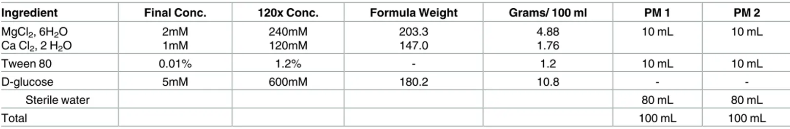
adjusted to 81% transmittance using a turbidimeter (Biolog Inc). The PM-additive solutions
for each plate were prepared according to
Table 1
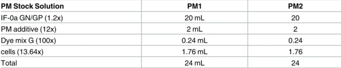
. The inoculating fluid (
Table 2
) consisted of
20 mL of IF-0a GN/GP (1.2 x), 0.24 mL of dye mix G (100x) and 2.0 mL of PM additive (12x)
added to the
M. ulcerans or M. marinum suspension in IF-0a GN/GP (1.76 mL). Each PM
plate was then inoculated in duplicate with 100
μL of inoculating fluid. The PM plates were
incubated in the OmniLog PM System (Biolog Inc.) which measures the growth of
mycobacte-ria every fifteen minutes for eight days at 30˚C. In each well the substrate was reduced to a
pur-ple color which was directly proportional to the growth of the mycobacteria. The intensity of
the purple color was recorded as dye reduction value, which was then plotted as area under the
curve (AUC) by Biolog’s parametric software. Negative control wells containing
non-inocu-lated additive solutions in each PM1 and PM2 plates were run at the same time as a quality
control element. The threshold separating the wells which exhibited a positive reaction from
those with a negative reaction was set for each plate according to the value of the area under
the curve (AUC) of the negative control Well (NCW). We defined moderately positive
grow-ing wells (MPW) and highly positive growgrow-ing wells (HPW) as follows: MPW is when the AUC
Table 1. Composition and preparation of 12 x PM additive solutions.
Ingredient Final Conc. 120x Conc. Formula Weight Grams/ 100 ml PM 1 PM 2
MgCl2, 6H2O Ca Cl2, 2 H2O 2mM 1mM 240mM 120mM 203.3 147.0 4.88 1.76 10 mL 10 mL Tween 80 0.01% 1.2% - 1.2 10 mL 10 mL D-glucose 5mM 600mM 180.2 10.8 - -Sterile water 80 mL 80 mL Total 100 mL 100 mL doi:10.1371/journal.pntd.0005303.t001
value of the well is equal to or lower than 1.25 times the AUC value of the negative control
well, and HPW is when the AUC value of the well is equal to or higher than 1.50 times the
AUC value of the negative control. PM plates were further examined visually at the end of
each incubation period to ensure an independent verification of the results.
Environmental sources of substrates metabolized by all tested M.
ulcerans strains
In order to find the potential environmental origin of the carbon substrates metabolized by
M.
ulcerans, we used the PubMed database to obtain information on the environmental sources
for each of the 190 carbon substrates present in the PM1and PM2 plates. The environmental
sources were organized in 10 categories (plants, fruits and vegetables, bacteria, algae, fungi,
nematodes, mollusks, mammals, insects and the inanimate environment). The Chi-square test
was used to compare the proportion of each category for substrates not metabolized by
M.
ulcerans versus substrates metabolized by all tested M. ulcerans strains; a P value < 0.05 was
used as the criterion for statistical significance. We then used the PubMed database to match
each substrate, used as a key-word, with all environmental sources significantly associated
with substrates metabolized by all tested
M. ulcerans strains, used as the second key-word (e.g.,
D-glucosamine and fungi). We calculated the number of hits obtained in this research and
compared it to the number of hits obtained by searching only for the key word corresponding
to the environmental sources (e.g., fungi).
Results
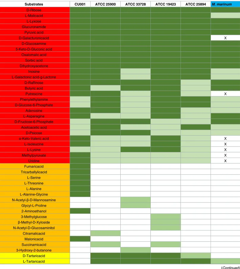
Carbone substrate profiling in M. marinum and M. ulcerans
The negative control wells remained negative in all the PMs plates, and results obtained with
the five
M. ulcerans strains and the M. marinum strain were duplicated. A total of 131/190
(69%) carbon substrates were metabolized by at least one of the five
M. ulcerans strains,
includ-ing 28/190 (15%) carbon substrates common to the five
M. ulcerans strains and 16/190 (8%)
carbon substrates metabolized by only one
M. ulcerans strain (
Table 3
). A total of 21/28 (75%)
substrates metabolized by all tested
M. ulcerans strains were also metabolized by M. marinum
(
Table 3
). In detail, 17/95 (18%) carbon sources in PM1 plates were metabolized by all
M.
ulcerans strains and comprised glucose-6-phosphate, ribose, L-asparagine, uridine,
fructose-6-phosphate, adenosine, inosine, acetoacetic acid, methyl pyruvate, L-malic acid,
D-psicose, L-lyxose, glucuronamide, pyruvic acid, L-galactonic acid-g-lactone, D-galacturonic
acid and phenylethylamine. Six of these substrates exhibited a strong positive reaction
(D-ribose, L-malic acid, L-lyxose, glucuronamide, pyruvic acid and D-galacturonic acid). Then,
11/95 (11.5%) carbon sources in PM2 plates metabolized by all
M. ulcerans strains comprised
D-raffinose, butyric acid, D-glucosamine,
α-keto-valeric acid, 5-keto-D-gluconic acid,
oxalo-malic acid, sorbic acid, L-isoleucine, L-lysine, putrescine and dihydroxyacetone. Five of these
Table 2. Recipe for 1x PM inoculating fluids from stock solutions.
PM Stock Solution PM1 PM2 IF-0a GN/GP (1.2x) 20 mL 20 PM additive (12x) 2 mL 2 Dye mix G (100x) 0.24 mL 0.24 cells (13.64x) 1.76 mL 1.76 Total 24 mL 24 doi:10.1371/journal.pntd.0005303.t002
Table 3. Carbone substrates metabolized by at least one of the five tested M. ulcerans strains compared with carbon substrates metabolized by
Mycobacterium marinum on Biolog PM1 & PM2 plates.
Substrates CU001 ATCC 25900 ATCC 33728 ATCC 19423 ATCC 25894 M. marinum D-Ribose L-Malicacid L-Lyxose Glucuronamide Pyruvic acid D-Galacturonicacid X D-Glucosamine 5-Keto-D-Gluconic acid Oxalomalic acid Sorbic acid Dihydroxyacetone Inosine L-Galactonic acid-g-Lactone D-Raffinose Butyric acid Putrescine X Phenylethylamine D-Glucose-6-Phosphate Adenosine L-Asparagine D-Fructose-6-Phosphate Acetoacetic acid D-Psicose α-Keto-Valeric acid X L-Isoleucine X L-Lysine X Methylpyruvate X Uridine X Fumaricacid Tricarballylicacid L-Serine L-Threonine L-Alanine L-Alanine-Glycine N-Acetyl-β-D-Mannosamine Glycyl-L-Proline 2-Aminoethanol 3-Methylglucose β-Methyl-D-Xyloside N-Acetyl-D-Glucosaminitol Citramalicacid Malonicacid Succinamicacid 3-Hydroxy-2-butanone D-Tartaricacid L-Tartaricacid (Continued )
Table 3. (Continued)
Substrates CU001 ATCC 25900 ATCC 33728 ATCC 19423 ATCC 25894 M. marinum Acetamide L-Arginine Glycine L-Histidine L-Homoserine Hydroxy-L-Proline L-Leucine L-Methionine L-Ornithine L-Phenylalanine L-Pyroglutamicacid L-Valine D,L-Carnitine sec-Butylamine D,L-Octopamine 2,3-Butanediol 2,3-Butanedione Itaconicacid D-Lactic acid Methyl Ester
Melibionicacid Oxalicacid Quinicacid D-Ribono-1,4-Lactone Sebacicacid Salicin Sedoheptulosan L-Sorbose Stachyose D-Tagatose Turanose Xylitol γ-Amino-N-Butyric acid
δ-Amino Valeric acid Capricacid Caproicacid 4-Hydroxybenzoic acid β-Hydroxybutyricacid γ-Hydroxybutyricacid Pectin N-Acetyl-D-Galactosamine N-Acetyl-Neuraminicacid β-D-Allose D-Arabinose 2-Deoxy-D-Ribose 3-O-β-D-Galactopyranosyl-D-Arabinose Gentiobiose L-Glucose (Continued )
Table 3. (Continued)
Substrates CU001 ATCC 25900 ATCC 33728 ATCC 19423 ATCC 25894 M. marinum D-Lactitol D-Melezitose Maltitol α-Methyl-D-Glucoside 2-Deoxyadenosine Glycyl-L-Aspartic acid Citricacid Bromosuccinicacid Propionicacid Mucicacid Glycolicacid Glyoxylicacid D-Cellobiose Glycyl-L-Glutamic acid Mono-Methylsuccinate D-Malicacid Tyramine D-Asparticacid 1,2-Propanediol Tween 40 α-Ketoglutaricacid α-Ketobutyricacid L-Glutamine Tween 80 α-Hydroxybutyric acid β-Methyl-D-Glucoside Adonitol Maltotriose Dulcitol D-Serine
D-Galactonic acid-γ-Lactone DL-Malicacid Tween 20 L-Rhamnose D-Fructose Aceticacid α-D-Glucose Thymidine
Carbone substrates metabolized by at least one of the five tested M. ulcerans strains. carbon substrates metabolized by only one of the five tested M. ulcerans strains. carbon substrates metabolized by all testedM. ulcerans strains.
Moderately positive wells Highly positive wells
X Carbon substrates which are not metabolized by M. marinum and metabolized by all tested M. ulcerans strains.
substrates exhibited a strong positive reaction (D-glucosamine, 5-keto-D-gluconic acid,
oxalo-malic acid, sorbic acid and dihydroxyacetone). A total of 21/28 carbon substrates were also
metabolized by
M. marinum leaving D-galacturonic acid, uridine, methyl pyruvate,
α-keto-valeric acid, L-isoleucine, L-lysine and putrescine as the only substrates specific to
M. ulcerans
(
Table 3
).
Environmental sources for substrates metabolized by all tested M.
ulcerans strains
Comparing the potential environmental sources in search of substrates metabolized by all
tested
M. ulcerans strains versus non-metabolized substrates, we found a significant
associa-tion between
M. ulcerans metabolized substrates and bacteria (p = 0.000), fungi (p = 0.001),
algae (p = 0.003) and mollusks (p = 0.007). The differences were not significant for plants
(p = 0.535), fruits and vegetables (p = 0.870), mammals (p = 0.064), insects (p = 0.234) and the
inanimate environment (p = 0.477). No carbon source was found to be associated with
nema-todes. Further MedLine research incorporating bacteria, fungi, algae and mollusks as keywords
disclosed that 16/28 (57%) metabolized substrates were associated with bacteria, 5/28 (18%)
were associated with alga, 3/28 (11%) were associated with mollusks and 2/28 with fungi.
Dis-carding bacteria because of a potential bias since Biolog was designed for the study of bacterial
metabolism, 15/28 (54%) metabolized substrates were associated with fungi whereas 6/28
(21%) were associated with the algae and 6/28 (21%) with mollusks (
Table 4
).
Discussion
We determined that five different strains of
M. ulcerans could use 28 different substrates as
sources of carbon. These results were authenticated by the negativity of the negative controls
introduced in every plate and the reproduction of data over two replicates. Moreover, stringent
criteria were used to ensure the predictive value of the positive results. However, only seven of
these 28 substrates were found to be specifically used by
M. ulcerans and not by the
phyloge-netically closest species
M. marinum. Three of these seven carbon sources indeed contain
indispensable amino-acids.
The carbon sources here determined for
M. ulcerans may by incorporated in culture media
in the perspective of enhancing the isolation and culture of this pathogen. Indeed,
M. ulcerans
is a slow-growing mycobacterium and the availability of an improved method for its culture
would improve the diagnosis of Buruli ulcer patients and the quest for environmental
reser-voirs [
32
]. As an example, it has been shown that the incorporation of chitin into the
Middleb-rook 7H9 broth enhances the growth of
M. ulcerans [
37
]. Accordingly, our study points
towards a possible association of
M. ulcerans with fungi as a potential source of chitin, a
poly-saccharide possibly degraded by
M. ulcerans’ genome-encoded chitinase [
42
]. Likewise, the
other carbon sources here disclosed should be tested for their potential to increase the
cultiva-tion of
M. ulcerans.
Moreover, our analyses suggested that
M. ulcerans may have found some sources of carbon
in microbial communities including alive and dead bacteria, fungi and algae. As for bacteria, it
has been previously reported that
M. ulcerans was isolated in environments where 17 other
mycobacteria species were also isolated, including
M. fortuitum as a constant co-inhabitant [
3
,
32
,
33
]. These results suggest cross-feeding between various bacterial complexes including
mycobacteria, for the acquisition of carbon. Likewise, green algae extracts have been shown to
halve the
in vitro doubling time of M. ulcerans and promote the formation of biofilm [
31
]. We
observed that
M. ulcerans metabolizes D-galacturonic acid, the main component of pectin
contained in the primary cell walls of terrestrial plants, and putrescine, a foul-smelling
chemical derived from the decomposition of dead plants, which indicates that
M. ulcerans may
live in assemblages of dead aquatic plants. This finding is reinforced by the observation that
M.
ulcerans’s genome encodes five putative cutinases. Cutinases are mainly produced by
phyto-pathogenic fungi to hydrolyze cutin (a main component of the cuticle which covers the aerial
surfaces of plants) during plant colonization process [
43
].
Green algae are among the main food of freshwater mollusks pointed out in our study;
mol-lusks are herbivores like other species of the freshwater snail family [
44
]. The principal genera
of mollusks met in freshwater in West Africa are
Bulinus, Planorbis, Pila, Lanistes, Melania,
Bithynia, Lymnaea, Biomphalaria, Mutela, Aspatharia and Sphaerium [
23
,
45
]. Previous
molec-ular investigations reported the detection of specific
M. ulcerans DNA sequences in Bulinus
spp. [
23
,
46
], in
Planorbis spp. [
23
] and in mollusks of different Gastropoda order, Bivalvia
order and Basommatophora order [
26
]. Furthermore, the experimental infection of
Pomacea
canaliculata (Ampullariidae) and Planorbis planorbis (Planorbidae) by plants contaminated by
M. ulcerans- showed through optic microscopy digestive tract observation that snails remained
infected by viable mycobacteria up to 25 days [
23
]. Small mollusks are also known to be a prey
for water bugs which are involved in the transmission of
M. ulcerans in Buruli ulcer endemic
Table 4. Cross-search of the Medline database (May, 2016) for fungi, mollusks and algae as potential sources of carbon substrates; and sub-strates metabolized by all tested M. ulcerans strains. The total number of hits for fungi, mollusks and algae is indicated into brackets. Each cell contains
the number of cross-hits and green cells indicate the higher relative hit for each carbon source.
Algae (19292) fungi (1392904) Molluscs (52885) Bacteria (1934745)
D-ribose 24/19292 2571/1392904 114/52885 5133/1934745 Glucuronamide 0/19292 4/1392904 0/52885 7/1934745 D-Galacturonicacid 0/19292 79/1392904 3/52885 218/1934745 D-Glucosamine 8/19292 498//1392904 28/52885 4499/1934745 Oxalomalic acid 0/19292 0/1392904 0/52885 0/1934745 Sorbic acid 0/19292 357/1392904 2/52885 411/1934745 Dihydroxyacetone 7/19292 294/1392904 3/52885 504/1934745 L-Galactonic acid-g-Lactone 0/19292 0/1392904 0/52885 0/1934745 D-Raffinose 3/19292 646/1392904 4/52885 728/1934745 Butyric acid 26/19292 3089/1392904 266/52885 5689/1934745 Putrescine 17/19292 1174/1392904 27/52885 2226/1934745 Phenylethylamine 10/19292 683/1392904 225/52885 908/1934745 D-Psicose 1/19292 14/1392904 0/52885 77/1934745 L-Malicacid 1/19292 107/1392904 0/52885 301/1934745 L-Lyxose 0/19292 4/1392904 0/52885 28/1934745 Pyruvic acid 19/19292 686/1392904 19/52885 1484/1934745 5-Keto-D-Gluconic acid 0/19292 5/1392904 0/52885 21/1934745 Inosine 2/19292 999/1392904 37/52885 1540/1934745 D-Glucose-6-Phosphate 1/19292 57/1392904 2/52885 94/1934745 Adenosine 182/19292 14109/1392904 1029/52885 27257/1934745 L-Asparagine 17/19292 1980/1392904 37/52885 3428/1934745 D-Fructose-6-Phosphate 2/19292 7/1392904 0/52885 92/1934745 Acetoacetic acid 0/19292 20/1392904 1/52885 62/1934745 a-Keto-Valeric acid 0/19292 2/1392904 0/52885 10/1934745 L-Isoleucine 4/19292 1400/1392904 40/52885 3296/1934745 L-Lysine 65/19292 6989/1392904 254/52885 11894/1934745 Methyl pyruvate 7/19292 4/1392904 0/52885 7/1934745 Uridine 30/19292 3408/1392904 112/52885 6435/1934745 doi:10.1371/journal.pntd.0005303.t004
regions [
3
]. In West Africa, approximately 76% of the population lives next to rivers, lakes,
and other water bodies contaminated with intermediate hosts such as snails [
47
].
In conclusion, our study is suggesting paths to improve culture media for the enhanced
iso-lation of
M. ulcerans by mimicking the natural ecosystem of M. ulcerans which is probably
liv-ing in microbial communities with other bacteria, fungi and algae. These data support the
recent hypothesis that mollusks could be part of a larger food chain including several hosts
giv-ing appropriate shelters to
M. ulcerans, as recently reported [
48
]. Small mollusks should be
fur-ther investigated using culture-based appropriate methods in the search for
M. ulcerans.
Acknowledgments
The authors acknowledge Pablo Gluschankof for his fruitful discussion of the manuscript’s
content.
Author Contributions
Conceptualization: MD.
Methodology: DZ AB.
Software: MM.
Validation: DZ AB MM.
Writing – original draft: DZ AB MD.
References
1. Doig KD, Holt KE, Fyfe JAM, Lavender CJ, Eddyani M, Portaels F, et al. On the origin of Mycobacterium ulcerans, the causative agent of Buruli ulcer. BMC Genomics 2012; 13, 258. doi: 10.1186/1471-2164-13-258PMID:22712622
2. Josse R, Guedenon A, Darie H, Anagonou S, Portaels F, Meyers WM. [Mycobacterium ulcerans cuta-neous infections: Buruli ulcers]. Medecine tropicale: revue du Corps de sante colonial 1995; 55(4), 363–373.
3. Portaels F, Meyers WM, Ablordey A, Castro AG, Chemlal K, de Rijk P, et al. First Cultivation and Char-acterization of Mycobacterium ulcerans from the Environment. PLoS Negl Trop Dis. 2008; 2(3), e178. doi:10.1371/journal.pntd.0000178PMID:18365032
4. Portaels F, Fonteyene PA, de Beenhouwer H, de Rijk P, Guedenon A, Hayman J, et al. Variability in 3’ end of 16S rRNA sequence of Mycobacterium ulcerans is related to geographic origin of isolates. J Clin Microbiol. 1996; 34(4), 962–965. PMID:8815117
5. Maccallum P, Tolhurst JC, Buckle G, Sissons HA A new mycobacterial infection in man. J Pathol Bac-teriol. 1948; 60(1), 93–122.
6. Johnson PD, Azuolas J, Lavender CJ, Wishart E, Stinear TP, Hayman JA, et al. Mycobacterium ulcer-ans in mosquitoes captured during outbreak of Buruli ulcer, southeastern Australia. Emerg Infect Dis. 2007; 13(11), 1653–1660. doi:10.3201/eid1311.061369PMID:18217547
7. Stinear TP, Seemann T, Pidot S, Frigui W, Reysset G, Garnier T, et al. Reductive evolution and niche adaptation inferred from the genome of Mycobacterium ulcerans, the causative agent of Buruli ulcer. Genome Res. 2007; 17(2), 192–200. doi:10.1101/gr.5942807PMID:17210928
8. WHO. World: Distribution of buruli ulcer, 2014 WHO Annual meeting on Buruli ulcer 2015.
9. Benbow ME, Williamson H, Kimbirauskas R, McIntosh MD, Kolar R, Quaye C, et al. Aquatic inverte-brates as unlikely vectors of Buruli ulcer disease. Emerg Infect Dis. 2008; 14(8), 1247–1254. doi:10. 3201/eid1408.071503PMID:18680648
10. Garchitorena A, Ngonghala CN, Texier G, Landier J, Eyangoh S, Bonds MH, et al. Environmental trans-mission of Mycobacterium ulcerans drives dynamics of Buruli ulcer in endemic regions of Cameroon. Sci Rep. 2015; 5, 18055. doi:10.1038/srep18055PMID:26658922
11. Kenu E, Nyarko KM, Seefeld L, Ganu V, Ka¨ser M, Lartey M, et al. Risk Factors for Buruli Ulcer in Ghana —A Case Control Study in the Suhum-Kraboa-Coaltar and Akuapem South Districts of the Eastern Region. PLoS Negl Trop Dis. 2014; 8(11), e3279. doi:10.1371/journal.pntd.0003279PMID:25411974
12. Narh CA, Mosi L, Quaye C, Dassi C, Konan DO, Tay SCK, et al. Source Tracking Mycobacterium ulcer-ans Infections in the Ashanti Region, Ghana. PLoS Negl Trop Dis. 2015; 9, e0003437. doi:10.1371/ journal.pntd.0003437PMID:25612300
13. Addo P, Adu-Addai B, Quartey M, Abbas M, Okang I, Owusu E, et al. Clinical and Histopathological Pre-sentation of Buruli Ulcer in Experimentally Infected Grasscutters (Thryonomys swinderianus). Intern J Trop Med. 2006; 3(2).
14. Mensah-Quainoo EK. A study of the magnitude and determinants of Buruli ulcer disease in the Ga Dis-trict of Ghana. International Conference on Buruli ulcer Control and Research; 6–8 July 1998; Yamous-soukro, Cote d’Ivoire 1998.
15. Peraudin ML, Herrault A, Desbois JC Ulcère cutane´eàMycobacterium ulcerans (ulcère de Buruli). Annales de Pe´diatrie 1980; 27(10), 687–692. PMID:7212557
16. Brou T, Broutin H, Elguero E, Asse H, Guegan JF Landscape diversity related to Buruli ulcer disease in Cote d’Ivoire. PLoS Negl Trop Dis. 2008; 2(7), 0000271.
17. Johnson RC, Makoutode´ M, Sopoh GE, Elsen P, Gbovi J, Pouteau LH, et al. Buruli Ulcer Distribution in Benin. Emerg Infect Dis. 2005; 11(3), 500–501. doi:10.3201/eid1103.040597PMID:15789490
18. N’krumah RTAS, Kone´ B, Tiembre I, Cisse´ G, Pluschke G, Tanner M, et al. Socio-Environmental Fac-tors Associated with the Risk of Contracting Buruli Ulcer in Tiassale´, South Coˆte d’Ivoire: A Case-Con-trol Study. PLoS Negl Trop Dis. 2016; 10(1), e0004327. doi:10.1371/journal.pntd.0004327PMID:
26745723
19. Aiga H, Amano T, Cairncross S, Domako JA, Nanas OK, Coleman S Assessing water-related risk fac-tors for Buruli ulcer: a case-control study in Ghana. Am J Trop Med Hyg. 2004; 71(4), 387–392. PMID:
15516631
20. Tian RB, Niamke S, Tissot-Dupont H, Drancourt M. Detection of Mycobacterium ulcerans DNA in the Environment, Ivory Coast. PLoS One 2016; 11(3): e0151567 doi:10.1371/journal.pone.0151567
PMID:26982581
21. Ahoua L, Guetta A, Ekanza E, Bouzid S, N’Guessan R, Dosso M Risk factors for Buruli ulcer in Coˆte d’Ivoire: Results of a cas-control study, August 2001. Afr J Biotechnol. 2009; 8, 536–546.
22. Vandelannoote K, Durnez L, Amissah D, Gryseels S, Dodoo A, Yeboah S, et al. Application of real-time PCR in Ghana, a Buruli ulcer-endemic country, confirms the presence of Mycobacterium ulcerans in the environment. FEMS Microbiol Lett. 2010; 304(2), 191–194. doi:10.1111/j.1574-6968.2010.01902. xPMID:20146745
23. Marsollier L, Severin T, Aubry J, Merritt RW, Saint Andre JP, Legras P, et al. Aquatic snails, passive hosts of Mycobacterium ulcerans. Appl Environ Microbiol. 2004; 70(10), 6296–6298. doi:10.1128/ AEM.70.10.6296-6298.2004PMID:15466578
24. Marsollier L, Robert R, Aubry J, Saint Andre JP, Kouakou H, Legras P, et al. Aquatic insects as a vector for Mycobacterium ulcerans. Appl Environ Microbiol. 2002; 68(9), 4623–4628. doi:10.1128/AEM.68.9. 4623-4628.2002PMID:12200321
25. Konan KL, Doannio JM, Coulibaly NG, Ekaza E, Marion E, Asse H, et al. [Detection of the IS2404 inser-tion sequence and ketoreductase produced by Mycobacterium ulcerans in the aquatic Heteroptera in the health districts of Dabou and Tiassale in Cote d’Ivoire]. Med Sante Trop. 2015; 25(1), 44–51. doi:
10.1684/mst.2014.0363PMID:25499000
26. Williamson HR, Benbow ME, Nguyen KD, Beachboard DC, Kimbirauskas RK, McIntosh MD, et al. Dis-tribution of Mycobacterium ulcerans in buruli ulcer endemic and non-endemic aquatic sites in Ghana. PLoS Negl Trop Dis. 2008; 2(3), e205. doi:10.1371/journal.pntd.0000205PMID:18365034
27. Zogo B, Djenontin A, Carolan K, Babonneau J, Guegan J-F, Eyangoh S, et al. A Field Study in Benin to Investigate the Role of Mosquitoes and Other Flying Insects in the Ecology of Mycobacterium ulcerans. PLoS Negl Trop Dis. 2015; 9(7), e0003941. doi:10.1371/journal.pntd.0003941PMID:26196901
28. Amissah NA, Gryseels S, Tobias NJ, Ravadgar B, Suzuki M, Vandelannoote K, et al. Investigating the Role of Free-living Amoebae as a Reservoir for Mycobacterium ulcerans. PLoS Negl Trop Dis. 2014; 8 (9), e3148. doi:10.1371/journal.pntd.0003148PMID:25188535
29. Williamson HR, Benbow ME, Campbell LP, Johnson CR, Sopoh G, Barogui Y, et al. Detection of Myco-bacterium ulcerans in the Environment Predicts Prevalence of Buruli Ulcer in Benin. PLoS Negl Trop Dis. 2012; 6(1), e1506. doi:10.1371/journal.pntd.0001506PMID:22303498
30. Tian RD, Lepidi H, Nappez C, Drancourt M Experimental Survival of Mycobacterium ulcerans in Watery Soil, a Potential Source of Buruli Ulcer. Am J Trop Med Hyg. 2016; 94(1), 89–92. doi:10.4269/ajtmh. 15-0568PMID:26526927
31. Marsollier L, Stinear T, Aubry J, Saint Andre´ JP, Robert R, Legras P, et al. Aquatic Plants Stimulate the Growth of and Biofilm Formation by Mycobacterium ulcerans in Axenic Culture and Harbor These Bac-teria in the Environment. App Environm Microbiol. 2004; 70(2), 1097–1103.
32. Aboagye SY, Danso E, Ampah KA, Nakobu Z, Asare P, Otchere ID, et al. Isolation of Nontuberculous Mycobacteria from the Environment of Buruli Ulcer Endemic Communities in Ghana. App Environ Microbiol. 2016; 6, 2016.
33. Marsollier L, Aubry J, Saint-andre´ J, Robert R, Legras P, Manceau A. Ecology and transmission of Mycobacterium ulcerans. 2003; 51:490–5.
34. Garland JAYL, Mills AL. Classification and Characterization of Heterotrophic Microbial Communities on the Basis of Patterns of Community-Level Sole-Carbon-Source Utilization. 1991; 57(8):2351–9.
35. Khatri B, Fielder M, Jones G, Newell W, Abu-Oun M, Wheeler PR High Throughput Phenotypic Analysis of Mycobacterium tuberculosis and Mycobacterium bovis Strains’ Metabolism Using Biolog Phenotype Microarrays. PLoS ONE 2013; 8(1), e52673. doi:10.1371/journal.pone.0052673PMID:23326347
36. Chen J-W, Scaria J, Chang Y-F Phenotypic and Transcriptomic Response of Auxotrophic Mycobacte-rium avium subsp. paratuberculosis leuD Mutant under Environmental Stress. PLoS ONE 2012; 7(6), e37884. doi:10.1371/journal.pone.0037884PMID:22675497
37. Sanhueza D, Chevillon C, Colwell R, Marion E, Marsollier L. Chitin promotes Mycobacterium ulcerans growth. J er. 2016; 2010(October 2015):1–6.
38. Schoro¨der K. H. Investigation into the relatioship of M. ulcerans to M. burili and other mycobacteria. American review of resperatory disease 1975; 111, 559–562.
39. Bouricha M, Castan B, Duchene-Parisi E, Drancourt M Mycobacterium marinum infection following con-tact with reptiles: vivarium granuloma. Int J Infect Dis. 2014; 21, 17–18. doi:10.1016/j.ijid.2013.11.020
PMID:24530276
40. Bochner BR Global phenotypic characterization of bacteria. Fems Microbiol Rev. 2009; 33(1), 191– 205. doi:10.1111/j.1574-6976.2008.00149.xPMID:19054113
41. Bochner BR, Gadzinski P, Panomitros E Phenotype MicroArrays for High-Throughput Phenotypic Test-ing and Assay of Gene Function. Genome Res. 2001; 11(7), 1246–1255. doi:10.1101/gr.186501
PMID:11435407
42. Stinear TP, Seemann T, Pidot S, Frigui W, Reysset G, Garnier T, et al. Reductive evolution and niche adaptation inferred from the genome of Mycobacterium ulcerans, the causative agent of Buruli ulcer. Genome Res. 2007; 17(2):192–200. doi:10.1101/gr.5942807PMID:17210928
43. Coˆtes K, Bakala N’Goma JC, Dhouib R, Douchet I, Maurin D, Carrière F, et al. Lipolytic enzymes in Mycobacterium tuberculosis. Appl Microbiol Biotechnol. 2008; 78(5):741–9. doi: 10.1007/s00253-008-1397-2PMID:18309478
44. El-Assal FM, Shanab SMM, Abou-El-Hassan AA, Mahmoud KMA. Effect of some algal species on the snail intermediate hosts of schistosomiasis in Egypt II. Growth, infection and mortality rates. Egypt J Phycol. 2005; 6(1), 93–110.
45. Thiam N, Diallo A Inte´gration de la biodiversite´ d’eau douce dans le processus de de´veloppement en Afrique: Module de formation des formateurs sur Le suivi des Mollusques d’eau douce. Wetlands Inter-national Afrique [serial on the Internet]. 2010.https://cmsdata.iucn.org/downloads/module_
mollusques_fr.pdf.
46. Kotlowski R, Martin A, Ablordey A, Chemlal K, Fonteyne P-A, Portaels F One-tube cell lysis and DNA extraction procedure for PCR-based detection of Mycobacterium ulcerans in aquatic insects, molluscs and fish. J Med Microbiol. 2004; 53(9), 927–933.
47. Hotez PJ, Kamath A Neglected Tropical Diseases in Sub-Saharan Africa: Review of Their Prevalence, Distribution, and Disease Burden. PLoS Negl Trop Dis. 2009, 3(8), e412. doi:10.1371/journal.pntd. 0000412PMID:19707588
48. Morris AL, Gue´gan J, Andreou D, Marsollier L, Carolan K, Le Croller M, et al. Deforestation-driven food-web collapse linked to emerging tropical infectious disease, Mycobacterium ulcerans. 2016;1–7.