HAL Id: hal-01163945
https://hal.archives-ouvertes.fr/hal-01163945
Submitted on 15 Jun 2015
HAL is a multi-disciplinary open access
archive for the deposit and dissemination of
sci-entific research documents, whether they are
pub-lished or not. The documents may come from
teaching and research institutions in France or
abroad, or from public or private research centers.
L’archive ouverte pluridisciplinaire HAL, est
destinée au dépôt et à la diffusion de documents
scientifiques de niveau recherche, publiés ou non,
émanant des établissements d’enseignement et de
recherche français ou étrangers, des laboratoires
publics ou privés.
3D cancer cell migration in collagen matrices
L Laforgue, Valérie M. Laurent, Alain Duperray, Claude Verdier
To cite this version:
L Laforgue, Valérie M. Laurent, Alain Duperray, Claude Verdier.
3D cancer cell migration
in collagen matrices.
Congrès de la société de Biomécanique, SB, Oct 2015, Paris, France.
�10.1080/10255842.2015.1069628�. �hal-01163945�
*Corresponding author. Email: [email protected]
3D cancer cell migration in collagen matrices
L. Laforgue
a,b, V.M. Laurent
a,b, A. Duperray
c,dand C. Verdier
a,b*aCNRS, Laboratoire Interdisciplinaire de Physique (LIPHY), F-38000 Grenoble; bUniv. Grenoble Alpes, LIPHY, F-38000
Grenoble; cINSERM, Institut Albert Bonniot (IAB), F-38000 Grenoble; dUniv. Grenoble Alpes, IAB, F-38000 Grenoble
Keywords: migration; cancer; collagen; MSD; velocity
1. Introduction
Cell migration is a widely studied subject but often limited to 2D motion (Sheetz et al., 1999) on various substrates for it allows to simplify cell-substrate interactions. The important relevant parameters to 2D cell migration are substrate rigidity, adhesion, and the role of the cell cytoskeleton. More recently, new studies on 3D cell migration have been performed thanks to the development of new visualization tools such as reflectance confocal microscopy (Iordan et al. 2010, Wolf et al. 2013). In particular, it is possible to image cell and the extra-cellular matrix at the same time. This leads to interesting applications on cancer cell migration, and could possibly open new routes to the understanding of cancer development, like the mechanisms used by cancer cells to invade new tissues. Thus, the aim of this study is to study cancer migration in collagen matrices, as a model of soft tissue, to investigate their motility within the surrounding collagen matrix. Different collagen concentrations will be used and the ability of these cancer cells to move through such a complex structure will be addressed. To this aim, first results of cell migration velocity as well as the Mean Squared Displacement (MSD) will be analyzed.
2. Methods
2.1 Cancer cells and collagen
T24 epithelial cell lines (an invasive bladder cancer type) were used and cultured in RPMI culture medium supplemented with Fetal Bovine Serum (FBS). Collagen gels were prepared from rat tail collagen solutions (BD Biosciences) and mixed with culture medium, then neutralized to reach pH=7.4 at 4°C. Then, they were polymerized at 37°C for 30 min. Different concentrations C (mg/mL) were used.
2.2 Confocal microscopy
Experiments were carried out using a confocal microscope (Zeiss, LSM 510 model) as previously described (Iordan et al. 2010). We make use of the natural reflection of collagen fibres to obtain clear images of cells embedded in the collagen matrix (see Figure 1). Regarding cells, we use an actin-GFP transfected cell line.
Figure 1 Typical confocal microscopy image representing T24 cancer cell (actin-GFP) embedded in the collagen matrix (red network). C=0.95mg/mL. Scale bar: 14µm.
2.3 Cell velocity and MSD
Following experiments, images were treated to identify the centre of geometry and follow it in time to obtain its position vector r(t). Instantaneous migration velocities were obtained and averaged in time. The Mean Squared Displacement (MSD) was calculated as <|r(t+τ)-r(τ)|2>τ, where τ is the lag time, and
between 0 and the longest time T. It is common to assume MSD(t) ~ tα, where the exponent α varies between 0 and 2 (Peschetola et al. 2013).
3. Results and discussion
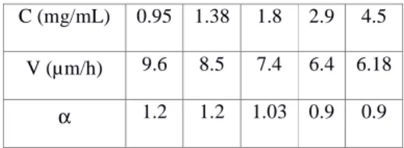
The velocities of migration of T24 cancer cells in various collagens (different concentrations) are shown in Table 1, whereas Figure 2 illustrates two cases of MSD calculation for low and high concentrations of collagen.
C (mg/mL) 0.95 1.38 1.8 2.9 4.5 V (µm/h) 9.6 8.5 7.4 6.4 6.18
α 1.2 1.2 1.03 0.9 0.9
Table 1 Values of cell velocity and MSD exponent α as a function of collagen concentration C.
Figure 2 MSD for three T24 cells migrating in low (0.95 mg/mL), medium (1.8 mg/mL) and high concentration collagen (4.5 mg/mL).
It appears from the data that the velocity of cells is a decreasing function of concentration C. This result is comparable to the previously mentioned data (Wolf et al. 2013). Also we note (Table 1) that the power exponent α of the MSD is a decreasing function of concentration. This result is new, and could be explained as follows. When embedded into 3D collagen gels, cells need to bind (adhesion) and modify their cytoskeleton accordingly to attach to binding sites, but they have much less space as they are confined and cannot deform as in the 2D case. Therefore, two possible effects are needed for cells to migrate efficiently: good adhesion to the collagen matrix fibres and cell ability to deform and squeeze through matrix pores. It is essentially the competition between these two effects which
guides their motility (Iordan et al. 2010). Therefore, the velocity of migration should be slow at very low concentrations, then increase with C to reach a maximum, then should decrease again as space becomes more and more limited. According to our experiments, the range studied [0.95mg/mL-4.5mg/mL] corresponds to decreasing velocities because space is already limited as shown in the low concentration case of Figure 1 (pore sizes were estimated to range between 4µm and 15µm in our concentration range). One thus observes that the filamentous network is already quite dense. In this context, it can be easily understood that cells in high concentrated collagen do not have a persistent direction so that the exponent α remains small, corresponding to sub-diffusive motion, whereas α is higher than 1 for low concentrated collagen.
4. Conclusions
Thanks to confocal imaging of cancer cells embedded in various collagen gels, we show that the migration velocity decreases with concentration, associated with sub-diffusive motion at large concentration and super-diffusive for low concentration.
Acknowledgements
We thank the ANR for grant No 12-BS09-020-01
(TRANSMIG). The LIPhy laboratory is part of the LabeX Tec21 (Investissements d’Avenir, grant agreement No
ANR-11-LABX-0030).
References
Iordan A, Duperray A, Gérard A, Grichine A, Verdier C. 2010. Breakdown of cell-collagen networks through collagen remodelling. Biorheology. 47:277-295. Peschetola V, Laurent VM, Duperray A, Michel R, Ambrosi
D, Preziosi L, Verdier C, Time-dependent traction force microscopy for cancer cells as a measure of invasiveness. 2013. Cytoskeleton. 70:201-214.
Sheetz MP, Felsenfeld D, Galbraith CG, Choquet D. 1999. Cell migration as a five-step cycle. Biochem Soc Symp. 65:233-243.
Wolf K, Lindert MT, Krause M, Alexander S, Riet JT, Willis AL, Hoffman RM, Figdor CG, Weiss SJ, Friedl P. 2013. Physical limits of cell migration: control by ECM space and nuclear deformation and tuning by proteolysis and traction force. J Cell Biol. 201:1069-1084.