Publisher’s version / Version de l'éditeur:
Frontiers in Immunology, 2017-11-20
READ THESE TERMS AND CONDITIONS CAREFULLY BEFORE USING THIS WEBSITE.
https://nrc-publications.canada.ca/eng/copyright
Vous avez des questions? Nous pouvons vous aider. Pour communiquer directement avec un auteur, consultez la
première page de la revue dans laquelle son article a été publié afin de trouver ses coordonnées. Si vous n’arrivez pas à les repérer, communiquez avec nous à PublicationsArchive-ArchivesPublications@nrc-cnrc.gc.ca.
Questions? Contact the NRC Publications Archive team at
PublicationsArchive-ArchivesPublications@nrc-cnrc.gc.ca. If you wish to email the authors directly, please see the first page of the publication for their contact information.
NRC Publications Archive
Archives des publications du CNRC
This publication could be one of several versions: author’s original, accepted manuscript or the publisher’s version. / La version de cette publication peut être l’une des suivantes : la version prépublication de l’auteur, la version acceptée du manuscrit ou la version de l’éditeur.
For the publisher’s version, please access the DOI link below./ Pour consulter la version de l’éditeur, utilisez le lien DOI ci-dessous.
https://doi.org/10.3389/fimmu.2017.01589
Access and use of this website and the material on it are subject to the Terms and Conditions set forth at
Camelid single-domain antibodies: historical perspective and future
outlook
Arbabi-Ghahroudi, Mehdi
https://publications-cnrc.canada.ca/fra/droits
L’accès à ce site Web et l’utilisation de son contenu sont assujettis aux conditions présentées dans le site LISEZ CES CONDITIONS ATTENTIVEMENT AVANT D’UTILISER CE SITE WEB.
NRC Publications Record / Notice d'Archives des publications de CNRC:
https://nrc-publications.canada.ca/eng/view/object/?id=e01626cc-59b9-436b-9d95-8396354ba2ae https://publications-cnrc.canada.ca/fra/voir/objet/?id=e01626cc-59b9-436b-9d95-8396354ba2ae
doi: 10.3389/immu.2017.01589
Edited by: Marc H. V. Van Regenmortel, Centre national de la recherche scientiique (CNRS), France
Reviewed by: Serge Muyldermans, Vrije Universiteit Brussel, Belgium Etienne Weiss, Ecole Supérieure de Biotechnologie de Strasbourg, France
*Correspondence: Mehdi Arbabi-Ghahroudi mehdi.arbabighahroudi@ nrc-cnrc.gc.ca
This is NRC publication number: 53362.
Specialty section: This article was submitted to Vaccines and Molecular Therapeutics, a section of the journal Frontiers in Immunology Received: 29 September 2017 Accepted: 03 November 2017 Published: 20 November 2017 Citation: Arbabi-Ghahroudi M (2017) Camelid Single-Domain Antibodies: Historical Perspective and Future Outlook. Front. Immunol. 8:1589. doi: 10.3389/immu.2017.01589
Camelid Single-Domain Antibodies:
Historical Perspective and Future
Outlook
Mehdi Arbabi-Ghahroudi1,2*
1 Human Health Therapeutics Research Centre, National Research Council Canada, Ottawa, ON, Canada, 2Department of Biology, Carleton University, Ottawa, ON, Canada
Tremendous effort has been expended over the past two and a half decades to under-stand many aspects of camelid heavy chain antibodies, from their biology, evolution, and immunogenetics to their potential applications in various ields of research and medicine. In this article, I present a historical perspective on the development of camelid single-domain antibodies (sdAbs or VHHs, also widely known as nanobodies) since their
discovery and discuss the advantages and disadvantages of these unique molecules in various areas of research, industry, and medicine. Commercialization of camelid sdAbs exploded in 2001 with a lurry of patents issued to the Vrije Universiteit Brussel (VUB) and later taken on by the Vlaams Interuniversitair Instituut voor Biotechnologie (VIB) and, after 2002, the VIB-founded spin-off company, Ablynx. While entrepreneurial spirit has certainly catalyzed the exploration of nanobodies as marketable products, IP restrictions may be partially responsible for the relatively long time span between the discovery of these biomolecules and their entry into the pharmaceutical market. It is now anticipated that the irst VHH-based antibody drug, Caplacizumab, a bivalent
anti-vWF antibody for treating rare blood clotting disorders, may be approved and commercialized in 2018 or shortly thereafter. This elusive irst approval, along with the expiry of key patents, may substantially alter the scientiic and biomedical landscape surrounding camelid sdAbs and pave the way for their emergence as mainstream biotherapeutics.
Keywords: camelid single-domain antibody, heavy chain antibody, VHH, nanobody, antibody engineering, therapeutic antibody
INTRODUCTION
he canonical view of antibodies as molecules composed of two heavy chains and two light chains was forever changed one day in 1989 following analysis of total and fractionated immunoglobulin G (IgG) molecules in the serum of a dromedary camel in the laboratory of Professor Raymond Hamers at the Vrije Universiteit Brussel (VUB). he serendipitous discovery of antibodies lacking a light chain [heavy chain-only antibodies (HCAbs)] occurred as part of a student-run project aimed at developing a serodiagnostic test for trypanosome infection in camels and water bufalos. he pre-liminary data showed that besides conventional IgG1 (MW ~150 kDa), two other immunoglobulin fractions (thereater called IgG2 and IgG3; MW ~90 kDa) were present which contributed up to 75% of all serum IgGs (1–3). Comparative studies on the sera of new world camelids (Lama glama and Lama pacos) subsequently conirmed the presence of HCAbs, albeit at concentrations between
2
Arbabi-Ghahroudi Historical Perspective on Camelid sdAbs
Frontiers in Immunology | www.frontiersin.org November 2017 | Volume 8 | Article 1589
30 and 50% (1, 4–8). Following these exciting indings, it became essential to analyze the antigen-binding properties of these IgG fractions since the presence of truncated forms of heavy chain antibodies with no light chains, classically described as “heavy chain disease,” had been reported in human patients (9, 10). No functional activity was reported for the pathogenic heavy chain antibodies in these patients, as these proteins were shown to bear extensive internal deletions in the variable (VH) and the irst constant region (CH1) domains. By contrast, antibodies from camelids exposed to Trypanosoma evansi demonstrated strong binding activity in the IgG2 and IgG3 heavy chain-only fractions as shown by radio-immunoprecipitation and blotting experiments (1).
In two subsequent reports, phage-display technology and high-resolution crystallography were utilized to (a) build a phage-display library from the lymphocytes of immunized
camels and isolate monomeric antigen-speciic VHH domains
in the absence of the constant regions (11) and (b) solve crystal structures of an unliganded VHH (12) and a VHH:lysozyme
complex, reported simultaneously by the VUB team and a Dutch–French research group (13). he term VHH was originally
introduced by the VUB team in 1994 to indicate a VH domain derived from camelid heavy chain antibodies. he feasibility of isolating stable and soluble VHH domains with nanomolar
aini-ties against lysozyme and tetanus toxoid showed very early on the promise of these molecules as high-ainity binding moieties. Crystallography studies revealed additional salient features of an anti-lysozyme VHH, including deep penetration of its long third
complementarity-determining region (CDR3) into the active site of the enzyme; this feature had rarely been seen with conventional antibodies and required a fundamental deviation from known human canonical CDR1 structure (13). Further evidence of the unique antigen recognition behavior of VHH domains (including
enzyme inhibition) was published over the next several years (11, 14, 15), suggesting that VHHs might probe diferent sets of
epitopes on proteins compared with conventional antibodies.
Key proof of concept for producing bivalent/bispeciic VHH
modalities via genetic fusion (using camelid short and long hinge sequences) of anti-lyzozyme and/or anti-tetanus toxin VHHs was
also established very early on (14).
MOLECULAR ONTOGENY OF CAMELID
HCAbs
Molecular biology techniques were subsequently applied to decipher the DNA sequences of HCAbs. he sequencing results showed that nature had designed HCAbs as an additional arm of the immune systems of camelid ungulates over the course of their evolutionary history. he consensus of these studies suggested camelid HCAbs possessed: (a) no CH1 domain, and therefore, a direct connection of the rearranged VHH exon to the
hinge region; (b) one of two types of long (IgG2) and short (IgG3) hinge isotypes; (c) speciic conserved amino acid substitutions in framework region 2 (FR2), mainly at VH positions that make contact with the VL in classical antibodies, including Kabat positions 37, 44, 45, and 47; and (d) potentially diferent CDR3
amino acid composition and a broader length distribution for CDR3 compared to the heavy chains of conventional antibodies (1, 16, 17).
Later genomic studies shed light on the origin of HCAbs in dromedary camels and alpacas. It is now established that HCAbs are produced from the same igh locus as conventional antibodies but with distinct sets of genes for the generation of HCAbs. It is estimated that alpaca and dromedary genomes contain ~17 and ~40 VHH genes, respectively, with an identical organization
of the genes that produce conventional antibodies (18, 19). he CH1 exon is present in the genomic DNA of HCAbs but a point mutation (G to A) at the 5′ end of the CH1-hinge intron disrupts the consensus splicing site (GT) and causes omission of this region during splicing (3, 18, 20–22). A complete picture of camelid germline V gene repertoires of heavy and light chains and the classiication of VH and VHH genes is still missing.
Published genomic and cDNA data have so far shown that
camelid VHH genes are highly homologous to the human VH3
family of clan III with the exception of several key amino acid substitutions in FR2, namely, Val37 → Phe/Tyr, Gly44 → Glu, Leu45 → Arg, and Trp47 → Gly (Kabat numbering), and are encoded by a distinct subset of germline V genes. Preliminary investigations of published llama VHH sequences classiied them
into four subfamilies by sequence similarity, and many of the earliest-described VHH features such as long CDR3s, additional
disulide bridges, and particular canonical structures of CDR1–3 were shown to be subfamily speciic (17, 23). Subsequent studies in alpaca identiied at least three V gene subgroups of the alpaca igh locus: IGHV1, IGHV2, and IGHV3 which are equivalent to the human IGHV families within clan I (VH families 2, 4, 6), II (VH families 1, 5, 7), and III (VH family 3), respectively, based on sequence homology. he alpaca VHH genes clustered into six
subsets by sequence similarity, but all are homologous to human
IGHV3 genes (18). Furthermore, recent investigations have
demonstrated the presence of genes belonging to IGHV families 1, 3, and 4 (human clan I and III) in llama and alpaca, and in addition, uncovered new camelid V genes highly homologous to the human IGHV5 and IGHV7 families (human clan II); how-ever, no genes similar to human families 2 or 6 (within human clan I) were found (24). Interestingly, a novel promiscuous class of V genes in camelids was identiied that is closely related to the human VH4 family (clan I). hese VH4 homologs contribute largely to the classical antibody repertoire and lack the hallmark solubilizing VHH residues in FR2. Nevertheless, antigen-speciic
VH4-family fragments with VHH-like stability and solubility
were isolated from an immune llama library (25). In the absence of a complete set of camelid germline VH and VHH genes, most
immunogenetic studies have relied on comparisons with human germline genes.
he consensus of immunogenetic studies of camelid HCAbs is that repertoire diversiication of these molecules may involve (a) a large number of unique VHH gene segments recombining
with DH and JH minigenes, possibly with additional non-templated nucleotide insertions leading to longer CDR3 loops; (b) somatic hypermutation, potentially of extended CDR1 regions compared with conventional antibodies; (c) acquisition of non-canonical cysteine residues in the CDRs and FR2; and
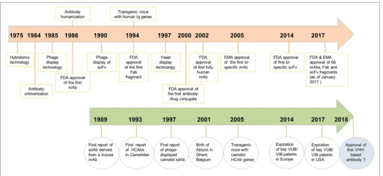
FIGURE 1 | Chronological timeline of major scientiic developments in the ield of antibody engineering since the discovery of monoclonal antibodies (mAbs) in 1975 leading to the regulatory approval of mAbs, antigen-binding fragments (Fabs) and scFvs as therapeutics. Developments for mAbs are shown in orange and developments of VHHs/heavy chain-only antibodies (HCAbs) in green. Regulatory approval of the irst VHH-based antibody drug is expected in 2018.
(d) involvement of FR2 residues in antigen binding and in structuring the CDR3 loop (3, 22, 26, 27). In agreement with immunogenetic analyses, several structural studies have sug-gested that due to the loss of VL domains, VHH paratopes have
acquired a higher structural complexity by involving more residues in antigen binding compared to classical VHs (27). As for the evolutionary origin of HCAbs, it is diicult to draw solid conclusions but several hypotheses have been proposed. A com-mon theme acom-mong most of these has been the need for generat-ing or expandgenerat-ing a new antigen-bindgenerat-ing repertoire in Camelidae to address certain antigenic challenges, e.g., cryptic epitopes of commonly encountered pathogens. Phylogenetic analyses have conirmed that HCAbs diverged from conventional antibodies as a result of recent adaptive changes (22, 27–29).
HISTORY OF THE DEVELOPMENT OF
CAMELID SINGLE-DOMAIN ANTIBODIES
(sdAbs) AS THERAPEUTICS
Prior to the discovery of HCAbs, a single report describing the concept of sdAbs was published by Sally Ward and colleagues
in 1989 (30), when they showed that VH domains from an
immunized mouse, in the absence of a VL domain, could bind speciically to lysozyme and keyhole limpet hemocyanin. However, poor VH domain stability and solubility, as well as weak antigen-binding ainity compared to its fragment variable region counterpart (Fv) or to the parent antibody, were major impedi-ments to any commercial applications (Figure 1).
From a historical perspective, development of camelid VHHs as drugs has gone through three major phases. he irst
10 years (1993–2003) can be classiied as the exploratory phase,
which coincided with the founding of Ablynx in December 2001 as a spin-of company from the Vlaams Interuniversitair Instituut voor Biotechnologie. he main developments in the irst decade included: (i) the irst description of VHHs (1); (ii)
sequence analyses of VHHs with identiication of VHH germline
gene segments and classiication of VHH gene subfamilies (16,
20, 23); (iii) adaptation of phage-display technology to VHHs
(11) and isolation of antigen-speciic VHHs, including several
enzyme inhibitors (12, 15); (iv) solving the crystal structure of several VHH:antigen complexes (13, 31–34); (v) development of
methods for expression of VHHs in bacteria and yeast systems
and for biophysical characterization of VHHs (35, 36); and (vi)
the use of VHHs as reagents in immunoainity puriication and
immuno-perfusion (37).
During the second phase of development (2003–2013), VHHs
began to receive more attention and publications in this area grew dramatically, surpassing 1,000 by 2013 [Ref. (38) and personal investigation on Web of Science]. Interestingly, a large and diverse group of countries and institutions (close to 50) were responsible for research on camelid VHHs during this time, mainly for the
purpose of exploring their potential applications in research, biotechnology, and medicine (38). he major hallmark of this decade was the start of preclinical and clinical studies of several nanobodies by Ablynx and others as therapeutics and imaging reagents (39, 40), including VHHs against (i) blood glycoprotein
vWF to control platelet aggregation and clot formation; (ii) viral infection (RSV); (iii) venom toxins; (iv) IL6-R for treatment of rheumatoid arthritis; and (v) the use of radiolabeled nanobod-ies for Her2+ tumor imaging. here were major technological advancements made in the expression of VHHs in heterologous
systems and in creating an array of bi- and multivalent VHHs with
4
Arbabi-Ghahroudi Historical Perspective on Camelid sdAbs
Frontiers in Immunology | www.frontiersin.org November 2017 | Volume 8 | Article 1589
Now in the third phase of development (2014–present), publications continue to grow and more VHHs have entered into
clinical trials or advanced closer to the market. he main patent claims on camelid antibody fragments expired in the summer of 2014 in Europe and in the summer of 2017 in America. Ablynx has expanded its collaborations with large biophama players, such as Merck, Boehringer Ingelheim, Sanoi, and so on, with more than 20 preclinical and clinical programs. It is expected that the irst VHH-based drug (Caplacizumab; bivalent anti-vWF
nanobody for treating rare blood clotting disorders) will reach the market sometime in 2018 (www.ablynx.com). Meanwhile, IP limitations on the composition of matter of VHHs are diminishing
and more biotechnology companies (39) are showing interest in commercialization of these domain antibodies as therapeutics, diagnostics, and research reagents (Figure 1).
CAMELID sdAbs: PROS, CONS, AND
APPLICATIONS
Immunization of Camelidae against targets of interest leads to the in vivo maturation of HCAb and conventional antibody repertoires. Construction of phage-display libraries is performed by cloning of ampliied VHH repertoires with minimal
modiica-tion, thus presenting an authentic picture of in vivo-matured heavy chain repertoire diversity. By contrast, in both scFv librar-ies (requiring the artiicial joining of VH and VL domains by a synthetic linker) and antigen-binding fragment (Fab) libraries derived from conventional antibody repertoires, natural VH–VL pairings are usually lost. he potential for direct cloning of VHH
repertoires from immunized camelids, the smaller library sizes required to capture the immune VHH repertoire, the stability
of the libraries, the feasibility of displaying VHHs on a phage
or alternative display formats, and the ease of sub-cloning and expression of antigen-speciic VHHs are among the major
techni-cal advantages of the camelid VHH platform over conventional
antibody platforms.
Key characteristics of VHHs include their high ainity and
speciicity (equivalent to conventional antibodies), high thermo-stability, good solubility and strictly monomeric behavior, small size (2.5 nm in diameter and about 4 nm in length; ~15 kDa), relatively low production cost, ease of genetic engineering, for-mat lexibility or modularity, low immunogenicity, and a higher penetration rate into tissues (3, 41–44). he short half-life of VHHs in blood circulation is well suited to certain applications
such as tumor imaging or delivery of toxin or radioisotopes to diseased tissues where rapid clearance is required. However,
the pharmacokinetic behavior of VHHs can also be improved
by extending their half-lives using diferent formatting options, including PEGylation or fusion to serum albumin or an anti-serum albumin moiety (43, 45, 46). he immunogenicity of VHHs domains can also be minimized by humanization (47–49).
As with all antibodies of non-human species origin (and even fully human antibodies), immunogenicity and toxicity must be investigated empirically for humanized VHHs. A complete
pic-ture of the immunogenicity of non-humanized and humanized camelid VHHs is lacking due to insuicient data, but anti-drug
immune responses may have been a major reason for the clinical failure of a humanized tetravalent Nanobody®
targeting the DR5 receptor (50). As of 2016, VHHs have been isolated against more
than 120 therapeutically important targets relevant to oncology, in vivo imaging, hematology, infectious diseases, neurological, and inlammatory disorders, with some in advanced stages of clinical trials (39).
One of the unique characteristics of VHHs is their ability
to target antigenic epitopes at locations which are diicult to access by large molecules such as conventional monoclonal antibodies (mAbs). Examples include intracellular targets (51,
52) or epitopes concealed from mAbs in protein structures
(53), G protein-coupled receptors (54, 55), and ion channels (3). VHHs are ideally suited for such applications due to their
small size, target speciicity, and long CDR3 loops, bypassing many drawbacks related to small-molecule synthetic drugs such as ine speciicity and of-target toxicity (56). As “intrabodies,” VHHs are also ideally suited for cytosolic expression due to their
ability to fold in the reducing intracellular environment. his feature likely relects the single disulide linkage present in the
VHH domain, as compared to the multi-domain structure and
multiple disulide linkages of conventional antibodies, and may not be completely general to all VHHs but appears to be quite
common; intracellular expression of VHHs has been widely and
productively exploited for in vivo cellular imaging (5, 57) as well as to inhibit the function of viral proteins (58, 59). here have been several excellent reviews covering VHH applications
in diferent areas of basic and applied research and a detailed description of each application is beyond the scope of this article (3, 39, 41, 43, 57, 60–65).
VHHs are also well suited in the generation of bi- and
multi-speciic antibodies. In the ield of antibody therapeutics, it is now widely accepted that monotherapy of cancer and other diseases may not result in efective outcomes, in particular due to the problem of acquired resistance (66, 67). Bispeciic antibodies provide a possible solution in which they could bind simulta-neously to a tumor-associated antigen and another activating molecule, e.g., CD3 on T cells, leading to tumor killing/lysis through lymphocyte recruitment, or alternatively, could target two or more tumor epitopes (bi-paratopic) or antigens simulta-neously. Bispeciic VHHs may be uniquely positioned for these
applications given their simple design and small size relative to other antibody fragments, which may result in better solid tumor penetration rates, homogeneous production at high yield in microbial systems, and ease of fusion to a heterodimeriza-tion motif, therefore bypassing issues related to some linker peptides such as aggregation and immunogenicity (45, 66, 68,
69). Interestingly, all of the VHH-based therapeutic candidates
in clinical trials are composed of bivalent, trivalent, or higher valency formats (39). It has been shown that some VHHs, when
properly selected, are able to transmigrate through human brain endothelial cell layers spontaneously and, possibly through
a receptor-mediated process (70–72); bispeciic molecules
incorporating these VHHs can, thus, deliver attached cargo (e.g.,
therapeutics) into the brain in rodents (73).
Despite the many advantages of VHHs, there are several
paratope of camelid HCAbs has been restricted to a single domain of about 110 amino acids will automatically put more
weight on each and every residue in the VHH domain. he
extended CDR1, longer CDR3, involvement of FR2 in antigen binding and shaping the CDR3 loop, the role of the “CDR4” (residues 76–80) loop in antigen binding, and extensive somatic hypermutation are some of the evolutionary mechanisms adapted to compensate repertoire diversity due to the lack of a VL domain (3). herefore, there may be limitations on the extent of manipulation and engineering that can be tolerated by antigen-speciic VHHs. For example, complete humanization
of camelid VHHs involving the mutation of residues outside the
binding loops oten drastically compromises antigen-binding ainity, VHH stability, and the expression yield
(unpub-lished data). A survey of the literature clearly demonstrates that almost all VHHs isolated to date have originated from direct
camelid immunization, or from large naïve camelid libraries, although recently, successful isolation of VHHs from synthetic
or semi-synthetic libraries against a number of protein antigens has also been reported (74–77). All available pieces of evidence support the notion that the VHH domain is a highly complex
molecule and that each amino acid (depending on its position) may have direct and indirect efects on the molecule’s stability and structural integrity, as well as on antigen-binding ainity and speciicity.
Another limitation of VHHs is their low propensity to bind
small molecules, likely due to their dominant convex surface topology as compared to the lat or concave topologies found on conventional antibody fragments (e.g., scFv, Fab). In a number of llama immunization trials, we and others have been able to generate strong conventional immune responses, but rather weak HCAb responses, against several haptens and carbohydrate antigens (unpublished data). However, repeated immunization of camelids with small molecules conjugated or fused to larger pro-teins has led to the successful isolation of VHHs against cafeine
(78), red dye (79), and linear peptides (80, 81) with ainities rang-ing from micromolar to low nanomolar. he biophysicochemical properties of VHHs suggest that they would be well suited to many
immunodiagnostic platforms for detecting small molecules and environmental chemicals; however, isolation of high-ainity VHHs suitable for such applications seems to be a diicult task,
although not impossible (3, 64, 65, 78, 82, 83). Immunization of large animals and heterogeneity in immune responses among individual outbred animals is another consideration which is important when alternative immunization techniques such as DNA immunization are applied. DNA immunization has had limited success in camelid and other large animals and reproduc-ibility is oten a major issue to be tackled (84–87). To overcome this limitation, transgenic mice bearing either a rearranged dromedary γ2a chain or hybrid llama/human antibody loci have been generated that produce a form of dromedary or human heavy chain antibodies (88–90).
CAMELID sdAbs VERSUS mAbs
he irst therapeutic mAb, Orthoclone OKT3, a murine IgG2a for the prevention of kidney transplant rejection, hit the market
little more than a decade ater the discovery of hydridoma technology in 1975 (91–94). Currently, mAbs constitute about half of marketed biological products and, as of January 2017, 68 mAbs have been approved by the Food and Drug Administration (FDA) in the USA and/or by the European Medicine Agency (EMA) in Europe. he projected global sales of mAbs will be close to $100 billion in 2017 (44, 95). he lack of restrictive IP on the original technology is considered by many as a driving force that allowed researchers to develop efective research tools and diagnostic mAb-based reagents without limitation. he introduction of antibody fragments, such as Fab and scFv (the “second generation” of antibodies), combined with the power of phage-display technology in the late 1990s, opened new horizons in the world of antibodies and empowered researchers with the ability to clone the entire immunoglobulin repertoire of mammalian immune B cells and to isolate speciic antibody fragments virtually against any target (96–98). his technology led to the development of the irst FDA-approved fully human mAb, Humira, which was obtained from a phage-displayed human antibody library 12 years ater the initial paper by McCaferty and co-workers on the construction of phage-displayed human antibody libraries (99–101). Further developments in antibody engineering have so far resulted in three FDA-approved therapeutic Fabs (95).
Overwhelming evidence in the literature suggests that camelid VHHs, as the so-called “third generation” of antibodies,
have many added features that supersede those of conventional
mAbs and antibody fragments (Fab and scFv). Although VHHs
have already been commercialized for non-medical applica-tions (63, 102), the research and medical communities eagerly await the irst VHH-based therapeutic to gain approval. If we
consider the 9- to 13-year time span between the discovery of the key technology enabling conventional mAbs (hybridoma technology) and the FDA-approval of a mAb or an antibody fragment, a longer time has been required for the development of the irst VHH-based therapeutic. It is unclear if technical
challenges, regulatory hurdles, or the need to deine a unique niche/indication for VHHs, have been involved in the prolonged
delay of the irst VHH-based therapeutic. It is obvious that issues
related to downstream processing, stability, immunogenic-ity, toxicimmunogenic-ity, safety, and potency of a VHH-based therapeutic
product will be doubly scrutinized by FDA and EMA since it would represent the irst product of its kind to enter the market. he fact that the irst potential Ablynx product is an engineered bivalent anti-vWF nanobody and is produced in a microbial system may have raised additional red lags for the approving regulatory bodies.
CONCLUDING REMARKS
Over a quarter century has passed since the irst observation by Hamers and colleagues of camelid HCAbs. his inding was a signiicant milestone in the ield of antibody engineering and opened many new opportunities and applications. It was also instrumental in reviving the concept of sdAbs, which had been originally suggested by Ward et al. a few years earlier. he unique and extraordinary features of HCAbs and their antigen-binding
6
Arbabi-Ghahroudi Historical Perspective on Camelid sdAbs
Frontiers in Immunology | www.frontiersin.org November 2017 | Volume 8 | Article 1589
domains (VHHs) have with no doubt attracted many researchers
and commercial entities to the ield of antibody engineering. VHHs
are now closer than ever to approval as pharmaceutical drugs to ight a wide range of diseases, including cancer, inlammation, hematology, and respiratory diseases, with ive VHH-based drugs
in various stages of clinical development. VHHs have also been
shown to be efective as therapeutics against infectious disease, particularly in viral therapy, as well as robust reagents in the ield of diagnostic and imaging applications. While the commercial applications of VHHs have been slowed by IP limitations, it is
probable that demand, as well as extensive research on these antibody domains, will ultimately supersede these limitations and bring many more of these molecules into use as biopharmaceuti-cal reagents within the next decade.
AUTHOR CONTRIBUTIONS
MA-G conceived and wrote the manuscript.
ACKNOWLEDGMENTS
he author gratefully acknowledges Greg Hussack, Roger MacKenzie, Kevin Henry, and Kristin Kemmerich for reading and providing comments on the text.
FUNDING
his work was supported by funding from the National Research Council Canada.
REFERENCES
1. Hamers-Casterman C, Atarhouch T, Muyldermans S, Robinson G, Hamers C, Songa EB, et al. Naturally occurring antibodies devoid of light chains. Nature (1993) 363:446–8. doi:10.1038/363446a0
2. Wernery U. Camelid immunoglobulins and their importance for the new-born – a review. J Vet Med B Infect Dis Vet Public Health (2001) 48:561–8. doi:10.1111/j.1439-0450.2001.00478.x
3. Muyldermans S. Nanobodies: natural single-domain antibodies. Annu Rev Biochem (2013) 82:775–97. doi:10.1146/annurev-biochem-063011- 092449
4. van der Linden R, de Geus B, Stok W, Bos W, van Wassenaar D, Verrips T, et al. Induction of immune responses and molecular cloning of the heavy chain antibody repertoire of Lama glama. J Immunol Methods (2000) 240:185–95. doi:10.1016/S0022-1759(00)00188-5
5. Rothbauer U, Zolghadr K, Tillib S, Nowak D, Schermelleh L, Gahl A, et al. Targeting and tracing antigens in live cells with luorescent nanobodies. Nat Methods (2006) 3:887–9. doi:10.1038/nmeth953
6. Maass DR, Sepulveda J, Pernthaner A, Shoemaker CB. Alpaca (Lama pacos) as a convenient source of recombinant camelid heavy chain antibodies (VHHs). J Immunol Methods (2007) 324:13–25. doi:10.1016/j.jim.2007.
04.008
7. De Simone EA, Saccodossi N, Ferrari A, Leoni J. Development of ELISAs for the measurement of IgM and IgG subclasses in sera from llamas (Lama glama) and assessment of the humoral immune response against diferent antigens. Vet Immunol Immunopathol (2008) 126:64–73. doi:10.1016/j. vetimm.2008.06.015
8. Blanc MR, Anouassi A, Ahmed Abed M, Tsikis G, Canepa S, Labas V, et al. A one-step exclusion-binding procedure for the puriication of functional heavy-chain and mammalian-type gamma-globulins from camelid sera. Biotechnol Appl Biochem (2009) 54:207–12. doi:10.1042/BA20090208 9. Franklin EC, Lowenstein J, Bigelow B, Meltzer M. Heavy chain disease – a
new disorder of serum gamma-globulins: report of the irst case. Am J Med (1964) 37:332–50. doi:10.1016/0002-9343(64)90191-3
10. Alexander A, Steinmetz M, Barritault D, Frangione B, Franklin EC, Hood L, et al. gamma heavy chain disease in man: cDNA sequence supports partial gene deletion model. Proc Natl Acad Sci U S A (1982) 79:3260–4. doi:10.1073/ pnas.79.10.3260
11. Arbabi Ghahroudi M, Desmyter A, Wyns L, Hamers R, Muyldermans S. Selection and identiication of single domain antibody fragments from camel heavy-chain antibodies. FEBS Lett (1997) 414:521–6. doi:10.1016/ S0014-5793(97)01062-4
12. Spinelli S, Frenken L, Bourgeois D, de Ron L, Bos W, Verrips T, et al. he crystal structure of a llama heavy chain variable domain. Nat Struct Biol (1996) 3:752–7. doi:10.1038/nsb0996-752
13. Desmyter A, Transue TR, Ghahroudi MA, hi MH, Poortmans F, Hamers R, et al. Crystal structure of a camel single-domain VH antibody fragment in complex with lysozyme. Nat Struct Biol (1996) 3:803–11. doi:10.1038/nsb0996-803
14. Arbabi Ghahroudi M. Generation and Characterization of Phage-Displayed Camel Single-Domain Antibodies [Ph.D. Dissertation]. Brussels (Belgium): Vrije Universiteit Brussel (VUB) (1996).
15. Lauwereys M, Arbabi Ghahroudi M, Desmyter A, Kinne J, Holzer W, De Genst E, et al. Potent enzyme inhibitors derived from dromedary heavy-chain antibodies. EMBO J (1998) 17:3512–20. doi:10.1093/emboj/17.13.3512 16. Muyldermans S, Atarhouch T, Saldanha J, Barbosa JA, Hamers R. Sequence and structure of VH domain from naturally occurring camel heavy chain immunoglobulins lacking light chains. Protein Eng (1994) 7:1129–35. doi:10.1093/protein/7.9.1129
17. Vu KB, Ghahroudi MA, Wyns L, Muyldermans S. Comparison of llama VH sequences from conventional and heavy chain antibodies. Mol Immunol (1997) 34:1121–31. doi:10.1016/S0161-5890(97)00146-6
18. Achour I, Cavelier P, Tichit M, Bouchier C, Lafaye P, Rougeon F. Tetrameric and homodimeric camelid IgGs originate from the same IgH locus. J Immunol (2008) 181:2001–9. doi:10.4049/jimmunol.181.3.2001
19. Nguyen VK, Hamers R, Wyns L, Muyldermans S. Camel heavy-chain antibodies: diverse germline VHH and speciic mechanisms enlarge the
antigen-binding repertoire. EMBO J (2000) 19:921–30. doi:10.1093/ emboj/19.5.921
20. Nguyen VK, Muyldermans S, Hamers R. he speciic variable domain of camel heavy-chain antibodies is encoded in the germline. J Mol Biol (1998) 275:413–8. doi:10.1006/jmbi.1997.1477
21. De Genst E, Saerens D, Muyldermans S, Conrath K. Antibody reper-toire development in camelids. Dev Comp Immunol (2006) 30:187–98. doi:10.1016/j.dci.2005.06.010
22. Conrath KE, Wernery U, Muyldermans S, Nguyen VK. Emergence and evolution of functional heavy-chain antibodies in Camelidae. Dev Comp Immunol (2003) 27:87–103. doi:10.1016/S0145-305X(02)00071-X 23. Harmsen MM, Ruuls RC, Nijman IJ, Niewold TA, Frenken LG, de Geus B.
Llama heavy-chain V regions consist of at least four distinct subfamilies revealing novel sequence features. Mol Immunol (2000) 37:579–90. doi:10.1016/S0161-5890(00)00081-X
24. Klarenbeek A, El Mazouari K, Desmyter A, Blanchetot C, Hultberg A, de Jonge N, et al. Camelid Ig V genes reveal signiicant human homology not seen in therapeutic target genes, providing for a powerful therapeutic antibody platform. MAbs (2015) 7:693–706. doi:10.1080/19420862.2015.10 46648
25. Deschacht N, De Groeve K, Vincke C, Raes G, De Baetselier P, Muyldermans S. A novel promiscuous class of camelid single-domain antibody contributes to the antigen-binding repertoire. J Immunol (2010) 184:5696–704. doi:10.4049/ jimmunol.0903722
26. Muyldermans S. Single domain camel antibodies: current status. J Biotechnol (2001) 74:277–302.
27. Nguyen VK, Su C, Muyldermans S, van der Loo W. Heavy-chain antibodies in Camelidae; a case of evolutionary innovation. Immunogenetics (2002) 54:39–47. doi:10.1007/s00251-002-0433-0
28. Daley LP, Gagliardo LF, Dufy MS, Smith MC, Appleton JA. Application of monoclonal antibodies in functional and comparative investigations
of heavy-chain immunoglobulins in new world camelids. Clin Diagn Lab Immunol (2005) 12:380–6.
29. Flajnik MF, Deschacht N, Muyldermans S. A case of convergence: why did a simple alternative to canonical antibodies arise in sharks and camels? PLoS Biol (2011) 9:e1001120. doi:10.1371/journal.pbio.1001120
30. Ward ES, Gussow D, Griiths AD, Jones PT, Winter G. Binding activities of a repertoire of single immunoglobulin variable domains secreted from Escherichia coli. Nature (1989) 341:544–6. doi:10.1038/341544a0
31. Spinelli S, Desmyter A, Frenken L, Verrips T, Tegoni M, Cambillau C. Domain swapping of a llama VHH domain builds a crystal-wide beta-sheet
structure. FEBS Lett (2004) 564:35–40. doi:10.1016/S0014-5793(04)00304-7 32. Decanniere K, Desmyter A, Lauwereys M, Ghahroudi MA, Muyldermans S, Wyns L. A single-domain antibody fragment in complex with RNase A: non-canonical loop structures and nanomolar ainity using two CDR loops. Structure (1999) 7:361–70. doi:10.1016/S0969-2126(99)80049-5
33. Desmyter A, Spinelli S, Payan F, Lauwereys M, Wyns L, Muyldermans S, et al. hree camelid VHH domains in complex with porcine pancreatic
α-amylase: inhibition and versatility of binding topology. J Biol Chem (2002) 277:23645–50. doi:10.1074/jbc.M202327200
34. Desmyter A, Decanniere K, Muyldermans S, Wyns L. Antigen speciicity and high ainity binding provided by one single loop of a camel single-domain antibody. J Biol Chem (2001) 276:26285–90. doi:10.1074/jbc.M102107200 35. Perez JM, Renisio JG, Prompers JJ, van Platerink CJ, Cambillau C, Darbon H,
et al. hermal unfolding of a llama antibody fragment: a two-state reversible process. Biochemistry (2001) 40:74–83. doi:10.1021/bi0009082
36. Dumoulin M, Conrath K, Van Meirhaeghe A, Meersman F, Heremans K, Frenken LG, et al. Single-domain antibody fragments with high conforma-tional stability. Protein Sci (2002) 11:500–15. doi:10.1110/ps.34602 37. Verheesen P, ten Haat MR, Lindner N, Verrips CT, de Haard JJ. Beneicial
properties of single-domain antibody fragments for application in immu-noainity puriication and immuno-perfusion chromatography. Biochim Biophys Acta (2003) 1624:21–8. doi:10.1016/j.bbagen.2003.09.006 38. Eyer L, Hruska K. Single-domain antibody fragments derived from
heavy-chain antibodies: a review. Vet Med (2012) 9:439–513.
39. Steeland S, Vandenbroucke RE, Libert C. Nanobodies as therapeutics: big opportunities for small antibodies. Drug Discov Today (2016) 21:1076–113. doi:10.1016/j.drudis.2016.04.003
40. D’Huyvetter M, Aerts A, Xavier C, Vaneycken I, Devoogdt N, Gijs M, et al. Development of 177Lu-nanobodies for radioimmunotherapy of
HER2-positive breast cancer: evaluation of diferent bifunctional chelators. Contrast Media Mol Imaging (2012) 7:254–64. doi:10.1002/cmmi.491
41. Wesolowski J, Alzogaray V, Reyelt J, Unger M, Juarez K, Urrutia M, et al. Single domain antibodies: promising experimental and therapeutic tools in infection and immunity. Med Microbiol Immunol (2009) 198:157–74. doi:10.1007/s00430-009-0116-7
42. Saerens D, Ghassabeh GH, Muyldermans S. Single-domain antibodies as building blocks for novel therapeutics. Curr Opin Pharmacol (2008) 8:600–8. doi:10.1016/j.coph.2008.07.006
43. Chakravarty R, Goel S, Cai W. Nanobody: the “magic bullet” for molecular imaging? heranostics (2014) 4:386–98. doi:10.7150/thno.8006
44. Fernandes CFC, Pereira SDS, Luiz MB, Zuliani JP, Furtado GP, Stabeli RG. Camelid single-domain antibodies as an alternative to overcome challenges related to the prevention, detection, and control of neglected tropical dis-eases. Front Immunol (2017) 8:653. doi:10.3389/immu.2017.00653 45. Holt LJ, Herring C, Jespers LS, Woolven BP, Tomlinson IM. Domain
antibod-ies: proteins for therapy. Trends Biotechnol (2003) 21:484–90. doi:10.1016/j. tibtech.2003.08.007
46. Harmsen MM, van Solt CB, Fijten HP, van Keulen L, Rosalia RA, Weerdmeester K, et al. Passive immunization of guinea pigs with llama single-domain antibody fragments against foot-and-mouth disease. Vet Microbiol (2007) 120:193–206. doi:10.1016/j.vetmic.2006.10.029
47. Vaneycken I, D’Huyvetter M, Hernot S, De Vos J, Xavier C, Devoogdt N, et al. Immuno-imaging using nanobodies. Curr Opin Biotechnol (2011) 22:877–81. doi:10.1016/j.copbio.2011.06.009
48. Hassanzadeh-Ghassabeh G, Devoogdt N, De Pauw P, Vincke C, Muyldermans S. Nanobodies and their potential applications. Nanomedicine (Lond) (2013) 8:1013–26. doi:10.2217/nnm.13.86
49. Vincke C, Loris R, Saerens D, Martinez-Rodriguez S, Muyldermans S, Conrath K. General strategy to humanize a camelid single-domain antibody
and identiication of a universal humanized nanobody scafold. J Biol Chem (2009) 284:3273–84. doi:10.1074/jbc.M806889200
50. Papadopoulos KP, Isaacs R, Bilic S, Kentsch K, Huet HA, Hofmann M, et al. Unexpected hepatotoxicity in a phase I study of TAS266, a novel tetrava-lent agonistic Nanobody(R) targeting the DR5 receptor. Cancer Chemother
Pharmacol (2015) 75:887–95. doi:10.1007/s00280-015-2712-0
51. McGonigal K, Tanha J, Palazov E, Li S, Gueorguieva-Owens D, Pandey S. Isolation and functional characterization of single domain antibody modula-tors of caspase-3 and apoptosis. Appl Biochem Biotechnol (2009) 157:226–36. doi:10.1007/s12010-008-8266-4
52. Staus DP, Wingler LM, Strachan RT, Rasmussen SG, Pardon E, Ahn S, et al. Regulation of β2-adrenergic receptor function by conformationally selective single-domain intrabodies. Mol Pharmacol (2014) 85:472–81. doi:10.1124/ mol.113.089516
53. Stijlemans B, Conrath K, Cortez-Retamozo V, Van Xong H, Wyns L, Senter P, et al. Eicient targeting of conserved cryptic epitopes of infectious agents by single domain antibodies: African trypanosomes as paradigm. J Biol Chem (2004) 279:1256–61. doi:10.1074/jbc.M307341200
54. Bradley ME, Dombrecht B, Manini J, Willis J, Vlerick D, De Taeye S, et al. Potent and eicacious inhibition of CXCR2 signaling by biparatopic nanobodies combining two distinct modes of action. Mol Pharmacol (2015) 87:251–62. doi:10.1124/mol.114.094821
55. Manglik A, Kobilka BK, Steyaert J. Nanobodies to study G protein-coupled receptor structure and function. Annu Rev Pharmacol Toxicol (2017) 57:19–37. doi:10.1146/annurev-pharmtox-010716-104710
56. Baker M. Upping the ante on antibodies. Nat Biotechnol (2005) 23:1065–72. doi:10.1038/nbt0905-1065
57. Beghein E, Gettemans J. Nanobody technology: a versatile toolkit for microscopic imaging, protein-protein interaction analysis, and protein function exploration. Front Immunol (2017) 8:771. doi:10.3389/immu.2017. 00771
58. Rossey I, Gilman MS, Kabeche SC, Sedeyn K, Wrapp D, Kanekiyo M, et al. Potent single-domain antibodies that arrest respiratory syncytial virus fusion protein in its prefusion state. Nat Commun (2017) 8:14158. doi:10.1038/ ncomms14158
59. Darling TL, Sherwood LJ, Hayhurst A. Intracellular crosslinking of iloviral nucleoproteins with Xintrabodies restricts viral packaging. Front Immunol (2017) 8:1197. doi:10.3389/immu.2017.01197
60. Holliger P, Hudson PJ. Engineered antibody fragments and the rise of single domains. Nat Biotechnol (2005) 23:1126–36. doi:10.1038/nbt1142 61. Vanlandschoot P, Stortelers C, Beirnaert E, Ibanez LI, Schepens B, Depla E,
et al. Nanobodies(R): new ammunition to battle viruses. Antiviral Res (2011)
92:389–407. doi:10.1016/j.antiviral.2011.09.002
62. Unciti-Broceta JD, Del Castillo T, Soriano M, Magez S, Garcia-Salcedo JA. Novel therapy based on camelid nanobodies. her Deliv (2013) 4:1321–36. doi:10.4155/tde.13.87
63. De Meyer T, Muyldermans S, Depicker A. Nanobody-based products as research and diagnostic tools. Trends Biotechnol (2014) 32:263–70. doi:10.1016/j.tibtech.2014.03.001
64. Helma J, Cardoso MC, Muyldermans S, Leonhardt H. Nanobodies and recombinant binders in cell biology. J Cell Biol (2015) 209:633–44. doi:10.1083/jcb.201409074
65. Bever CS, Dong JX, Vasylieva N, Barnych B, Cui Y, Xu ZL, et al. VHH
anti-bodies: emerging reagents for the analysis of environmental chemicals. Anal Bioanal Chem (2016) 408:5985–6002. doi:10.1007/s00216-016-9585-x 66. Li J, Zhu Z. Research and development of next generation of antibody-based
therapeutics. Acta Pharmacol Sin (2010) 31:1198–207. doi:10.1038/ aps.2010.120
67. Mazor Y, Sachsenmeier KF, Yang C, Hansen A, Filderman J, Mulgrew K, et al. Enhanced tumor-targeting selectivity by modulating bispeciic antibody binding ainity and format valence. Sci Rep (2017) 7:40098. doi:10.1038/ srep40098
68. Holliger P, Winter G. Engineering bispeciic antibodies. Curr Opin Biotechnol (1993) 4:446–9. doi:10.1016/0958-1669(93)90010-T
69. Rozan C, Cornillon A, Petiard C, Chartier M, Behar G, Boix C, et al. Single-domain antibody-based and linker-free bispeciic antibodies targeting FcγRIII induce potent antitumor activity without recruiting regulatory T cells. Mol Cancer her (2013) 12:1481–91. doi:10.1158/1535-7163.MCT- 12-1012
8
Arbabi-Ghahroudi Historical Perspective on Camelid sdAbs
Frontiers in Immunology | www.frontiersin.org November 2017 | Volume 8 | Article 1589
70. Muruganandam A, Tanha J, Narang S, Stanimirovic D. Selection of phage-displayed llama single-domain antibodies that transmigrate across human blood-brain barrier endothelium. FASEB J (2002) 16:240–2. 71. Abulrob A, Sprong H, Van Bergen en Henegouwen P, Stanimirovic D. he
blood-brain barrier transmigrating single domain antibody: mechanisms of transport and antigenic epitopes in human brain endothelial cells. J Neurochem (2005) 95:1201–14. doi:10.1111/j.1471-4159.2005.03463.x 72. Li T, Bourgeois JP, Celli S, Glacial F, Le Sourd AM, Mecheri S, et al.
Cell-penetrating anti-GFAP VHH and corresponding luorescent fusion protein
VHH-GFP spontaneously cross the blood-brain barrier and speciically
rec-ognize astrocytes: application to brain imaging. FASEB J (2012) 26:3969–79. doi:10.1096/j.11-201384
73. Webster CI, Caram-Salas N, Haqqani AS, hom G, Brown L, Rennie K, et al. Brain penetration, target engagement, and disposition of the blood-brain barrier-crossing bispeciic antibody antagonist of metabotropic glutamate receptor type 1. FASEB J (2016) 30:1927–40. doi:10.1096/j.201500078 74. Moutel S, Bery N, Bernard V, Keller L, Lemesre E, de Marco A, et al. NaLi-H1:
a universal synthetic library of humanized nanobodies providing highly functional antibodies and intrabodies. Elife (2016) 5:e16228. doi:10.7554/ eLife.16228
75. Monegal A, Ami D, Martinelli C, Huang H, Aliprandi M, Capasso P, et al. Immunological applications of single-domain llama recombinant anti-bodies isolated from a naive library. Protein Eng Des Sel (2009) 22:273–80. doi:10.1093/protein/gzp002
76. Goldman ER, Anderson GP, Liu JL, Delehanty JB, Sherwood LJ, Osborn LE, et al. Facile generation of heat-stable antiviral and antitoxin single domain antibodies from a semisynthetic llama library. Anal Chem (2006) 78:8245–55. doi:10.1021/ac0610053
77. Yan J, Li G, Hu Y, Ou W, Wan Y. Construction of a synthetic phage-dis-played nanobody library with CDR3 regions randomized by trinucleotide cassettes for diagnostic applications. J Transl Med (2014) 12:343. doi:10.1186/ s12967-014-0343-6
78. Ladenson RC, Crimmins DL, Landt Y, Ladenson JH. Isolation and character-ization of a thermally stable recombinant anti-cafeine heavy-chain antibody fragment. Anal Chem (2006) 78:4501–8. doi:10.1021/ac058044j
79. Spinelli S, Frenken LG, Hermans P, Verrips T, Brown K, Tegoni M, et al. Camelid heavy-chain variable domains provide eicient combining sites to haptens. Biochemistry (2000) 39:1217–22. doi:10.1021/bi991830w 80. Smolarek D, Hattab C, Hassanzadeh-Ghassabeh G, Cochet S, Gutierrez C,
de Brevern AG, et al. A recombinant dromedary antibody fragment (VHH or
nanobody) directed against human Dufy antigen receptor for chemokines. Cell Mol Life Sci (2010) 67:3371–87. doi:10.1007/s00018-010-0387-6 81. Traenkle B, Emele F, Anton R, Poetz O, Haeussler RS, Maier J, et al.
Monitoring interactions and dynamics of endogenous β-catenin with intra-cellular nanobodies in living cells. Mol Cell Proteomics (2015) 14:707–23. doi:10.1074/mcp.M114.044016
82. van der Linden RH, Frenken LG, de Geus B, Harmsen MM, Ruuls RC, Stok W, et al. Comparison of physical chemical properties of llama VHH
antibody fragments and mouse monoclonal antibodies. Biochim Biophys Acta (1999) 1431:37–46. doi:10.1016/S0167-4838(99)00030-8
83. Doyle PJ, Arbabi-Ghahroudi M, Gaudette N, Furzer G, Savard ME, Gleddie S, et al. Cloning, expression, and characterization of a single-domain antibody fragment with ainity for 15-acetyl-deoxynivalenol. Mol Immunol (2008) 45:3703–13. doi:10.1016/j.molimm.2008.06.005
84. Maussang D, Mujic-Delic A, Descamps FJ, Stortelers C, Vanlandschoot P, Stigter-van Walsum M, et al. Llama-derived single variable domains (nano-bodies) directed against chemokine receptor CXCR7 reduce head and neck cancer cell growth in vivo. J Biol Chem (2013) 288:29562–72. doi:10.1074/ jbc.M113.498436
85. McCoy LE, Rutten L, Frampton D, Anderson I, Granger L, Bashford- Rogers R, et al. Molecular evolution of broadly neutralizing llama antibod-ies to the CD4-binding site of HIV-1. PLoS Pathog (2014) 10:e1004552. doi:10.1371/journal.ppat.1004552
86. Peyrassol X, Laeremans T, Gouwy M, Lahura V, Debulpaep M, Van Damme J, et al. Development by genetic immunization of monovalent antibodies (nanobodies) behaving as antagonists of the human ChemR23 receptor. J Immunol (2016) 196:2893–901. doi:10.4049/jimmunol.1500888 87. Liu S, Wang S, Lu S. DNA immunization as a technology platform for
mono-clonal antibody induction. Emerg Microbes Infect (2016) 5:e33. doi:10.1038/ emi.2016.27
88. Nguyen VK, Zou X, Lauwereys M, Brys L, Bruggemann M, Muyldermans S. Heavy-chain only antibodies derived from dromedary are secreted and displayed by mouse B cells. Immunology (2003) 109:93–101. doi:10.1046/j. 1365-2567.2003.01633.x
89. Zou X, Smith JA, Nguyen VK, Ren L, Luyten K, Muyldermans S, et al. Expression of a dromedary heavy chain-only antibody and B cell develop-ment in the mouse. J Immunol (2005) 175:3769–79. doi:10.4049/jimmunol. 175.6.3769
90. Janssens R, Dekker S, Hendriks RW, Panayotou G, van Remoortere A, San JK, et al. Generation of heavy-chain-only antibodies in mice. Proc Natl Acad Sci U S A (2006) 103:15130–5. doi:10.1073/pnas.0601108103
91. Kohler G, Milstein C. Continuous cultures of fused cells secreting antibody of predeined speciicity. Nature (1975) 256:495–7. doi:10.1038/256495a0 92. Prentice HG, Blacklock HA, Janossy G, Bradstock KF, Skeggs D, Goldstein G,
et al. Use of anti-T-cell monoclonal antibody OKT3 to prevent acute grat-versus-host disease in allogeneic bone-marrow transplantation for acute leukaemia. Lancet (1982) 1:700–3. doi:10.1016/S0140-6736(82)92619-8 93. Cosimi AB, Colvin RB, Burton RC, Rubin RH, Goldstein G, Kung PC, et al.
Use of monoclonal antibodies to T-cell subsets for immunologic moni-toring and treatment in recipients of renal allograts. N Engl J Med (1981) 305:308–14. doi:10.1056/NEJM198108063050603
94. Beck A, Wurch T, Bailly C, Corvaia N. Strategies and challenges for the next generation of therapeutic antibodies. Nat Rev Immunol (2010) 10:345–52. doi:10.1038/nri2747
95. Ecker DM, Jones SD, Levine HL. he therapeutic monoclonal antibody market. MAbs (2015) 7:9–14. doi:10.4161/19420862.2015.989042
96. Winter G, Griiths AD, Hawkins RE, Hoogenboom HR. Making antibod-ies by phage display technology. Annu Rev Immunol (1994) 12:433–55. doi:10.1146/annurev.iy.12.040194.002245
97. Hoogenboom HR. Selecting and screening recombinant antibody libraries. Nat Biotechnol (2005) 23:1105–16. doi:10.1038/nbt1126
98. Nelson AL, Reichert JM. Development trends for therapeutic antibody fragments. Nat Biotechnol (2009) 27:331–7. doi:10.1038/nbt0409-331 99. McCaferty J, Griiths AD, Winter G, Chiswell DJ. Phage antibodies:
ilamentous phage displaying antibody variable domains. Nature (1990) 348:552–4. doi:10.1038/348552a0
100. Jespers LS, Roberts A, Mahler SM, Winter G, Hoogenboom HR. Guiding the selection of human antibodies from phage display repertoires to a single epitope of an antigen. Biotechnology (N Y) (1994) 12:899–903.
101. Kempeni J. Preliminary results of early clinical trials with the fully human anti-TNF-α monoclonal antibody D2E7. Ann Rheum Dis (1999) 58 (Suppl 1):I70–2. doi:10.1136/ard.58.2008.i70
102. Wang Y, Fan Z, Shao L, Kong X, Hou X, Tian D, et al. Nanobody-derived nanobiotechnology tool kits for diverse biomedical and biotechnology appli-cations. Int J Nanomedicine (2016) 11:3287–303. doi:10.2147/IJN.S107194
Conlict of Interest Statement: he author declares that the research was con-ducted in the absence of any commercial or inancial relationships that could be construed as a potential conlict of interest.
Copyright © 2017 Arbabi-Ghahroudi. his is an open-access article distributed under the terms of the Creative Commons Attribution License (CC BY). he use, distribu-tion or reproducdistribu-tion in other forums is permitted, provided the original author(s) or licensor are credited and that the original publication in this journal is cited, in accordance with accepted academic practice. No use, distribution or reproduction is permitted which does not comply with these terms.