Biomedical applications of holographic microscopy
Texte intégral
Figure

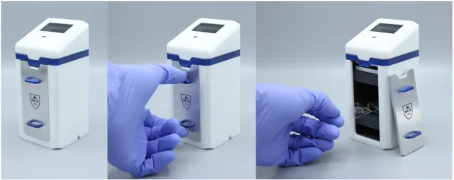
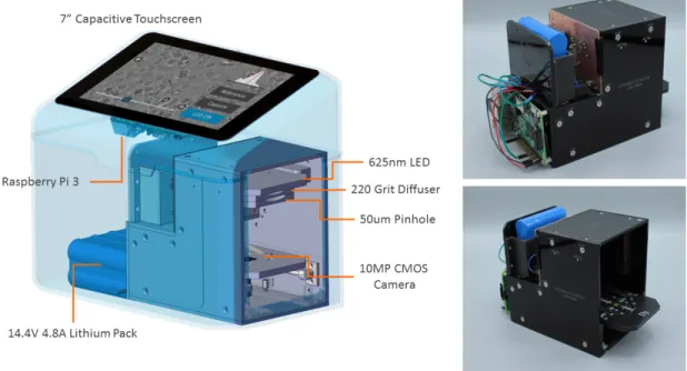
![Figure 3-1: Digital Holographic Imager [27, 7]](https://thumb-eu.123doks.com/thumbv2/123doknet/14165462.473802/24.918.303.622.106.305/figure-digital-holographic-imager.webp)
![Figure 3-6: Twin Image Problem - Latychevskaia et al [16]](https://thumb-eu.123doks.com/thumbv2/123doknet/14165462.473802/27.918.269.654.377.641/figure-twin-image-problem-latychevskaia-et-al.webp)

Documents relatifs
optical complex fields; MO: microscope oil objective; USAF: USAF target located in plane U’ that is imaged in plane U by MO; C: camera plane; C’: plane of the image of the camera
3D tracking the Brownian motion of colloidal particles using digital holographic microscopy and joint reconstruction... 3D tracking the Brownian motion of colloidal particles
This access to the real-time evolution of concen- tration can be used to measure diffusion coefficients, Soret coefficients or dissolution coefficients.. Temperature fringes can be
Specifically, we show that one of the simplest instances of such kernel methods, based on a single layer of image patches followed by a linear classifier is already
We must note that, at high illumination level (p), the bead detection dynamic range is limited by the parasitic light background (arrow 1) which is several orders of magni- tude
Considering an object located in the CCD image plane and diffracting a plane wave, the wave impinging the CCD plane is a spherical wave emerging form F’.. For most of
Our approach using MNPs as stochastic probes thereby presents many advantages. The main obstacle, however, is the light scattered to the far field by the plasmonic object
A coherent source (a monomode laser diode, λ = 635nm) is transformed into a partial spatial coherent source by focusing the incident beam, with lens L1, close to the plane of a

![Figure 4-1: D3 Binding and Reconstruction - Im et al [11]](https://thumb-eu.123doks.com/thumbv2/123doknet/14165462.473802/32.918.226.718.115.396/figure-d-binding-reconstruction-im-et-al.webp)


