Short communication
Cationic Pd(II) complexes acting as topoisomerase II inhibitors:
Synthesis, characterization, DNA interaction and cytotoxicity
Fillipe V. Rocha
a,b,⁎
, Carolina V. Barra
a, Saulo S. Garrido
c, Francine A. Manente
d, Iracilda Z. Carlos
d,
Javier Ellena
e, Andrea S.C. Fuentes
f, Arnaud Gautier
g, Laurent Morel
h, Antonio E. Mauro
a, Adelino V.G. Netto
a aDepartamento de Química Geral e Inorgânica, Instituto de Química, UNESP, CEP 14801-970 Araraquara, SP, Brazil
b
Department of Chemistry, Federal University of São Carlos, São Carlos, SP CEP 13.565-905, Brazil
c
Departamento de Bioquímica e Tecnologia Química, Instituto de Química, UNESP, CEP 14801-970 Araraquara, SP, Brazil
d
Departamento de Análises Clínicas, Faculdade de Ciências Farmacêuticas de Araraquara, UNESP— Univ. Estadual Paulista, P. O. Box 502, Araraquara, São Paulo 14801-902, Brazil
eInstituto de Física, Universidade de São Paulo, São Carlos 13560-970, SP, Brazil f
Laboratory of Molecular Biology, Department of Genetics and Evolution, Federal University of São Carlos, Rodovia Washington Luis Km 235, 13565-905 São Carlos, SP, Brazil
g
Clermont–Université, ICCF, Université Blaise Pascal, CNRS, UMR 6296, 63177 Aubière Cedex, France
hClermont–Université, Université Blaise Pascal, GreD UMR 6247 CNRS, INSERM U931, 24 Avenue des Landais, 63177 Aubière Cedex, France
Pd(II) 4-methyl-3-thiosemicarbazide complexes with interesting in vitro antitumor activity were synthesized. These compounds are un-able to interact to the DNA in low concentration, but are capun-able of
inhibiting human topoisomerase IIα and cathepsin B in the same
range of concentration.
Type II topoisomerases are a class of ubiquitous enzymes that alter DNA topology by catalyzing the passing of an intact DNA double helix through a transient double-stranded break made in a second helix[1]. The topoisomerase IIα (topo IIα) isoform is considered to be the prima-ry pharmacological target for some of the most active drugs currently available for the treatment of human malignancies, since topo IIα is highly up-regulated in transformed and cancer cells[2–5]. All topoisom-erase II-targeting drugs are able to interfere with at least one step of the catalytic cycle[6]. Agents able to stabilize the covalent DNA topo IIα complex are traditionally called topo IIα poisons (e.g. doxorubicin, dau-norubicin, mitoxantrone, amsacrine, and etoposide) whereas those act-ing on any of the other steps in the catalytic cycle are called catalytic inhibitors[6,7]. The cytotoxic effects of these agents may be mediated by the formation of an intermediate covalent DNA-topo IIα complex, which is highly effective in triggering tumor cell apoptosis[8]. There is now a growing body of evidence to support that topo IIα is highly sen-sitive to thiol-reactive agents such as quinones[9], selenium com-pounds [10], cadmium [11], thimerosal [12] and cisplatin [13]. According to Hasinoff and coworkers[13], topo IIα monomer has at leastfive free cysteines that could potentially react with a thiophilic metal center. Given that the electrophilic reaction of soft metals with critical sulfhydryl groups on topo IIα is thought, in part, to be responsi-ble for its inhibition, this suggests that other metal based compounds
bearing thiophillic metal can be investigated as new potential inhibitor of topo IIα.
In this sense, palladium(II) derivatives soon appeared to be excellent candidates because of its high affinity for sulfur-containing ligands[14]. An important breakthrough reported by Caires' group has revealed that the cyclopalladated [Pd2(S(-)C2,N-dmpa)2(μ-dppe)Cl2] {dmpa =
N,N-di-methyl-1-phenethylamine; dppe = 1.2-bis(diphenylphosphino)ethane} interacts with protein thiol groups in the mitochondrial membrane[15, 16]. In addition, this class of compounds has also displayed the ability to inhibit cathepsin B[17], a cysteine protease whose active site is composed of cysteine, histidine, and asparagine residues in a catalytic triad[18]. It is believed that the Cys residue in the cathepsin B catalytic site might be able to bind to Pd(II) center replacing one of its ligands (e.g. Cl−, H2O), while
the remaining ligand moiety might establish favourable interactions with other residues in the active site cavity[19]. Surprisingly, very few studies on the topo IIα inhibition activity of Pd(II) compounds have been described so far[20].
Recently, we have synthesized cationic complexes of the type [PdX(4-PhT)(PPh3)]X (PPh3= triphenylphosphine; 4-PhT =
4-phe-nyl-3-thiosemicarbazide; X = Cl, Br, I, SCN) and evaluated their cyto-toxic effects on breast (LM3) and lung (LP07) tumor murine cells[21]. These Pd(II) compounds were more cytotoxic towards LM3 cell line than cisplatin (30.3 ± 3.7μM), displaying IC50values in the range
2.79–8.84 μM. For the LP07 cells, most of these complexes exhibited cy-totoxic effects similar to that of cisplatin (4.3 ± 0.4μM).
Spectroscopic studies of model reactions with guanosine and aga-rose gel mobility shift assay showed that [PdX(4-PhT)(PPh3)]X
com-plexes have a limited reactivity towards nucleobases and DNA. We therefore hypothesized that one plausible molecular target for this type of complexes might be thiol-containing molecules, such as topo IIα and cathepsin B. As a part of our continuing research into the coor-dination and biological chemistry of metal-based compounds[22–24], we present in this work the synthesis, characterization, DNA interaction
Journal of Inorganic Biochemistry 159 (2016) 165–168
⁎ Corresponding author at: Department of Chemistry, Federal University of São Carlos, São Carlos, SP CEP 13.565-905, Brazil
E-mail address:fillipe@ufscar.br(F.V. Rocha).
http://dx.doi.org/10.1016/j.jinorgbio.2016.02.039
0162-0134/© 2016 Elsevier Inc. All rights reserved.
Contents lists available atScienceDirect
Journal of Inorganic Biochemistry
studies, in vitro cytotoxicity and inhibitory activity evaluation (topoisomerases I and II, cathepsin B) of four cationic compounds of the type [PdX(PPh3)(4-MeT)]X {PPh3= triphenylphosphine;
4-MeT = 4-methyl-3-thiosemicarbazide; X = Cl(1), Br(2), I(3), SCN(4)}. The synthesis of the [PdX(4-MeT)(PPh3)]X was achieved starting
from bis(acetonitrile)dichloropalladium(II) (see synthetic procedures, ESI†). First, PPh3and 4-methyl-3-thiosemicarbazide displace the labile
ligand acetonitrile and one of the two chloro-ions to obtain 1. In a sec-ond step, the Cl ions are easily replaced by Br, I and SCN ions by the ad-dition of two equivalents of their appropriate potassium salt to afford 2– 4 (Scheme 1).
The formation of the [PdX(4-MeT)(PPh3)]X compounds was
con-firmed by elemental analysis, IR and1H NMR spectroscopies and ESI/
MS spectra (ESI†). Formation of the N,S-chelated products was proved by spectroscopic data. IR spectra show a significant shift of 30 cm−1to
lower frequency for theνC_S band after coordination. Variation of ~ 4 ppm downfield of the chemical shift (1
H NMR) was observed for the two N2 protons after complexation. ESI/MS spectra confirmed the expected molecular mass of the compounds. After several attempts, sin-gle crystals suitable for X-ray diffraction studies were obtained for com-plexes 3 and 4 by vapor diffusion of diethyl ether at 4 °C into a saturated methanol. The ORTEP representations of cationic compounds 3 and 4
with the atom labeling scheme are presented in Fig. 1A and B,
respectively.
Thiosemicarbazide acts as a neutral bidentate ligand leading to cis
coordination through its S1 and N2 atoms to form afive membered
ring. The remaining two coordination sites of the palladium atom are
occupied by an iodide ion (3) or a thiocyanate group (4) coordinated trans to S1 and a triphenylphosphine ligand coordinated trans to N2. The palladium atom adopts a distorted square-planar geometry with bond lengths and angles in the usual range expected[25][ESI†].
In vitro cytotoxic activity of ligand 4-MeT and complexes 1–4 was determined towards LM3 (murine mammary adenocarcinoma), LP07 (murine lung adenocarcinoma) and MCF-7 (human breast adenocarci-noma) cell lines by means of the colorimetric MTT assay. The corre-sponding IC50values are listed inTable 1.
Our data for the LM3 cell line show that all the Pd(II) compounds are more cytotoxic than cisplatin, exhibiting IC50values in the range
3.22–6.05 μM. Complex 3 was ca. 9.5 times more active than the refer-ence drug. The complexes 1–4 displayed remarkable cytotoxic levels over the 7.66–11.03 μM concentration range, being ca. 5-fold more po-tent than etoposide against MCF-7 tumor cells. Cisplatin showed no drug response at concentrationsb50 μM. Concerning the cytotoxic ac-tivity on LP07 cells, compounds 1–4 showed interesting cytotoxicity levels (4.69–6.98 μM), values similar to cisplatin (3.9–4.7 μM).
The interaction between complexes 1–4 and purine bases was evalu-ated employing guanosine (Gua) as a model system. Initially such interac-tion has been carried out directly in water media. However, complexes 1– 4 did not react with guanosine in this solvent. In addition, all attempts to remove coordinated halide/pseudohalide by using AgNO3in water were
unsuccessful. Aiming at generating more reactive and soluble complexes capable to readily interact with guanosine, we have employed the AgNO3/
dmf method[26]to afford in situ reactive and soluble solvento/nitrato Pd(II) complexes. The abstraction of anionic groups was performed by the addition of 2 equiv. of AgNO3in dmf leading to the precipitation of
sil-ver halide and silsil-ver thiocyanate, thenfiltered through a Millipore filter. The resulting solution was combined with Gua (2.0 equiv.) and the gua-nosine binding was confirmed by1H NMR spectroscopy in [D
4]MeOD
(after solvent removal) and mass spectrometry (ESI†). The observed
Scheme 1. Synthesis of complexes 1–4.
Fig. 1. Structures of 3 (A) and 4 (B). Table 1
Cytotoxicity values for complexes 1–4. Compound IC50(μM) LM3 LP07 MCF-7 [PdCl(PPh3)(4-MeT)]Cl (1) 5.92 ± 0.59 5.61 ± 1.32 8.78 ± 1.12 [PdBr(PPh3)(4-MeT)]Br (2) 6.05 ± 0.76 6.98 ± 0.71 9.04 ± 1.27 [PdI(PPh3)(4-MeT)]I (3) 3.22 ± 0.84 4.69 ± 0.14 9.54 ± 1.01 [Pd(SCN)(PPh3)(4-MeT)]SCN (4) 5.55 ± 0.20 4.83 ± 0.91 10.63 ± 0.40 4-MeT N100 N100 N100 Cisplatin 30.3 ± 3.7 4.3 ± 0.4 N50 Etoposide nd nd 44.87 ± 4.87 nd., not determined. 166 F.V. Rocha et al. / Journal of Inorganic Biochemistry 159 (2016) 165–168
downfield shift of the1H NMR signal corresponding to guanosine H8
nu-cleus agrees well with the Gua coordination through N7[27]. The mass spectra showed the expected molecular peak at m/z = 755, correspond-ing to the monocharged adduct [Pd(Gua)(4-MeT)(PPh3)]+. Thisfinding
indicates that triphenylphosphine is maintained as the ligand and a pro-ton is abstracted during guanosine coordination, probably through the acidic thiosemicarbazide proton.
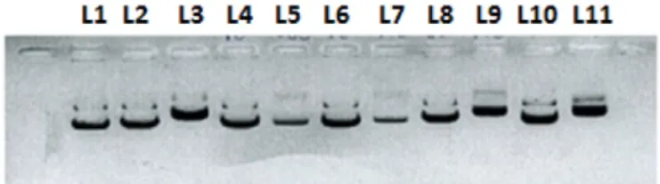
The effect of 1–4 on the structure of a supercoiled DNA was evaluat-ed by their ability to modify the mobility of the circular pNFkB-luc plas-mid in a gel electrophoresis assay (Fig. 2).
All complexes were capable of changing electrophoretic mobility of circular DNA. Nevertheless, it has only occurred at high concentrations (100μM). These data suggested that the cytotoxicity mechanisms of
the compounds 1–4 may be associated with additional molecular
targets. However, it is important to emphasize that the scarce reactivity of these complexes towards plasmid DNA is not sufficient to rule out the involvement of DNA as a molecular target. To assess the mechanism of the cytotoxicity of 1–4, an in vitro study of the inhibitory effects of each complex on topo I and II was undertaken.
The inhibitory ability of complexes 1–4 on the activity of topo I (Fig. 3) and IIα (Fig. 4) has been evaluated by incubating topo and circu-lar plasmideal DNA pBR320 in a concentration-dependent manner [ESI†]. In addition, we have also investigated the inhibitory activity in-duced by analogous compounds of the type [PdX(PPh3)(4-PhT)]X
{4-PhT = 4-phenyl-3-thiosemicarbazide; X = Cl(5), Br(6), I(7), SCN(8)}
[21]aiming at evaluating the influence of substituent at N-3 position of thiosemicarbazide ligand on the observed activity. With regard to DNA-topoisomerase I (topo I), gel shift assay results (Fig. 3) clearly showed that supercoiled pBR320 DNA was fully relaxed by the enzyme (Lane 1, topo I) while none of the Pd(II) compounds were able to inhibit the action of Topo I at 50μM.
As shown inFig. 4, with increasing the concentration of complexes (5, 25, 50, 100μM), the levels of relaxed form were inhibited successive-ly andfinally the activity of topo IIα was totally inhibited at 5 μM
(com-pounds 3, 6, 7 and 8), 25μM (1 and 4) or 50 μM (2). Complex
[PdCl(PPh3)(4-PhT)]Cl (5) appeared unable to inhibit the enzyme at
any tested concentration. The ability of compounds 3, 6, 7 and 8 to in-hibit the relaxation of DNA was six fold more potent than etoposide (35μM), an usual anticancer drug[28].
At this point, it is very difficult to rationalize the observed inhibitory concentration ranges in terms of structure–activity relationship. As a whole, substitution of coordinated chlorido with more polarizable and bulkier anionic ligands, such as I, tend to increase the inhibitory effect. Thisfinding implies that the strength of the Pd–X bond and/or the kinet-ic differences between the coordinated anionkinet-ic ligands play an impor-tant role in topo II inhibition. In addition, we assumed that the steric hindrance at N-3 position of the thiosemicarbazide ligand may also af-fect the observed inhibitory efaf-fect since most of
4-phenyl-3-Fig. 2. Plasmid incubation with 1–4 and cisplatin after 24 h. Line 1: plasmid in water. Line 2: plasmid in water/dmf (2.5%). Line 3: cisplatin (20μm). Line 4: 2 (10 μm). Line 5: 2 (100μm). Line 6: 3 (10 μm). Line 7: 3 (100 μm). Line 8: 4 (10 μm). Line 9: 4 (100 μm). Line 10: 5 (10μm). Line 11: 5 (100 μm).
Fig. 3. Topoisomerase I relaxation assay with 1–8 and camptothecin (CPT) at 100 μM. C− (supercoiled plasmid). C+ (supercoiled plasmid and topoisomerase). 1–4 (A) and 5–8 (B).
Fig. 4. Topoisomerase II relaxation assay. C− (supercoiled plasmid). C+ (supercoiled plasmid, topoisomerase and ATP). 1 and 2 (A). 3 and 4 (B). 5 and 6 (C). 7 and 8 (D). Table 2
IC50values for the cathepsin B inhibition of 3 and 7.
Complex IC50(μM)
[PdI(4-MeT)(PPh3)]I (3) 5.53 ± 0.66
[PdI(4-PhT)(PPh3)]I (7) 8.81 ± 0.80
167 F.V. Rocha et al. / Journal of Inorganic Biochemistry 159 (2016) 165–168
thiosemicarbazide derivatives (6, 7 and 8) demonstrated to be more ac-tive than the majority of their 4-MeT analogues.
We have selected the iodo-complexes 3 and 7 to evaluate their abil-ity to induce the inhibition of cathepsin B. It was determined through the inhibition of cathepsin B activity, which was measured spectrofluorometrically, based on the procedure using the fluorogenic substrate carbobenzoxy-Phe-Arg-7-amido-4-methylcoumarin(Z-Phe-Arg-MCA) (Calbiochem, La Jolla, CA, USA). Fluorescence was measured in a Hitachi F-2500 spectrofluorometer at λex= 380 nm andλem=
460 nm[29]. Inhibitory activity of iodo-complexes 3 and 7 was deter-mined by measuring the residual hydrolytic activity of cysteine pepti-dase after pre-incubation with different inhibitor concentrations. The inhibitory activity against the cysteine protease cathepsin B and the ob-tained IC50values are listed inTable 2.
Both complexes inhibited the enzyme activity, exhibiting IC50values
inferior than 10μM. The inactivation of cathepsin B by compounds 3 and 7 may be attributed to the high nucleophilic character of the thiolate group at the active site of cathepsin B combined with the high thiophilic character of the palladium atom[14]. A nucleophilic attack at the metal center by the thiolate followed by the displacement of the iodide ion or other leaving group coordinated to palladium is ex-pected to be an important step in the interaction of these Pd(II) com-pounds and cathepsin B.
The molecular mechanism underlying the topo II inhibition effect of these palladium(II) compounds is unclear. However, taking into ac-count that they were unable to inhibit topo I, whose action is indepen-dent of the ATP, we hypothesized that these complexes may act as catalytic inhibitors of topo II, competing with ATP for the ATPase do-main[30]. On the other hand, the well-known ability of Pd compounds to interact with biological thiolato groups may also suggest the involve-ment of thiol modification in topo II cleavage complex formation[31]. Nevertheless, further studies are required in order to elucidate the mechanisms of action of these compounds.
Acknowledgements
We thank the São Paulo State University (UNESP), Institute of Chem-istry of Araraquara. This work was sponsored by FAPESP 2013/20156-5, CNPq and CAPES.
Appendix A. Supplementary data
Supplementary data to this article can be found online athttp://dx. doi.org/10.1016/j.jinorgbio.2016.02.039.
References
[1] K.D. Corbett, J.M. Berger, Annu. Rev. Biophys. Biomol. Struct. 33 (2004) 95–118.
[2] A.K. McClendon, N. Osheroff, Mutat. Res. 623 (2007) 83–97.
[3] J.M. Fortune, N. Osheroff, Prog. Nucleic Acid Res. Mol. Biol. 64 (2000) 221–253.
[4] T.K. Li, L.F. Liu, Annu. Rev. Pharmacol. Toxicol. 41 (2001) 53–77.
[5] G. Chen, D. Templeton, D.P. Suttle, D.W. Stacey, Oncogene 18 (1999) 7149–7160.
[6] A.K. Larsen, A.E. Escargueil, A. Skladanowski, Pharmacol. Ther. 99 (2003) 167–181.
[7] K.R. Hande, Update on Cancer Therapeutics, 32008 13–26.
[8] D.A. Burden, N. Osheroff, Biochim. Biophys. Acta 1400 (1998) 139–154.
[9] B.B. Hasinoff, X. Wu, A. Begleiter, L.J. Guziec, F. Guziec Jr., A. Giorgianni, S. Yang, Y. Jiang, J.C. Yalowich, Cancer Chemother. Pharmacol. 57 (2006) 221–233.
[10] N. Zhou, H. Xiao, T.-K. Li, A. N.-E-Kamal, L.F. Liu, J. Biol. Chem. 278 (2003) 29532–29537.
[11] X. Wu, J.C. Yalowich, B.B. Hasinoff, J. Inorg. Biochem. 105 (2011) 833–838.
[12] X. Wu, H. Liang, K.A. O'Hara, J.C. Yalowich, B.B. Hasinoff, Chem. Res. Toxicol. 21 (2008) 483–493.
[13]B.B. Hasinoff, X. Wu, O.V. Krokhin, W. Ens, K.G. Standing, J.L. Nitiss, T. Sivaram, A. Giorgianni, S. Yang, Y. Jiang, J.C. Yalowich, Mol. Pharmacol. 67 (2005) 937–947.
[14] S. Kawanishi, A. Yokoyama, H. Tanaka, Chem. Pharm. Bull. 20 (1972) 262–268.
[15]D.P. Santana, P.A. Faria, E.J. Paredes-Gamero, A.C.F. Caires, I.L. Nantes, T. Rodrigues, Biochem. J. 417 (2009) 247–256.
[16]F.A. Serrano, A.L. Matsuo, P.T. Monteforte, A. Bechara, S.S. Smaili, D.P. Santana, T. Rodrigues, F.V. Pereira, L.S. Silva, J. Machado Jr., E.L. Santos, J.B. Pesquero, R.M. Martins, L.R. Travassos, A.C.F. Caires, E.G. Rodrigues, BMC Cancer 11 (2011) 296–311.
[17]C. Bincoletto, I.L.S. Tersariol, C.R. Oliveira, S. Dreher, D.M. Fausto, M.A. Soufen, F.D. Nascimento, A.C.F. Caires, Bioorg. Med. Chem. 13 (2005) 3047–3055.
[18] R. Mosi, I.R. Baird, J. Cox, V. Anastassov, B. Cameron, R.T. Skerlj, S.P. Fricker, J. Med. Chem. 49 (2006) 5262–5272.
[19] J. Spencer, A. Casini, O. Zava, R.P. Rathnam, S.K. Velhanda, M. Pfeffer, S.K. Callear, M.B. Hursthouse, P.J. Dyson, Dalton Trans. (2009) 10731–10735.
[20]J. Chen, Y.-W. Huang, G. Liu, Z. Afrasiabi, E. Sinn, S. Padhye, Y. Ma, Toxicol. Appl. Pharmacol. 197 (2004) 40–48.
[21]F.V. Rocha, C.V. Barra, A.E. Mauro, I.Z. Carlos, L. Nauton, M. El Ghozzi, A. Gautier, L. Morel, A.V.G. Netto, Eur. J. Inorg. Chem. (2013) 4499–4505.
[22]C.V. Barra, F.V. Rocha, A. Gautier, L. Morel, M.B. Quilles, I.Z. Carlos, O. Treu-Filho, R.C.G. Frem, A.E. Mauro, A.V.G. Netto, Polyhedron 65 (2013) 214–220.
[23] F.V. Rocha, C.V. Barra, A.V.G. Netto, A.E. Mauro, I.Z. Carlos, R.C.G. Frem, S.R. Ananias, M.B. Quilles, A. Stevanato, M.C. da Rocha, Eur. J. Med. Chem. 45 (2010) 1698–1702.
[24] A.C. Moro, A.E. Mauro, A.V.G. Netto, S.R. Ananias, M.B. Quilles, I.Z. Carlos, F.R. Pavan, C.Q.F. Leite, M. Horner, Eur. J. Med. Chem. 44 (2009) 4611–4615.
[25] P. Kalaivani, R. Prabhakaran, E. Ramachandran, F. Dallemer, G. Paramaguru, R. Renganathan, P. Poornima, V. Vijaya Padma, K. Natarajan, Dalton Trans. 41 (2012) 2486–2499.
[26] L.S. Hollis, A.R. Amundsen, E.W. Stern, J. Med. Chem. 32 (1989) 128; G. Zhao, H. Lin, S. Zhu, H. Sun, Y. Chen, J. Inorg. Biochem. 70 (1998) 219–226.
[27] A. Chevry, M.-L. Teyssot, A. Maisonial, P. Lemoine, B. Viossat, M. Traïkia, D.J. Aitken, G. Alves, L. Morel, L. Nauton, A. Gautier, Eur. J. Inorg. Chem. (2010) 3513–3519.
[28] F. Gao, H. Chao, L.-N. Ji, Chem. Biodivers. 5 (2008) 1962–1979.
[29] A. Gianotti, C.A. Sommer, A.K. Carmona, F. Henrique-Silvia, Biol. Chem. 389 (2008) 447–453.
[30] H. Huang, Q. Chen, X. Ku, L. Meng, L. Lin, X. Wang, C. Zhu, Y. Wang, Z. Chen, M. Li, H. Jiang, K. Chen, J. Ding, H. Liu, J. Med. Chem. 53 (2010) 3048–3064.
[31]H. Wang, Y. Mao, A.Y. Chen, N. Zhou, E.J. LaVoie, L.F. Liu, Biochemistry 40 (2001) 3316–3323.