Publisher’s version / Version de l'éditeur:
Biochimica et Biophysica Acta, 1814, 2, pp. 283-289, 2011-02-01
READ THESE TERMS AND CONDITIONS CAREFULLY BEFORE USING THIS WEBSITE. https://nrc-publications.canada.ca/eng/copyright
Vous avez des questions? Nous pouvons vous aider. Pour communiquer directement avec un auteur, consultez la première page de la revue dans laquelle son article a été publié afin de trouver ses coordonnées. Si vous n’arrivez pas à les repérer, communiquez avec nous à PublicationsArchive-ArchivesPublications@nrc-cnrc.gc.ca.
Questions? Contact the NRC Publications Archive team at
PublicationsArchive-ArchivesPublications@nrc-cnrc.gc.ca. If you wish to email the authors directly, please see the first page of the publication for their contact information.
NRC Publications Archive
Archives des publications du CNRC
This publication could be one of several versions: author’s original, accepted manuscript or the publisher’s version. / La version de cette publication peut être l’une des suivantes : la version prépublication de l’auteur, la version acceptée du manuscrit ou la version de l’éditeur.
For the publisher’s version, please access the DOI link below./ Pour consulter la version de l’éditeur, utilisez le lien DOI ci-dessous.
https://doi.org/10.1016/j.bbapap.2010.11.004
Access and use of this website and the material on it are subject to the Terms and Conditions set forth at
Cystine-mediated oligomerization of the Atlantic salmon serum C-type
lectin
Hudson, David M.; Mattatall, Neil R.; Uribe, Elke; Richards, Robert C.; Gong,
Huansheng; Ewart, K. Vanya
https://publications-cnrc.canada.ca/fra/droits
L’accès à ce site Web et l’utilisation de son contenu sont assujettis aux conditions présentées dans le site LISEZ CES CONDITIONS ATTENTIVEMENT AVANT D’UTILISER CE SITE WEB.
NRC Publications Record / Notice d'Archives des publications de CNRC:
https://nrc-publications.canada.ca/eng/view/object/?id=2bd4ee6d-db04-48da-baac-e678f105495b
https://publications-cnrc.canada.ca/fra/voir/objet/?id=2bd4ee6d-db04-48da-baac-e678f105495b
Cystine-mediated oligomerization of the Atlantic salmon serum C-type lectin
David M. Hudson
a,1, Neil R. Mattatall
b, Elke Uribe
a, Robert C. Richards
b,
Huansheng Gong
b,2, K. Vanya Ewart
b,⁎
aDepartment of Biochemistry and Molecular Biology, Dalhousie University, Halifax, NS, B3H 1H7, Canada bNRC Institute for Marine Biosciences, 1411 Oxford St., Halifax, NS, B3H 3Z1, Canada
a b s t r a c t
a r t i c l e
i n f o
Article history:
Received 16 June 2010
Received in revised form 8 November 2010 Accepted 10 November 2010
Available online 23 November 2010
Keywords:
Atlantic salmon C-type lectin Oligomerization Disulfide bond
The Atlantic salmon (Salmo salar) serum lectin (SSL) is a C-type lectin that binds to bacteria including salmon pathogens. SSL has been shown to be oligomeric in salmon serum and it displays a stoichiometric band-laddering pattern when analyzed by SDS-PAGE under non-reducing conditions. In this study, a model was generated for SSL isoform 2 in silico in order to identify cysteines that are available to form intermolecular disulfide bonds facilitating oligomerization. Then, recombinant SSL was expressed in E. coli and mutants were produced at positions Cys72 and Cys149. The SSL preparations were purified by metal-affinity chromatography and shown to be functional by carbohydrate-affinity chromatography. The recombinant SSL formed oligomers, which were evident by non-reducing covalent cross-linking and non-reducing SDS-PAGE; however, the band patterns were different for the mutants, with the maximal and predominant multimer sizes distinct from the wild-type recombinant lectin. Further examination of oligomerization by size exclusion chromatography revealed a subunit number from 35 to at least 110 for the wild-type recombinant SSL and subunit numbers below 9 for each mutant SSL oligomer. Thus, both cysteines were found to contribute to oligomerization of SSL.
Crown Copyright © 2010 Published by Elsevier B.V. All rights reserved.
1. Introduction
Lectins are carbohydrate-binding proteins found in all kingdoms of life. Animal lectins have been classified into types based on structure, function, subcellular localization, and to a lesser extent carbohydrate specificity[1]. C-type lectins are the largest and most complex group of animal lectins[2]. This superfamily of lectins shares a tightly folded C-type lectin-like domain (CTLD) that binds a Ca2+ion. C-type lectins have been shown to play roles in cell–cell recognition, cell–cell adhesion, cellular uptake and innate immunity[3].
The roles of specific C-type lectins in innate immunity depend upon their carbohydrate ligand specificity. Individual CTLDs normally have low affinities for sugar ligands; however, the oligomerization of these proteins allows multivalent interactions, which increase the overall avidity for complex carbohydrates. A lone CTLD isolated from human serum was shown to have a relatively poor affinity for a single monosaccharide, with a dissociation constant (Kd) of about 10−3M[4].
When the CTLDs were allowed to cluster, as occurs during oligomer-ization, the binding affinity increased substantially (Kd~10−9M)[5]. Since lectins are frequently oligomeric, the spatial arrangement and oligomerization of the protein structures have roles in ligand specificity
[6]. Therefore, determinants of oligomeric structure are important components of carbohydrate selectivity of intact lectins.
A salmon serum lectin (SSL) was previously isolated from the blood serum of Atlantic salmon (Salmo salar) using mannose-agarose beads
[7]. Mannose binding was shown to be Ca2+-dependent[7]. SSL was determined to be the predominant C-type lectin in salmon serum and was approximated to have a serum concentration of 5 μg/mL[8]. This lectin was shown to recognize, bind, and opsonize salmonid pathogens
Vibrio (Listonella) anguillarum and Aeromonas salmonicida, and it has
therefore been suggested to play a role in innate immunity[7–9]. SSL consists of a 17 kDa subunit that oligomerizes into a multimeric structure. Using non-reducing sodium dodecyl sulfate polyacrylamide gel electrophoresis (SDS-PAGE), the protein was shown to associate into a range of oligomer sizes, resulting in a ladder-like banding pattern[7]. This pattern was not the result of purification conditions, as the same ladder was detected in salmon serum by immunoblotting using an anti-SSL antibody[10]. The full size of the oligomer was not clear on SDS-PAGE; however, it is apparent that disulfide bonds are required for full oligomerization of SSL, as the laddering pattern on SDS-PAGE gives way to the single 17 kDa monomer band when analyzed under reducing conditions[7,10]. The laddering pattern of
⁎ Corresponding author. Tel.: +1 902 426 7091; fax: +1 902 423 9413.
E-mail address:vanya.ewart@nrc-cnrc.gc.ca(K.V. Ewart).
1Present address: Department of Orthopaedics and Sports Medicine, University of
Washington, 1959 NE Pacific St., Seattle, WA, 98195, USA.
2Present address: Division of Infectious Diseases, Department of Medicine,
University of British Columbia, Vancouver BC, Canada, V5Z 1 L8.
1570-9639/$ – see front matter. Crown Copyright © 2010 Published by Elsevier B.V. All rights reserved. doi:10.1016/j.bbapap.2010.11.004
Contents lists available atScienceDirect
Biochimica et Biophysica Acta
SSL evident upon SDS-PAGE analysis has also been observed for other C-type lectins, specifically in the cases of the chicken (Gallus
domesticus) serum mannan-binding protein [11] and the rainbow trout (Oncorhynchus mykiss) ladderlectin[12], suggesting that these proteins all form variable-subunit oligomers dependent upon disulfide bonds.
Sequence analysis of the protein encoded by the SSL cDNA showed it to be a soluble long-form C-type lectin with a CTLD and an N-terminal extension[13]. As such, the SSL was classified as a group VII C-type lectin, which has no distinct associated domains[13]. The SSL was found to be part of a multigene family with at least five distinct cDNA clones and a fifth isoform was identified by gene sequencing
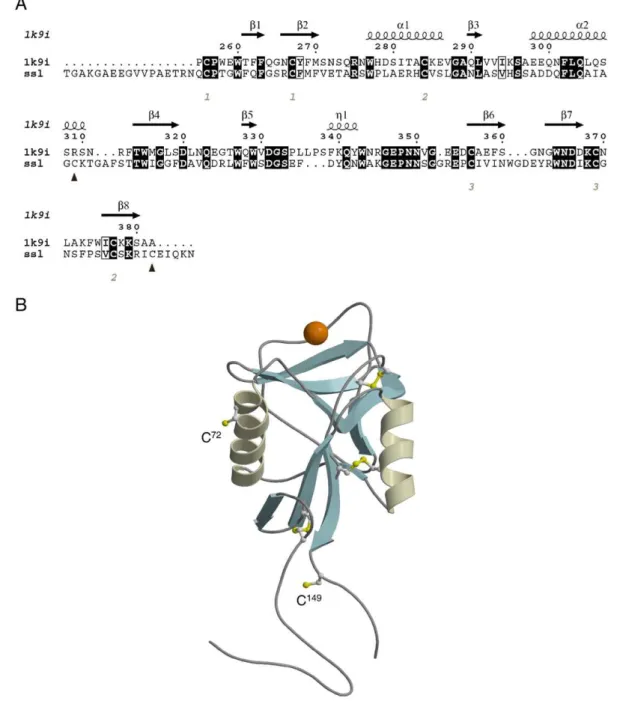
[13]. All were very similar, showing only minor sequence micro-heterogeneity. Sequence alignments between the SSL and represen-tative CTLDs revealed the presence of six highly conserved cysteine residues that form three disulfide bonded cystines in the long-form CTLDs[13,14]. Sequences of the SSL isoforms also revealed cysteine residues that were predicted to be unpaired because they did not align with the conserved cysteine residues forming intramolecular cystine disulfides in the C-type lectin superfamily[13]. In the present study, oligomerization of SSL was examined with particular attention to the roles of unpaired cysteines. The structure of the predominant serum isoform of the SSL was homology modeled to determine the positions of the cysteines relative to the CTLD. Recombinant SSL and site-directed mutants were then produced in E. coli and examined in order to determine the roles of these cysteine residues in oligomer formation. 2. Materials and methods
2.1. Homology modelling of SSL
Homology modelling followed a procedure similar to that used previously for another C-type CTLD[15]. The isoform used was SSL-2, which will hereafter be referred to as SSL except when otherwise specified. Eight iterations of PSI-BLAST[16] were performed using amino acids 1–154 of the sequence (GenBank accession number AAO43606.1) against the NCBI nr database[17]using the Blosum62 matrix[18]with an e-value threshold of 0.001. The resulting position-specific scoring matrix was used to BLAST search sequences in the NCBI pdb database for a structural template on which to homology model SSL. The structure with the highest number of identical amino acids in the BLAST alignment, and with the highest resolution, was dendritic cell specific intercellular adhesion molecule-3 grabbing nonintegrin (DC-SIGN1B; 1k9i)[19], a type II C-type receptor lectin. The alignment was drawn using ESPript[20]. Models of SSL were generated using MODELLER (version 7v7 [21]) with a standard optimization schedule and the PSI-BLAST-generated alignment. Twenty candidate models were screened by objective function score and evaluated using PROCHECK (version 3.5.4 [22]) and PROSA II (version 3.0[23]). Takeoff points for problematic loop regions were modified by hand and were re-evaluated using PROSA II Z-scores. SCWRL3 [24] was used to predict side chain conformations of amino acids in SSL that were not identical with DC-SIGN1B. Amino acids 1–17 and 150–154 of SSL extend beyond the structural template and are represented by random coil. High quality pictures were generated using MOLSCRIPT (version 2.1.2 [25]) and RASTER3D (version 2.7c[26]).
2.2. DNA manipulation
The cDNA encoding isoform SSL-2 (SSL) was prepared as described previously[13]. Briefly, total RNA was isolated from salmon kidney using the RNeasy mini kit (Qiagen, Mississauga, Canada). Residual DNA was removed by DNase-treating the total RNA using the RNase-free DNase set (Qiagen, Mississauga, Canada). First strand cDNA synthesis was prepared using gene-specific primer pairs
complemen-tary to specific 5′ and 3′ untranslated regions of the SSL mRNA in accordance with the Ready-to-Go™ RT-PCR Beads system (GE Healthcare Life Sciences, Baie D'Urfé, Canada). The cDNA insert was amplified using Pfu DNA polymerase using primers 5′-ATA CCA TGG ATA CAG GAG CTA AGG GGG CAG A-3′ and 5′-ATA CTC GAG GTT TTT CTG GAT TTC ACA GAT CT-3′, which contain sites for Nco1 and Xho1 cleavage (shown underlined). The SSL PCR product was gel-purified and extracted using the Qia1Quick gel extraction kit (Qiagen, Mississauga, Canada) and cloned into the pET-22 vector (Novagen, Madison, U.S.A.). The recombinant SSL (rSSL) was propagated in NovaBlue (DE3) competent cells (Novagen, Madison, U.S.A.). All DNA sequences were confirmed on an ABI 377 automated sequencer with Big Dye Terminator Cycle Sequencing Ready Reaction (ABI Prism Applied Sciences, Warrington, United Kingdom) using T7 promoter and T7 terminator primers. Three mutant rSSLs (C149A, C72A and C72S) were constructed using the QuikChange® site-directed muta-genesis kit from Stratagene (La Jolla, U.S.A.) and the oligonucleotides inTable 1.
2.3. Expression of rSSL
SSL and its mutants were expressed in E. coli using the system previously described for the Atlantic salmon C-type lectin receptor C (SCLRC)[15]. Briefly, the cDNA fragment encoding the mature SSL-2 isoform was cloned in frame with a sequence encoding the PelB leader sequence and six-histidine-tag of the pET22b plasmid (Novagen, Madison, U.S.A.), allowing expression of rSSL as a fusion protein directed toward the periplasm. The pDsbABCD1 plasmid, encoding disulfide bond isomerisation enzymes (DSBs), was also expressed concurrently to promote efficient disulfide formation in the periplasm of the bacteria[27]. For protein production, Tuner™ (DE3) competent cells (Novagen, Madison, U.S.A.) were co-transformed with both the rSSL sequence-containing plasmid and the pDsbABCD1 plasmid and grown in LB broth to an OD of 0.6 at 37 °C. The cultures were rapidly cooled on ice to room temperature in order to induce the production of endogenous E. coli chaperones, such as trigger factor[28], and 10 mM CaCl2 was added to provide Ca2+ ions necessary for CTLD folding. The rSSL and the DSBs were induced with 20 μM IPTG and 200 μg/mLL-arabinose, respectively and the cultures were grown for an additional 5 h at room temperature. Cells containing expressed rSSL were collected by centrifugation. The cells were resuspended in 30 mM Tris–HCl pH 8.0, 20% sucrose. EDTA (pH 8.0) was added to a final concentration of 1.0 mM and the mixture was stirred at room temperature for 10 min. The cells again were collected and resus-pended in ice-cold 5 mM MgSO4for 10 min to release the periplasmic proteins by osmotic shock.
2.4. Purification of rSSL
The rSSL was purified by immobilized metal affinity chromatog-raphy (IMAC) on Ni-NTA beads (Novagen, Madison, U.S.A.) using an
Table 1
Primers used to generate mutations in the recombinant SSL. Primer name Primer sequence C149A
Sense primer 5′-CCC TCG GTG TGC TCC AAA AGA ATC GCT GAA ATC CAG AAA AAC-3′
Antisense primer 5′-GTT TTT CTG GAT TTC AGC GAT TCT TTT GGA GCA CAC CGA GGG-3′
C72A
Sense primer 5′-GCA ATT GCG GGG GCC AAG ACT GGC GC-3′ Antisense primer 5′-GC GCC AGT CTT GGC CCC CGC AAT TGC-3′ C72S
Sense primer 5′-GCA ATT GCG GGG TCC AAG ACT GGC GC-3′ Antisense primer 5′-GC GCC AGT CTT GGA CCC CGC AAT TGC-3′
AKTA FPLC (Amersham Biosciences, Piscataway, U.S.A.). The concen-trated periplasmic fraction was buffer exchanged into phosphate-buffered saline (PBS), 20 mM sodium phosphate, 300 mM NaCl, pH 7.8. Elution of rSSL from the Ni-NTA was achieved using a gradient of 0–250 mM imidazole in 20 mM sodium phosphate, 300 mM NaCl, pH 7.8 and analyzed on 15% SDS-PAGE. The fractions containing purified protein were dialyzed into TCS buffer (10 mM Tris–HCl pH 7.5, 1.0 mM CaCl2, 150 mM NaCl). The rSSL mutants were purified by the same method as the wild-type protein. Protein concentrations were determined by the Bradford protein assay using Bradford Reagent (Protein Assay Dye Reagent Concentrate, Bio-Rad Laboratories, Hercules, U.S.A.).
2.5. Electrophoretic and disulfide analysis
Standard SDS-PAGE was performed using a Novex system (Invitrogen, Carlsbad, U.S.A.). Gels were either stained using GelCode Blue™ Coomassie blue (Pierce, Rockford, U.S.A.) or transferred to Hybond-P PVDF (Amersham Biosciences, Piscataway, U.S.A.) for western blot analysis. Western blots were probed with 1:1000 anti-His primary antibody (Tetra-anti-His™ antibody, mouse monoclonal IgG, Qiagen, Mississauga, Canada) and visualized using 1:5000 secondary antibody (anti-mouse IgG whole molecule peroxidase conjugate, Sigma-Aldrich, St. Louis, U.S.A.) and chemiluminescent enzyme substrate (ECL-Plus, Amersham Biosciences, Piscataway, U.S.A.).
SDS-PAGE using high-resolution gels was performed using a Bio-Rad mini-gel system (Bio-Bio-Rad Laboratories, Hercules, U.S.A.) with Next Gel solution (Amresco, Solon, OH, U.S.A.) and silver stained[29]. For comparison, SSL from salmon serum was obtained as previously described[7]and analyzed under the same conditions as rSSL.
The rSSL was subject to analysis using 5,5′-dithio-bis-(2-nitrobenzoic acid) (DTNB), purchased along with cysteine hydrochloride standards from Pierce (Thermo Fisher Scientific, Rockford, U.S.A.). Reactions were carried out following the manufacturer's recommendations and absor-bances were read at 412 nm.
2.6. Carbohydrate-binding assays
The sugar-binding properties of the rSSL proteins were assessed using both mannose- and mannan-binding assays. Mannan-Sepharose beads were prepared by covalently coupling yeast mannan to Sepharose 4B (Amersham Biosciences, Piscataway, U.S.A.) [30]. Mannose-agarose beads were purchased from Sigma-Aldrich (St. Louis, U.S.A.). Each rSSL protein sample (0.8 μg) was incubated with the beads for 30 min at room temperature in TCS buffer. Beads were washed with TCS buffer and bound proteins were eluted in 1 mL fractions with TE buffer (10 mM Tris–HCl, pH 7.5, 10 mM EDTA).
2.7. Cross-linking assay
Optimal chemical cross-linking conditions were determined empirically using the homobifunctional chemical cross-linking reagent bis(sulfosuccinimidyl)suberate (BS3) (Sigma-Aldrich, St. Louis, U.S.A.). Proteins (0.4 μg) were incubated for 15 min at room temperature in HCS buffer (25 mM HEPES, pH 7.8, 10 mM CaCl2, 150 mM NaCl) and then treated with 5 mM BS3and an additional incubation for 1 h at room temperature. Samples were analyzed on 4–12% NuPAGE™ Novex Bis-Tris gradient gels (Novagen, Madison, U.S.A.).
2.8. Size exclusion chromatography
Size exclusion chromatography was performed using a BioSep-Sec-S4000 column (300 × 7.80 mm) (Phenomenex, Torrance, U.S.A.) on a HP-1090 HPLC (Waters, Milford, U.S.A.). All analyses were performed at room temperature with a flow rate of 1 mL/min in HCS
buffer. Proteins were detected by UV absorbance at 230 nm. All standards and samples were run in duplicate and the average values were used for analyses.
3. Results
3.1. Modeled SSL structure
A homology model of SSL was generated in order to confirm the three disulfide bond positions of the three conserved cystines and to examine the two remaining cysteines. The alignment of SSL and DC-SIGN1B used to construct the model was produced using BLAST and then modified to accommodate structural stress in unstructured loop regions (Fig. 1A). It showed the predicted positions of Cys72 and Cys149 (Fig. 1B). Both Cys residues were located well away from the Ca2+- and carbohydrate-binding site of the lectin such that intermolecular disulfides at those residues would not be expected to impede carbohydrate ligand binding. Additionally, each free cysteine was in an exposed location, with Cys72 located on the external face of the second alpha helix and Cys149 located in the unstructured C-terminal extension just beyond the last beta strand.
3.2. Production of rSSL and mutagenesis
Wild-type and mutant rSSL were produced as six-histidine-tagged proteins in E. coli. The average yield of purified rSSL was approxi-mately 2.0 mg/L of culture. Each rSSL had an apparent molecular mass of 18 kDa on SDS-PAGE, which was larger than the 17 kDa monomer observed with purified native SSL[7]. This mass was consistent with the predicted protein mass determined from its sequence with an additional C-terminal methionine-aspartate from periplasmic proces-sing and an N-terminal six-histidine-tag, which is 18.4 kDa.
The disulfide-mediated oligomerization of the wild-type rSSL was compared with the native SSL from salmon serum by SDS-PAGE under denaturing, non-reducing conditions. The laddering patterns of both SSL preparations were equivalent (Fig. 2). Slight differences in the migration of bands may reflect the distinct masses and sequences of these proteins because of tag addition to the rSSL. The rSSL was analyzed for binding to DTNB and showed a 1:0.93 molar ratio, implying a single free sulfhydryl group per subunit on average. This suggests that free sulfhydryls are present in the oligomerized protein, which is consistent with laddering.
3.3. Evidence of native folding in rSSL and mutants
A mannan-binding assay was used to further examine Ca2+ -dependent sugar binding and oligomerization of rSSL. Individual CTLDs normally have low affinities for their sugar ligands[31]and strong interaction typically results from avidity achieved by the interaction of multiple CTLD subunits in an oligomeric lectin. As such, binding to mannan can be interpreted as evidence of proper folding and oligomerization. The wild-type rSSL and the C72A and C72S mutants showed binding to the mannan and each was eluted using EDTA (Fig. 3A, C and D). In contrast, although the C149A mutant was present in the EDTA elution fractions like with the C72A mutants, a proportion of this protein eluted earlier, suggesting that it did not bind fully to the mannan beads (Fig. 3B). Binding to mannan could be compromised by incomplete CTLD folding and/or incomplete oligo-merization. A carbohydrate-binding assay using mannose monomer was therefore performed in order to determine whether the C149A mutant CTLD could bind mannose. Binding of the C149A mutant to mannose was indistinguishable from that of the wild-type rSSL (Fig. 3E and F), suggesting that the individual CTLDs of each mutant were properly folded and functional. Taken together, these results suggest that the C149A mutant does not oligomerize to the same extent as the other mutants and wild-type rSSL.
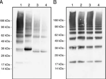
3.4. Electrophoretic and chemical cross-linking analysis of oligomerization in rSSL
Initial comparison of oligomerization patterns for the rSSL proteins (wild-type, C149A, C72A, and C72S) was made by SDS-PAGE under non-reducing conditions. All four proteins oligomerized into larger disulfide-bonded structures evident under non-reducing conditions (Fig. 4, lanes 1–4). The observed patterns of the C72A and C72S mutants were distinct from the wild-type rSSL, with loss of the apparent trimer. In addition, larger oligomers appeared far less abundant than those for the wild-type protein. The C149A mutant pattern was different from all the others as it appeared to oligomerize into different units than the wild-type or the C72 mutants, with predominant bands corresponding to predicted dimers and tetramers. These findings suggest that all four proteins have some capacity to oligomerize below 200 kDa. When the proteins were
reduced, the wild-type and mutant rSSLs were monomeric as expected (data not shown).
To determine the predominant oligomeric state of rSSL proteins in their native forms, the proteins were covalently cross-linked using BS3 and then analyzed by reducing SDS-PAGE. The wild-type and mutant proteins produced comparable stoichiometric banding patterns on SDS-PAGE following cross-linking (Fig. 4B, lanes 1–4). After cross-linking the wild-type and mutant rSSL, proteins appeared to form units with every stoichiometry ranging from monomers to assemblies with sizes exceeding the maximal resolvable size for the gel used, which was approximately 200 kDa. This finding is consistent with the formation of oligomers of various sizes in which monomers associate by disulfide and/or non-covalent interaction. This experiment indicates that the distribution of oligomer sizes up to approximately 200 kDa does not differ between the wild-type and mutant proteins.
Fig. 1. Homology model of wild-type SSL model. (A) The alignment of SSL with 1k9i is shown with identical residues in black boxes and similar residues in white boxes. Cysteines signalled with numbers in italics correspond to disulfide bonds with the numbers specifying the bonds. Black triangles show the cysteines that do not appear to be positioned to form disulfide bonds in the model. Secondary structure of 1k9i is shown above the sequences. (B) Ribbon diagram of the wild-type SSL model. The structure of SSL is shown with alpha helices depicted as coiled ribbons and beta strands as arrows with the rest of the structure depicted as coils. Ca2+is shown as an orange sphere. Cysteines and disulfide-bonded
cystine are shown in ball-and-stick representation. The unpaired cysteine residues (C72 and C149) are labeled.
3.5. Examination of oligomerization in rSSL by size exclusion chromatography
Size exclusion chromatography was employed to further examine oligomerization. A comparison of the HPLC chromatograms for the rSSLs revealed similar peaks at approximately 10 min (Fig. 5) that eluted slightly ahead of the blank buffer peak; they were very weak in the wild-type rSSL and more distinct in the mutants. The C149A, C72A and C72S peaks were at 9.82, 10.08 and 10.07 min, respectively. These peaks were beyond the resolvable range of the column and therefore they could not be sized accurately. The wild-type rSSL emerged as a predominant peak at 5.56 min, which indicated a size greater than 2000 kDa and fell within the void volume of the column. An extended shoulder was evident, which continued to elute until 7.5 min, corresponding to a mass of 650 kDa. The data suggested that the wild-type rSSL exists predominantly as oligomers ranging from 35 to at least 110 subunits, whereas the mutants appear to form much smaller oligomers (below 9 subunits).
4. Discussion
This study was designed to better define the oligomeric structure of native SSL and the roles of intermolecular cysteines therein. The SSL-2 isoform was used in this study for two reasons. First, the SSL-2 cDNA encodes a protein with a sequence that exactly matches the N-terminal sequence of an SSL previously isolated from salmon serum
[7], suggesting that it is a predominant isoform. Second, SSL-2 is the single isoform identified with two cysteines appearing to be unpaired, as there is a Ser in position 72 in all others. An unpaired cysteine aligning with C149 is present in other fish group VII C-type lectins including the egg lectins of shishamo smelt (Osmerus (Spirinchus)
lanceolatus) [32,33], the skin mucus lectin of conger eel (Conger
myriaster) [34], the serum lectin of spotted halibut (Verasper
variegatus)[35], and the serum ladderlectin of rainbow trout[36]. In contrast, C72 not only appears unique to SSL-2 among SSL isoforms, but it is absent from all other reported group VII C-type lectins of fish. Nonetheless, both cysteines were predicted to be on the surface of SSL-2 in the structural model and therefore both are potentially available for intermolecular disulfides mediating oligomerization. Therefore, mutations in this study included C149A, C72A, with Ala
Fig. 2. Comparison of native and recombinant SSL purified by mannan-affinity chromatog-raphy. Non-reducing SDS-PAGE and silver staining were carried out as described in the
Materials and methods. Gel panels are (A) native SSL salmon blood serum and (B) rSSL.
Fig. 3. Carbohydrate-binding analysis of wild-type and mutant rSSLs. Analyses were performed by western blotting under reducing conditions and detected using the anti-tetra-His antibody as described in theMaterials and methods. For each blot, lanes are (1) 0.8 μg of rSSL applied to column, (2) flow through, (3) wash 1, (4) wash 2, (5) wash 3, (6) wash 4, (7) wash 5, (8) elution 1, (9) elution 2, and (10) elution 3. Mannan-binding assays are shown in panels A (rSSL), B (C149A), C (C72A) and D (C72S). Mannose-binding assays are shown in panels E (rSSL) and F (C149A).
Fig. 4. Oligomerization in the cross-linked and reduced rSSLs and the non cross-linked and non-reduced rSSLs. Sample treatments, SDS-PAGE and western blotting were carried out as described in theMaterials and methods. In each image, lanes are (1) rSSL, (2) C149A, (3) C72A, and (4) C72S. Samples in panel A were not cross-linked prior to analysis and they were run under non-reducing conditions in SDS-PAGE. Samples in panel B were cross-linked prior to analysis and then run under reducing conditions in SDS-PAGE.
chosen as the substitution as it is unlikely to disrupt protein–protein interaction and C72S, with S chosen because it is the residue at that position in the other SSL isoforms.
The two C72 mutants recognized mannan effectively, suggesting proper folding and oligomerization; however, the investigation of low molecular mass oligomers by non-reducing SDS-PAGE showed a lower average size of disulfide-bonded chains, with tetramers as the predominant form and abundant dimers present. Analysis of the same protein by size-exclusion chromatography revealed clear differences between this mutant and wild-type rSSL in larger-range oligomer formation. The oligomers formed by the C72 mutants had apparent sizes below that of aldolase (158 kDa), whereas the wild-type lectin formed much larger oligomers with at least 35 subunits predicted. The native size range of rSSL oligomers is consistent with the 67-subunit MBL oligomers detected by atomic force microscopy[37], whereas the smaller sized oligomers formed by the C72 mutants are more in line with those found in other fish type VII lectins. The oligomers of the C72 mutants were consistent in size with most other fish group VII lectins, which lack cysteine at that position. Examples include the lectins of spotted halibut, and smelt that form disulfide-linked dimers
[32,35], and the conger eel lectin that forms tetramers composed of two disulfide-linked dimers[34]. The oligomerization behaviour of trout ladderlectin is difficult to reconcile with the current results. Although reported sequences for ladderlectin lack a cysteine corresponding to C72 in SSL, the lectin forms a similar laddering pattern to that of SSL on non-denaturing PAGE, suggesting the
formation of a range of larger oligomers[12]. If the lectin is encoded by a multigene family, it is possible that the trout produces isoforms with and without this cysteine, as is the case for SSL and further cloning may reveal such an isoform. Screening for a C72-bearing isoform in ladderlectin would shed light on the extent of structural and functional similarity between these two lectins.
The C149A mutant showed both structural and functional features of impaired oligomerization. Size exclusion chromatography of C149A showed a reduced oligomer size, which was indistinguishable from those of the C72 mutants; however, non-reducing SDS-PAGE analysis revealed dimers to be the predominant form of this mutant. The binding of C149A to mannose was indistinguishable from that of wild-type rSSL, whereas its binding to mannan was incomplete in comparison with the wild-type protein. This result suggested a functional difference between C149A and the other SSL forms studied here. In the case of another C-type lectin, the mammalian mannan-binding lectin (MBL), which is an oligomeric CTLD-bearing lectin with a collagen tail, functional differences of this nature reflect a structural difference. The use of beads conjugated with mannan instead of mannose was found to lead to selection for the larger oligomers, since these would bind with greater avidity to the mannan polymer than the smaller oligomers with fewer subunits available for recognition of individual mannose units within the mannan[38]. The current results for C149A appear to reflect a similar difference in avidity. The predominantly dimeric C149A mutant appears to have very limited avidity, whereas the other mutants and the wild-type rSSL, which bind mannan effectively, appear to assemble into tetramers and larger structures with greater frequency. This would suggest that the minimal SSL stoichiometry for avidity in mannan binding is the four units of a tetramer exhibited by the C72 mutants and wild-type rSSL. It is not clear whether there is an advantage of forming larger assemblies of SSL in ligand binding or in opsonisation. Nonetheless, different sized oligomers of MBL have shown differences in binding to carbohydrate and to the MBL-associated serine protease that induces complement [39,40]. Further suggestion of a role of C149 in full oligomer formation comes from the study of dimeric group VII lectins of fish, in which a similar mutation appears to have resulted in monomeric lectin. The three C-terminal cysteine residues of smelt egg lectin were mutated together and the resulting triple mutant failed to dimerize[33]. It is unclear which combination of the three cysteines was responsible for the intermolecular disulfide linkage in that case; however, one cysteine corresponded to the C149 in the rSSL, raising the possibility that this cysteine is involved in the covalent association of subunits for both proteins.
In the current analyses, both C72 and C149 are shown to have roles in oligomerization. This is a logical result because the formation of ladders on non-reducing SDS-PAGE implies a covalent disulfide network among subunits and such a network would require that each subunit be able to form a minimum of 2 separate intermolecular disulfides. It is not clear whether the intermolecular disulfides are between corresponding cysteines (72–72 and 149–149) in adjacent lectins or whether disulfides also form between the two positions (C72 and C149). A mixture of the corresponding and non-corresponding disulfides between subunits would account for the laddering exhibited on non-reducing SDS-PAGE. Assuming steric differences among the various associations of SSL subunits, a mixture of these disulfides would result in an assortment of covalent oligomers that reach different subunit stoichiometries before further addition becomes sterically unfavourable. It is difficult to predict how many subunits in such a structure would be able to bind at once to a complex ligand; however, mannan-binding assay results on the mutant rSSLs suggest that tetramers are the minimal unit for meaningful avidity.
Non-covalent interaction among the subunits in SSL oligomers was not directly investigated in this study; however, cross-linking of the rSSL and mutants followed by reducing SDS-PAGE has shown non-covalent interaction to have a role in oligomerization. Although the size
Fig. 5. Size exclusion analysis of wild-type and mutant rSSLs. Chromatogram panels are (A) wild-type rSSL, (B) C149A, (C) C72A, (D) C72S and (E) buffer blank analyzed as described in theMaterials and methods. Elution times for molecular mass standards are shown with letters as follows: (D) dextran blue (2000 kDa), (T) thyroglobulin (669 kDa), (F) ferritin (440 kDa), (C) catalase (232 kDa), and (A) aldolase (158 kDa).
distribution of bands in the non-reducing SDS-PAGE analysis revealed the covalent linkages to be predominantly dimers for C149A and slightly larger units for C72A and S, these mutants formed a very clear and evenly distributed ladder structure within the resolving range of the gel when the lectin was covalently cross-linked in vitro to stabilize the units prior to reducing SDS-PAGE. This indicates that all forms of the rSSL examined were able to oligomerize over a substantial range of sizes due in part to covalent interactions under native conditions. The non-covalent interactions are not sufficient to allow the disulfide mutants to oligomerize to the extent that the wild-type SSL does, as shown by the size exclusion chromatography results. Nonetheless, in the lower size ranges, non-covalent association of subunits is evident. This is consistent with the non-covalent dimerization reported for other long-form CTLDs in fish, such as the dimeric smelt antifreeze protein[41]. Non-covalent interactions among subunits are also prevalent throughout the C-type lectin superfamily, lending support to the notion that these interactions could be anticipated in rSSL.
In summary, the current study has revealed that SSL forms very large oligomers that span a range of sizes. Two cysteines were shown to mediate the covalent association of subunits in these oligomers through intermolecular disulfide bonds and there was also evidence of non-covalent interaction. The very large structures formed by SSL stand in contrast to the majority of fish group VII lectins, which occur as dimers, tetramers and octamers. The formation of dimers is required for agglutination; however, the biological implications of larger units formed by the different fish type VII lectins remain unclear and these may be informative in determining the roles of these lectins in the animals that produce them.
Acknowledgements
We thank Kelly Soanes (NRC-IMB) for review of the manuscript. This work was supported by an NSERC Discovery grant (KVE) and by the NRC Institute for Marine Biosciences. This is NRC publication number 51771. References
[1] K. Drickamer, M.E. Taylor, Evolving views of protein glycosylation, Trends in Biochemical Sciences 23 (1998) 321–324.
[2] K. Drickamer, A.J. Fadden, Genomic analysis of C-type lectins, Biochemical Society Symposium (2002) 59–72.
[3] K. Drickamer, M.E. Taylor, Biology of animal lectins, Annual Review of Cell Biology 9 (1993) 237–264.
[4] W.I. Weis, K. Drickamer, Structural basis of lectin-carbohydrate recognition, Annual Review of Biochemistry 65 (1996) 441–473.
[5] N. Kawasaki, T. Kawasaki, I. Yamashina, Isolation and characterization of a mannan-binding protein from human serum, Journal of Biochemistry 94 (1983) 937–947. [6] D.C. Kilpatrick, Animal lectins: a historical introduction and overview, Biochimica
et Biophysica Acta 1572 (2002) 187–197.
[7] K.V. Ewart, S.C. Johnson, N.W. Ross, Identification of a pathogen-binding lectin in salmon serum, Comparative Biochemistry and Physiology. Part C: Pharmacology, Toxicology & Endocrinology 123 (1999) 9–15.
[8] C.A. Ottinger, S.C. Johnson, K.V. Ewart, L.L. Brown, N.W. Ross, Enhancement of
anti-Aeromonas salmonicida activity in Atlantic salmon (Salmo salar) macrophages by a
mannose-binding lectin, Comparative Biochemistry and Physiology. Part C: Pharmacology, Toxicology & Endocrinology 123 (1999) 53–59.
[9] K.V. Ewart, S.C. Johnson, N.W. Ross, Lectins of the innate immune system and their relevance to fish health, ICES Journal of Marine Science 58 (2001) 380–385. [10] L. Stratton, S. Wu, R.C. Richards, K. Vanya Ewart, Oligomerisation and
carbohydrate binding in an Atlantic salmon serum C-type lectin consistent with non-self recognition, Fish & Shellfish Immunology 17 (2004) 315–323. [11] S.B. Laursen, J.E. Hedemand, S. Thiel, A.C. Willis, E. Skriver, P.S. Madsen, J.C. Jensenius,
Collectin in a non-mammalian species: isolation and characterization of mannan-binding protein (MBP) from chicken serum, Glycobiology 5 (1995) 553–561. [12] L.E. Jensen, S. Thiel, T.E. Petersen, J.C. Jensenius, A rainbow trout lectin with multimeric
structure, Comparative Biochemistry and Physiology 116 (1997) 385–390. [13] R.C. Richards, D.M. Hudson, P. Thibault, K.V. Ewart, Cloning and characterization of
the Atlantic salmon serum lectin, a long-form C-type lectin expressed in kidney, Biochimica et Biophysica Acta 1621 (2003) 110–115.
[14] M. Meier, M.D. Bider, V.N. Malashkevich, M. Spiess, P. Burkhard, Crystal structure of the carbohydrate recognition domain of the H1 subunit of the asialoglycopro-tein receptor, Journal of Molecular Biology 300 (2000) 857–865.
[15] K.H. Soanes, K.V. Ewart, N.R. Mattatall, Recombinant production and character-ization of the carbohydrate recognition domain from Atlantic salmon C-type lectin receptor C (SCLRC), Protein Expression and Purification 59 (2008) 38–46. [16] M. Krauthammer, A. Rzhetsky, P. Morozov, C. Friedman, Using BLAST for
identifying gene and protein names in journal articles, Gene 259 (2000) 245–252. [17] D.L. Wheeler, D.M. Church, A.E. Lash, D.D. Leipe, T.L. Madden, J.U. Pontius, G.D. Schuler, L.M. Schriml, T.A. Tatusova, L. Wagner, B.A. Rapp, Database resources of the National Center for Biotechnology Information, Nucleic Acids Research 29 (2001) 11–16.
[18] S. Henikoff, J.G. Henikoff, Performance evaluation of amino acid substitution matrices, Proteins 17 (1993) 49–61.
[19] H. Feinberg, D.A. Mitchell, K. Drickamer, W.I. Weis, Structural basis for selective recognition of oligosaccharides by DC-SIGN and DC-SIGNR, Science (New York, N.Y.) 294 (2001) 2163–2166.
[20] P. Gouet, X. Robert, E. Courcelle, ESPript/ENDscript: Extracting and rendering sequence and 3D information from atomic structures of proteins, Nucleic Acids Research 31 (2003) 3320–3323.
[21] A. Sali, T.L. Blundell, Comparative protein modelling by satisfaction of spatial restraints, Journal of Molecular Biology 234 (1993) 779–815.
[22] M.M.W. Laskowski, R.A. Moss, J.M. Thornton, PROCHECK: a program to check the stereochemical quality of protein structures, Journal of Applied Crystallography 26 (1993) 283–291.
[23] M.J. Sippl, Recognition of errors in three-dimensional structures of proteins, Proteins 17 (1993) 355–362.
[24] A.A. Canutescu, A.A. Shelenkov, R.L. Dunbrack Jr., A graph-theory algorithm for rapid protein side-chain prediction, Protein Science 12 (2003) 2001–2014. [25] P.J. Kraulis, MOLSCRIPT: a program to produce both detailed and schematic plots
of protein structures, Journal of Applied Crystallography 24 (1991) 946–950. [26] E.A. Merritt, M.E. Murphy, Raster3D Version 2.0. A program for photorealistic
molecular graphics, Acta Crystallographica 50 (1994) 869–873.
[27] Y. Kurokawa, H. Yanagi, T. Yura, Overexpression of protein disulfide isomerase DsbC stabilizes multiple-disulfide-bonded recombinant protein produced and transported to the periplasm in Escherichia coli, Applied and Environmental Microbiology 66 (2000) 3960–3965.
[28] O. Kandror, A.L. Goldberg, Trigger factor is induced upon cold shock and enhances viability of Escherichia coli at low temperatures, Proceedings of the National Academy of Sciences of the United States of America 94 (1997) 4978–4981.
[29] M. Swain, N.W. Ross, A silver stain protocol for proteins yielding high resolution and transparent background in sodium dodecyl sulfate-polyacrylamide gels, Electrophoresis 16 (1995) 948–951.
[30] O. Bertrand, S. Cochet, J.-P. Cartron, Expanded bed chromatography for one-step purification of mannose binding lectin from tulip bulbs using mannose immobilized on DEAE Streamline, Journal of Chromatography 25 (1998) 19–28. [31] W.I. Weis, M.E. Taylor, K. Drickamer, The C-type lectin superfamily in the immune
system, Immunological Reviews 163 (1998) 19–34.
[32] M. Hosono, S. Sugawara, Y. Ogawa, T. Kohno, M. Takayanagi, K. Nitta, Purification, characterization, cDNA cloning, and expression of asialofetuin-binding C-type lectin from eggs of shishamo smelt Osmerus [Spirinchus] lanceolatus, Biochimica et Biophysica Acta 1725 (2005) 160–173.
[33] S. Sugawara, M. Hosono, Y. Ogawa, M. Takayanagi, K. Nitta, Molecular and sugar-binding heterogeneity of C-type lectins from Osmerus (Spirinchus) lanceolatus eggs, Biological & Pharmaceutical Bulletin 28 (2005) 791–796.
[34] S. Tsutsui, K. Iwamoto, O. Nakamura, T. Watanabe, Yeast-binding C-type lectin with opsonic activity from conger eel (Conger myriaster) skin mucus, Molecular Immunology 44 (2007) 691–702.
[35] A. Hatanaka, N. Umeda, N. Hirazawa, Characterization of highly concentrated serum lectins in spotted halibut Verasper variegatus, Parasitology 135 (2008) 359–369. [36] S. Russell, K.M. Young, M. Smith, M.A. Hayes, J.S. Lumsden, Cloning, binding
properties, and tissue localization of rainbow trout (Oncorhynchus mykiss) ladderlectin, Fish & Shellfish Immunology 24 (2008) 669–683.
[37] H. Jensenius, D.C. Klein, M. van Hecke, T.H. Oosterkamp, T. Schmidt, J.C. Jensenius, Mannan-binding lectin: structure, oligomerization, and flexibility studied by atomic force microscopy, Journal of Molecular Biology 391 (2009) 246–259. [38] T. Vorup-Jensen, E.S. Sorensen, U.B. Jensen, W. Schwaeble, T. Kawasaki, Y. Ma, K.
Uemura, N. Wakamiya, Y. Suzuki, T.G. Jensen, K. Takahashi, R.A. Ezekowitz, S. Thiel, J.C. Jensenius, Recombinant expression of human mannan-binding lectin, International Immunopharmacology 1 (2001) 677–687.
[39] R. Wallis, J.Y. Cheng, Molecular defects in variant forms of mannose-binding protein associated with immunodeficiency, Journal of Immunology 163 (1999) 4953–4959.
[40] M.R. Dahl, S. Thiel, M. Matsushita, T. Fujita, A.C. Willis, T. Christensen, T. Vorup-Jensen, J.C. Jensenius, MASP-3 and its association with distinct complexes of the mannan-binding lectin complement activation pathway, Immunity 15 (2001) 127–135.
[41] J.C. Achenbach, K.V. Ewart, Structural and functional characterization of a C-type lectin-like antifreeze protein from rainbow smelt (Osmerus mordax), European Journal of Biochemistry 269 (2002) 1219–1226.