REVIEW
Connexins participate in the initiation and progression
of atherosclerosis
Sandrine Morel&Laurent Burnier&Brenda R. Kwak
Received: 27 January 2009 / Accepted: 7 April 2009 / Published online: 30 April 2009 # Springer-Verlag 2009
Abstract Connexins are members of a large family of transmembrane proteins that form hemichannels or gap junctions. These channels allow the exchange of ions and small metabolites between the cytosol and extracellular space or between neighboring cells. Connexins are impor-tant in vascular physiology; they support radial and longitudinal cell-to-cell communication in the vascular wall. Four connexins are expressed in the vascular wall: Cx37, Cx40, Cx43, and Cx45. Their expression is not uniform in all blood vessels and varies with vascular territory and species. Significant changes in the expression pattern of vascular connexins have been described during the development of atherosclerosis, a progressive inflam-matory disease. In this review, we provide an overview of
(1) the tools used to study the involvement of connexins in atherosclerosis, (2) the participation of connexins in atherogenesis, (3) the increasing interest of a polymorphism in the human connexin37 gene as marker of cardiovascular disease, and (4) the possible therapeutic implications of connexins.
Keywords Connexins . Hemichannels . Gap junctions . Atherosclerosis . Restenosis
Abbreviations
ADP Adenosine diphosphate AMI Acute myocardial infarction ApoE Apolipoprotein E
ATP Adenosine triphosphate CAC Carotid artery compliance CAD Coronary artery disease
CL Cytoplasmic loop
CT COOH-termini
Cx Connexin
ECs Endothelial cells
EL Extracellular loop
ECM Extracellular matrix FMD Flow-mediated dilatation GFP Green fluorescent protein
GJIC Gap junctional intercellular communication HMG-CoA 3-Hydroxy-3-methylglutaryl-CoA
IMT Intima-media thickness
LDLR Low density lipoprotein receptor
MC Monocytes/macrophages
NT NH2-termini
PCI Percutaneous coronary intervention SMCs Smooth muscle cells
TNF-alpha Tumor necrosis factor-alpha DOI 10.1007/s00281-009-0147-6
S. Morel
:
L. Burnier:
B. R. KwakDivision of Cardiology, Department of Internal Medicine, Geneva University Hospitals, University of Geneva, Geneva, Switzerland
L. Burnier
Division of Angiology and Hemostasis, Department of Internal Medicine,
Geneva University Hospitals, University of Geneva, Geneva, Switzerland
L. Burnier
Service and Central Laboratory of Hematology,
Centre Hospitalier Universitaire Vaudois, University of Lausanne, Geneva, Switzerland
B. R. Kwak (*)
Foundation for Medical Research, Division of Cardiology, Geneva University Hospitals,
64 Av. De la Roseraie, 1211 Geneva 4, Switzerland
Introduction
Atherosclerosis is a progressive disease characterized by accumulation of lipids, macrophages, T lymphocytes, and smooth muscle cells (SMCs) in large- and medium-sized arteries. Clinical and experimental observations have led to the notion that the initiating step of this disease is an endothelial dysfunction [1]. This dysfunction leads to an increase in the expression of various cell adhesion molecules and to the secretion of chemoattractants. As a consequence, monocytes transmigrate between endothelial cells (ECs) to infiltrate into the arterial intima where they propagate and mature. These intimal macrophages ingest lipids and transform into macrophage foam cells, forming the earliest atherosclerotic plaque. This initial atheroscle-rotic lesion is then covered by SMCs that migrate from the media to the intima. In the intima, SMCs proliferate and secrete extracellular matrix (ECM) components that partic-ipate to the formation of a strong fibrous cap. In the advanced atherosclerotic plaque, foam cells die and release lipids that form the necrotic core of the lesion. In time, the fibrous cap might rupture inducing the formation of a thrombus at the site of the lesion [2, 3]. This process is implicated in 60% of sudden death by thrombosis [4].
It is now well recognized that inflammation is central in all stages of atherosclerosis [5, 6]. Similar to other inflammatory diseases, paracrine intercellular communica-tion involving cytokines, chemokines, and growth factors is known to play an important role in the development of the atherosclerotic lesions. In this review, we summarize the evidence that another form of intercellular communication involving connexins (Cx) might also be implicated in the development of the disease.
Connexins, connexons, and gap junctions Connexins
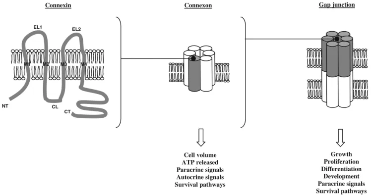
Connexins are members of a family consisting of 20 proteins in mice and 21 in humans. Cx genes are composed of a 5′-untranslated exon, an intron of variable length, an exon harboring the complete coding region, and the 3′-untranslated exon [7]. In some cases, the untranslated exon can be spliced. Two nomenclatures exist to distinguish the different Cx. The first one is based on the molecular mass deduced from their cDNA sequences (for example, the protein with a molecular weight about 43 kDa is called Cx43) The second one is based on sequence similarity and length of the cytoplasmic loop (CL) and separates Cx in four groups: alpha, beta, gamma, and delta (in this system, Cx43 is named“alpha 1” because it has been the first alpha Cx found) [8,9]. As shown in Fig.1, a Cx exhibits four
α-helical transmembrane domains (M1–M4), two extracel-lular loops (EL1 and EL2) that are linked by two disulfide bonds, a short CL, and cytoplasmic NH2- and
COOH-termini (NT and CT, respectively). The EL1 and EL2 have highly conserved amino acid sequences and are involved in docking and recognition of compatible Cx [10]. In contrast, the CL is more variable. The CT is characteristic for each Cx; it varies significantly in both length and composition. This domain acts as a substrate for specific kinases or as a partner for other proteins. As a consequence, this domain is involved in the modulation of channel activity in response to appropriate biochemical stimuli [11–14]. Cx work in concert and may have some overlap in function, but the function of one Cx can often not be replaced completely by another Cx isoform [15–17].
Connexons
Cx are synthesized in the endoplasmic reticulum where they form hexameric connexons. This process is completed in the Golgi apparatus after which connexons traffic to the plasma membrane (for reviews, see [10, 18]). The con-nexon is named homomeric when made of identical Cx and heteromeric when multiple Cx isoforms are involved. During intracellular transit, connexons are associated with microtubules to improve the efficiency of the delivery process [19, 20]. During this process, connexons likely remain in the closed configuration to avoid exchange between cytosol and intracellular compartments.
Once integrated in the plasma membrane, the connexons generally stay in a closed configuration under normal conditions, but they may open upon different stimuli such as removal of extracellular calcium, hypoxic or ischemic stress, mechanical stimulation, and dephosphorylation [21, 22]. These hemichannels allow the passage of ions and small molecules (~1,000 Da) such as ATP or NAD+ between cytoplasm and extracellular space (Fig. 1). Such exchanges are implicated in regulation of cell volume, in paracrine or autocrine signaling, and activation of survival pathways [23].
Gap junction intercellular channels
Once inserted in plasma membrane, connexons can diffuse laterally and dock with another connexon from a neighboring cell. This association between two connexons occurs via noncovalent interactions between the extracel-lular loops and permits the formation of a gap junction intercellular channel. The channel is named homotypic if connexons are identical and heterotypic if the two connexons are different. These gap junction channels allow the exchange of ions, small metabolites, second messengers, linear peptides, or small silencing RNA
between connected cells [23–25] (Fig.1). Connexins have a half-lives ranging from 1 to 5 h [25]. Gap junctional intercellular communication (GJIC) allows not only for fast coordinated activities such as contraction of cardiac cells or transmission of neuronal signals at electrical synapses but also for slower physiological processes such as cell growth and development.
Tools to study connexins Chimeric connexins
Chimeric connexins are Cx tagged at the CT with different compounds like, for example, chemiluminescent aequorin or green fluorescent protein (for reviews, see [10, 26]). These protein reporters induce an increase of the molecular mass of the Cx and may limit the flexibility of the CT. However, these tags do generally not change the trafficking characteristics of the proteins and do also not inhibit the formation of gap junction channels. Chimeric connexins have been used to visualize the intracellular trafficking of Cx on their way to form gap junctions.
Transfected cells
Transfection of cultured cell lines is often used to study gap junction channel characteristics and possible functions of Cx. In general, experiments are realized with communication-incompetent HeLa cells [27], N2A cells [28], or SKHep1
cells [29] transfected with human or mouse Cx. In these cell lines, specific permeability and charge selectivity of each type of gap junction channel is conserved. In the case of the study of atherosclerosis, Wong et al. [30] performed transfection of H36.12j mouse peritoneal macrophage cell line to prove the implication of Cx37 in monocytic cell adhesion.
Transgenic mice
In vivo studies toward atherosclerosis are currently performed by the use of two well-characterized mouse models: the apolipoprotein E (ApoE−/−) knockout mice and the low-density lipoprotein receptor knockout mice (LDLR−/−). ApoE−/−mice present high plasma cholesterol concentration (400 to 500 mg/dL) and develop spontane-ously foam cell-rich depositions throughout the arterial tree [31]. LDLR−/− mice have a lower increase of plasma NT
CT
Connexin Connexon Gap junction
Cell volume ATP released Paracrine signals Autocrine signals Survival pathways Growth Proliferation Differentiation Development Paracrine signals Survival pathways Electrical signals M1 M2 M3 M4 CL EL1 EL2
Fig. 1 Schematic representation of connexin topology, a connexon, and a gap junction channel. Connexins have four transmembrane domains (M1, M2, M3, and M4), two extracellular loops (EL1 and EL2), a cytoplasmic loop (CL), and cytoplasmic NH2- and COOH-termini (NT and CT, respectively). Connexons or hemichannels are
formed by the association of six connexins. They mediate trans-membranous exchange of ions and small metabolites. The associations of connexons from two neighboring cells form gap junction channels. They allow exchange of metabolites or second messengers up to 1-kDa molecular mass between cells in contact
cholesterol level (175 to 225 mg/dL) and they develop only minimal atherosclerotic lesions in aortic roots when fed a normal chow diet [32]. Upon feeding these mouse models with a high-cholesterol diet, they both rapidly develop advanced lesions throughout the vascular tree. Atheroscle-rosis is also studied in Watanabe heritable hyperlipidemic rabbit or in pigs. However, mice are preferred because they can be interbred with other knockout mice to study the effects of those specific molecules.
To date, 20 Cx have been identified in mice, and 18 Cx-deficient mice exist [33]. An important problem with mice knockout for vascular Cx is that these deletions are often lethal. For example, Cx43−/− and Cx45−/− die in utero or shortly after birth. Cx45 knockout mice display normal vasculogenesis but subsequent transformation into mature vessels is interrupted [34]. Cx43 knockout mice present a swelling and a blockage of the right ventricular outflow tract from the heart that lead to failure in pulmonary gas exchange [35]. As a consequence, heterozygous Cx43 mice have been used to study the implication of Cx43 in atherosclerosis. Cx43+/− mice express 50% of the normal Cx43 level [36]. Cx40−/−mouse are viable but sometimes develop arrhythmias [37,38]. Indeed, Cx40 is prominent in the atrium and its lack leads to a slow conduction within the atrium increasing the risk for atrial flutter. Furthermore, the absence of Cx40 in the His-Purkinje system may lead to a bundle branch block, preferably in the right bundle. In addition, these mice are hypertensive [39]. Cx37−/−mouse are viable as well and their heart function is normal, but females are infertile [40]. Cx37 and Cx40 are co-expressed in ECs. As a consequence, a double deletion of these Cx induces embryonic lethality in these mice due to an excessive dilation of blood vessels [41]. Gene deletion of Cx in mice is frequently used to study disease processes; however, the absence of one Cx may lead to a decreased or
increased expression of another Cx. Cx are differentially expressed in different vascular cell types (see below), and cell-specific Cx deletion may be used to study the implication of the Cx in a particular cell type. For example, the Cre-loxP system under the control of the Tie2 promoter has been used to create mice in which Cx40 or Cx43 has been deleted from the endothelium only [42–44].
Connexin antisense, blocking peptides, and enhancing peptides
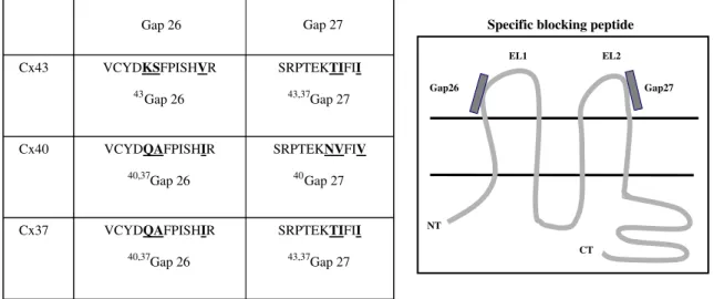
Different compounds such as heptanol, octanol, 18α-glycyrrhetinic acid, carbonoxolone, or oleamide are known to inhibit GJIC, but their actions are nonspecific [45–47]. As a consequence, specific peptides to block Cx have been generated. These short peptides have a sequence homology with the conserved extracellular loops of Cx, and they can selectively inhibit the activity of one type of gap junction channels in cells containing multiple Cx [48]. They have a rapid and reversible mode of action, and they are nontoxic for the cells (for review, see [49]). Initially, two blocking peptides have been designed, Gap26 and Gap27, corresponding respectively to the sequence of the first and the second extracellular loop of Cx43 (Fig. 2). These blocking peptides have been used in studies toward gap junctional communication between ECs and SMCs and between ECs and macrophages [50–52]. Derivatives of these first blocking peptides have been created to be more or less specific for Cx37 and Cx40 (Fig. 2). They are efficient in both rodent and human cells, thus reflecting the high degree of amino acid conservation in the extracellular loops. In a study concerning atherosclerosis, blocking peptides have been used to prove the implication of Cx37 in the adhesive property of macrophages [30]. Furthermore, reducing conductivity of Cx43 channels with 43Gap26
Gap26
EL1
NT
CT
Specific blocking peptide
Gap27 EL2 Gap 26 Cx43 VCYDKSFPISHVR 43 Gap 26 SRPTEKTIFII 43,37 Gap 27 Cx40 VCYDQAFPISHIR 40,37Gap 26 SRPTEKNVFIV 40Gap 27 Cx37 VCYDQAFPISHIR 40,37 Gap 26 SRPTEKTIFII 43,37 Gap 27 Gap 27
Fig. 2 Connexin-specific blocking peptides. Gap26 and Gap27 sequences correspond, respectively, to the sequence of the first and the second extracellular loops (EL1 and EL2) of connexins. These synthetic peptides inhibit direct intercellular communication in a connexin-specific manner
decreased the adhesion of neutrophils to ECs in vitro and reduced neutrophil recruitment in a mouse model of acute lung inflammation in vivo [53].
There is increasing attention for peptides that selectively open gap junctions. Such peptides might be of particular interest to treat cardiac arrhythmias [54]. More than a decade ago, the first peptide enhancing gap junctional communication (AAP10) has been identified [55, 56]. Stable analogs have been developed since then (ZP123, rotigaptide), although the exact molecular target of AAP10-derived peptides remains to be identified [57]. Moreover, Shibayama et al. [58] identified by phage display a series of RXP peptides capable of binding to the CT of Cx43. One of these peptides, RXP-E, prevents heptanol- and acidosis-induced closure of Cx43 gap junction channels in trans-fected cells and neonatal cardiomyocytes [58,59].
The function of connexins in tissues or cells is also investigated using siRNA or antisense oligonucleotides. In experiments toward skin repair, a Cx43 antisense has been prepared in a gel and used in combination with various types of skin lesion. The application of the Cx43 antisense decreased inflammation, lessened scarring, and improved wound closure [60,61]. In a recent work, we tested Cx43 antisense to inhibit in vitro the dedifferentiation and migration of SMCs, processes implicated in restenosis after ballooning injury [62].
Connexins and gap junctions in healthy vessels
Vascular function is dependent on radial and longitudinal cell-to-cell communication in the vascular wall [63,64]. It has been extensively reviewed how paracrine molecules such as nitric oxide and prostaglandins secreted by ECs control the vascular tone by their effects on SMCs [65,66]. In addition, cell-to-cell communication via gap junctions may also be implicated in the control of vascular function [67, 68]. Homomeric and heteromeric channels and homocellular and heterocellular gap junctions are described in the vascular wall (EC–EC, SMC–SMC, EC–SMC gap junctions) [64, 69, 70]. Four Cx are expressed in the vascular wall: Cx37, Cx40, Cx43, and Cx45. Their expression is not uniform in all blood vessels and varies with vascular territory and species. Usually, Cx37 and Cx40 are co-expressed in ECs, whereas Cx43 and Cx45 are present in SMCs (for a review, see [64]). Nevertheless, Cx37 and Cx40 are also found in SMCs of small elastic or resistance arteries or during development [71, 72], and Cx43 is described in ECs at branch points of arteries [73]. The importance of vascular gap junctions has been demonstrated by the fact that Cx deletion alters normal vascular functioning (as described above). Thus, gap junctions interconnecting neighboring ECs allow the spread
of signals along the vessel wall, which serve to coordinate vessel behavior. For example, Cx provide the molecular basis for ascending dilatations (i.e., conducted dilations) in arterioles that are required for substantial increases in blood flow during exercise [74, 75]. Among the vascular Cx known to be expressed in ECs, Cx40 appears to play a central role in the arterial conducted response [76,77]. This is supported by evidence showing that Cx40-deficient mice display impaired conduction of vasodilatation along arterio-les in mouse cremaster muscle. Of note, the effect of the absence of one Cx on the expression of other Cx is unclear. In a study on mouse aorta, deletion of Cx40 induced a decrease of Cx37 in ECs, but an increase of Cx37 and Cx43 in the media [78]. In contrast, other investigators reported that deletion of Cx40 was associated with upregulation and redistribution of Cx37 in ECs [79]. Finally, the deletion of Cx37 did not significantly modify the expression of Cx40 in the mouse aortic endothelium [30].
Thus, Cx and gap junctions are central in vascular physiology. In addition, the expression of Cx is modified and implicated in pathological situations such as diabetes, hypertension, or atherosclerosis.
Connexins in atherosclerosis
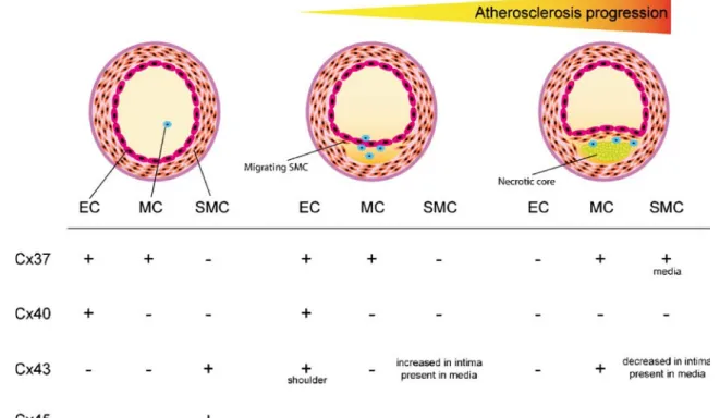
As mentioned earlier, atherosclerosis is usually studied in mouse deficient in ApoE−/−or for the LDLR−/−[80]. The additional deletion of Cx37, Cx40, and Cx43 in these atherosclerotic-susceptible mice permits to determine the implication of each Cx in atherogenesis. Moreover, the use of the Cre-LoxP system [81] allows for studying the importance of Cx in specific cell types. As shown in Fig. 3, significant changes in the expression pattern of vascular Cx have been described during the formation of atherosclerotic plaques (reviewed in [82]). Moreover, the expression of Cx in vascular wall is influenced by atherosclerotic risk factors, such as turbulent flow, hyper-tension, and hypercholesterolemia, which act on ECs, on SMC activation, and proliferation and on the inflammatory process (for a review, see [64]). By affecting Cx expression, these risk factors modify gap junction channel- or hemichannel-mediated communication between cells and influence the progression of atherosclerosis.
Connexin37
Cx37 is expressed in healthy ECs, but disappears from these cells in the advanced atherosclerotic plaque [83]. A similar observation has been reported in mice subjected to a high-cholesterol diet for several months [84]. Moreover, Cx37 expression is found in macrophages in early and late atheroma [83, 85]. Taking into account that ECs and
monocytes/macrophages have central roles in atherogenesis, Cx37 was expected to play a role during atherosclerotic lesion development. Although Cx37−/−mice are infertile due to the absence of ovulation [40], their vascular function is normal [77] and they can be used to study atherosclerosis.
In a study performed in our laboratory, Cx37−/− mice have been crossed with ApoE−/− mice and subjected to a high-cholesterol diet for 10 weeks [30]. The deletion of Cx37 accelerated atherosclerotic lesion development in thoracic–abdominal aorta and in aortic sinus in comparison with control (Cx37+/+ApoE−/−) mice. Thus, Cx37 appeared to have a protective effect against atherosclerosis in ApoE−/− mice. The initiating event in atherosclerosis is endothelial dysfunction that leads to monocyte recruitment at the site of the injury in response to chemotactic factors. Monocytes adhere to the ECs, transmigrate across them, and penetrate in the arterial intima where they proliferate, mature, and accumulate lipids to finally progress into macrophage foam cells. As Cx37 is expressed in ECs and monocytes and gap junctions between ECs and leukocytes have been demonstrated (for reviews, see [85,86]), the role of Cx37 in transmigration was then investigated. For this purpose, fluorescent control and Cx37-deficient monocytes or macrophages were introduced by adoptive transfer in control and Cx37-deficient hypercholesterolemic mice, and the number of fluorescent leukocytes within atherosclerotic
plaques was determined [30]. These experiments showed that the deletion of Cx37 in monocytes/macrophages increased the number of leukocytes in atherosclerotic plaques. Interestingly, the presence or the absence of Cx37 in ECs did not influence the transmigration of leukocytes. Thus, the recruitment of leukocytes appeared dependent on the presence of Cx37 in monocytes/macro-phages rather than on the existence of gap junction between these cells and ECs, or on intercellular communication within the endothelium. Next, in vitro experiments showed that the deletion of Cx37 in monocytes/macrophages enhanced adhesion of these cells. Similar results were obtained using α-glycyrrhetic acid and connexin blocking peptides. Together, these results demonstrated the implica-tion of funcimplica-tional hemichannels in the adhesion of monocytes/macrophages during atherosclerotic plaque de-velopment. Inflammation is mediated in part by extracellu-lar purines (ATP, ADP, adenosine), and ATP is known to pass through various types of gap junctions and hemi-channels [87]. The absence of Cx37 or the inhibition of Cx37 by blocking peptides reduced the release of ATP by monocytes/macrophages and increased their adhesion [30]. The use of extracellular ATP scavenger confirmed this result. We therefore proposed that Cx37 protects against atherosclerosis by regulating ATP-dependent monocyte adhesion [30].
Fig. 3 Evolution of connexin expression during atherosclerosis progression. Atherosclerosis is a progressive vascular pathology implicating endothelial cells (ECs), monocytes/macrophages (MCs), and smooth muscle cells (SMCs). Four connexins are expressed in the
vascular wall. Cx37, Cx40, and Cx43 have dynamic expression patterns in healthy vessels and during atherogenesis. Relatively little information is available on Cx45
Cx37 is also expressed in medial SMCs beneath advanced atherosclerotic lesions in mice [83]. A similar Cx37 expression pattern is observed in advanced athero-sclerotic plaques in human carotid artery. The role of Cx37 in these SMCs remains to be established.
Connexin40
Similar to Cx37, Cx40 is present in ECs of healthy vessels, and this Cx disappears from the endothelium covering advanced atherosclerotic plaques [83]. Endothelial Cx40 expression and function is influenced by different factors such as oxidative stress, prothrombotic molecules, pro-inflammatory cytokines, and classic cardiovascular risk factors [88]. Recent studies have shown that abrupt reoxygenation following hypoxia reduces gap junctional coupling between microvascular ECs of wild type but not of Cx40-deficient mice. The reduction in GJIC involves a protein kinase A-dependent pathway and reactive oxygen species [89]. Hyperhomocysteinemia is associated with impaired endothelial-dependent vasodilation and increased risk of atherosclerosis and thrombosis. In a rat model of hyperhomocysteinemia, a downregulation of Cx40 mRNA is described [90]. Tumor necrosis factor alpha (TNF-alpha) is a potent pro-inflammatory cytokine that activates ECs during pathological situations. In human umbilical vein endothelial cells, TNF-alpha treatment decreases Cx40 [91]. Furthermore, a recent study on streptozotocin diabetic mice suggests that downregulation of Cx40 expression and the resultant inhibition of GJIC contribute to coronary vascular dysfunction in diabetes [92].
As mentioned earlier, Cx40-deficient mice are hyperten-sive. This hypertension is, in part, due to the requirement of Cx40 for longitudinal transmission of endothelium-dependent vasodilator responses [76]. Moreover, blood pressure is controlled by the renin–angiotensin–aldosteron system. In the juxtaglomerular apparatus, Cx40 gap junctions link the ECs of the afferent arteriole to the renin-secreting cells. Two distinct studies have shown that the deletion of Cx40 increases the number of renin-secreting cells and enhances the renal production and release of renin [93, 94]. The role of Cx40 in the renal barosensor mechanism controlling renin synthesis and secretion has been demonstrated with a pharmacological gap junction blocker [94]. The hypertension observed in mice with ubiquitous Cx40 deletion prevents an in vivo study of the implication of Cx40 in atherosclerosis. To avoid this deleterious effect, we have made atherosclerosis-susceptible ApoE−/− mice with specific Cx40 deletion in ECs. Indeed, these mice are not hypertensive and have a normal heart rate [43]. Preliminary data indicate that the EC-specific deletion of Cx40 induced increased atheroscle-rotic plaque development compared to control mice [43].
These results suggest an atheroprotective role of Cx40, but the mechanisms implicated remain to be investigated. Connexin43
In healthy vessels, Cx43 is mostly expressed in SMCs. Coronary arteries of hearts removed from patients under-going cardiac transplantation show markedly increased Cx43 expression in gap junctions between intimal SMCs compared with undiseased vessels [95]. In advanced atherosclerotic plaques, the intimal expression of Cx43 declines. In LDLR−/− mice fed a cholesterol-rich diet, Cx43 increased in intimal SMCs in early atherosclerotic lesions [83]. Cx43 expression was also shown in macro-phage foam cells of mouse aorta and of human carotid artery [83, 96], in ECs covering the shoulder region of atherosclerotic lesions [83], and in ECs at branch points of large arteries [73].
Atherosclerotic plaques are generally formed at branch points or at curved areas of large arteries that are regions associated with turbulent blood flow [97]. Oscillatory shear stress induces a high and rapid increase of endothelial Cx43 expression [98]. The effects of unidirectional shear stress on endothelial Cx43 expression are less clear. This shear stress is associated with an increase or with no change in Cx43 expression dependent on the experimental conditions used [98, 99]. Increased hydrostatic pressure does not modify the Cx43 level in ECs [98].
As previously mentioned, Cx43 knockout mice die in utero or shortly after birth [35]. As a consequence, we have studied the implication of Cx43 in the development of the atherosclerotic plaques by interbreeding atherosclerotic-susceptible LDLR−/− mice with heterozygous Cx43+/− mice. The expression of Cx43 was reduced by half in Cx43+/−mice [36]. Ten-week-old Cx43+/+ LDLR−/− and Cx43+/−LDLR−/−mice were fed a cholesterol-rich diet for 14 weeks to evaluate the progression of atherosclerosis. Cx43+/−LDLR−/− mice showed reduced atherosclerotic plaque development in the thoracic–abdominal aorta and in the aortic sinus by about 50% in comparison to Cx43+/+LDLR−/− mice [100]. Moreover, atherosclerotic lesions in Cx43+/−LDLR−/− mice have smaller lipid cores and fewer macrophages, whereas leukocyte counts in peripheral blood were similar between both groups of mice. In addition, the fibrous cap of atherosclerotic plaques in Cx43+/−LDLR−/−mice contained more SMCs and intersti-tial collagen. During the development of the atherosclerotic lesion, SMCs migrate from the media to the intima where they multiply and produce components of the ECM. During this process, SMCs are transformed from the differentiated contractile state to the activated synthetic state. Curiously, synthetic SMCs have been described to express higher levels of Cx43 than the contractile phenotype [62,101].
The vulnerability of atherosclerotic lesions to rupture is dependent of the content of SMCs and macrophages, the extent of collagen within the lesion and the size of the lipid core. As plaque rupture might lead to acute myocardial infarction, targeting Cx43 may be promising for stabiliza-tion of the plaque. Actually, mechanisms by which Cx43 influences atherosclerotic lesion formation and plaque stability are not clearly identified. It has been hypothesized that the effect of Cx43 might depend on specific atheroma-associated cell types [102]. In ECs, Cx43 might induce or enhance endothelial dysfunction. In leukocytes, Cx43 might enhance their migration and proliferation, or might decrease their apoptosis in the atherosclerotic plaque. In SMCs, Cx43 might limit their activation, proliferation, and migration from the media to the intima, or might increase their apoptosis in the plaque. These hypotheses concerning the effects of Cx43 in ECs, leukocytes, and SMCs during atherogenesis are currently investigated in mice with cell-specific deletion of Cx43. Preliminary data showed that endothelial-specific deletion of Cx43 in mice provided beneficial effects on both the natural progression and composition of atherosclerotic lesions [44].
Connexin37 polymorphism and atherosclerosis
Krutovskikh et al. have discovered in 1996 a first polymorphism in the human Cx37 gene while investigating lung and breast carcinoma for mutations [103]. This polymorphism results in an amino acid change at codon 130, which is situated in the cytoplasmic loop of Cx37. Following this first description of a Cx37 gene
polymor-phism in the human population, Boerma and coworkers [104] have described a second polymorphism in the human Cx37 gene in 1999. This polymorphism corresponds to a cytosine-to-thymine replacement at the position 1019 in the Cx37 gene, resulting in an amino acid alteration in the CT of the protein; a proline residue at position 319 (Cx37-319P) is replaced by a serine residue (Cx37-319S). In this study, authors showed that Cx37-319P was correlated with the occurrence of significant atherosclerotic plaques in carotid arteries in the Swedish population [104]. These results have been confirmed in coronary arteries in other studies performed in Taiwan [105] and Switzerland [106]. In contrast, Collings et al. showed that C1019T polymor-phism was not related with markers of subclinical athero-sclerosis in young adults in Finland [107]. When myocardial infarction was used as a clinical endpoint, a study in Japanese population showed that Cx37-319S was associated with increased risk in men [108]. This result has been confirmed in a Sicilian population [109]. Discrepant results obtained between the different studies might depend on various reasons such as the chosen clinical endpoint (coronary stenosis versus acute myocardial infarction), the sample size, phenotypic heterogeneity, racial differences, or environment interactions. The different studies concerning Cx37 polymorphism are listed in Table 1. A recent study described the influence of smoking on atherosclerosis in relation with the Cx37-C1019T polymorphism. The authors observed that variation in the Cx37 gene might modify the effects of smoking on the vascular function [110].
As previously mentioned, Cx37 interferes with leukocyte adhesion by releasing ATP. Monocytes transfected with Cx37-319P or Cx37-319S present different adhesion Table 1 Cx37 polymorphism studies in relation to artery disease and myocardial infarction
Population Pathology Cx37 polymorphism prognostic marker References
Hypertensive Swedish men Carotid disease P [104]
Taiwanese patients receiving coronary catheterization CAD P [105]
Japanese patients with myocardial infarction MI S in men [108]
No relation in women
Sicilian young men with acute MI MI S [109]
Irish population with premature onset CAD CAD, MI No relation [118]
American patients with acute coronary syndrome 3-year mortality S [119]
Swiss patients requiring angiographic evaluation CAD P [106]
Centenarian Sicilian men MI S [120]
Finnish children and adolescents IMT, CAC, FMD No relation [107]
Northern Han Chinese patients with CAD CAD P in men [121]
No relation in women
The most studied polymorphism in the human Cx37 gene corresponds to a cytosine-to-thymine replacement at the position 1019 (Cx37-1019C and Cx37-1019T), which leads to a replacement of proline residue at position 319 319P) in the carboxyl tail by a serine residue (Cx37-319S)
CAD coronary artery disease, MI myocardial infarction, IMT intima-media thickness, CAC carotid artery compliance, FMD flow-mediated dilatation
properties and this difference seems due to different ATP permeability [30]. Indeed, Cx37-319P transfected mono-cytes release more ATP than Cx37-319S transfected monocytes and have lower adhesive properties. These differences may explain the protective effect on acute myocardial infarction conferred by this polymorphic vari-ant. In a larger context, other inflammatory pathologies in which monocytes/macrophages are involved may also be associated with this Cx37 polymorphism. In general, the identification of predisposing genetic factors might help to identify individuals with increased risk for the development of atherosclerosis or other inflammatory pathologies.
Therapeutic implications of connexins in the treatment of atherosclerosis
Regulation of connexin expression by statin treatment Reduction of atherosclerosis-related morbidity and mortal-ity is possible by lowering plasma cholesterol with statins (inhibitors of 3-hydroxy-3-methylglutaryl-CoA (HMG-CoA) reductase) [111]. In addition, in vivo and in vitro studies suggest that statins modulate atherogenesis and plaque rupture by mechanisms independent of the decrease of plasma cholesterol concentration [112]. Interestingly, various types of statins dose dependently inhibited Cx43 expression in human vascular cells [100]. In addition, the presence ofL-mevalonate abolished the effect of statins on Cx43 expression, confirming that HMG-CoA reductase was responsible for this reduction. The reduction in Cx43 expression was associated with reduction in GJIC. In mice, statin treatment does not reduce plasma lipid levels due to a compensatory upregulation of HMG-CoA reductase. The maintenance of high plasma lipids allows the study of the pleiotropic effects of statins independently of their effects on plasma cholesterol. Statins reduce Cx43 expression in atherosclerotic plaque of LDLR−/− mice and displays beneficial changes in plaque morphology [100]. These observations are comparable to the observations in Cx43+/−LDLR−/−mice. Otherwise, long-term hyperlipidemia in mice decreased Cx37 and Cx40 expression in aorta [84]. Treatment with simvastatin reversed this hyperlipidemia-induced decrease in Cx37 and Cx40. Thus, the statin-induced regulation of Cx expression might be classified as one more pleiotropic beneficial effect of these compounds. Connexin expression and percutaneous coronary
interventions
Coronary atherosclerosis might lead to the occlusion of the artery and to myocardial infarction. This vascular problem is often treated by percutaneous coronary intervention (PCI)
consisting of balloon dilatation with or without stent implantation. Clinical studies have shown, however, that the long-term efficacy of PCI is limited by restenosis or renarrowing of the arteries at the site of intervention [113]. Indeed, the stretching of a diseased artery can induce an exaggerated response to injury that involves the recruitment and infiltration of leukocytes into the damaged site and a surge in cytokines and growth factors. Moreover, medial SMCs undergo a phenotypic modulation from a contractile to a synthetic phenotype, proliferate, and migrate toward the intima. Together, these events induce the formation of the neointima. Drug-eluting stents prevent restenosis by inhibiting neointimal hyperplasia. Unfortunately, they also delay re-endothelialization, which increases the period of time during which the stent remains thrombogenic leading to late in-stent thrombosis [114]. Yeh and colleagues have described an upregulation of Cx43 between medial and intimal SMCs after balloon catheter injury in the rat carotid artery [115]. To investigate a possible role of Cx43 in neointima formation, we have performed carotid balloon distension injury in hyper-cholesterolemic Cx43+/−LDLR−/−mice [116]. This technique induced endothelial denudation and activation of medial SMCs. Neointima formation, macrophage infiltration, SMCs migration, and proliferation were reduced in Cx43+/−LDLR−/− mice, and endothelial repair was accelerated as compared to Cx43+/+LDLR−/− mice. Furthermore, recent in vitro studies showed that Cx43 antisense prevented platelet-derived growth factor-BB-induced deleterious phenotypic changes of porcine SMCs [117]. Together, these results suggest that targeting Cx43 may be a promising strategy for reducing restenosis after PCI. In this respect, recent in vivo applications of Cx43 antisense gel to increase wound healing and to limit burn extension in the mouse skin [60,61] are of particular interest.
Conclusion
In this review, we provide an overview of the implication of Cx in atherosclerosis and describe pilot work toward possible future therapeutic strategies involving these pro-teins. The importance of each Cx is revealed by the use of transgenic mice, transfected cells, specific blocking pep-tides, and antisense. Clearly, further investigations are needed to better understand the exact role of each Cx in the various cell types involved in atherogenesis. Therapeu-tic targeting of Cx might become promising to limit deleterious consequences of percutaneous coronary inter-ventions. Otherwise, studies toward Cx polymorphisms as marker of cardiovascular diseases is of increasing interest. Acknowledgments This work was supported by grants from the Swiss National Science Foundation (#PPOOA-116897/1) and the Leenaards Foundation.
References
1. Ross R (1995) Cell biology of atherosclerosis. Annu Rev Physiol 57:791–804. doi:10.1146/annurev.ph.57.030195.004043
2. Libby P (2002) Inflammation in atherosclerosis. Nature 420 (6917):868–874. doi:10.1038/nature01323
3. Hansson GK (2005) Inflammation, atherosclerosis, and coronary artery disease. N Engl J Med 352(16):1685–1695. doi:10.1056/ NEJMra043430
4. Virmani R, Burke AP, Farb A, Kolodgie FD (2006) Pathology of the vulnerable plaque. J Am Coll Cardiol 47(8 Suppl):C13–C18.
doi:10.1016/j.jacc.2005.10.065
5. Tedgui A, Mallat Z (2006) Cytokines in atherosclerosis: pathogenic and regulatory pathways. Physiol Rev 86(2):515– 581. doi:10.1152/physrev.00024.2005
6. Milioti N, Bermudez-Fajardo A, Penichet ML, Oviedo-Orta E (2008) Antigen-induced immunomodulation in the pathogenesis of atherosclerosis. Clin Dev Immunol 2008:723539
7. Sohl G, Willecke K (2004) Gap junctions and the connexin protein family. Cardiovasc Res 62(2):228–232. doi:10.1016/j. cardiores.2003.11.013
8. Kumar NM, Gilula NB (1996) The gap junction communication channel. Cell 84(3):381–388. doi:10.1016/S0092-8674(00)81282-9
9. Sohl G, Willecke K (2003) An update on connexin genes and their nomenclature in mouse and man. Cell Commun Adhes 10 (4–6):173–180. doi:10.1080/714040423
10. Martin PE, Evans WH (2004) Incorporation of connexins into plasma membranes and gap junctions. Cardiovasc Res 62 (2):378–387. doi:10.1016/j.cardiores.2004.01.016
11. Thomas MA, Huang S, Cokoja A, Riccio O, Staub O, Suter S, Chanson M (2002) Interaction of connexins with protein partners in the control of channel turnover and gating. Biol Cell 94(7– 8):445–456. doi:10.1016/S0248-4900(02)00015-1
12. Duffy HS, Delmar M, Spray DC (2002) Formation of the gap junction nexus: binding partners for connexins. J Physiol (Paris) 96(3–4):243–249. doi:10.1016/S0928-4257(02)00012-8
13. Giepmans BN (2004) Gap junctions and connexin-interacting proteins. Cardiovasc Res 62(2):233–245. doi:10.1016/j. cardiores.2003.12.009
14. Chanson M, Kwak BR (2007) Connexin37: a potential modifier gene of inflammatory disease. J Mol Med 85(8):787–795.
doi:10.1007/s00109-007-0169-2
15. Plum A, Hallas G, Magin T et al (2000) Unique and shared functions of different connexins in mice. Curr Biol 10(18):1083– 1091. doi:10.1016/S0960-9822(00)00690-4
16. White TW (2003) Nonredundant gap junction functions. News Physiol Sci 18:95–99
17. Wolfle SE, Schmidt VJ, Hoepfl B, Gebert A, Alcolea S, Gros D, de Wit C (2007) Connexin45 cannot replace the function of connexin40 in conducting endothelium-dependent dilations along arterioles. Circ Res 101(12):1292–1299. doi:10.1161/ CIRCRESAHA.107.163279
18. Laird DW (2006) Life cycle of connexins in health and disease. Biochem J 394(Pt 3):527–543. doi:10.1042/BJ20051922
19. Thomas T, Jordan K, Laird DW (2001) Role of cytoskeletal elements in the recruitment of Cx43-GFP and Cx26-YFP into gap junctions. Cell Commun Adhes 8(4–6):231–236.
doi:10.3109/15419060109080729
20. Johnson RG, Meyer RA, Li XR et al (2002) Gap junctions assemble in the presence of cytoskeletal inhibitors, but enhanced assembly requires microtubules. Exp Cell Res 275(1):67–80.
doi:10.1006/excr.2002.5480
21. John S, Cesario D, Weiss JN (2003) Gap junctional hemi-channels in the heart. Acta Physiol Scand 179(1):23–31.
doi:10.1046/j.1365-201X.2003.01197.x
22. Derouette JP, Desplantez T, Wong CW, Roth I, Kwak BR, Weingart R (2008) Functional differences between human Cx37 polymorphic hemichannels. J Mol Cell Cardiol 46:499–507 23. Goodenough DA, Paul DL (2003) Beyond the gap: functions of
unpaired connexon channels. Nat Rev Mol Cell Biol 4(4):285– 294. doi:10.1038/nrm1072
24. Evans WH, Martin PE (2002) Gap junctions: structure and function. Mol Membr Biol 19(2):121–136. doi:10.1080/
09687680210139839Review
25. Saez JC, Berthoud VM, Branes MC, Martinez AD, Beyer EC (2003) Plasma membrane channels formed by connexins: their regulation and functions. Physiol Rev 83(4):1359–1400 26. Verselis VK, Bukauskas FF (2002) Connexin-GFPs shed light on
regulation of cell-cell communication by gap junctions. Curr Drug Targets 3(6):483–499. doi:10.2174/1389450023347272
27. Elfgang C, Eckert R, Lichtenberg-Frate H, Butterweck A, Traub O, Klein RA, Hulser DF, Willecke K (1995) Specific perme-ability and selective formation of gap junction channels in connexin-transfected HeLa cells. J Cell Biol 129(3):805–817.
doi:10.1083/jcb.129.3.805
28. Beblo DA, Wang HZ, Beyer EC, Westphale EM, Veenstra RD (1995) Unique conductance, gating, and selective permeability properties of gap junction channels formed by connexin40. Circ Res 77(4):813–822
29. Kwak BR, Hermans MM, De Jonge HR, Lohmann SM, Jongsma HJ, Chanson M (1995) Differential regulation of distinct types of gap junction channels by similar phosphorylating conditions. Mol Biol Cell 6(12):1707–1719
30. Wong CW, Christen T, Roth I et al (2006) Connexin37 protects against atherosclerosis by regulating monocyte adhesion. Nat Med 12(8):950–954. doi:10.1038/nm1441
31. Zhang SH, Reddick RL, Piedrahita JA, Maeda N (1992) Spontaneous hypercholesterolemia and arterial lesions in mice lacking apolipoprotein E. Science 258(5081):468–471.
doi:10.1126/science.1411543
32. Ishibashi S, Brown MS, Goldstein JL, Gerard RD, Hammer RE, Herz J (1993) Hypercholesterolemia in low density lipoprotein receptor knockout mice and its reversal by adenovirus-mediated gene delivery. J Clin Invest 92(2):883–893. doi:10.1172/JCI116663
33. Dobrowolski R, Willecke K (2009) Connexin-caused genetic diseases and corresponding mouse models. Antioxid Redox Signal 11(2):283–295. doi:10.1089/ars.2008.2128
34. Kruger O, Plum A, Kim JS et al (2000) Defective vascular development in connexin 45-deficient mice. Development 127 (19):4179–4193
35. Reaume AG, de Sousa PA, Kulkarni S et al (1995) Cardiac malformation in neonatal mice lacking connexin43. Science 267 (5205):1831–1834. doi:10.1126/science.7892609
36. Guerrero PA, Schuessler RB, Davis LM, Beyer EC, Johnson CM, Yamada KA, Saffitz JE (1997) Slow ventricular conduction in mice heterozygous for a connexin43 null mutation. J Clin Invest 99(8):1991–1998. doi:10.1172/JCI119367
37. Simon AM, Goodenough DA, Paul DL (1998) Mice lacking connexin40 have cardiac conduction abnormalities characteristic of atrioventricular block and bundle branch block. Curr Biol 8 (5):295–298. doi:10.1016/S0960-9822(98)70113-7
38. Kirchhoff S, Nelles E, Hagendorff A, Kruger O, Traub O, Willecke K (1998) Reduced cardiac conduction velocity and predisposition to arrhythmias in connexin40-deficient mice. Curr Biol 8(5):299–302. doi:10.1016/S0960-9822(98)70114-9
39. de Wit C, Roos F, Bolz SS, Pohl U (2003) Lack of vascular connexin 40 is associated with hypertension and irregular arteriolar vasomotion. Physiol Genomics 13(2):169–177 40. Simon AM, Goodenough DA, Li E, Paul DL (1997) Female
infertility in mice lacking connexin 37. Nature 385(6616):525– 529. doi:10.1038/385525a0
41. Simon AM, McWhorter AR (2002) Vascular abnormalities in mice lacking the endothelial gap junction proteins connexin37 and connexin40. Dev Biol 251(2):206–220. doi:10.1006/ dbio.2002.0826
42. Theis M, de Wit C, Schlaeger TM et al (2001) Endothelium-specific replacement of the connexin43 coding region by a lacZ reporter gene. Genesis 29(1):1–13. doi:10.1002/1526-968X (200101)29:1<1::AID-GENE1000>3.0.CO;2-0
43. Chadjichristos CE, Roth I, Hoepfl B, van Veen T, Deutsch U, Van Kempen M, De Wit C, Kwak BR (2005) Increased development of atherosclerosis in mice with endothelial-specific deletion of connexin40. Circulation 112:U196 44. Morel S, Sutter E, Roth I, Foglia B, Deutsch U, Theis M, Kwak BR
(2008) Endothelial-specific deletion of the gap junction proteincon-nexin43 reduces atherosclerosis in mice. Circulation 118:S473 45. Takens-Kwak BR, Jongsma HJ, Rook MB, Van Ginneken AC
(1992) Mechanism of heptanol-induced uncoupling of cardiac gap junctions: a perforated patch-clamp study. Am J Physiol 262 (6 Pt 1):C1531–C1538
46. Guan X, Cravatt BF, Ehring GR, Hall JE, Boger DL, Lerner RA, Gilula NB (1997) The sleep-inducing lipid oleamide deconvo-lutes gap junction communication and calcium wave transmis-sion in glial cells. J Cell Biol 139(7):1785–1792. doi:10.1083/ jcb.139. 7.1785
47. Guo Y, Martinez-Williams C, Gilbert KA, Rannels DE (1999) Inhibition of gap junction communication in alveolar epithelial cells by 18alpha-glycyrrhetinic acid. Am J Physiol 276(6 Pt 1): L1018–L1026
48. Kwak BR, Jongsma HJ (1999) Selective inhibition of gap junction channel activity by synthetic peptides. J Physiol 516 (Pt 3):679–685. doi:10.1111/j.1469-7793.1999.0679u.x
49. Evans WH, Leybaert L (2007) Mimetic peptides as blockers of connexin channel-facilitated intercellular communication. Cell Commun Adhes 14(6):265–273. doi:10.1080/15419060801891034
50. Griffith TM (2004) Endothelium-dependent smooth muscle hyperpolarization: do gap junctions provide a unifying hypothesis? Br J Pharmacol 141(6):881–903. doi:10.1038/sj.bjp. 0705698
51. Zahler S, Hoffmann A, Gloe T, Pohl U (2003) Gap-junctional coupling between neutrophils and endothelial cells: a novel modulator of transendothelial migration. J Leukoc Biol 73 (1):118–126. doi:10.1189/jlb.0402184
52. Isakson BE, Duling BR (2005) Heterocellular contact at the myoendothelial junction influences gap junction organization. Circ Res 97(1):44–51. doi:10.1161/01.RES.0000173461.36221.2e
53. Sarieddine MZ, Scheckenbach KL, Foglia B, Maass K, Garcia I, Kwak BR, Chanson M (2009) Connexin43 modulates neutrophil recruitment to the lung. J Cell Mol Med (in press). doi:10.1111/ j.1582-4934.2008.00654.x
54. Eloff BC, Gilat E, Wan X, Rosenbaum DS (2003) Pharmaco-logical modulation of cardiac gap junctions to enhance cardiac conduction: evidence supporting a novel target for antiarrhyth-mic therapy. Circulation 108(25):3157–3163. doi:10.1161/01. CIR. 0000101926.43759.10
55. Muller A, Gottwald M, Tudyka T, Linke W, Klaus W, Dhein S (1997) Increase in gap junction conductance by an antiarrhyth-mic peptide. Eur J Pharmacol 327(1):65–72. doi: 10.1016/S0014-2999(97)89679-3
56. Muller A, Schaefer T, Linke W, Tudyka T, Gottwald M, Klaus W, Dhein S (1997) Actions of the antiarrhythmic peptide AAP10 on intercellular coupling. Naunyn Schmiedebergs Arch Pharma-col 356(1):76–82. doi:10.1007/PL00005031
57. Axelsen LN, Haugan K, Stahlhut M, Kjolbye AL, Hennan JK, Holstein-Rathlou NH, Petersen JS, Nielsen MS (2007) Increas-ing gap junctional couplIncreas-ing: a tool for dissectIncreas-ing the role of gap junctions. J Membr Biol 216(1):23–35. doi: 10.1007/s00232-007-9026-z
58. Shibayama J, Lewandowski R, Kieken F, Coombs W, Shah S, Sorgen PL, Taffet SM, Delmar M (2006) Identification of a novel peptide that interferes with the chemical regulation of connexin43. Circ Res 98(11):1365–1372. doi:10.1161/01. RES.0000225911.24228.9c
59. Lewandowski R, Procida K, Vaidyanathan R, Coombs W, Jalife J, Nielsen MS, Taffet SM, Delmar M (2008) RXP-E: a connexin43-binding peptide that prevents action potential prop-agation block. Circ Res 103(5):519–526. doi:10.1161/CIRCRE SAHA.108.179069
60. Qiu C, Coutinho P, Frank S, Franke S, Law LY, Martin P, Green CR, Becker DL (2003) Targeting connexin43 expression accel-erates the rate of wound repair. Curr Biol 13(19):1697–1703.
doi:10.1016/j.cub.2003.09.007
61. Coutinho P, Qiu C, Frank S, Wang CM, Brown T, Green CR, Becker DL (2005) Limiting burn extension by transient inhibition of Connexin43 expression at the site of injury. Br J Plast Surg 58(5):658–667. doi:10.1016/j.bjps.2004.12.022
62. Chadjichristos CE, Morel S, Derouette JP, Sutter E, Roth I, Brisset AC, Bochaton-Piallat ML, Kwak BR (2008) Targeting connexin 43 prevents platelet-derived growth factor-BB-induced phenotypic change in porcine coronary artery smooth muscle cells. Circ Res 102(6):653–660. doi:10.1161/CIRCRE SAHA.107.170472
63. Figueroa XF, Duling BR (2009) Gap junctions in the control of vascular function. Antioxid Redox Signal 11(2):251–266.
doi:10.1089/ars.2008.2117
64. Brisset AC, Isakson BE, Kwak BR (2009) Connexins in vascular physiology and pathology. Antioxid Redox Signal 11(2):267– 282. doi:10.1089/ars.2008.2115
65. Villar IC, Francis S, Webb A, Hobbs AJ, Ahluwalia A (2006) Novel aspects of endothelium-dependent regulation of vascular tone. Kidney Int 70(5):840–853. doi:10.1038/sj.ki.5001680
66. Bellien J, Thuillez C, Joannides R (2008) Contribution of endothelium-derived hyperpolarizing factors to the regulation of vascular tone in humans. Fundam Clin Pharmacol 22(4):363– 377. doi:10.1111/j.1472-8206.2008.00610.x
67. de Wit C, Boettcher M, Schmidt VJ (2008) Signaling across myoendothelial gap junctions—fact or fiction? Cell Commun Adhes 15(3):231–245. doi:10.1080/15419060802440260
68. Sandow SL, Haddock RE, Hill CE, Chadha PS, Kerr PM, Welsh DG, Plane F (2009) What'S where and why at a vascular myoendothelial microdomain signalling complex. Clin Exp Pharmacol Physiol 36 (1):67–76. doi:10.1111/j.1440-1681.2008.05076.x
69. Haefliger JA, Nicod P, Meda P (2004) Contribution of connexins to the function of the vascular wall. Cardiovasc Res 62(2):345– 356. doi:10.1016/j.cardiores.2003.11.015
70. de Wit C, Hoepfl B, Wolfle SE (2006) Endothelial mediators and communication through vascular gap junctions. Biol Chem 387 (1):3–9. doi:10.1515/BC.2006.002
71. Little TL, Beyer EC, Duling BR (1995) Connexin 43 and connexin 40 gap junctional proteins are present in arteriolar smooth muscle and endothelium in vivo. Am J Physiol 268(2 Pt 2):H729–H739
72. Haefliger JA, Polikar R, Schnyder G, Burdet M, Sutter E, Pexieder T, Nicod P, Meda P (2000) Connexin37 in normal and pathological development of mouse heart and great arteries. Dev Dyn 218(2):331–344. doi:10.1002/(SICI)1097-0177(200006) 218:2<331::AID-DVDY7>3.0.CO;2-4
73. Gabriels JE, Paul DL (1998) Connexin43 is highly localized to sites of disturbed flow in rat aortic endothelium but connexin37 and connexin40 are more uniformly distributed. Circ Res 83 (6):636–643
74. De Wit C (2004) Connexins pave the way for vascular communication. News Physiol Sci 19:148–153. doi:10.1152/ nips.01520.2004
75. Segal SS (2005) Regulation of blood flow in the microcirculation. Microcirculation 12(1):33–45. doi:10.1080/10739680590895028
76. de Wit C, Roos F, Bolz SS, Kirchhoff S, Kruger O, Willecke K, Pohl U (2000) Impaired conduction of vasodilation along arterioles in connexin40-deficient mice. Circ Res 86(6):649–655 77. Figueroa XF, Duling BR (2008) Dissection of two Cx37-independent conducted vasodilator mechanisms by deletion of Cx40: electrotonic versus regenerative conduction. Am J Physiol Heart Circ Physiol 295(5):H2001–H2007. doi:10.1152/ ajpheart.00063.2008
78. Simon AM, McWhorter AR (2003) Decreased intercellular dye-transfer and downregulation of non-ablated connexins in aortic endothelium deficient in connexin37 or connexin40. J Cell Sci 116(Pt 11):2223–2236. doi:10.1242/jcs.00429
79. Kruger O, Beny JL, Chabaud F, Traub O, Theis M, Brix K, Kirchhoff S, Willecke K (2002) Altered dye diffusion and upregulation of connexin37 in mouse aortic endothelium deficient in connexin40. J Vasc Res 39(2):160–172. doi:10.1159/000057764
80. Zadelaar S, Kleemann R, Verschuren L, de Vries-Vander Weij J, van der Hoorn J, Princen HM, Kooistra T (2007) Mouse models for atherosclerosis and pharmaceutical modifiers. Arterioscler Thromb Vasc Biol 27(8):1706–1721. doi:10.1161/ATVBAHA.107.142570
81. Schwenk F, Baron U, Rajewsky K (1995) A cre-transgenic mouse strain for the ubiquitous deletion of loxP-flanked gene segments including deletion in germ cells. Nucleic Acids Res 23 (24):5080–5081. doi:10.1093/nar/23.24.5080
82. Burnier L, Fontana P, Angelillo-Scherrer A, Kwak BR (2009) Intercellular communication in atherosclerosis. Physiology (Bethesda) 24:36–44. doi:10.1152/physiol.00036.2008
83. Kwak BR, Mulhaupt F, Veillard N, Gros DB, Mach F (2002) Altered pattern of vascular connexin expression in atheroscle-rotic plaques. Arterioscler Thromb Vasc Biol 22(2):225–230.
doi:10.1161/hq0102.104125
84. Yeh HI, Lu CS, Wu YJ et al (2003) Reduced expression of endothelial connexin37 and connexin40 in hyperlipidemic mice: recovery of connexin37 after 7-day simvastatin treatment. Arterioscler Thromb Vasc Biol 23(8):1391–1397. doi:10.1161/ 01.ATV.0000083508.21989.15
85. Wong CW, Christen T, Kwak BR (2004) Connexins in leukocytes: shuttling messages? Cardiovasc Res 62(2):357– 367. doi:10.1016/j.cardiores.2003.12.015
86. Chanson M, Derouette JP, Roth I, Foglia B, Scerri I, Dudez T, Kwak BR (2005) Gap junctional communication in tissue inflammation and repair. Biochim Biophys Acta 1711(2):197– 207. doi:10.1016/j.bbamem.2004.10.005
87. Leybaert L, Braet K, Vandamme W, Cabooter L, Martin PE, Evans WH (2003) Connexin channels, connexin mimetic peptides and ATP release. Cell Commun Adhes 10(4–6):251– 257. doi:10.1080/714040436
88. Hou CJ, Tsai CH, Yeh HI (2008) Endothelial connexins are down-regulated by atherogenic factors. Front Biosci 13:3549– 3557. doi:10.2741/2948
89. Bolon ML, Ouellette Y, Li F, Tyml K (2005) Abrupt reoxygenation following hypoxia reduces electrical coupling between endothelial cells of wild-type but not connexin40 null mice in oxidant- and PKA-dependent manner. FASEB J 19(12):1725–1727
90. Heil SG, De Vriese AS, Kluijtmans LA, Mortier S, Den Heijer M, Blom HJ (2004) The role of hyperhomocysteinemia in nitric oxide (NO) and endothelium-derived hyperpolarizing factor (EDHF)-mediated vasodilatation. Cell Mol Biol Noisy-le-grand 50(8):911–916
91. van Rijen HV, van Kempen MJ, Postma S, Jongsma HJ (1998) Tumour necrosis factor alpha alters the expression of con-nexin43, connexin40, and connexin37 in human umbilical vein endothelial cells. Cytokine 10(4):258–264. doi:10.1006/ cyto.1997.0287
92. Makino A, Platoshyn O, Suarez J, Yuan JX, Dillmann WH (2008) Downregulation of connexin40 is associated with coronary endothelial cell dysfunction in streptozotocin-induced diabetic mice. Am J Physiol Cell Physiol 295(1):C221–C230.
doi:10.1152/ajpcell.00433.2007
93. Krattinger N, Capponi A, Mazzolai L et al (2007) Connexin40 regulates renin production and blood pressure. Kidney Int 72 (7):814–822. doi:10.1038/sj.ki.5002423
94. Wagner C, de Wit C, Kurtz L, Grunberger C, Kurtz A, Schweda F (2007) Connexin40 is essential for the pressure control of renin synthesis and secretion. Circ Res 100(4):556–563. doi:10.1161/ 01.RES.0000258856.19922.45
95. Blackburn JP, Peters NS, Yeh HI, Rothery S, Green CR, Severs NJ (1995) Upregulation of connexin43 gap junctions during early stages of human coronary atherosclerosis. Arterioscler Thromb Vasc Biol 15(8):1219–1228
96. Polacek D, Lal R, Volin MV, Davies PF (1993) Gap junctional communication between vascular cells. Induction of connexin43 messenger RNA in macrophage foam cells of atherosclerotic lesions. Am J Pathol 142(2):593–606
97. Davies PF (2009) Hemodynamic shear stress and the endothe-lium in cardiovascular pathophysiology. Nat Clin Pract Cardio-vasc Med 6(1):16–26. doi:10.1038/ncpcardio1397
98. Kwak BR, Silacci P, Stergiopulos N, Hayoz D, Meda P (2005) Shear stress and cyclic circumferential stretch, but not pressure, alter connexin43 expression in endothelial cells. Cell Commun Adhes 12(5–6):261–270. doi:10.1080/15419060500514119
99. Cowan DB, Lye SJ, Langille BL (1998) Regulation of vascular connexin43 gene expression by mechanical loads. Circ Res 82 (7):786–793
100. Kwak BR, Veillard N, Pelli G, Mulhaupt F, James RW, Chanson M, Mach F (2003) Reduced connexin43 expression inhibits atherosclerotic lesion formation in low-density lipoprotein receptor-deficient mice. Circulation 107(7):1033–1039.
doi:10.1161/01.CIR.0000051364.70064.D1
101. Rennick RE, Connat JL, Burnstock G, Rothery S, Severs NJ, Green CR (1993) Expression of connexin43 gap junctions between cultured vascular smooth muscle cells is dependent upon pheno-type. Cell Tissue Res 271(2):323–332. doi:10.1007/BF00318619
102. Wong CW, Burger F, Pelli G, Mach F, Kwak BR (2003) Dual benefit of reduced Cx43 on atherosclerosis in LDL receptor-deficient mice. Cell Commun Adhes 10(4–6):395–400.
doi:10.1080/714040458
103. Krutovskikh V, Mironov N, Yamasaki H (1996) Human connexin 37 is polymorphic but not mutated in tumours. Carcinogenesis 17(8):1761–1763. doi:10.1093/carcin/17.8.1761
104. Boerma M, Forsberg L, Van Zeijl L et al (1999) A genetic polymorphism in connexin 37 as a prognostic marker for atherosclerotic plaque development. J Intern Med 246(2):211– 218. doi:10.1046/j.1365-2796.1999.00564.x
105. Yeh HI, Chou Y, Liu HF, Chang SC, Tsai CH (2001) Connexin37 gene polymorphism and coronary artery disease in Taiwan. Int J Cardiol 81(2–3):251–255. doi: 10.1016/S0167-5273(01)00574-5
106. Wong CW, Christen T, Pfenniger A, James RW, Kwak BR (2007) Do allelic variants of the connexin37 1019 gene polymorphism differentially predict for coronary artery disease and myocardial infarction? Atherosclerosis 191(2):355–361.
doi:10.1016/j.atherosclerosis.2006.03.031
107. Collings A, Islam MS, Juonala M et al (2007) Associations between connexin37 gene polymorphism and markers of subclinical atherosclerosis: the Cardiovascular Risk in Young Finns study. Atherosclerosis 195(2):379–384. doi:10.1016/j. atherosclerosis.2006.10.016
108. Yamada Y, Izawa H, Ichihara S et al (2002) Prediction of the risk of myocardial infarction from polymorphisms in candidate
genes. N Engl J Med 347(24):1916–1923. doi:10.1056/ NEJMoa021445
109. Listi F, Candore G, Lio D, Russo M, Colonna-Romano G, Caruso M, Hoffmann E, Caruso C (2005) Association between C1019T polymorphism of connexin37 and acute myocardial infarction: a study in patients from Sicily. Int J Cardiol 102 (2):269–271. doi:10.1016/j.ijcard.2004.05.031
110. Collings A, Raitakari OT, Juonala M et al (2008) The influence of smoking and homocysteine on subclinical atherosclerosis is modified by the connexin37 C1019T polymorphism—The Cardiovascular Risk in Young Finns Study. Clin Chem Lab Med 46(8):1102–1108. doi:10.1515/CCLM.2008.216
111. Vaughan CJ, Gotto AM Jr, Basson CT (2000) The evolving role of statins in the management of atherosclerosis. J Am Coll Cardiol 35(1):1–10. doi:10.1016/S0735-1097(99)00525-2
112. Arnaud C, Braunersreuther V, Mach F (2005) Toward immuno-modulatory and anti-inflammatory properties of statins. Trends Cardiovasc Med 15(6):202–206. doi:10.1016/j.tcm.2005.07.002
113. Serruys PW, de Jaegere P, Kiemeneij F et al (1994) A comparison of balloon-expandable-stent implantation with bal-loon angioplasty in patients with coronary artery disease. Benestent Study Group. N Engl J Med 331(8):489–495.
doi:10.1056/NEJM199408253310801
114. Newsome LT, Kutcher MA, Royster RL (2008) Coronary artery stents: part I. Evolution of percutaneous coronary intervention. Anesth Analg 107(2):552–569. doi:10.1213/ ane.0b013e3181732049
115. Yeh HI, Lupu F, Dupont E, Severs NJ (1997) Upregulation of connexin43 gap junctions between smooth muscle cells after
balloon catheter injury in the rat carotid artery. Arterioscler Thromb Vasc Biol 17(11):3174–3184
116. Chadjichristos CE, Matter CM, Roth I, Sutter E, Pelli G, Luscher TF, Chanson M, Kwak BR (2006) Reduced connexin43 expression limits neointima formation after balloon distension injury in hypercholesterolemic mice. Circulation 113(24):2835– 2843. doi:10.1161/CIRCULATIONAHA.106.627703
117. Hao H, Ropraz P, Verin V, Camenzind E, Geinoz A, Pepper MS, Gabbiani G, Bochaton-Piallat ML (2002) Heterogeneity of smooth muscle cell populations cultured from pig coronary artery. Arterioscler Thromb Vasc Biol 22(7):1093–1099.
doi:10.1161/01.ATV.0000022407.91111.E4
118. Horan PG, Allen AR, Patterson CC, Spence MS, McGlinchey PG, McKeown PP (2006) The connexin 37 gene polymorphism and coronary artery disease in Ireland. Heart 92(3):395–396.
doi:10.1136/hrt.2004.055665
119. Lanfear DE, Jones PG, Marsh S, Cresci S, Spertus JA, McLeod HL (2007) Connexin37 (GJA4) genotype predicts survival after an acute coronary syndrome. Am Heart J 154(3):561–566.
doi:10.1016/j.ahj.2007.04.059
120. Listi F, Candore G, Balistreri CR, Caruso M, Incalcaterra E, Hoffmann E, Lio D, Caruso C (2007) Connexin37 1019 gene polymorphism in myocardial infarction patients and centenar-ians. Atherosclerosis 191(2):460–461. doi:10.1016/j. atherosclerosis.2006.08.009
121. Han YL, Xi SY, Zhang XL, Yan CH, Yang Y, Kang J (2007) Association of C1019T polymorphism in the connexin 37 gene and coronary artery disease in Chinese Han population. Zhonghua Yi Xue Za Zhi 87(2):100–104