Université de Montréal
Assessment and Modulation of the Lymphatic Function
Throughout the Onset and Progression of Atherosclerosis
par
Andreea Milasan
Sciences Biomédicales, Université de Montréal Médecine
Thèse présentée à la Faculté des études supérieures en vue de l’obtention du grade de Doctorat (PhD)
en Sciences Biomédicales option Médecine Expérimentale
3 Juin 2019
Université de Montréal Faculté des études supérieures
Cette thèse intitulée:
Assessment and modulation of the lymphatic function throughout the onset and progression of atherosclerosis
Présentée par: Andreea Milasan
A été évaluée par un jury composé des personnes suivantes:
Éric Thorin, PhD Président-rapporteur Catherine Martel, PhD Directrice de recherche Matthieu Ruiz, PhD Membre du jury
Pierre-Yves von der Weid, PhD Examinateur externe Guy Rousseau, PhD Représentant du doyen de la FESP
Résumé
L'athérosclérose est la principale cause de maladies coronariennes, affectant les artères de grand et moyen calibre. C'est une maladie inflammatoire chronique caractérisée par des plaques situées dans la couche de l’intima, composées de cellules inflammatoires, de cellules musculaires lisses, de composants fibreux et de lipides. Qu'il provienne de source alimentaire ou hépatique, le cholestérol qui s'accumule dans les macrophages des tissus périphériques, comme la paroi artérielle, engendre une réaction inflammatoire et doit être conséquemment mobilisé à l'aide d’accepteurs de cholestérol comme les lipoprotéines de haute densité (HDL). Ce processus spécifique est appelé transport inverse du cholestérol (mRCT). Des études ont démontré que l'apolipoprotéine A-I (apoA-I) pourrait être un acteur clé dans la régulation du mRCT, exerçant des effets différents de ceux du HDL. Plus important encore, le système lymphatique a récemment été identifié comme un nouvel acteur essentiel dans l'élimination du cholestérol de la lésion athérosclérotique (Martel et al., JCI 2013). Il a été démontré que sans vaisseaux lymphatiques fonctionnels, la mobilisation du cholestérol hors de la plaque ne peut pas être réalisée correctement et aggrave la maladie.
Le réseau lymphatique est parallèle au système sanguin et il est présent dans presque tous les tissus du corps. C'est un acteur essentiel dans le maintien de l'homéostase des fluides, dans le transport des cellules immunitaires de la périphérie vers les ganglions lymphatiques correspondants, ainsi que dans l’absorption des lipides alimentaires de l'intestin vers la circulation sanguine. Le système lymphatique comprend les vaisseaux lymphatiques (LVs) initiaux et collecteurs, ainsi que les ganglions lymphatiques, qui ont une anatomie spécifique et des rôles distincts. La lymphe, le liquide clair qui circule dans les LVs, se jette dans la circulation sanguine au niveau de la veine sous-clavière. Les plaquettes sont responsables de la régulation de cette séparation des vaisseaux sanguins et lymphatiques via la formation d’un thrombus formé lors de l’interaction de leur récepteur CLEC-2 avec la podoplanine présente sur les cellules endothéliales lymphatiques. Il a également été démontré que l’activité plaquettaire était nécessaire tout au long de la vie pour maintenir l’intégrité des jonctions des LVs.
L'athérosclérose est également caractérisée par une activation cellulaire et une apoptose accrue. Par conséquent, ces activités cellulaires peuvent entraîner la formation de
souvent néfastes, sur l'endothélium sanguin et l'évolution de la plaque. La maladie cardiovasculaire a été associée à une augmentation du nombre des vésicules extracellulaires (EVs) en circulation, et nous croyons que ces véhicules pourraient être impliqués dans le dysfonctionnement lymphatique lié à l'athérosclérose.
D'après des données récentes publiées au cours de ma maîtrise, l'amélioration du transport lymphatique pourrait limiter la progression de l'athérosclérose et favoriser la régression de la plaque. Nous avons montré que le transport lymphatique est altéré chez les jeunes souris prédisposés à développer l'athérosclérose, même avant l'apparition de la plaque. Nous avons prouvé que cet effet est d’abord associé à un défaut au niveau des vaisseaux collecteurs et nous suggérons que l'amélioration de la liaison du VEGF-C/ VEGFR3 puisse supprimer ce défaut spécifique.
L'objectif global de cette thèse était de poursuivre dans cette voie et de mieux définir le rôle de l’important facteur de croissance lymphatique, VEGF-C, et de la lipoprotéine apoA-I dans la maintenance de l’intégrité et la fonction des vaisseaux lymphatiques. En outre, une meilleure description des composants de la lymphe, en particulier des agents libérés par les cellules, a été jugée nécessaire.
La première publication nous a permis de montrer que, lorsqu'elles étaient injectées avec un mutant du facteur de croissance VEGF-C ciblant spécifiquement le récepteur VEGFR-3 (VEGF-C 152s), avant l'administration d'une diète pro-athérogène, les souris Ldlr-/- étaient protégées contre l’accumulation excessive dans la plaque et celle-ci était plus stable à long terme. La capacité de contraction soutenue des vaisseaux lymphatiques collecteurs et l'expression accrue de VEGFR-3 et de FOXC2 observée chez ces souris traitées avec VEGF-C-152s ont contribué à la clairance des composants nocifs contenus dans les tissus périphériques tels que les macrophages et le cholestérol.
La deuxième publication a montré que des souris Ldlr-/- athérosclérotiques traitées à faible dose avec de l’apoA-I, présentaient un transport lymphatique accru et une hyperperméabilité des vaisseaux lymphatiques collecteurs abrogée, possiblement par une modulation de l’activité plaquettaire.
La troisième publication est la première à démontrer la présence de vésicules extracellulaires d'origines hétérogènes dans la lymphe des souris et que le nombre de différents sous-types augmente chez les souris athérosclérotiques.
Collectivement, ces études confirment la présence d'un dysfonctionnement lymphatique chez la souris avant même l'apparition de la plaque, et il est intéressant de noter que ce dysfonctionnement est principalement associé à un défaut des vaisseaux lymphatiques collecteurs, limitant ainsi le transport de la lymphe des tissus périphériques vers le sang. Différents traitements avec des facteurs de croissance et des lipoprotéines peuvent potentiellement moduler l’apparition et la progression de la lésion en améliorant la fonction lymphatique à différents stades de la maladie athérosclérotique. Nos découvertes concernant la présence de EVs dans la lymphe représentent leur potentiel en tant que biomarqueurs, mais également une nouvelle cible pour mieux comprendre la dysfonction lymphatique.
Mots-clés: athérosclérose, vaisseaux lymphatiques, cellules endothéliales lymphatiques, cellules musculaires lisses lymphatiques, lipoprotéines, transport cellulaire, inflammation, vésicules extracellulaires, plaquettes.
Abstract
Atherosclerosis is the principal cause of coronary artery disease (CAD), affecting large- and medium-sized arteries. It is a chronic inflammatory disease characterized by intimal plaques composed of inflammatory cells, smooth muscle cells, fibrous components and lipids. Cholesterol that accumulates within macrophages in peripheral tissues, like the arterial wall, whether from dietary or synthetic sources, promotes inflammatory responses and needs to be excreted with the help of the cholesterol acceptor high density lipoprotein (HDL). This specific process is termed macrophage reverse cholesterol transport (mRCT) and studies have demonstrated that lipid free apolipoprotein A-I (apoA-I) could be a key player in mRCT regulation, exuding different effects than HDL. More importantly, recently, the lymphatic system has been identified as a novel prerequisite player in the removal of cholesterol out of the atherosclerotic lesion (Martel et al., JCI 2013). It has been demonstrated that without functioning lymphatic vessels cholesterol mobilization from the plaque cannot be properly achieved and aggravates the disease.
The lymphatic network runs in parallel to the blood vasculature and is present in almost all the tissues of the body. It is a crucial player in maintaining fluid homeostasis, trafficking immune cells from the periphery to corresponding lymph nodes, as well as transporting lipids from the intestine to the circulation. The lymphatic system comprises the initial and collecting lymphatic vessels (LVs), as well as lymph nodes, all with a specific anatomy and distinctive roles. Lymph, the clear fluid that circulates within LVs drains towards the bloodstream at the level of the subclavian vein. Platelets are responsible to regulate this blood/lymphatic vessel separation by forming a clog, upon the interaction of their C-type lectin-like receptor 2 (CLEC-2) with podoplanin, present on lymphatic endothelial cells. Platelet activity has also been shown to be required throughout life in order to maintain LV junction integrity.
Atherosclerosis is also characterized by increased cellular activation and apoptosis. Consequently, these cellular activities may result in the formation of submicron particles called extracellular vesicles (EVs) that have variable effects on the blood endothelium and subsequent plaque evolution. CAD has been associated with increased circulating EVs, and we suspect that these EVs might be involved in atherosclerosis-related lymphatic dysfunction.
Based on recent data collected during my master’s degree, there is evidence that enhancing lymphatic transport could limit atherosclerosis progression and favour plaque regression. We showed that lymphatic transport is impaired in young, atherosclerosis-prone mice, even before atherosclerosis onset. We believe it to be potentially associated with a defect in the lymphatic pumping capacity, and we suggest that enhancing VEGF-C/VEGFR-3 binding can abolish this specific defect.
The global objective of this thesis was to pursue along this path and better delineate the role of the important lymphatic-specific growth factor, VEGF-C and the lipoprotein apoA-I, on collecting LVs function. Furthermore, a better understanding of lymph components, especially cellular releasants was deemed necessary.
The first publication allowed us to show that when injected with VEGF-C 152s, before the administration of a pro-atherogenic regimen, Ldlr-/- mice were protected from excessive plaque formation and long-term, had a more stable plaque. The sustained contraction capacity of the collecting lymphatic vessels and the enhanced expression of VEGFR-3 and FOXC2 observed in these VEGF-C-152s treated mice contributed to the clearance of harmful components contained in peripheral tissues such as the macrophages and cholesterol.
The second publication showed that atherosclerotic Ldlr-/- mice treated with low-dose
lipid-free apoA-I had enhanced lymphatic transport and abrogated collecting LV permeability possibly through modulation of platelet activity.
The third publication is the first ever to demonstrate the presence of extracellular vesicles of heterogeneous origins in the lymph of mice, and that their levels differ in atherosclerosis.
Collectively, these studies confirm that lymphatic dysfunction is present before the onset of atherosclerosis, and particularly of interest, that this dysfunction is primarily associated with a defect in the collecting vessels, thereby limiting the lymph transport from peripheral tissues to the blood. Different treatments with growth factors and lipoproteins have the potential to modulate the lesion onset and progression through the enhancement of lymphatic function, while our findings regarding the presence of EVs in lymph represents their potential as biomarkers, but also a new venue to better understand lymphatic dysfunction.
Keywords: atherosclerosis, cholesterol, lymphatic vessels, lymphatic endothelial cells, smooth muscle cells, lipoproteins, cellular transport, inflammation, extracellular vesicles, platelets.
Table of contents
Résumé... III Abstract ... VI Table of contents ... IX List of figures ... XIV List of acronyms ... XV Acknowledgements ... XIX
INTRODUCTION ... 1
1 CARDIOVASCULAR DISEASE ... 2
1.1 The circulatory system ... 2
1.1.1 The heart... 2
1.1.2 The blood vessels ... 4
1.2 Atherosclerosis ... 6 1.2.1 A historical perspective ... 8 1.2.2 Risk factors ... 9 1.3 Cholesterol ... 10 1.3.1 Function... 10 1.3.2 Biosynthesis... 10 1.3.3 Dietary sources ... 11
1.3.4 Lipid metabolism and lipoprotein transport ... 11
1.3.5 Recycling and excretion ... 15
1.3.6 Dyslipidemia ... 15
1.4 Evolution of an atheroma ... 16
1.4.1 Endothelial dysfunction ... 18
1.4.2 Lipoprotein modification ... 20
1.4.3 Immune cell infiltration ... 22
1.4.6 Platelet adhesion and aggregation ... 28
1.4.7 Plaque vulnerability ... 30
1.4.8 Reverse cholesterol transport ... 32
1.5 Treatment ... 34
1.5.1 Diet and exercise... 34
1.5.2 Low Density Lipoprotein management ... 35
1.5.3 High Density Lipoprotein pharmacotherapeutic strategies ... 40
1.5.4 Triglyceride management ... 42
1.5.5 Antiplatelet and atherothrombosis drugs ... 43
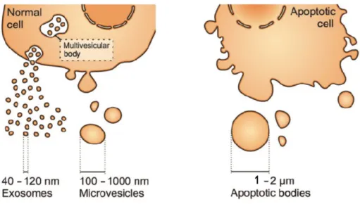
2 EXTRACELLULAR VESICLES ... 45 2.1 Classification ... 45 2.1.1 Exosomes ... 46 2.1.2 Microvesicles ... 47 2.1.3 Apoptotic bodies ... 48 2.2 Internalization ... 49 2.3 Intercellular communication ... 53
2.4 EVs and atherosclerosis ... 54
2.4.1 EVs released by platelets ... 55
2.4.2 EVs released by red blood cells ... 57
2.4.3 EVs released by endothelial cells ... 58
2.4.4 EVs released by immune cells ... 59
2.5 Methods and challenges in studying EVs... 60
2.5.1 Isolation ... 61
2.5.2 Quantification ... 62
3 THE LYMPHATIC SYSTEM ... 65
3.1 Development ... 67
3.1.1 Origin and specification ... 67
3.1.2 Formation of lymph sacs ... 69
3.1.3 Proliferation and Migration ... 70
3.1.5 Lymphangiogenesis in the adult ... 75
3.1.6 Separation of the venous and lymphatic vasculature ... 75
3.1.7 Lymph formation and ultrafiltrates ... 81
3.2 Physiological functions ... 82
3.2.1 Tissue fluid homeostasis ... 82
3.2.2 Immune surveillance ... 84
3.2.3 Uptake of dietary lipids ... 88
3.3 Mechanobiology of lymphatic contraction ... 90
3.3.1 Intrinsic lymphatic pump ... 90
3.3.2 The extrinsic lymphatic pump ... 93
3.3.3 Lymphatic relaxation: endothelial derived relaxing factors and fluid shear stress .. 94
RESULTS ... 98
4 GENERAL THESIS OBJECTIVES ... 99
4.1 Presentation of the first article ... 100
4.1.1 First article... 101
4.1.2 Supplementary data... 135
4.2 Presentation of the second article ... 142
4.2.1 Second article ... 144
4.2.2 Supplementary data... 186
4.3 Presentation of the third article ... 191
4.3.1 Third article ... 193
DISCUSSION ... 216
5 Fundamentals of the thesis ... 217
6 The blood and the lymphatic vasculature: interconnected ... 220
7 Potential therapies for plaque prevention ... 222
7.1 VEGF-C/VEGFR-3 axis ... 222
7.2 Lymphatic and muscle cell innervation ... 224
9 ApoA-I and lymphatic integrity ... 233
9.1 ApoA-I and intercellular junctions ... 235
9.2 ApoA-I and lipid raft modulation ... 236
9.3 ApoA-I and cytokine modulation ... 237
9.4 ApoA-I and EVs... 238
9.4.1 EVs adherence/internalization on the lymphatic endothelium ... 238
9.4.2 EVs contents ... 241
9.4.3 EVs cellular origin ... 243
10 Rethinking atherosclerosis therapies ... 245
10.1 Antiplatelets ... 245
10.2 Statins ... 246
11 Translational perspective ... 248
11.1 Lymphatic dysfunction as a biomarker of cardiovascular disease ... 248
11.1.1 Lymphatic imaging differences in mice and humans ... 250
11.1.2 Taking into account sex differences ... 252
11.2 Other inflammatory diseases ... 253
CONCLUSION ... 257
REFERENCES ... 261
List of tables
List of figures
Figure 1. Structure of the artery wall. ... 5
Figure 2. Brief overview of atherosclerosis development. ... 7
Figure 3. Lipoprotein transport pathways. ... 12
Figure 5. The evolution of an atherosclerotic lesion. ... 17
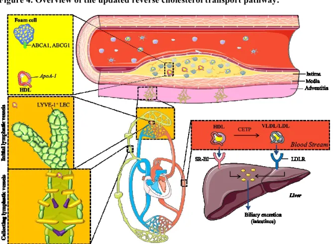
Figure 4. Overview of the updated reverse cholesterol transport pathway. ... 32
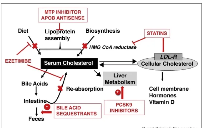
Figure 6. Summary of the mechanisms of action of cholesterol lowering drugs. ... 39
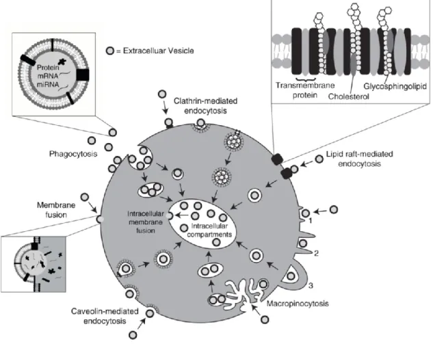
Figure 8. Internalization pathways of extracellular vesicles within target cells. ... 53
Figure 9. Extracellular vesicles in vascular inflammation and atherosclerosis. ... 55
Figure 10. Brief introduction to the molecules that are most central to the molecular biology of the lymphatic system. ... 66
Figure 11. Organization of the lymphatic vasculature. ... 73
Figure 12. Model of the development of the lymphovenous valve in the murine embryo. ... 76
Figure 13. Platelets are required throughout life to maintain proper blood-lymphatic separation. ... 78
Figure 14. Effect of fluid sheer stress on lymphatic function. ... 95
Figure 15. Impairment in the collecting lymphatic vessel pumping capacity under inflammatory conditions... 96
Figure 16. Lipid-free apoA-I treatment in vitro modulates LEC permeability. ... 234
Figure 17. PdEVs adhesion/internalization in cultured lymphatic endothelial cells. ... 240
Figure 18. Atorvastatin decreases VEGFR-3 expression at the surface of LECs in a dose-dependent manner. ... 247
List of acronyms
Akt Protein kinase B
AngII Angiotensin II
ApoA-I Apolipoprotein A-1
ApoB Apolipoprotein B
ApoE Apolipoprotein E
ApoE-/- Mouse strain deficient in apoE
Ca2+ Calcium ion
CAD Coronary artery disease
CD62P P-selectin
CETP Cholesterylester transfer protein
CFDA Carboxyfluorescein diacetate
CFSE Carboxyfluorescein succinimidyl ester CLEC-2 C-type lectin-like receptor-2
CM Chylomicron
CO2 Carbon dioxide
CVD Cardiovascular disease
Connexin-37 Cx-37
EC Endothelial cell
ECM Extracellular matrix
eNOS Endothelial nitric oxide synthase
Erk1/2 Extracellular-signal-regulated kinase 1/2
ET-1 Endothelin 1
EVs Extracellular vesicles
FITC Fluorescein isothiocyanate
GM-CSF Granulocyte-macrophage colony-stimulating factor GPIa/IIa Glycoprotein Ia/IIa
GPIIb/IIIa Glycoprotein IIb/IIIa
hApoB-100 Human apolipoprotein B-100
HDL High density lipoprotein
HMG-CoA 3-hydroxy-3-methylglutaryl CoA
HMG-CoA reductase 3-hydroxy-3-methyl-glutaryl-coenzyme A reductase HMVEC-dLyAd Human dermal microvascular lymphatic endothelial cells
HSP Heat shock protein
IBD Inflammatory bowel disease
ICAM-1 Intercellular Adhesion Molecule 1
IDL Intermediate-density lipoprotein
IL-(1,8,…) Interleukin-(1,8,…)
iNOS Inducible nitric oxide
IVUS Intravascular ultrasound
LCAT Lecithin–cholesterol acyltransferase
Ldlr-/- Mouse strain deficient in LDLR
LEC Lymphatic endothelial cell
LMC Lymphatic muscle cell
LN Lymph node
LOX-1 Lectin-like oxidized low-density lipoprotein receptor-1
Lp(a) Lipoprotein(a)
LPL Lipoprotein lipase
LPS Lipopolysaccharides
LV Lymphatic vessel
LYVE-1 Lymphatic vessel hyaluronan receptor 1
MAPK Mitogen-activated protein kinase
MCP-1 Monocyte chemoattractant protein 1
MHC Major histocompatibility complex
MI Myocardial infarction
MMP Metalloproteinase
MLC20 Myosin light chain of 20 kDa
MLCK Myosin light chain kinase
MLCP Myosin light chain phosphatase
mRCT Macrophage reverse cholesterol transport
mRNA Messenger ribonucleic acid
miRNA Micro ribonucleic acid
MV Microvesicle
NF-B Nuclear factor kappa-light-chain-enhancer of activated B cells
NIRF Near infrared fluorescence
NO Nitric oxide
NOS Nitric oxide synthase
nNOS Neuronal nitric oxide synthase
OVA488 Ovalbumin 488
oxLDL Oxidized LDL
PAF Platelet activating factor
PAMP Pathogen-associated molecular complexes
PCSK9 Proprotein convertase subtilisin/kexin type 9
Pcsk9-/- Mouse strain deficient in PCSK9
pdEV Platelet-derived extracellular vesicle PDGF Platelet-derived growth factor
PDPN Podoplanin
PE Phenylephrine
PI3K Phosphatidylinositol 3-kinase
PKC Protein kinase C
PROX-1 Prospero homeobox 1
PUFA Polyunsaturated fatty acids
qPCR Quantitative polymerase chain reaction
RBC Red blood cell
RNAi RNA interference
ROS Reactive oxygen species
siRNA Single interfering ribonucleic acid
SMC Smooth muscle cell
SREBP Sterol regulatory element-binding protein SR-B1 Scavenger receptor class B type 1
TF Tissue factor
TG Triglyceride
TGF Transforming growth factor beta
TLR Toll-like receptor
TNF- Tumor necrosis factor alpha
Treg Regulatory T cell
TXA2 Thromboxane A2
VCAM-1 Vascular cell adhesion protein 1 VEGF-C Vascular endothelial growth factor-C
VEGFR-3 Vascular endothelial growth factor receptor-3
VLDL Very low-density lipoprotein
vWF von Willebrand factor
You can do anything as long as you have the drive, the passion, the focus, and especially the support.
Acknowledgements
Dear thesis committeeI would first and foremost like to thank the members of my thesis defense panel who have accepted with great interest to read and comment my thesis. I certainly do know how busy a researcher's schedule is, so dedicating your precious time to review this work is greatly appreciated. Your impressive knowledge in each of your respective fields and scientific opinions will certainly improve and deepen the quality of my work and will help me submit a thesis of upmost quality. Thank you for playing an important part of this journey!
Dear Catherine
I would like to thank you from the bottom of my heart for your guidance, insightful ideas, constant encouragement and support. You were much more than my supervisor, you were the mentor I always look up to and I am so very grateful for everything we have accomplished these past years. You are always interested to see me succeed not only academically, but in all my other endeavours as well. Under your wings, I have evolved into a young professional and all the confidence, knowledge and experience I now possess is in large part due to your mentorship. I could not have dreamt of anyone better to guide me through this unbelievable journey. Needless to say, I will always remain Martel lab’s #1 fan!
Dear Montreal Heart Institute lab mates/friends
I could not be happier to have met all of you amazing people! We have had such an incredibly fun time together, and these past few years just flew by. We really defined “Work hard, play hard”. While we gave our hearts out to the projects we advanced together, we also never forgot to have a lot of laughs together and make each day in the lab even more fun than the last.
François Dallaire, my dear Franky boy, thank you so much for all your help and support during my MSc and PhD degrees. Your passion for science is contagious, so I’m glad I’m not the only science nerd at all our get-togethers haha! Thanks so much for always being there for
me and encouraging me time and time again. I couldn’t be happier when I look back on all our achievements.
Gab Jean, your happy nature and funny jokes were greatly missed in the lab after you graduated, not to mention our epic random dance sessions. On the bright side, at least I didn’t get spooked anymore by Kaaris or Post Malone sneaking up on me while I was working :p Looking forward to more karaoke nights so we can continue to recreate passionate renditions of DJ Khaled’s new hits!
Ali Smaani, my honorary lil bro, doing experiments together was unbelievably fun even if it would take us hours on end. (While you still have a few flasks in the incubator, please don’t forget to prep me about 52 petri dishes for tomorrow :p). Thank you for your help, as well as constant support and encouragements! And never forget, casses-toi pas la lymphe! ;) Bye Bang!
Maya Farhat, ma guuuurl Mayoush! My last year in the lab would not have been the same without you. Thank you for your constant support and encouragements. You never forgot to always send me a thought before every single exam I had, oral, presentation, etc. Can’t wait for the next dance semester and going out every weekend so we can be scientists in heels together ;)
Carl Fortin, thank you so much for all your precious help towards the end of my PhD! You are so wise, and your calming nature reassured me numerous times while doing experiments. Also, glad to see someone else who enjoys sushi as much as I do and constantly thinks about it haha!
Laurent Vachon and Stéphanie Jarry, I’m so glad I got to know you guys! While we barely overlapped in the lab, the time that we did was great, and it was wonderful to see how passionate you guys are about the projects. Happy to hear you represent the new generation and will both be pursuing your graduate studies in the awesome Martel lab!
Julien Renaud, you were my first friend at the Institute and sorry again for stealing your bench haha! So happy you joined our crew and socialized. Happy to see you’re doing so well
and since you are our honorary Martel lab student, I’m looking very much forward to more Martel lab reunions!
My dear Fanny Toussaint (aka Fannita), we have been through so much together. While we were even collaborators at some point (hope you don’t still hate me for having to open up minuscule collectors), thank you for always being there for me and giving me precious advice. We were neighbors and quickly became great friends, and I’m so happy to have had so many fun times. With your encouragements, we even ran a half marathon together! Organizing activities at the institute like the happy hours and the escape games was such a blast, and I couldn’t have asked for a better partner.
Kevin Kojok, I feel like just yesterday we were in class together and yet, here we are! So happy we are graduating around the same time and got to explore career choices together. Cheers to us! So happy you found such a fun job already.
Dear Adeline Raignault (aka Adelina), miss you but I’m so happy you are doing so well! We had such a great time working at the Institute and I’m so happy we got to meet even if it was for a short while. You rock girlie!
My dear Olivia de Montgolfier, my graduation twin! We started together and we are finishing this journey together. It feels like just yesterday we sat next to each other in class and realized we both worked at the Institute. Since, I couldn’t be happier with everything we have achieved and all the fun times we had together. You also taught us how to use the pressurizer, and I’m still impressed with your skills!
Dear friends
Whether we know each other since we were kids, or just a few months ago, thank you for always being there for me. Your friendship has been priceless throughout the years and helped me stay focused on my graduate studies. Thank you for understanding my absences and last-minute bails when deadlines were fast approaching. Your constant encouragements have greatly motivated me to push myself and perform even better. Love you all!
Dear family
An enormous thank you to my incredible parents Nina Milasan and Eugen Milasan, and my younger brother Andrei Milasan for giving me the strength to reach for the stars and chase my dreams. Thank you for your endless love, encouragements and always being there for me no matter how stressful things became. Without your priceless love and advice you gave me, I would not be the person I am today. I owe it all to you.
Last, but certainly not least, special thanks to Gabriel Moreau for everything you do to always put a smile on my face. You seem so cool, calm and collected, but you’re always ready to be my partner in crime and join in on any crazy activities I have planned for us. Can’t wait for both of us to finally be adults together and not students anymore! Love you and I am looking forward to plenty more adventures together!
1 CARDIOVASCULAR DISEASE
Cardiovascular disease (CVD), characterized by disorders of the heart and blood vessels, is the number one cause of death globally[1]. Its prevalence is increasing with the age of the population, and the incidence is five times more common in men than in women. This difference, between the sexes, decreases with age[2]. Most deaths are from heart attacks caused by sudden blood clots in the heart’s arteries[3]. CVDs include coronary artery disease (CAD), which affects the coronary vessels supplying the heart muscle; cerebrovascular disease, which affects the vessels supplying the brain; peripheral arterial disease, that affects arteries of the arms and legs; rheumatic heart disease, triggered by rheumatic fever that is caused by streptococcus; cardiac birth defects; and venous thrombosis which is a blood clot that forms in a vein[4].
1.1 The circulatory system
To keep the body alive, each of its cells must be able to benefit from a continuous supply of nutrients and oxygen. In addition, carbon dioxide (CO2) and other metabolic waste produced by the cells must be collected and disposed of. This task belongs to the circulatory system, a network of vessels that allows the heart to circulate blood throughout the body[5]. Deoxygenated blood from the periphery is transported back to the heart by the capillaries, to the venules, to the veins, all the way to the right side of the heart, to ultimately make its way to the lungs. Oxygenated blood from the lungs is sent back to the left side of the heart from where it is ejected into the aorta, and makes its way to the arteries, arterioles, and finally reaches the capillaries where the exchange of nutrients occurs[6].
1.1.1 The heart
The heart is at the core of the circulatory system and acts as a pump responsible of circulating blood throughout the body to supply the necessary physiological needs. Blood carries the essential elements required by the cells, namely nutrients, oxygen and hormones. In addition, blood contributes to the elimination of cellular waste, the protection of the body via the immune system, and regulates internal homeostasis[7].
The wall of the heart consists of three tunics, the epicardium, located on the outside, the myocardium, located in the central part of the wall, and the endocardium lining the cavities of the heart. The latter consists of endothelium and a thin layer of connective tissue. Thus, this layer allows the cavities of the heart and the valves to be smooth in order to reduce friction of the blood during each propulsion[8]. The myocardium is mainly composed of cardiomyocytes which contribute to cardiac contraction, and therefore, to the pumping action of the heart[9]. The epicardium is made up of mesothelial cells and connective tissue, which makes the surface of the heart smooth and slippery, thus avoiding friction[10]. Moreover, the epicardium is a constituent of the pericardium, which is a double-walled sac, representing the inner layer called the visceral pericardium. As a whole, the pericardium’s main function is to protect the heart and avoid sudden dilation, especially caused by the right chamber, while it also helps keep the heart in place within the mediastinum which is a division of the thoracic cavity[11]. Since it contains pericardial fluid, it also provides the heart with the freedom of movement necessary for contractions that occur quickly and vigorously[12].
The heart is composed of four different cavities, the right and left atria, as well as the right and left ventricles. The right atrium serves to receive deoxygenated blood from the systemic circulation via the superior and inferior vena cava, and the coronary sinus that carries blood from the heart. The blood then passes to the right ventricle when opening the right atrioventricular valve, also called the tricuspid valve[11]. In both the right ventricle and the left ventricle, atrioventricular valves are connected to papillary muscles via tendinous cords. Thus, the opening of these valves occurs when the blood pressure of the atria is greater than that of the ventricles, resulting in the relaxation of the papillary muscles and tendinous cords[13]. Subsequently, the blood is propelled from the right ventricle to the pulmonary trunk tendinous ropes. This allows the blood to be delivered to the lungs via the right and left pulmonary arteries. Once cleared of CO2 and reoxygenated, the blood returns to the heart via the pulmonary veins that carry the blood to the left atrium. The blood then passes from the left atrium to the left ventricle following the opening of the left atrioventricular valve, also called bicuspid or mitral valve. Finally, the oxygenated blood is propelled to the systemic circulation through the opening of the aortic valve, which carries the blood into the aortic arch, and then the ascending and descending aortas[14].
Both ventricles contract simultaneously to propel similar volumes of blood. However, their morphology is distinct. The left ventricle is much thicker, since it must propel blood towards the systemic circulation, where it is opposed by a greater resistance and has more distance to cover. The right ventricle sends blood to the nearby lungs, which offer lower resistance, and this could explain why the right ventricular myocardium is less bulky[15]. Finally, the septum separates the right and left ventricles and includes elements of the cardiac conduction system[16].
1.1.2 The blood vessels
The circulatory system is composed of three main types of blood vessels: arteries, veins and capillaries. The aorta is the primary and largest artery in the human body and is emerging straight out of the heart. It represents an important conduit and an elastic reservoir that accommodates blood flow during contractions[17]. From the rising aorta, at the level of the aortic arch there is a bifurcation of three main branches, namely the brachiocephalic trunk, left common carotid artery, and left subclavian artery. From the ascending aorta, two other coronary arteries originate, the right coronary artery and the left main coronary artery, that supply the heart muscle with blood[18]. Blockade of these arteries or their branches, by pathological conditions such as atherosclerosis can lead to angina. The latter is due to insufficient blood supply and causes chest pain, and even a heart attack[19].
Blood vessels are composed of three different layers, except for the capillaries, and within each one of these layers there are differences in muscle and collagen content that varies with size and the location of the vessel[20]. From the inside outwards, are the intima, the media and the adventitia (Figure 1). The intima is the finest and innermost tunic, and it is at this level that atherosclerosis develops. It is composed of a single layer of endothelial cells (ECs) portraying different properties like metabolic activities, thromboresistance, immune functions and elasticity. It also contains an elastic membrane made of extracellular matrix (ECM) on which ECs reside[21]. Also referred to as a basement membrane, it is dotted with fenestrations whose number and appearance vary depending on the location of the vessel in the arterial tree or vascular bed[22]. Following, is the tunica media, which is the thickest. It is the main constituent of the artery and consists of smooth muscle cells (SMCs) surrounded by an ECM composed of elastic and fibrous proteins, such as collagen and elastin[21]. In majority, the media
is avascular, except in its external part that receives irrigation by the vasa vasorum of the adventitia (tunica externa)[23]. The latter is the external coat of the blood vessel that consists of the external elastic lamina with loosely organized connective tissue rich in collagen and elastic fibers, as well as fibroblasts and adipocytes[24]. The adventitia ensures the anchorage of the arteries to the surrounding structures and is sometimes also traversed by longitudinal smooth muscle fibers[23].
Figure 1. Structure of the artery wall.
Illustration showing the different layers of a blood vessel wall. Adapted from Servier Medical Art http://www.servier.com/slidekit/?item=16
In the arteries, blood circulates under very high pressure. As such, the structure of an artery must allow it to withstand these variations in pressure generated by the rhythmic pumping of the heart. This is why the arteries are covered with thick walls, are wrapped in elastic tissue, and contain less muscle, which allows them to expand. To withstand hemodynamic loads, elastin is present in these vessels to allow them to increase in size and modulate their diameter as needed. The more muscular arteries, like the brachial artery, the radial artery, and the femoral
The latter, which include the aorta and pulmonary arteries, since they are in close proximity to the heart, they are mainly composed of elastic tissue in the media. This allows them to maintain the necessary pressure gradients while supporting the continuous pumping of the heart[6].
Arterioles are blood vessels in the microcirculation that extend and branch out from arteries and are consequently smaller in diameter. They are composed of one to two layers of SMCs and due to lack of elastic tissue they represent the main site of vascular resistance[6].
Blood capillaries are the smallest subunit of the microcirculation and are composed of a single layer of ECs. Although they lack SMCs, capillaries can be surrounded by cells with a contractile function called pericytes[25] that control blood flow by varying the diameter of the capillaries[26]. Since the walls of the arteries and veins are too thick to allow diffusion of molecules, the thin walls of the capillaries allow for this exchange of nutrients and metabolites, to and from the bloodstream[6].
Venules emerge from veins and receive blood from capillaries. They also participate in the exchange of oxygen and nutrients for water products. Venules are fragile as they are very thin-walled, making them likely to break easily when the volume exceeds their normal capacity[6].
Blood will flow from venules into larger veins, similar to the arterial system. Contrarily, venous pressure is low and as such, veins are thin walled and less elastic. This permits veins to hold the majority of the blood in circulation, due to their high capacitance in accommodating a large volume of blood at relatively low pressure. Blood flow in the leg veins is helped by muscle contractions and the one from lower extremities is promoted by continuous respiratory changes that affect pressure gradients in the abdomen and chest cavity. Interestingly, one-way valves are present inside the veins to ensure forward flow of blood towards the heart[6].
1.2 Atherosclerosis
Atherosclerosis is derived from the Greek words ‘sclerosis’ meaning hardening and ‘athera’ that relates to gruel, which is the accumulation of lipids (Figure 2)[27].
Figure 2. Brief overview of atherosclerosis development.
Atherosclerosis is due to fatty deposition, which clogs the artery and causes the vessel to lose elasticity. With this plaque accumulation, lumen space is decreased and thus leads to reduced blood flow, or even worse, a total blockade of the vessel. This leads to a multitude of adverse reactions implicating different cells and altering their functions[28]. Illustration used from
http://medwords.blogspot.com/2013/09/atherosclerosis-disease-symptoms.html (Copyright ©
2011 Medicinal Words Powered by Blogger
Atherosclerosis is initially asymptomatic[29]. While clinically it often manifests later on in life, the onset of atheroma can begin during childhood, and the rate of progression is a function of risk factors that can be modulated, as well as unmodifiable factors such as age, sex and family history. Risk factors influence the development of atheroma, the frequency of occurrence of cardiovascular complications and their recurrence[30]. With time, atherosclerotic plaques can evolve to become particularly complex. While more advanced lesions can grow and
end up completely blocking blood flow, the most significant clinical complication is detachment of the thrombus that leads to acute vessel occlusion, thus resulting in myocardial infarction (MI), pulmonary embolism or stroke[30]. Additionally, the atherosclerotic plaque is under dynamic changes that result in increased cellular activation and apoptosis, thus resulting in the release of extracellular vesicles (EVs) of various cellular origins[31]. Despite being considered simple cellular debris for decades, EVs are now well documented to interact with neighboring cells and are suspected to be key players in many physiopathological processes such as thrombosis, autoimmune diseases and inflammation. EVs have been associated with several pathologies, including rheumatoid arthritis[32], tumor progression[33], angiogenesis[34], metastasis[35], diabetes[36], hypertension[37], metabolic syndrome[38], hypercholesterolemia and CVDs including atherosclerosis[39]. As EVs are increasingly associated with the key steps of atherosclerosis, and participate in vascular remodelling, they represent important new potential biomarkers and pharmacological targets [40]. A more thorough review of their characterization and specific functions will be described in the following chapters.
The effects of atherosclerosis can be fatal, so a more thorough understanding of the causes and diverse mechanisms of CAD pathogenesis is of upmost importance. As knowledge about all the players involved in atherosclerosis onset and progression improves, it will lead to superior optimization of disease management.
1.2.1 A historical perspective
Atherosclerosis is often thought to be characteristic of the modern world and largely due to contemporary lifestyles. The earliest known case of atherosclerosis was diagnosed by tomography in the aorta of the mummy of an Egyptian princess who died in her early 40s, more than 3500 years ago[41]. Calcification of an atherosclerotic lesion was also identified by computerized tomography scan in a mummified iceman who lived around 3000 years ago[42]. More evidence was supplied recently, when 44 out of 52 mummies had identifiable cardiovascular structures, with 20 of them categorized with either definite atherosclerosis, due to calcification within the wall of a specific artery, or probable atherosclerosis, with calcifications along the longitude of an artery[43]. Since Egyptian culture might have had unique attributes, analysis of other ancient societies was also performed. In fact, probable or definite atherosclerosis was found in 47 of the 137 mummies originating from four different
preindustrial populations such as, 29 ancient Egyptians, 13 ancient Peruvians, 2 Ancestral Puebloans, and 3 Unangan hunter gatherers[43]. While their diets were different, as well as their climates, what they all had in common is smoke inhalation from the fire used for warmth and cooking. Furthermore, a high level of chronic infections and inflammation present in premodern conditions could have promoted the inflammatory aspects of atherosclerosis[43].
The presence of atherosclerosis in pre-modern individuals suggests that the disease is part of human ageing more than it is characteristic of any specific diet or lifestyle[44]. While there may be a genetic predisposition to the disease, the longer you live and survive a multitude of threats, the more susceptible you are to a multitude of risk factors that cause atherosclerosis development.
1.2.2 Risk factors
While scientists thought that cardiovascular disease was primarily a consequence of age, the Framingham study, which began in 1948, showed the importance of other risk factors like tobacco, lack of exercise, stress, diabetes, high blood pressure, and a diet high in cholesterol, just to name a few[4]. In 1976, based on this study, a risk score, which was improved in 1998, was developed to detect the individuals most likely to trigger a cardiovascular event according to these different factors[45]. It was a longitudinal study that aimed to assess similarities in characteristics and factors between subjects, that may cause CAD. The studies included numerous participants who did not yet have any symptoms or any previous incidents of MI or stroke[4, 46]. Up to 90% of CVD may be preventable and improving some of the risk factors such as healthy eating, exercise, no smoking and limiting alcohol intake can make a significant difference[47]. Furthermore, treatment against predisposing factors such as high blood pressure, high content in blood lipids and lipoproteins, as well as diabetes is also beneficial[28]. Nonetheless, as clarified by the mummies studies, by living longer we are still prone to a multitude of advert situations that increase the risk to develop atherosclerosis. At the very least, by narrowing down some of the main factors that increase susceptibility, some prevention becomes possible.
1.3 Cholesterol
Cholesterol is an organic molecule and a hydrophilic lipid. Cholesterol plays an important role in the structure of the cell membrane, which is why it is the principal sterol synthesized by all the cells of an animal. Furthermore, several compounds are synthesized from cholesterol, such as sex hormones, bile acids and vitamin D[28]. Cholesterol is obtained from both synthesis and dietary sources, and in vertebrates, the hepatic cells produce the highest amount. It is absent in prokaryotes, with the exception of Mycoplasma which requires it for growth[48].
François Poulletier de la Salle is responsible for finding cholesterol in gallstones, back in 1769. More than a century later, the first link between cholesterol and atherosclerosis was brought to light by Adolf Windaus, where patients' aortic plaques were shown to contain 25 times more cholesterol than normal aortas[49].
1.3.1 Function
Cholesterol is a vital component of the body, which is why each cell is able to synthetize it in a complex process[50]. As organisms evolved, cells required membranes that would allow for a wide variety of integral membrane proteins, such as channels, transporters, and enzymes. As such, cholesterol maintains proper fluidity, contributes to intracellular transport and can modulate important properties of the vertebrate plasma membrane, as it composes it at around 30%[51]. Part of its structural role also includes modulating the permeability of the plasma membrane, to allow for proper regulation of ion leaks[52]. Cholesterol is also required for the structure and function of lipid rafts which are required for endocytosis of various substances inside cells, as well as facilitating certain signalling pathways[53, 54].
1.3.2 Biosynthesis
Endogenous synthesis of cholesterol takes place in the cytoplasm of the liver and intestinal cells, where the condensation of three acetyl-CoA forms 3-hydroxy-3-methylglutaryl CoA (HMG-CoA), a reaction catalyzed by HMG-CoA synthase. Following, HMG-CoA becomes mevalonate with help from the enzyme HMG-CoA reductase, a reaction that represents the rate-limiting step in cholesterol biosynthesis. Finally, mevalonate is converted to isopentenyl
pyrophosphate and further down leads to dimethylallyl pyrophosphate, both which are used to make isoprenoids, a diverse class of biomolecules such as cholesterol, heme and vitamin K[55].
All animal cells can produce cholesterol for diverse uses and this process differs between cell types depending on their needs, as well as the organ function. Primarily the liver newly synthesizes close to 70% of cholesterol, endogenously, while close to 30% comes from dietary intake, exogenously. In total, the human body contains approximatively 70 g of cholesterol[56].
1.3.3 Dietary sources
Since all animal cells produce cholesterol, animal-based foods such as red meat, eggs, fish oil, butter and even breast milk contain cholesterol in varying amounts[57]. It is at the level of the small intestine that cholesterol absorption takes places daily. Specialized lymphatic vessels called lacteals are located within the villi of the intestine and are responsible for the uptake of dietary fats, to then deliver them to the blood via the mesenteric LVs that drain into the thoracic duct[58]. Interestingly, following increased cholesterol ingestion, the body is able to compensate by reducing its own cholesterol synthesis. The intestine tightly regulates how much cholesterol of dietary origins enters the body and this ingested cholesterol is mainly esterified after reaction with fatty acids. The conversion of free cholesterol to cholesteryl ester is catalyzed by lecithin:cholesterol acyltransferase (LCAT) in peripheral tissues, while dietary cholesterol absorbed by enterocytes, the intestinal cells, is esterified by acyl-coenzyme A: cholesterol acyltransferase 2. This conversion allows for more cholesterol to be packaged into the interior of lipoproteins, which allow for more efficient cholesterol transport through the blood stream[59].
1.3.4 Lipid metabolism and lipoprotein transport
Lipids, like cholesterol and triglycerides (TGs), are insoluble in plasma[59]. In the circulation, they must be transported in association with proteins, to undertake a broad array of functions such as to serve as energy for cells, deposition of lipids and to form bile acids. Their classification, along with their function and pathways of metabolism, play an important role in different disorders and how they can promote the development of atherosclerosis.
1.3.4.1 Classification
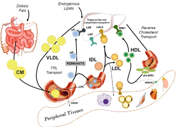
There are five major classes of particles that transport lipids between organs, tissues, and cells, listed from larger and less dense to smaller and denser: chylomicron (CM), very low-density lipoprotein (VLDL), intermediate-low-density lipoprotein (IDL), low-low-density lipoprotein (LDL), and high-density lipoprotein (HDL). Lipoproteins are larger and less dense when their lipid to protein ratio is high[60]. A thorough comprehension of the main functions of all the lipoproteins is necessary, as they each play a critical role in proper metabolism and lipid handling, among other various responsibilities (Figure 3).
Figure 3. Lipoprotein transport pathways.
A brief overview of the different pathways involved in lipoprotein transport is illustrated by de Guia et al.[61] with permission and will be discussed with more depth in the sections below. Triglyceride-rich lipoprotein (TRL). Permission obtained from © 2017 Elsevier Ltd.
Chylomicrons are the largest lipoproteins that form during digestion and are composed of several apolipoproteins such as A-I, B-48, C-II, C-III, and E[59]. Their main role is to transport exogenous lipids like cholesterol and fatty acids from the small intestine to peripheral adipose tissues where they are reprocessed. Within enterocytes, free fatty acids combine with glycerol and form TGs, while cholesterol is esterified[62]. Following, in order to form CMs, TGs and cholesterol are assembled, and the main apolipoproteins contained are B-48, along with C-II and E. Importantly, apoB-48 does not bind to LDL receptor (LDLR) who has an apoB-100 and not an apoB-48 binding site, and whose main ligand is LDL[59]. Along the way, CMs exchange apoproteins with HDL, as they acquire apoE and apoC-II from HDL to convert into a mature form[63]. Lipoprotein lipase (LPL) reduces CM size by hydrolyzing core TGs and releasing free fatty acids that can then be directly used as energy, in combination to form more TGs or deposited in adipose tissue[59, 64]. At the end, CMs are broken down to CM remnants that have a smaller core of lipids and are cleared away by liver CM remnant receptors that bind to apoE[65].
Conversely, the endogenous pathway of lipid metabolism commences with the synthesis of VLDL by the liver, which contains a core composed in major part of TGs and also some cholesterol esters (CEs). The surface apolipoproteins for VLDL include apo C-II, which activates LPL; apoC-III, which inhibits LPL; and apoB-100 and E, which bind to the LDL receptor[59]. Similar to CMs, the TG core of nascent VLDL is hydrolyzed by LPL, thus generating IDL. Circulating cholesteryl ester transfer protein (CETP) mediates the transfer of cholesteryl esters from HDL particles to VLDL, while TGs are transferred in the opposite direction, which promotes cholesterol removal from peripheral cells and uptake by the liver. IDL remnants can then be cleared from the circulation by remnant receptors on the liver, or remodeled by hepatic lipase to form LDL particles[66].
Two of the main cholesterol carriers in the body are LDL, clinically often referred to as the “bad” type of cholesterol, and HDL, considered as the “good” cholesterol. A high ratio of HDL: LDL in the body is associated with a lower risk of CAD[67], and both entities play a pivotal, yet opposite role, in the modulation of atherosclerosis. The main function of LDL is to transport cholesterol and deliver it to cells, where it is used for a variety of functions. LDL contains a core of CEs, lesser amounts of TGs, and its most important and atherogenic
present, cholesterol uptake by the cells is reduced, leading to increased cholesterol circulating in the blood vessels[69]. Knocking out LDLR in transgenic mice leads to a significant increase in total cholesterol levels which can be reversed by restoring the Ldlr gene[70].
LDL internalization is thoroughly regulated by the body through a negative feedback control mechanism depending on cellular cholesterol requirements. Decreased HMG CoA reductase activity, which leads to a fall in de novo cholesterol synthesis by the cell, upregulates LDLR expression and cholesterol uptake from the circulation. The main protein responsible for the regulation of cholesterol in the endoplasmic reticulum is the sterol regulatory element-binding protein (SREBP)[71].
LDLR is also modulated by the proprotein convertase subtilisin/kexin type-9 (PCSK9), which binds to it and targets it for lysosomal degradation in cells, leading to decreased hepatic clearance of plasma LDL[72]. In addition to its influence on cholesterol transport, overexpression of PCSK9 also affects production of apoB-containing lipoproteins in the intestine by increasing apoB mRNA and enhancing apoB protein stability through activation of the microsomal triglyceride transfer protein (MTP), which is required for the assembly and secretion of VLDL in the liver and also participates in the association of TGs with apoB-48 in CMs[73]. Since PCSK9 significantly impairs the clearance of LDL from the blood, its loss-of-function mutations led to nearly 85% lower plasma LDL levels[74] and as such, offer an important mechanism for protection from CVDs like atherosclerosis[75]. PCSK9 is now considered a potential target for cholesterol-lowering therapies that are especially beneficial for patients who do not tolerate other medications such as statins which have pleiotropic effects[72, 76].
HDL is the smallest and most dense lipoprotein, as it contains the highest proportion of proteins to lipids. Its most abundant apolipoprotein is apoA-I, followed by apoE[77]. However, more than ninety-five proteins are believed to be associated with HDL, ranging up to two hundred twenty-five, as compiled by Dr. Sean Davidson and his team to date. Its main lipoprotein, apoA-I, is synthesized in the liver and the intestine. ApoA-I interacts with receptors in various cell types, including hepatocytes, enterocytes, and macrophages[78]. While apoA-I is lipid-free or at most lipid-poor, which is the preferred substrate of the ABCA1 receptor, in plasma, the preferred state of apoA-I is in association with HDL[79]. ApoA-I in circulation interacts with phospholipids to form nascent discoidal HDL which, once generated, promotes
cholesterol efflux in macrophages present within the lesion[80]. Externalized cholesterol is absorbed by nascent discoidal HDL, and then esterified by LCAT, which sequesters it and eventually makes the newly synthesized HDL spherical. This esterification of cholesterol is particularly important for HDL uptake by the liver and any impairment leads to detrimental HDL dysfunction[81]. Further discussions regarding HDL and apoA-I are detailed in chapter 3 and study #2.
1.3.5 Recycling and excretion
Bile acid synthesis occurs in the liver following cholesterol oxidation. This process is responsible for a daily turnover of a majority of the cholesterol present in humans. Following, bile acids get secreted into the bile and can be stored temporarily in the gallbladder[82]. Bile salts solubilize fats in the digestive tract and facilitate digestion and absorption of fat molecules in the small intestine. In humans, around 50% of the cholesterol is reabsorbed by the small intestine and brought back into the bloodstream, while the rest pursues the path of fecal excretion [83]. Around 95% of bile acids secreted by the hepatocytes are reabsorbed from the intestines, via the portal circulation, while the remainder are excreted in the feces[84]. On a daily basis, close to 1g of cholesterol enters the colon, originating from the diet, bile, or intestinal cells and is metabolized by bacteria present in the colon[85]. Under conditions where cholesterol is more concentrated, as in the gallbladder, it crystallizes and forms gallstones[86].
1.3.6 Dyslipidemia
Dyslipidemia is characterized by an abnormally high amount of lipids, such as total cholesterol, LDL or TGs and low HDL or apoA-I in the blood. Prevalence of dyslipidemia is highest in patients with premature CAD and the disturbance is most often familial[87].
Although polygenic, dyslipidemia is strongly influenced by obesity, particularly visceral adiposity, as well as the amount of saturated fat and cholesterol from the diet. A commonly known genetic disorder, familial hypercholesterolemia (FH), is the leading clinical phenotype resulting from dominantly inherited defects in LDL catabolism. Genetic variations in Ldlr, apoB or Pcsk9 genes are the main ones associated with FH. Genetic variations in the Ldlr gene are mostly due to loss-of-function mutations, thus leading to increased plasma LDL levels. Defects
rise to the same lipid homeostasis functional defects. In addition, other genes are also associated with lipid control and regulatory regions such as Upstream Transcription Factor 1,
apoE, LPL, Fibrinogen Beta Chain, and Hepatic Lipase, which all lead to hypercholesterolemia
and have been shown to predispose to premature cardiovascular diseases[88]. The apoE
-/-mouse is particularly representative of familial combined hyperlipidemia, in that it leads to both elevated LDL and TG plasma levels[89]. Similarly, in a Japanese family, heterozygous LPL deficiency led to increased LDL levels in plasma despite normal LDLR activity[90].
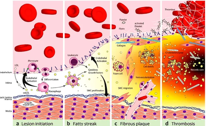
1.4 Evolution of an atheroma
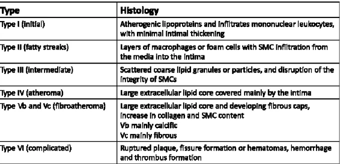
Atherosclerotic plaque formation is a continuous process and can extend longitudinally along the entire circumference of the vessel (Figure 5). A thorough description of each underlying step of atherosclerosis onset and progression follows.
Figure 5. The evolution of an atherosclerotic lesion.
Gargiulo et al. provide us with a general summary of the main steps that characterize the atherosclerotic plaque initiation and progression. (a) In the first stage, endothelial cell injury allows for LDL to gain entry and then be modified by oxidation thus becoming oxidized LDL. The latter then promotes leukocyte recruitment and differentiation into macrophages, by upregulating adhesion molecules and various chemokines. (b) The modified LDL is ingested by macrophages to create foam cells that form a fatty streak in the arterial wall and secrete pro-inflammatory cytokines.(c) Smooth muscle cells then migrate to the surface of the plaque and create a fibrous cap further reinforced by collagen, fibrin and activated platelets. (d) When the cap is thick, plaque is considered stable. However, proteases secreted from foam cells can destabilize plaque, making it prone to rupture and thrombus formation. As well, foam cells undergo apoptosis where they release various debris and lipids, resulting in the formation of a necrotic core[91]. Open Access article distributed under the terms of the Creative Commons Attribution-Noncommercial 3.0 Unported License
1.4.1 Endothelial dysfunction
The vascular endothelium has long been perceived as a mere physical barrier between blood and the components of the vessel wall[92]. However, it does not only physically limit the access of blood components to cells, tissues and organs, it is also significantly implicated in atherosclerosis development[93].
A broad array of regulatory functions of the vascular endothelium have been highlighted by Robert F. Furchgott, among others. In 1980, he demonstrated the existence of an endothelium-derived gas, later identified as nitric oxide (NO)[93], that led to vasorelaxation following achetylcholine addition to the endothelium[94]. Acetylcholine is recognized to date as a potent vasodilator in vivo. Conversely, phenylephrine (PE) binds to α1-receptors and leads to vasoconstriction which increases blood pressure[95], while exerting a minimal relaxing effect at concentrations higher than 1 μM in the mouse aorta[96].
NO has both protective and toxic properties depending on its concentration and is an important regulator of vascular tone. It is also involved in inhibiting platelet adhesion and aggregation, leukocyte adherence, SMC proliferation and LDL uptake[27, 97, 98]. Reduced bioavailability of NO leads to impairment of endothelial vasodilation that in turn leads to detrimental effects. The amino acid L-arginine is the biological precursor of NO, which is provided by NO synthases (NOSs), either the neuronal (nNOS), endothelial (eNOS) or inducible (iNOS) isoforms[99]. Both nNOS and eNOS constitutively produce low levels of NO when intracellular calcium concentrations increase[100]. More complex, iNOS which is present in macrophages and SMCs among other types, starts releasing NO under inflammatory conditions as induced by certain cytokines[27, 101, 102]. Being a free radical, NO produces endothelial damage, as it can act as both an oxidant and an antioxidant[102]. In the presence of reactive oxygen species (ROS), which cause cellular damage and alter DNA, NO combines with them to form peroxynitrite which is believed to be implicated in lipoprotein oxidation, a key early stage in the development of atherosclerosis[103]. ROS are produced by a variety of the risk factors associated with atherosclerosis such as smoking, stress and radiation[104]. Therefore, during atherosclerosis, the damage sustained to the endothelium impairs eNOS function and NO release is altered. Administration of L-Arginine demonstrated favorable effects in atherosclerosis and disturbed shear stress by increasing eNOS expression in cells and in
vivo[105, 106]. Other potent vasodilators such as prostacyclin and tissue-type plasminogen
activator are also produced by the endothelium[107]. While NO in itself cannot be simply categorized as good or bad, it is important to delineate the underlying problem of the endothelial dysfunction that attenuates its protective role. NO insufficiency due to inefficient eNOS activity may be at play in some disease states, however, insensitivity of the SMCs or altered responsiveness despite sufficient NO release are also likely[108].
Atherosclerotic lesions occur mainly at sites that are exposed to disturbed blood flow, which cause low or oscillatory shear stress on the vessel wall. Such areas occur at bends, branches and bifurcations of the arterial tree[109]. Disturbed flow alters EC function[110] and impairs its atheroprotective role[111]. These changes can be mediated by stimulation of the release of NO from the ECs[112]. In addition, low shear stress was shown to increase the intima-media thickness of the common carotid artery in healthy men[113].
When the endothelium is altered due to damage and increased activation, ECs release vasoconstrictor factors such as endothelin-1 (ET-1) and angiotensin II (AngII)[27]. ET-1 can significantly contribute to the pathogenesis of atherosclerosis and can also stimulate SMC migration and growth[114]. ET-1 is released from intracellular and extracellular compartments of human coronary atherosclerotic tissue in response to mechanical stress[115]. Furthermore, increased plasma concentrations of AngII also promote the development and severity of atherosclerosis, especially in cases of hyperlipidemia[27]. AngII modulates vascular SMC proliferation and the production of ECM[116]. Both these effectors can promote leukocytes and platelets recruitment and increase several adhesion molecules at the surface of ECs[117, 118]. Intercellular adhesion molecule-1 (ICAM-1) and vascular cell adhesion molecule-1 (VCAM-1) are cell surface glycoproteins induced at endothelial sites of inflammation able to mediate the adherence of leukocytes to the endothelium. While a low level of ICAM-1 is expressed on normal endothelial cells, VCAM-1 expression occurs during inflammation and is present in the microvessels of human atherosclerotic lesions[119]. As such, although both adhesion molecules increase in atherosclerotic lesions, VCAM-1 seems more important in the initiation of atherosclerosis[120]. P-selectin (CD62P) is an activated platelet and blood EC (BEC) receptor that mediates adhesion between vascular cells and was also shown to promote migration of inflammatory cells into early and advanced atherosclerotic lesions[121].
At sites of injury and inflammation, a variety of proinflammatory cytokines such as interleukin-1 (IL-1) and tumor necrosis factor alpha (TNF-) promote leukocyte adhesion and activation, as well as neutrophil activators, such as granulocyte macrophage colony stimulating factor (GM-CSF), plasminogen-activating factor (PAF) and interleukin-8 (IL-8)[122]. These markedly potentiate neutrophil activation by increasing their adhesiveness, which then contributes to further endothelial damage and injury by producing ROS[123]. As such, antibodies that block adhesion molecules could improve the inflammatory response in atherosclerotic plaques[124].
1.4.2 Lipoprotein modification
Abnormal lipoprotein metabolism is a major predisposing factor to atherosclerosis. Dyslipidemia is estimated to be present in over 70% of patients with premature CAD[87]. Different players have an instigative role in atherosclerotic lesion development and modulation. LDL particles contain cholesterol, TGs, phospholipids, apoB-100, apoE and apoC, among the twenty-two associated proteins identified to date, a number that may range up to sixty as reflected when studies were compiled by Dr. Sean Davidson and his team. Since all LDL particles contain one copy of apoB-100, and only 10 to 20 percent contain apoC-III, there is a direct relationship between apoB-100 and LDL particle number[59]. Interestingly, elevated plasma concentrations of apoB-100 containing lipoproteins can induce the development of atherosclerosis even in the absence of other risk factors. This is due to LDLs getting trapped in the subendothelium of the vessel via a charge-mediated interaction with proteoglycans in the ECM[125] and are more prone to ROS modification of surface phospholipids and unesterified cholesterol[126]. Compared to mice with regular LDL particles, mice expressing LDL with defective proteoglycan binding developed significantly less atherosclerosis[127].
Oxidized LDL (OxLDL), produced when LDL cholesterol is damaged by chemical interactions with free radicals, is responsible for a panoply of aggravating effects and elevated plasma concentrations are associated with CAD[128]. OxLDL promotes recruitment of proinflammatory factors[27] and increases monocyte binding through activation of monocyte ß1 integrin[129]. Macrophage mobility is thus reduced, trapping them within the vessel wall. In a study performed on patients with FH, hyperlipidemia displayed elevated levels of ICAM-1 and VCAM-1, which upregulate endothelial adhesiveness, but these levels were reduced