1
Supporting Information for
Article
Facile and Rapid Formation of Giant Vesicles from
Glass Beads
Radu Tanasescu
1, Ute Mettal
1, Adai Colom
2,3, Aurélien Roux
2,3and Andreas Zumbuehl
1,3,*
1 Department of Chemistry, University of Fribourg, Chemin du Musée 9, 1700 Fribourg, Switzerland;
[email protected] (R.T.); [email protected] (U.M.)
2 Department of Biochemistry, University of Geneva, 30, Quai Ernest-Ansermet, 1211 Geneva, Switzerland;
[email protected] (A.C.); [email protected] (A.R.)
3 National Centre of Competence in Research in Chemical Biology, 1211 Geneva, Switzerland
* Correspondence: [email protected]; Tel.: +41-26-300-8794
Glass beads used after 1 year in the freezer
Figure S1. Representative confocal micrographs of DPPC/DOPC vesicles hydrated at 65 °C and 500 rpm
from glass beads that stayed 1 year in the freezer at −20 °C. Each square is sized to 20 µm × 20 µm. The micrographs were recorded using 1 wavelength only, therefore no general polarization image is provided. The vesicles were artificially colored green for increased visibility.
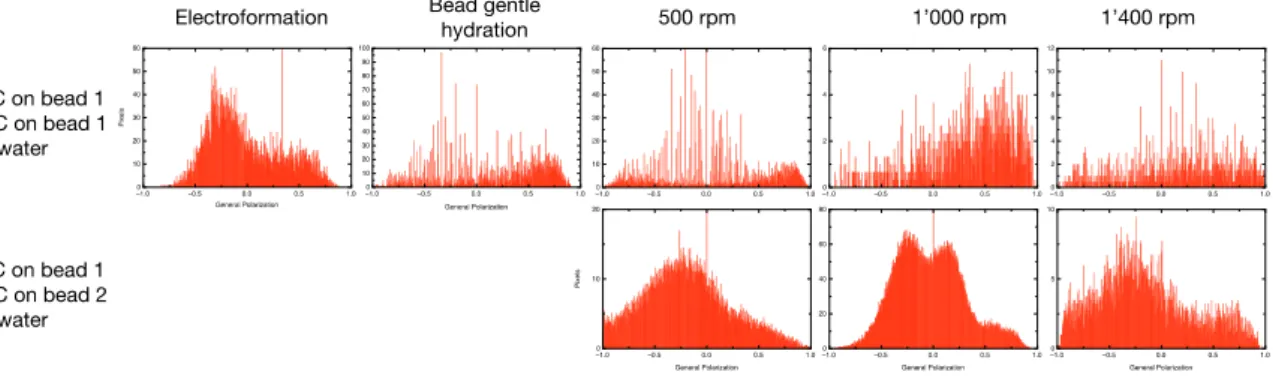
Complete set of general polarization histograms
Figure S2. Complete set of general polarization histograms of vesicles formulated in pure water
2
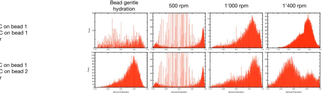
Figure S1. Complete set of general polarization histograms of vesicles formulated in PBS buffer in a
sucrose/glucose gradient. The data were calculated from overview micrographs of the GV. Each histogram summarizes at least 20 GVs.