References
(/) PETERS U , WITHERS HR, THAMES HD JR:
Tumor radioresistance in clinical radiotherapy. Int J Radiat Oncol Biol Phys 8:101-108, 1982
(2) PETERS LJ, HOPWOOD LE, WITHERS HR, ET AL:
Predictive assays of tumor radiocurability. Cancer Treat Sympos 1:67-74, 1984
(3) DEACON J, PECKHAM MJ, STEELE GG: The
responsiveness of human tumors and the initial slope of the cell survival curve. Radiother Oncol 2:317-323, 1984
(4) FERTIL B, DERTINCER H, COURDI A, ET AL:
Mean inactivation dose: A useful concept for intercomparison of human cell survival curves. Radiat Res 99:73-84, 1984
(5) WEICHSELBAUM RR, DAHLBERG W, BECKETT
M, ET AL: Radiation-resistant and repair-profi-cient human tumor cells may be associated with radiotherapy failure in head and neck cancer patients. Proc Natl Acad Sci USA 83:2684-2688,1986
(6) WEICHSELBAUM RR, BECKETT MA, SCHWARTZ
JL, ET AL: Resistant tumor cells are present in head and neck carcinomas that recur after radiotherapy. Int J Radiat Oncol Biol Phys 15:575-579,1988
(7) GOFFMAN TE, RAUBITSCHEK A, MITCHELL JB,
ET AL: The emerging biology of modern radia-tion oncology. Cancer Res 50:7735-7744, 1990
(8) MCKINNON PJ: Ataxia telangiectasia: An in-herited disorder of ionizing radiation sen-sitivity in man. Hum Genet 75:197-208, 1987
(9) KASID U, PFEIFFER A, WEICHSELBAUM RR, ET
AL: The raf oncogene is associated with a radiation-resistant human laryngeal cancer. Science 237:1039-1041, 1987
(10) KASID U, PFEIFFER A, BRENNAN T, ET AL:
Ef-fect of antisense c-raf-1 on tumorigenicity and radiation sensitivity of a human squamous car-cinoma. Science 243:1354-1356, 1989
( / / ) CHANG EH, PIROLLO KH, ZOU ZQ, ET AL:
On-cogenes in radioresistant, noncancerous skin fibroblasts from a cancer-prone family. Science 237:1036-1039, 1987
(12) PIROLLO KF, GARNER R, YUAN SY, ET AL: Raf
involvement in the simultaneous genetic trans-fer of the radioresistant and transforming phenotypes. Int J Radiat Biol 55:783-796,
1989
(13) LITTLE CD, NAU MM, CARNEY DN, ET AL:
Amplification and expression of the c-myc on-cogene in human lung cancer cell lines. Nature 306:194-196,1983
(14) CARNEY DN, MITCHELL JB, KINSELLA TJ: In
vitro radiation and chemotherapy sensitivity of established cell lines of human small cell lung cancer and its large cell morphological vari-ants. Cancer Res 43:2806-2811, 1983 (15) SKLAR MD: The ras oncogenes increase the
in-trinsic resistance of NIH 3T3 cells to ionizing radiation. Science 239:645-647, 1988 (16) O'FARRELL PH: High resolution
two-dimen-sional electrophoresis of proteins. J Biol Chem 250:4007-4021.1975
(17) BOOTHMAN DA, BOUVARD I, HUGHES EN:
Identification and characterization of x-ray-in-duced proteins in human cells. Cancer Res 49:2871-2878,1989
(IS) LAMBERT M, BOREK C: X-ray-induced changes
in gene expression in normal and oncogene-transformed rat cell lines. J Natl Cancer Inst 80:1492-1497,1988
(19) WORLAND PJ, BRONZERT D, DICKSON RB, ET
AL: Secreted and cellular polypeptide patterns of MCF-7 human breast cancer cells following
either estrogen stimulation or v-H-ras transfec-tion. Cancer Res 49:51-57, 1989
(20) VANDEKERCKHOVE J, BAUW G,
VANCOMPER-NOLLE K, ET AL: Comparative two-dimensional gel analysis and microsequencing identifies gelsolin as one of the most prominent downregulated markers of transformed human fibroblast and epithelial cells. J Cell Biol
111:95-102,1990
(21) PUCK TT, MARCUS PI: Clonal growth of
mam-malian cells in vitro. J Exp Med 103:237-284, 1956
(22) ANDERSON NG, ANDERSON NL: Analytical
techniques for cell fractions. XXI. Two-dimensional analysis of serum and tissue proteins: Multiple isoelectric focusing. Anal Biochem 85:331-340, 1978
(23) ANDERSON NL, ANDERSON NG: Analytical
techniques for cell fractions. XXII. Two-dimensional analysis of serum and tissue proteins: Multiple gradient-slab electro-phoresis. Anal Biochem 85:341-354, 1978 (24) MORRISEY JH: Silver stain for proteins in
poly-acrylamide gels: A modified procedure for en-hanced uniform sensitivity. Anal Biochem 117:307-310,1981
(25) OLSON AD, MILLER MJ: ELSIE-4:
Quantita-tive computer analysis of sets of two-dimen-sional gel electrophoretograms. Anal Biochem
169:49-70,1988
(26) DIXON WJ, MASSAY FJ JR: Inference: Two
populations. In Introduction to Statistical Analysis (Corrigan JJ, Wagley S, Amar JS, eds), chap 8. New York: McGraw-Hill Book Co, 1983, pp 116-137
Expression of Insulinlike
Growth Factor (IGF) and
IGF-Binding Protein Genes in
Human Lung Tumor Cell Lines
J. G. Reeve* A. Brinkman,
S. Hughes, J. Mitchell,
J. Schwander, N. M. Bleehen
Background: The presence of multiple, low-molecular-weight, insulinlike growth factor (IGF)-binding proteins in lung tumor cell-conditioned medium and lung cancer patient serum has been recently reported. Purpose: To begin to elucidate the genetic basis for these ob-servations, the present study examines the expression by lung tumor cell lines of three IGF-binding protein genes, namely, IGFBP-1, IGFBP-2, and IGFBP-3. Since IGF-binding proteins are thought to modulate the biologic action of the IGFs, the relationship be-tween the expression of IGF-binding protein genes and the genes encoding
IGF-I and IGF-II also has been inves-tigated. Methods: Gene expression was studied in four small-cell lung cancer (SCLC) and three non-small-cell lung cancer (NSCLC) cell lines using North-ern blot analysis and reverse transcriptase polymerase chain reaction (RT-PCR) for IGFBP-1. Results: IGFBP-1 gene ex-pression was detected by Northern blot analysis in one NSCLC cell line only. However, RT-PCR revealed that the IGFBP-1 gene was expressed in all four SCLC cell lines and in two of the three NSCLC lines. Northern blot analysis of IGFBP-2 gene expression demon-strated that all lung tumor cell lines ex-pressed this gene. A low level of IGFBP-3 gene expression was detected in one SCLC cell line and in all three NSCLC cell lines. All lung tumor cell lines expressed the IGF-II gene as determined by Northern blot analysis. In marked contrast, none of the lines showed evidence of IGF-I gene expres-sion using this method. However, RT-PCR revealed a low level of IGF-I gene expression in one SCLC and one NSCLC cell line only. Conclusions: These observations indicate 1) that IGF-binding proteins secreted by lung tumors are encoded by at least three different genes; 2) that there may be a close association between IGF-II and IGFBP-2 gene expression, such that, where there is production of IGF-II, IGFBP-2 is the principal BP; and 3) that the IGF-II gene is more widely ex-pressed than the IGF-I gene in human lung tumor cell lines. [J Natl Cancer Institute 84:628-634,1992]
An increasing number of proteins with insulinlike growth factor (IGF)-binding characteristics have been isolated from various body fluids, tissue extracts, and
Received August 5, 1991; revised December 19, 1991; accepted January 2. 1992.
J. G. Reeve, S. Hughes. J. Mitchell, N. M. Bleehen, Medical Research Council, Clinical Oncol-ogy and Radiotherapeutics Unit, Medical Research Council Center, Cambridge, England.
A. Brinkman, Pediatric Endocrinology, Erasmus University, Rotterdam, The Netherlands.
J. Schwander, Department Innere Medizin, Kan-tonsspital, Basel, Switzerland.
Correspondence to: J. G. Reeve, Ph.D., Medical Research Council, Clinical Oncology and Radio-therapeutics Unit, MRC Center, Hills Rd., Cambridge CB2 2QH, England.
cell lines (1). On the basis of extensive protein and complementary DNA (cDNA) sequencing studies, these pro-teins have been classified (2) into six dis-tinct groups. IGFBP-1, also named placental protein 12 (ppl2) (3), IBP-1 (4), BP-25 (5), BP-28 (6), and alpha pregnan-cy-associated endometrial globulin (7), has been purified from amniotic fluid and several other sources. Its cloned cDNA sequence predicts a molecular mass of 25 kd, and expression of the cDNA encoding IGFBP-1 in COS cells results in the syn-thesis of protein with a relative molecular mass (MT) of 30 kd on nonreduced
sodi-um dodecyl sulfate (SDS)-polyacryla-mide gel electrophoresis (PAGE) (4). IGFBP-2 (8) is the human homologue of a protein isolated from rat BRL-3a cells (9) and has been isolated from human serum (10). The cDNA sequence of the human protein predicts a molecular mass of 31 kd; under nonreducing conditions, the expressed protein has an Mr of 36 kd
(8). IGFBP-3, also known as BP-53 (//), is a growth hormone-dependent protein, originally purified from human plasma. On nonreduced SDS-PAGE, the protein appears as a glycoprotein doublet consist-ing of a major 53-kd and a minor 47-kd component (//). The cDNA for this protein predicts a molecular mass of 28 kd for the nonglycosylated protein (12). IGFBP-4, isolated from human osteoblas-toma- (13) and prostatic carcinoma- (14) conditioned media and human serum (10), has a predicted molecular mass of 22 kd and migrates as a 28- to 30-kd IGF-binding protein on nonreduced gels. A further IGF-binding protein, IGFBP-5, isolated from cerebrospinal fluid (15) and human serum (10), has a predicted molec-ular mass of 26 kd for the mature protein and an MT of 24 kd for the
nonglycosy-lated protein. Finally, a sixth IGF-binding protein, 1GFBP-6, has been purified re-cently from pig ovarian follicular fluid, and cDNA clones encoding rat and hu-man IGFBP-6 have also been isolated and characterized (16). All six IGF-binding proteins are distinct from the type I and type II receptors for IGFs; importantly, stimulatory (1,17-19) and/or inhibitory (1,20,21) effects on cell growth have been demonstrated for certain IGF-bind-ing proteins.
Recently, multiple IGF-binding pro-teins have been shown to be secreted by
human lung tumor cells both in vitro (22,23) and in vivo (23). These proteins, under nonreducing conditions, range in size from 12 kd to 30 kd and may be en-coded by one or more of the aforemen-tioned genes. To determine whether lung tumor cells secrete multiple different IGF-binding proteins, the present study examines the expression of the IGFBP-1, IGFBP-2, and IGFBP-3 genes in a panel of small-cell lung cancer (SCLC) and non-small-cell lung cancer (NSCLC) cell lines and examines the relationship be-tween IGF-binding protein gene expres-sion and expresexpres-sion of the genes en-coding IGF-I and IGF-II.
Materials and Methods
Cell Lines
Full details of the derivation and char-acterization of SCLC cell lines COR-L47, COR-L51, and COR-L88 and large-cell lung cancer cell line COR-L23 have been described (24). The classic SCLC cell line NCI-H69 was donated by Drs. D. Carney and A. Gazdar (National Cancer Institute Navy Medical Oncology Branch, Bethesda, Md.). The squamous cell lung carcinoma cell line BEN and the lung adenocarcinoma cell line MOR were from Dr. M. Ellison (Ludwig Institute, Sutton, Surrey, England). All cell lines were grown in RPMI-1640 medium sup-plemented with 10% fetal calf serum (both medium and serum from GIBCO BRL, Paisley, Scotland).
RNA Preparation
Cells in logarithmic phase of growth were collected by centrifugation at 300g for 10 minutes and suspended in 100 \xh of medium. A solution containing 6.0 M guanidine hydrochloride and 0.2 M sodium acetate (pH 5.5) was added to the cells (20 mL per 5 x 10⁷ cells), and the . DNA was sheared by vigorous homogeni-zation in a Virtis homogenizer (Virtis Co., Gardiner, N.Y.). RNA was pre-cipitated by the addition of a half volume of 95% ethanol followed by incubation at -20 °C overnight. The pelleted precipitate was dissolved in a solution containing 7.0 M urea, 0.35 M NaCl, 50 mM Tris (pH 7.5), 1 mM EDTA, and 0.2% SDS and then was extracted once with phenol-chloroform. RNA was precipitated from
the aqueous phase using two volumes of ethanol, washed with 70% ethanol, air dried, and dissolved in sterile, double-dis-tilled water.
Poly(A)+ RNA was prepared from total RNA using a messenger RNA (mRNA) purification kit (Pharmacia LKB Biotech-nology Inc., Piscataway, N.J.).
Northern Blot Analysis
Five micrograms of poly(A)+ RNA in 10 mM sodium phosphate buffer (pH 7.0) was denatured in 1.0M glyoxal for 1 hour at 50 °C. The RNA was electrophoresed in a 1.4% agarose gel in 10 mM sodium phosphate buffer and was transferred by Northern blotting to nylon filters. After treatment for 2 minutes with UV light, the nylon filters were baked at 80 °C for 2 hours before hybridization.
The IGFBP-3 (12) and the IGF-I and IGF-II cDNA probes (25) were supplied by Genentech Inc., San Francisco, Calif., and by Dr. G. Bell, Howard Hughes Medical Institute, Chicago, 111., respec-tively. The IGFBP-1 (4) and the IGFBP-2 (8) cDNAs, both cloned into the vector PTZ19 (Pharmacia LKB Biotechnology Inc.), the IGFBP-3 cDNA, cloned into the pUC119 vector (12), and the IGF-I and IGF-II cDNAs, cloned into the pKT218 vector (Pharmacia LKB Biotechnology Inc.), were separated from their vectors by treatment with EcoRl followed by agarose gel electrophoresis. The £coRI fragments, still in the gel slice, were radio-labeled by transcribing the fragments using mixed oligonucleotides to initiate transcription. The radiolabeled probes were separated from unincorporated nucleotide triphosphates using Sephadex G50 (Pharmacia LKB Biotechnology, Inc.) and boiled for 3 minutes before use. A mouse p actin probe, PRT3 (donated by Dr. John Rogers, Laboratory of Molecular Biology, Medical Research Council), was similarly labeled to con-firm equal loading of RNA.
The labeled probe, at a concentration of 106 counts per minute per milliliter, was hybridized to the filter in 1 M NaCl and 0.1 M trisodium citrate (6x SSC), 5% dextran sulfate, 0.02% Ficoll, 0.02% bovine serum albumin, 0.02% polyvinyl pyrrolidone, 0.1% SDS, and 150 ng/mL sonicated salmon sperm DNA at 65 °C for 18 hours. The filter was washed with 6x SSC and 0.1% SDS at 65 °C to
remove unhybridized probe prior to autoradiography.
Reverse Transcriptase Polymerase Chain Reaction Analysis of IGFBP-1 and IGF-I Gene Expression
Synthetic oligonucleotides, designed on the basis of the nucleotide sequence of the mRNA encoding IGFBP-1 and IGF-I, were synthesized using an Applied Bio-systems 380 DNA Synthesizer (Applied Biosystems, Warrington, England). The sequences of the IGFBP-1 specific pri-mers were the following:
(B1) 5'GCTCCCCATGCTGCAGAGG-CAGGG3' corresponding to nucleotides
386-409.
(B2) 5'TACATTAAAATACATCTGG-CAGTT3' complementary to nucleotides
823-800.
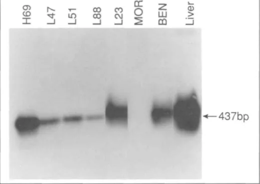
The 437-base-pair (bp) amplification product is unique to IGFBP-1.
The sequences of the primers used for reverse transcriptase polymerase chain reaction (RT-PCR) analysis of IGF-I gene expression were the following:
(IG-1) 5'TCTTGAAGGTGAAGAT-GCACACCA3' corresponding to nucleotides 238-261. (IG-2) 5'AGCGAGCTGACTTGGCA-GGCTTGA3' complementary to nucleotides 540-517.
The 302-bp amplification product is com-mon to IGF-IA and IGF-IB.
Ten micrograms of total RNA was reversed transcribed into first-strand cDNA by addition of 5 [ih 0.1 M dithio-threitol, 2.5 \iL 5 nW deoxyribo-nucleoside triphosphate (dNTP) (Pharmacia LKB, Milton Keynes, England), 20 pmol of either oligo-nucleotide B2 or IG-2, and 5 (iL 5x reverse transcriptase buffer (500 xnM Tris [pH 8.3], 60 m/W MgCl2, and 400 mA/ KC1). After heating for 10 minutes at 70 °C, the reaction mixture was cooled to 25 °C, and 2 units of avian myeloblastosis virus reverse transcriptase (Anglian Biotec Ltd., Colchester, England) were added. Fol-lowing incubation at 42 °C for 1 hour, a 5-|iL aliquot of first-strand cDNA was added to 20 pmol of either oligonucleo-tide primers Bl and B2 or IG-1 and IG-2, 5 iiL 5 mM dNTP, and 5 nL lOx Ther-mus aquaticus (Taq) polymerase buffer— 0.67 mM Tris (pH 8.8), 9.17 M
(NH4)2SO4, 0.1 M MgCl2, 0.1 M bromomercaptoethanol, and 2 mg/mL gelatin—in a total volume of 50 |j.L. Two units of Taq polymerase (ILS Ltd., Lon-don, England) were added, and amplifica-tion (35 cycles) was performed using a PHC-1 automated cycler (Techne Ltd., Duxford, England). Annealing was for 2 minutes at 55 °C, polymerization was at 72 °C for 3 minutes, and denaturation was at 95 °C for 1 minute. Forty microliters of the reaction mixture was then electro-phoresed on 1.4% agarose gels in the presence of ethidium bromide. Amplified products were detected by UV light trans-illumination and by autoradiography fol-lowing Southern blotting and hybridiza-tion with either IGFBP-1 cDNA or IGF-I cDNA.
Results
IGFBP-1 Gene Expression
Using a 32P-labeled £coRI fragment derived from cDNA clone W85 as a probe, a single transcript of approximate-ly 1.5 kb was detected in the squamous cell lung carcinoma cell line BEN only (data not shown). However, Southern blot analysis of amplification products pro-duced by RT-PCR (Fig. 1) revealed that the IGFBP-1 gene was expressed in all SCLC cell lines, the BEN cell line, and the large-cell lung carcinoma cell line COR-L23, as evidenced for each cell line by the hybridization of an amplification product having the expected molecular mass of 437 bp to the radiolabeled IGFBP-1 cDNA probe. Only the lung adenocarcinoma cell line MOR failed to show IGFBP-1 gene expression.
IGFBP-2 Gene Expression
Fig. 2 (panel a) shows that IGFBP-2 gene expression was detected by North-ern blot analysis in all SCLC and NSCLC cell lines. Two transcripts were present in all cell lines: a 4-kb and a 1.4-kb species. The 4-kb species could not be removed by washing filters in 0.1 x SSC and 0.1% SDS. Fig. 2 (panel b) shows hybridization of mRNAs with the mouse (3 actin cDNA probe.
IGFBP-3 Gene Expression
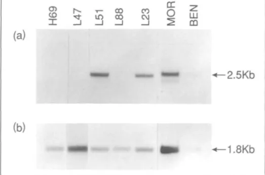
A single 2.5-kb IGFBP-3 gene trans-cript was detected by Northern blot analysis in one of the four SCLC cell
lines and in all NSCLC cell lines studied (Fig. 3, panel a). In addition, a smaller mRNA species was detected in the MOR cell line. Detection of IGFBP-3 gene ex-pression required long exposure times of up to 10 days. Hybridization of lung tumor mRNAs to the mouse (3 actin probe is shown in Fig. 3 (panel b). The apparent absence of IGFBP-3 transcripts in SCLC cell lines NCI-H69, L47, and COR-L88 was investigated further using RT-PCR. No evidence of IGFBP-3 gene expression in these cell lines was ob-served using this method (data not shown).
IGF-I and IGF-II Gene Expression The IGF-I cDNA probe used in this study hybridized to 1.1-kb and 6.3-kb transcripts in human liver. However, no evidence of IGF-I gene expression was obtained by Northern blot analysis in any of the lung tumor cell lines examined (data not shown). The expression of the IGF-I gene was investigated further using RT-PCR. Fig. 4 shows that, with this method, amplification products of the ex-pected size were detected in one of the four SCLC and in one of the three NSCLC cell lines.
Fig. 5 shows Northern blot analysis of IGF-II gene expression and demonstrates the presence of IGF-II gene transcripts in all lung tumor cell lines examined. A 5.3-kb transcript was detected in liver poly(A)+ RNA. Three transcripts, 3.5 kb, 4.8 kb, and 6 kb, were detected in SCLC cell lines NCI-H69 and COR-L47 and in the lung adenocarcinoma cell line MOR. Only the 6-kb and 4.8-kb transcripts were present in COR-L51. A weak but detec-table 4.8-kb transcript only was observed in NSCLC cell lines COR-L23 and BEN. SCLC cell line COR-L88 failed to show IGF-II gene expression.
Discussion
We have shown previously that SCLC and NSCLC cell lines produce multiple low-molecular-weight IGFBPs and that sera from lung cancer patients contain elevated levels of these proteins (25). Af-finity cross-linking studies indicated that lung tumor cells may secrete up to four different molecular-weight species, in-cluding 30-kd, 28-kd, 25-kd, and 12-kd proteins. The present study has
inves-Fig. 1. Detection of IGFBP-1 gene expression in SCLC and NSCLC cell lines by RT-PCR and Southern blotting, followed by hybridization with a 1.2-kb radiolabeled IGFBP-1 cDNA probe (clone w85).
Fig. 2. Northern blot analysis of IGFBP-2 gene expression in SCLC and NSCLC cell lines (panel a). Blots were probed with a 1.5-kb fragment containing approximately 60 bp of 5' untranslated and approximately 470 bp of 3' untranslated regions. In all cell lines, 4.0-kb and 1.4-kb IGFBP-2 transcripts are present. The actin signal for each cell line is shown in panel b.
tigated the expression of three genes en-coding IGFBPs, namely, IGFBP-1, IGFBP-2, and IGFBP-3, in these cells and demonstrates the concomitant expres-sion of two of these genes in four of seven cell lines and of all three genes in the remaining three cell lines studied. IGFBP-1 gene expression, though un-detectable in most cell lines by Northern
blot analysis, does occur in most SCLC and NSCLC lung tumor cells. This ex-pression is evidenced by hybridization of low-molecular-weight amplification prod-ucts generated from enzymatic amplifica-tion from IGFBP-1 mRNA via RT-PCR, with the IGFBP-1 cDNA probe. Expres-sion of this gene correlates with the secre-tion of a 25-kd IGFBP by lung tumor cell
lines, as indicated by the observation that this protein is secreted by all cell lines ex-cept MOR—the only line showing no IGFBP-1 gene expression. IGFBP-2 gene expression was readily detected by North-ern blot analysis in all cell lines ex-amined, and pilot studies using an IGFBP-2-specific radioimmunoassay con-firm the secretion of this protein by the lung tumor cell lines studied here. Fi-nally, all NSCLC cell lines and one of the four SCLC lines showed IGFBP-3 gene expression. The findings of the present study are in marked contrast to those of a recent report in which IGFBP-2 gene ex-pression only was detected in lung tumor cell lines (26). Detection of IGFBP-1 gene expression in the present study is at-tributable to the use of a sensitive RT-PCR method, and detection of IGFBP-3 transcripts is attributable to the use of greater amounts of poly(A)+ RNA than those used in the Northern blot analyses of the earlier study.
The findings of the present study indi-cate that, of the three IGF-binding pro-teins studied, IGFBP-2 is the principal one produced by lung tumor cell lines. The marked differences in the relative levels of 1, 2, and IGFBP-3 gene expression detected in the various lines examined may reflect differences in IGF-binding protein gene activation, vari-ation in mRNA stabilities, or expression of the IGFBP-1 and IGFBP-3 genes in a subset of cells only. A number of factors have been shown to influence the expres-sion of IGF-binding protein genes, in-cluding insulin which decreases IGFBP-2 gene expression (27), growth hormone which decreases transcription of the IGFBP-1 gene (28), and dexamethasone which increases both IGFBP-1 (29) and IGFBP-3 (30) mRNAs in hepatocytes. Studies are in progress to identify factors involved in the hormonal regulation of tu-mor-derived IGF-binding protein produc-tion.
The sizes of the IGFBP-1 and IGFBP-3 gene transcripts reported here for lung tumor cells are consistent with those detected in human liver (4,12). In con-trast, normal adult liver contains only a 1.4-kb IGFBP-2 mRNA, whereas most lung tumor cell lines contained this transcript and a 4-kb mRNA. We have recently detected this transcript in fetal lung fibroblasts (Reeve JG: unpublished
Fig. 3. Northern blot analysis of IGFBP-3 gene expression in SCLC and NSCLC cell lines (panel a). Blots
were probed with a 2.5-kb fragment including the full coding region of human IGFBP-3. A 2.5-kb transcript is present in only one SCLC cell line. In contrast, all three NSCLC cell lines express the IGFBP-3 gene. A 2.0-kb transcript is also detected in the lung adenocarcinoma cell line MOR. The actin signal for each cell line is given in panel b.
Fig. 4. Detection of IGF-I gene expression in lung tumor cell lines by RT-PCR followed by Southern blotting
and hybridization to the 0.66-kb phigf-I cDNA probe. Amplification products having the expected size of 302 bp were detected in the SCLC cell line NCI-H69 and the lung adenocarcinoma cell line MOR only.
data), and it is also present in fetal liver and in the HepG2 and the WRL-68 embryonic liver lines {31). In the liver, the presence of the 4-kb transcript ap-pears to be inversely related to the degree of differentiation, in that adult liver con-tains only the mature 1.4-kb mRNA, fetal liver contains both the 1.kb and the 4-kb transcripts, but cultured WRL-68 embryonic liver cells contain only the 4-kb mRNA. The presence of the 4-4-kb transcript in human lung tumor cells and fetal lung fibroblasts may indicate that the presence of this transcript is also dif-ferentiation related in the lung. Studies
are in progress to investigate the relative amounts of these two transcripts in a variety of normal fetal and adult tissues.
Although the biological significance of IGF-binding protein production by lung tumor cells is not known at this time, a number of studies have shown that these proteins can modulate cellular responses to IGF stimulation. IGF-binding proteins have been shown to inhibit the effects of IGF-I and IGF-II on fibroblast DNA syn-thesis (20,21), to increase the binding of IGF-I to its receptor (32), to potentiate markedly the replication of human, mouse, and chicken fibroblasts in response to
IGF-I stimulation (17), and to enhance the cellular DNA synthesis response of cultured porcine aortic smooth muscle cells to IGF-I (19). Given that both IGF-I and IGF-II have been shown to promote the proliferation of SCLC cells in vitro (33,34), IGF-binding proteins may regu-late the mitogenic action of the IGFs in these cells.
A number of studies have reported the secretion of immunoreactive IGF-I (2333) and the presence of IGF-I receptors (22, 34) in SCLC cell lines. Such observations, together with the mitogenic responsiveness of these cells to IGF-I stimulation and the growth-inhibitory effects of antibodies to the IGF-I receptor, have led to the con-clusion that IGF-I is an important auto-crine growth factor for SCLC (33-36). However, the present study is the first to examine the expression of the IGF-I gene in SCLC cell lines and shows that the majority of SCLC cell lines studied failed to express the IGF-I gene as determined by RT-PCR, perhaps challenging the im-portance of this peptide in the autocrine growth of SCLC. This finding, as well as the low level of IGF-I gene expression found in the only positive SCLC cell line, is surprising, given that several of the cell lines investigated have been shown to secrete low levels of immunoreactive IGF-I (25). However, the antiserum used to determine IGF-I secretion showed 3% cross-reactivity with IGF-II. Given the observed expression of the IGF-II gene in all the lines studied, secretion of IGF-II, and not IGF-I, seems more likely. Together with the observation that ex-ogenous IGF-II promotes SCLC cell proliferation (33) and stimulates DNA synthesis in NSCLC cell lines (Reeve JG, Schwander J, Bleehen NM: manuscript submitted for publication), the findings of the present study support the contention that IGF-II may be more widely involved in the autocrine growth of lung tumors than IGF-I.
IGF-II gene expression in lung tumor cell lines is particularly interesting, given that IGF-II is an embryonal mitogen (37-40) and is thought to play an important role in lung differentiation and maturation (41). Transcription of the IGF-II gene in the fetus is driven by three distinct promoters (42-44) and yields three major transcripts of 6.0 kb, 4.8 kb, and 1.9 kb (45). In adult tissues, including the lung,
Fig. 5. IGF-II gene expression in SCLC and NSCLC cell lines detected using the 1.1 -kb phigf-II cDNA probe and Northern blot analysis (panel a). Each track contains approximately 5 ng mRNA, and filters were exposed for 10 days. Panel b shows the actin signal for each track.
all three promoters are markedly sup-pressed (42,43,45). Hence, in the present study, the detection of abundant 6.0-kb and 4.8-kb mRNA species in lung tumor cells may represent re-activation of IGF-II fetal promoters during lung carcino-genesis. Recent circumstantial evidence has also implicated IGF-II in the genesis of developmental tumors, such as hepa-toblastoma, Wilms' tumor, and rhabdo-myosarcoma (45), but elevated ex-pression is also seen in hepatocellular carcinoma (46), colon carcinoma (47), liposarcoma (47), and fibrosarcoma (48).
The IGF-II gene has been assigned to Ilpl4.1 (49) and is in the immediate vicinity of the Wilms' tumor suscep-tibility gene locus. In the Wilms' tumor, increased expression of the IGF-II gene is thought to arise either through juxtaposi-tion of the Wilms' tumor locus and the IGF-II gene via chromosomal deletion or through re-activation of a set of em-bryonic genes, which includes the IGF-II gene, as a consequence of the recessive mutation in the Wilms' tumor locus (45). Since chromosome 1 lp is one of several
sites of frequent cytogenetic deletion and loss of heterozyosity in lung cancers (50£l), it is tempting to speculate that similar mechanisms may be responsible for re-expression of the IGF-II gene in lung tumors. Studies are in progress to in-vestigate this possibility.
References
(/) BAXTER RC, MARTIN JL: Binding proteins for
the insulin-like growth factors: Structure, regulation, and function. Prog Growth Factor Res 1:49-68, 1989
(2) BALLARD J, BAXTER R, BINOUX M, ET AL:
Let-ter: On the nomenclature of the IGF binding proteins. Acta Endocrinol (Copenh) 121:751-752, 1989
(3) KOISTINEN R, KALKKINEN N, HUHTALA ML, ET
AL: Placental protein 12 is a decidual protein that binds somatomedin and has an identical N-terminal amino acid sequence with soma-tomedin-binding protein from human amniotic fluid. Endocrinology 118:1375-1378, 1986
(4) BRINKMAN A, GROFFEN C, KORTLEVE DJ, ET
AL: Isolation and characterization of a cDNA encoding the low molecular weight insulin-like growth factor binding protein (IBP-1). EMBOJ 7:2417-2423, 1988
(5) LEE YL, HINTZ RL, JAMES PM, ET AL:
Insulin-like growth factor (IGF) binding protein com-plementary deoxyribonucleic acid from human HEP G2 hepatoma cells: Predicted protein
se-quence suggests an IGF binding domain dif-ferent from those of the IGF-I and IGF-II receptors. Mol Endocrinol 2:404-411, 1988
(6) BAXTER RC, MARTIN JL, WOOD MH: TWO
im-munoreactive binding proteins for insulin-like growth factors in human amniotic fluid: Relationship to fetal maturity. J Clin En-docrinol Metab 65:423^31, 1987
(7) BELL SC, KEYTE JW: N-terminal amino-acid
sequence of human pregnancy-associated en-dometrial alpha 1-globulin, an enen-dometrial in-sulin-like growth factor (IGF) binding protein —evidence for two small molecular weight IGF binding proteins. Endocrinology 123:1202-1204,1988
(8) BlNKERT C , LANDWEHR J, MARY J L , ET AL:
Cloning, sequence analysis, and expression of a cDNA encoding a novel insulin-like growth factor binding protein (IGFBP-2). EMBO J 8:2497-2502,1989
(9) MOTTOLA C, MACDONALD RG, BRACKETT JL,
ET AL: Purification and amino-terminal se-quence of an insulin-like growth factor-bind-ing protein secreted by rat liver BRL-3A cells. JBiolChem 261:11180-11188, 1986
(10) KIEFER MC, MASIARZ FR, BAUER DM, ET AL:
Identification and molecular cloning of two new 30-kDa insulin-like growth factor binding proteins isolated from adult human serum. J Biol Chem 266:9043-9049, 1991
(//) MARTIN JL, BAXTER RC: Insulin-like growth
factor-binding protein from human plasma: Purification and characterization. J Biol Chem 26:8754-8760,1986
(12) WOOD WI. CACHIANES G, HENZEL WJ, ET AL:
Cloning and expression of the growth hor-mone-dependent insulin-like growth factor-binding protein. Mol Endocrinol 2:1176-1185,
1988
(13) MOHAN S, BAUTISTA CM, WERGEDAL J, ET AL:
Isolation of an inhibitory insulin-like growth factor (IGF) binding protein from bone cell-conditioned medium; a potential local regulator of IGF action. Proc Natl Acad Sci USA 86:8338-8342, 1989
(14) PERKEL VS, MOHAN S, BAYLINK DJ, ETAL: An
inhibitory insulin-like growth factor binding protein (In-IGFBP) from human prostatic cell conditioned medium reveals N-terminal se-quence identity with bone derived In-IGFBP. J Clin Endocrinol Metab 71:533-535, 1990 (15) ROCHANl M, HOSSENLOPP P, BALLAND A, ET
AL: Isolation from human cerebrospinal fluid of a new insulin-like growth factor-binding protein with a selective affinity for IGF-II. FEBS Lett 255:253-258, 1989
(16) SHIMASAKI S, GAO L, SHIMONAKA M, ET AL:
Isolation and molecular cloning of insulin-like growth factor-binding protein-6. Mol En-docrinol 5:938-948, 1991
(17) ELGIN RG, BUSBY WH JR, CLEMMONS DR: An
insulin-like growth factor (IGF) binding pro-tein enhances the biologic response to IGF-I. Proc Natl Acad Sci USA 84:3254-3258, 1987
(18) BUSBY WH JR, KLAPPER DG, CLEMMONS DR:
Purification of a 31,000-dalton insulin-like growth factor binding protein from human am-niotic fluid. Isolation of two forms with dif-ferent biologic actions. J Biol Chem 263:14203-14210,1988
(19) CLEMMONS DR, CASCIERI MA,
CAMACHO-HUB-NER C, ET AL: Discrete alterations of the in-sulin-like growth factor I molecule which alter its affinity for insulin-like growth factor-bind-ing proteins result in changes in bioactivity. J Biol Chem 265:12210-12216, 1990
(20) KNAUER DJ, SMITH GL: inhibition of
ac-tiviiy by binding to its carrier protein. Proc Natl Acad Sci USA 77:7252-7256, 1989
(21) Liu L, BRINKMAN A, B L A T C , ETAL: IGFBP-1,
an insulin-like growth factor binding protein, is a cell growth inhibitor. Biochem Biophys ResCommun 174:673-679, 1991
(22) JAQUES G, KIEFER P, ROTSCH M, ET AL:
Production of insulin-like growth factor bind-ing proteins by small-cell lung cancer cell lines. Exp Cell Res 184:396-406, 1989
(23) REEVE JG, PAYNE JA, BLEEHEN NM:
Produc-tion of immunoreactive insulin-like growth factor I (IGF-I) and IGF-I binding proteins by human lung tumours. Br J Cancer 61:727-731,
1990
(24) BAILLIE-JOHNSON H, TWENTYMAN PR, Fox
NE, ET AL: Establishment and characterisation of cell lines from patients with lung cancer (predominantly small cell carcinoma). Br J Cancer 52:495-504, 1985
(25) BELL GI, MERRYWEATHER JP,
SANCHEZ-PES-CADOR R, ET AL: Sequence of a cDNA clone encoding human preproinsulin-like growth factor II. Nature 310:775-777, 1984
(26) KIEFER P, JAQUES G, SCHONEBERCER J, ET AL:
Insulin-like growth factor binding protein ex-pression in human small cell lung cancer cell lines. Exp Cell Res 192:414-417, 1991
(27) BONI-SCNETZLER M, SCHMID C, MARY JL, ET
AL: Insulin regulates the expression of the in-sulin-like growth factor binding protein 2 mRNA in rat hepatocytes. Mol Endrocrinol 4:1320-1326,1990
(28) SENEVIRANTE C, LUO J, MURPHY U :
Regula-tion of insulin-like growth factor binding protein-1 expression by growth hormone. Mol Endocrinol 4:1199-1204, 1990
(29) Luo J, REID RE, MURPHY LJ: Dexamethasone
increases hepatic insulin-like growth factor binding protein-1 (IGFBP-1) mRNA and serum IGFBP-1 concentrations in the rat. En-docrinology 127:1456-1469, 1990
(30) Luo J, MURPHY LJ: Regulation of insulin-like growth factor binding protein-3 expression by dexamethasone. Mol Cell Endocrinol 74:213-219,1990
(31) ZAPF J, KIEFER M, MERRYWEATHER J, ET AL:
Isolation from adult human serum of four in-sulin-like growth factor (IGF) binding proteins and molecular cloning of one of them that is increased by IGF I administration and in ex-trapancreatic tumor hypoglycemia. J Biol Chem 265:14892-14898.1990
(32) CLEMMONS DR, ELGIN RG, HAN VK, ET AL:
Cultured fibroblast monolayers secrete a protein that alters the cellular binding of somatomedin-C/insulinlike growth factor I. J Clin Invest 77:1548-1556, 1986
(33) JAQUES G, ROTSCH M, WEGMANN C, ET AL:
Production of immunoreactive insulin-like growth factor I and response to exogenous IGF-I in small cell lung cancer lines. Exp Cell Res 176:336-343, 1988
(34) NAKANISHI Y. MULSHINE JL, KASPRZYK PG, ET
AL: Insulin-like growth factor-I can mediate proliferation of human small cell lung cancer cell lines in vitro. J Clin Invest 82:354-359,
1988
(35) MACAULY VM, TEALE JD. EVERARD MJ, ET
AL: Somatomedin-C/insulin-like growth factor I is a mitogen for human small cell lung can-cer. BrJ Cancer 57:91-93, 1988
(36) MINUTO F, DEL MONTE P, BARRECA A, ETAL:
Evidence for autocrine mitogenic stimulation by somatomedin-C/insulin-like growth factor I on an established human lung cancer cell line. Cancer Res 48:3716-3719, 1988
(37) GRAY A, TAM AW, DULL TJ, ET AL:
Tissue-specific and developmentally regulated trans-cription of the insulin-like growth factor 2 gene. DNA 6:283-295, 1987
(38) BRICE AL, CHEETHAM JE, BOLTON VN, ET AL:
Temporal changes in expression of the insulin-like growth factor II gene associated with tis-sue maturation in the human fetus. Development 106:543-554, 1989
(39) HAN VK, LUND PK, LEE DC, ET AL:
Expres-sion of somatomedin/insulin-like growth factor messenger ribonucleic acids in the human fetus: Identification, characterization, and tis-sue distribution. J Clin Endocrinol Metab 66:422-429,1988
(40) DE CHIARA TM, EFSTRATIADIS A, ROBERTSON
EJ: A growth-deficiency phenotype in hetero-zygous mice carrying an insulin-like growth factor II gene disrupted by targeting. Nature 345:78-80,1990
(41) DAVENPORT ML, D'ERCOLE AJ, AZIZKHAN JC,
ET AL: Somatomedin-C/insulinlike growth tor I (Sm-C/IGF-I) and insulinlike growth fac-tor II (IGF-II) mRNAs during lung de-velopment in the rat. Exp Lung Res 14:607-618,1988
(42) SCHOFIELD PN, TATE VE: Regulation of
human IGF-II transcription in fetal and adult tissues. Development 101:793-803, 1987
(43) DE PAGTER-HOLTHUIZEN P, JANSEN M, VAN
SCHAIK FM, ET AL: The human insulin-like growth factor II gene contains two develop-ment specific promoters. FEBS Lett 214:259-264, 1987
(44) SUSSENBACH JS: The gene structure of the in-sulin-like growth factor family. Prog Growth Factor Res 1:33-48, 1989
(45) SCOTT J, COWELL J, ROBERTSON ME:
Insulin-like growth factor-II gene expression in Wilms' tumour and embryonic tissues. Nature 317:260-262, 1985
(46) Su TS, Liu WY, HAN SH, ET AL: Transcripts of the insulin-like growth factors I and II in human hepatoma. Cancer Res 49:1773-1777,
1989
(47) TRICOLI JV, RALL LB, KARAKOUSIS CP, ETAL:
Enhanced levels of insulin-like growth factor messenger RNA in human colon carcinomas and liposarcomas. Cancer Res 46:6169-6173,
1986
(48) SCHOFIELD PN, TURNER RC, CONNOR H, ET AL:
Tumour hypoglycaemia: Raised tumour IGF-II mRNA associated with reduced plasma soma-tomedins. BrJ Cancer 60:661-663. 1989
(49) REEVE AE, ECCLES MR, WILKINS RJ, ET AL:
Expression of insulin-like growth factor II transcripts in Wilms' tumour. Nature 317:258-260,1985
(50) SlIIRAISHI M, MORINAGA S, NOGUCHI M, ETAL:
Loss of genes in the short arm of chromosome 11 in human lung carcinomas. Jpn J Cancer Res 78:1302-1308, 1987
(5/) WESTON A, WILLEY JC, MODALI R, ET AL:
Dif-ferential DNA sequence deletions from chromosomes 3, I I . 13. and 17 in squamous cell carcinoma, large-cell carcinoma, and adenocarcinoma of the human luns. Proc Natl Acad Sci USA 86:5099-5103, 1989
Induction by Estrogen
Metabolite 16a-Hydroxyestrone of Genotoxic Damage and Aberrant Proliferation in Mouse Mammary Epithelial Cells
Nitin T. Telang* Akihiko Suto, George Y. Wong, Michael P. Osborne, H. Leon Bradlow
Background: Estrogens are potent mammary tumor promoters influenc-ing post-initiational events via epige-netic mechanisms. The upregulation (i.e., induction) of the C16a-hydroxyla-tion pathway during 17(3-estradiol (E2)
biotransformation has been associated with mammary cell transformation. The action of E2 metabolites on tumorigenic
transformation, however, is poorly un-derstood. Purpose: The newly estab-lished mammary epithelial cell line C57/MG, derived from the C57BL mouse strain, was used to examine whether E2 or its metabolites,
16-hydroxyestrone (16a-OHE|) and estriol (£_,), function as initiators of mammary cell transformation. Methods: DNA repair (hydroxyurea-insensitive thymi-dine uptake), estrogen metabolism (3H
exchange to form 3H20),
hyperprolifer-ation (increased cell number), and ac-quisition of anchorage-independent
Received October 23, 1991; revised January 3, 1992; accepted January 8, 1992.
Supported in part by Public Health Service grants R29 CA-44741 and POI CA-29502 from the Na-tional Cancer Institute, NaNa-tional Institutes of Health, Department of Health and Human Services (N. T. Telang); and by the Wanda Jablonski Fund (M. P. Osbome).
N. T. Telang, A. Suto, G. Y. Wong, M. P. Osbome, Breast Cancer Research Laboratory, Memorial Sloan-Kettering Cancer Center, New York, N.Y.
H. L. Bradlow, Institute for Hormone Research, New York.
Correspondence to: Nitin T. Telang, Ph.D, Division of Carcinogenesis and Prevention. Breast Cancer Research Laboratory, Memorial Sloan-Kettering Cancer Center. 1275 York Ave.. New York, NY 10021.