ORIGINAL ARTICLE
Static biofilm removal around ultrasonic tips in vitro
Thomas Thurnheer&Elodie Rohrer&Georgios N. Belibasakis&Thomas Attin&Patrick R. Schmidlin
Received: 9 September 2013 / Accepted: 25 November 2013 / Published online: 8 December 2013 # Springer-Verlag Berlin Heidelberg 2013
Abstract
Objectives This study aims to investigate the biofilm removal capacity of two ultrasonic tips under standardized conditions using a multi-species biofilm model.
Methods Six-species biofilms were grown on hydroxyapatite discs for 64.5 h and were treated for 15 s with a standardized load of 40 g with a piezoelectric or magnetostrictive device. Tips were applied either with the tip end or with the side facing downwards. Detached bacteria were determined in the supernatant and colony-forming units (CFUs) counted after 72 h of incubation. Untreated specimens served as con-trols. Moreover, the biofilms remaining on the hydroxyapatite surface after treatment were stained using the Live/Dead stain, and the pattern of their detachment was assessed by confocal laser scanning microscopy (CLSM).
Results As compared to the untreated control, it was found that only a side application of the magnetostrictive device was able to remove efficiently the biofilm. In contrast, its tip application as well as both applications of the piezoelectric device removed significantly less bacteria from the biofilm structure. These findings were corroborated by CLSM observation.
Conclusion Both ultrasonic tips under investigations led to bacterial detachment, but the action mode as well as the tip configuration and adaptation appeared to be influenced by the biofilm removal effectiveness.
Clinical relevance Biofilm removal remains a main goal of ultrasonic debridement. This should be reflected in respective laboratory investigations. The presented combination of methods applied on a multi-species biofilm model in vitro allows the evaluation of the effectiveness of different ultra-sonic scaler applications.
Keywords Biofilm . Periodontitis . Tooth cleaning . Ultrasonic scaler . Vibration
Introduction
The current approach to treat periodontitis is primarily focus-ing on the elimination of bacterial biofilms, which is still considered the primary etiologic factor of soft tissue inflam-mation [1]. Besides the traditional treatment using hand cu-rettes, ultrasonic devices have become a well-documented and effective treatment modality [2]. The removal of plaque from tooth surfaces with ultrasound is achieved primarily by a vibratory machining action of the instrument tip [3]. The latter is supported by cavitational activity [4] and acoustic microstreaming in water or within the associated cooling water supply [4,5]. The physical action is thereby related to the displacement amplitude of the instrument tip and an ellip-tical motion, which was demonstrated for both, piezoelectric and magnetostrictive ultrasonic devices [6–9]. Various factors influencing these movements have been identified, for exam-ple, loading, generator power or the amount of cooling water [4, 8,9], but also, the design and the length of the probe influence the amount of cavitation activity generated, but again, the application of load affects the production of cavita-tion at the most clinically relevant area—the tip [10].
Whereas the mechanical action of ultrasonic devices has been widely investigated under different laboratory settings, there is still a need to assess these effects on a laboratory T. Thurnheer
:
G. N. BelibasakisSection for Oral Microbiology and General Immunology, Institute of Oral Biology, Center of Dental Medicine, University of Zurich, Zurich, Switzerland
E. Rohrer
:
T. Attin:
P. R. Schmidlin (*)Clinic of Preventive Dentistry, Periodontology and Cariology, Center of Dental Medicine, University of Zurich, Plattenstrasse 11, 8032 Zürich, Switzerland
surrogate model, which provides insights in the resulting biofilm removal efficiency [11]. Therefore, we aimed to assess differences in hydrodynamic action in terms of biofilm re-moval and a fluorescence in situ hybridisation/confocal laser scanning microscopy (FISH/CLSM) analysis in an in vitro multi-species biofilm model. In this context, the well-established and validated“Zürich” biofilm model was select-ed, which consisted of six species [12]. This allowed for the formation of reproducible biofilms and treatment under stan-dardized conditions in vitro.
A magnetostrictive device and a piezoelectric ultrasound device with different action modes and tip designs were investigated. A positive control treatment consisting of man-ual scraping using a plastic curette allowed for the determina-tion of the complete biofilm mass. Therefore, as a primary outcome, we hypothesized that an effective static ultrasonic action using test devices would result in comparable total colony-forming unit (CFU) values when compared to the positive control, whereas any less effective treatment would leave more biofilms behind attached on the hydroxyapatite (HA) discs and lead to decreased total CFU values. In addi-tion, we hypothesized that a slim tip design of a magnetostric-tive device would lead to greater biofilm removal as compared to a more rigid piezoelectric tip. These differences may be-come evident not only in terms of quantitative removal of viable bacteria with the proposed method but also regarding a visual examination.
Material and methods Biofilm preparation
In this study, a modified multi-species biofilm model was used [13] in order to mimic more accurately the fast and feast periods experienced by natural dental plaque, rendering the biofilm more sticky and adherent. In brief, Actinomyces oris (formerly Actinomyces naeslundii ) OMZ 745, Veillonella dispar OMZ 493, Fusobacterium nucleatum OMZ 598, Streptococcus mutans OMZ 918, Streptococcus oralis OMZ 607 and Candi-da albicans OMZ 110 were used to grow biofilms in 24-well polystyrene cell culture plates on HA discs (Ø 9 mm; Clarkson Chromatography Products, South Williamsport, PA, USA) that had been preconditioned (pellicle coated) in 1 ml processed whole unstimulated pooled saliva and incubated for 4 h at room temperature. To start a biofilm experiment, discs were covered with 1 ml of growth medium (saliva/modified fluid universal medium (mFUM)) and 200μl of a microbial suspension pre-pared from equal volumes and densities of each strain. mFUM corresponds to a well-established tryptone–yeast-based broth medium designated as FUM [14] and modified by supplementing 67 mM Sorensen's buffer (final pH 7.2). The carbohydrate concentration in mFUM was 0.3 % (w/v), which
consisted of glucose for the first 16.5 h and, from then on, of a 1:1 (w/w) mixture of glucose and sucrose (see below). Biofilms were incubated anaerobically at 37 °C for 64.5 h. After inocu-lation, the discs remained for 45 min in the feeding solution containing 0.3 % glucose. Afterwards, they were subjected to three consecutive 1 min dip washes in 2 ml 0.9 % NaCl to remove growth medium and free floating cells, but not micro-organisms adhering firmly to the HA discs. The biofilms were then further incubated in new wells containing 1 ml of saliva only. After 16.5, 20.5, 24.5, 40.5, 44.5 and 48.5 h, biofilms were pulse fed by transferring the discs for 45 min into 30 % saliva/70 % mFUM with 0.15 % glucose and 0.15 % sucrose. They were washed again as described above and re-incubated in saliva. Fresh saliva was provided after 16.5 and 40.5 h. After 64.5 h, the biofilms were dip washed again prior to processing for further treatments and analyses (see below).
Treatments
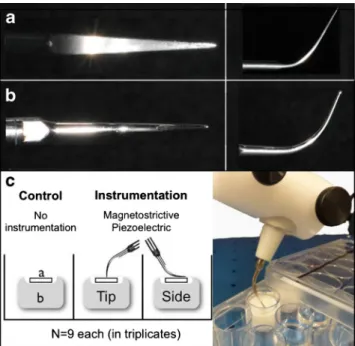
Biofilms were treated with two different ultrasonic scalers, namely, a piezoelectric miniMaster generator (EMS, Nyon, Switzerland) and a Cavitron Select SPS generator (Dentsply, York, PA, USA) at medium power. The insert designs used with these generators included the P tip (with the EMS gen-erator) and the straight Slimline insert (with the Dentsply generator; Fig.1).
Fig. 1 Tip design (a, b) and study set-up (c). The different insert designs used in this study as seen from above (left) and sideways (a P tip, b Slimline). First, HA discs with established biofilms (a) were put into sterile customized Teflon moulds (b), which were placed in 96-well plates, and were treated with either a magnetostrictive or piezoelectric ultrasonic device as described in the“Material and methods” section. All
tests were carried out in triplicates in three different experiments resulting in a total sample size of N =9 per group
Prior to treatment, the HA discs were fixed in Teflon moulds and put in wells of a 24-well cell culture plate that was placed on a balance in order to apply defined pressure during the treatment (Fig. 1). Treatments were randomly allocated, and four discs were used for each intervention. Four samples were treated for 15 s at a pressure of 40 g with the tip of the piezoelectric scaler, while another four biofilms where treated under the same conditions, but using the convex front part of the same scaler (referred as the“side” throughout the manuscript; Fig.1). The same procedures were performed for the magnetostrictive scaler. Standardized application force for each treatment method was achieved by mounting the teeth in a specially adapted pressure-sensitive electronic device (TM 503 Power Module, Tektronix®, Inc., Beaverton, Oregon, USA). After every treatment, the biofilms were rinsed with 1.6 ml sterile saline. Four control discs were left untreated except for rinsing. Biofilms of these samples were scraped off manually (control) in order to determine the total CFU of firmly adhering bacteria.
While three of the four discs were used for the analysis of the biofilm mass, the randomly selected fourth sample was used for CLSM analysis, as described below. Therefore, the experiments to assess the biofilm removal were carried out in triplicates, resulting in N =9 samples in total per group. Analysis of biofilm removal
To measure the amount of potentially growing bacteria with-out ultrasonic treatment, biofilms were manually scraped off (Perio Soft-Scaler, Kerr, Bioggio, Switzerland) the discs, and the latter were rinsed with 1.6 ml sterile saline to remove non-adherent bacteria.
After treatment, the supernatant was collected (1.6 ml), and serial dilutions of suspended biofilm bacteria were prepared in 0.9 % NaCl, and 50-μl aliquots were plated on Columbia blood agar supplemented with 5 % whole human blood to estimate total CFU, and agar plates were incubated anaerobi-cally at 37 °C for 72 h. Data were scored as total CFU per biofilm.
All microbiological tests and analyses were performed strictly blinded to the nature of the previous treatment of the individual discs.
Staining of biofilms and CLSM
For CLSM, treated and untreated biofilms were stained using the LIVE/DEAD BacLight bacterial viability assay (Invitrogen, Zug, Switzerland) according to the instructions of the manufacturer. After 20 min of staining, excess dye was gently aspirated from the discs without touching the biofilms. They were embedded upside down in 20μl of Mowiol [15] and stored at room temperature in the dark for at least 6 h prior to microscopic examination.
Stained biofilms were examined by CLSM at randomly selected positions using a Leica TCS SP5 (Leica Microsystems, Heidelberg GmbH, Germany) with a ×20/0.8 numerical aperture (NA) and ×63/1.4 NA oil immersion ob-jective lens in conjunction with 488-nm laser excitation and 530-nm emission filters for Syto 9 (live stain) and 561-nm laser excitation and 640-nm emission filters for propidium iodide (dead stain). Image acquisition was done in eight-line average mode, and the data were processed using Imaris 7.2.2 (Bitplane AG, Zurich, Switzerland).
Statistical analysis
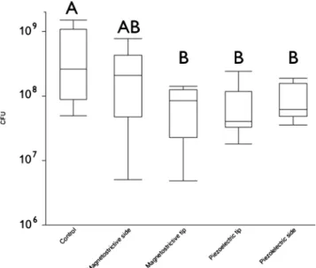
Descriptive statistics of the data were performed with SPSS (version 20.0) and illustrated with box plots. The log10
-trans-formed data met the requirements for parametric analysis. Hence, differences between treatments were analyzed using a one-way analysis of variance (ANOVA) followed by the Scheffe post hoc tests (significance level p <0.05).
Results
The effect of the different instrumentation procedures on the HA-grown biofilms was evaluated by means of defining the bacterial CFUs in the culture supernatants, following the treatments (Fig. 2). As compared to the untreated control, which displayed the maximum bacterial mass to be potentially dislodged, it was found that only a side application of the magnetostrictive device was able to remove a comparable amount of the biofilm. In contrast, its tip application as well
Fig. 2 Box plots of the total CFUs of the untreated control specimens (scrapped off the HA surface) and the total CFUs release into the super-natant after treatment of biofilms with the different devices. Identical capital letters represent results, which do not statistically significantly differ from each other (ANOVA, Scheffe)
as both applications of the piezoelectric device removed sig-nificantly less bacteria.
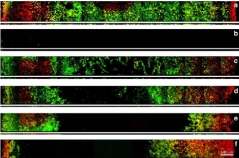
In Fig.3, a representative series of confocal images before and after treatment is presented. The broad image represents a section of the biofilm taken at 5μm distance from the surface of the disc, and the smaller image shows the corresponding cross section of the biofilm. The figure confirms the results of the CFU analyses and the Live/Dead staining demonstrates that ultrasonic scaling has no effect on the vitality of the biofilms except for the treatment with the magnetostrictive side application (Fig. 3f). Figure 3a shows the untreated biofilm. The HA disc was confluently colonized by a biofilm with a mean thickness of 38μm. In Fig.3b, the biofilm after manual scaling is shown, and the image confirms that the whole biofilm was eliminated. Figure3c, dshows the effect of the piezoelectric scaler: Treatment using the tip removed the biofilm only in the centre of the disc (Fig.3c), whereas a sideways application was somewhat more efficient. In
Fig. 3e, f, the treatment of the magnetostrictive device is
demonstrated. Apparently, this scaler was able to eliminate more of the in vitro biofilm than piezoelectric application mode.
Discussion
This study assessed the direct impact of ultrasonic scaler tips on biofilm removal. This was performed by both conventional culture techniques to determine the removed bacteria in the supernatant and by visualization using a combination of FISH
and CLSM. In general, there is still a great need to assess and standardize biofilm removal procedures for testing the (pre-)cleaning efficiency [16], and there are still limited data available concerning the biofilm removal capacity using dif-ferent protocols, devices and/or chemicals.
The main finding of this study is that only a side application of the magnetostrictive device was able to remove a consid-erable amount of the biofilm from the HA surface, as com-pared to the manually treated control, which displayed the maximum bacterial mass to be potentially dislodged. A tip application, in either the magnetostrictive or the piezoelectric device, in contrast, removed a significantly smaller amount of the biofilm, as evaluated by both CFU counting and CLSM visualization.
The in vitro biofilm used in this study is a well-established and validated biofilm model of standardized conditions, consisting of either five or six species representative for supragingival plaque [17,18]. This model has been proven to provide repeatable results on different materials and has been successfully used to evaluate the antimicrobial potential in vitro [12,19–21]. Although this method still represents a simplified laboratory plaque model, it mimics the complex in vivo situation more precisely than a mono-species biofilm, and due to a feeding model, the biofilm exerts a sticky con-sistency, which is comparable to the natural conditions. Hence, this is a suitable experimental model to test the effi-ciency of ultrasonic scalers in removing supragingival plaque, under standardized conditions.
Regarding the biofilm removal capacity, a previously pub-lished study showed comparable numbers of cultivatable
Fig. 3 CLSM images of in vitro biofilms on the HA surface before and after treatment. Images have been taken along the diameter of the discs and represent only a detail of the whole disc, whereas the broad and small strips represent transverse (xy) sections taken at 5μm above the HA surface and the corresponding cross sections (xz), respectively, with the surface of the biofilm facing downwards. The biofilms were stained using
the LIVE/DEAD Viability Kit; live cells appear green and dead cells red. Black areas on the HA surface resulted from complete removal of the biofilm. a Untreated biofilm. b After manual collection of biofilms. c Piezoelectric scaler tip. d Piezoelectric scaler side. e Magnetostrictive scaler tip. f Magnetostrictive scaler side. Scale bar = 500μm
bacteria on untreated samples, by investigating colonization and measuring total CFUs [11], which elucidates again the reproducibility of this model with regard to biofilm growth. In addition, the effectiveness of biofilm removal has proven to be largely dependent on the methods applied so far. The treat-ment with ozone and photodynamic therapy (PDT), for in-stance, showed only minute effects on the remaining biofilm [19]; the observed reduction of viable counts by both treat-ment options was less than one log10step. In another study,
where the efficiency of shock waves was investigated, they effectively removed biofilms by three log steps [11]. In the present study, both the biofilm structure remaining on the HA surface and the number of detached bacteria were evaluated, following the two treatment modalities. However, since only viable counts were determined, one can drive conclusions only on the viability after treatment, not on bacterial detach-ment per se. That is because non-viable bacteria could also be detached from the HA surface following treatment. This can-not be taken under consideration when measuring the CFUs. Hence, further combined usage of FISH and CLSM to identify the biofilm remaining on the surface is a suitable complemen-tary approach in the present study.
Relevant work also exists on the mechanisms of non-contact biofilm removal by sonic and other powered tooth-brushes, which elucidates the importance of this kind of evaluations to study the efficiency of bacteria removal by physimechanical means. A study by Busscher and co-workers showed that sonic brushing at contact removed 92 to 94 % of the coadhering and non-coadhering pair under investigation, respectively, but removal decreased with in-creasing distance between the brush and the pellicle surfaces [22]. Especially, non-contact biofilm removal must be regarded as an interplay of hydrodynamic energy transfer through the fluid [23]. The extent to which specific different hydrodynamic factors contribute is still dependent on the specifics of the instruments. On the other hand, He and co-workers found that powered brushing in non-contact mode changed the viscoelastic properties of the oral biofilm, resulting in an increased penetration of antimicrobial com-pounds into the biofilm [24,25]. Therefore, even if biofilms are not totally removed by sonic brushing, they may be more susceptible to antimicrobials thereafter.
Walmsley and co-workers mapped the occurrence of cav-itation around scaler tips under loaded conditions [10]. The vibration displacement amplitude of ultrasonic scalers in-creased with the occurrence of cavitation, but factors such as the length of the probe influence the amount of cavitation activity generated. In general, the application of load affects the production of cavitation at the most clinically relevant area, which is the tip of the device.
A standardized load of 40 g was applied in this study. It has been shown that magnetostrictive probes oscillated with great-er displacement amplitudes than piezoelectric probes but still
produced similar defects. This may be due to the cross-sectional shape of the probes [8]. In the present study, the same devices and tips were used, and these earlier findings could be reproduced in this biofilm model as well. However, the applied loads were higher in the latter case, ranging from 100 to 200 g. Future studies could use the presented biofilm model to study the contact free effectiveness of ultrasonic instrumentation using different action modes, power settings and geometries. However, one should keep in mind that the sample arrangement, i.e. the embedding or attachment of the samples, may influence the vibration transduction and, thus, the efficacy of the treatment. The disc material can as well play a role. With this respect, it can also be speculated that a slimmer tip may cut into the disc throwing up micron-sized particles, which may then add to a slurry increasing cavitation. In the present study, we used an artificial HA disc with a Knoop hardness number of 310, which is in the range of enamel, and no surface damage could be observed. However, this does not exclude the possibility of microscopic damage, and it would be of interest to assess this aspect on dentin or other biological surfaces and biomaterials in terms of biofilm removal in combination with surface damage in future studies. In summary, studies of ultrasonic devices provide valid documentation of their efficiency, particularly when using biofilm models, and this study provided first insights in the microbiological aspects of working action of ultrasonic scalers under standardized conditions.
Conclusions
Regarding the research hypothesis, it was found that both ultrasonic tips under investigations led to bacterial detach-ment, but the action mode as well as the tip configuration and adaptation appeared influenced the biofilm removal effectiveness.
Acknowledgments The authors would like to thank Andy Meier and Ruth Graf for their great support in the laboratory. We thank the Centre of Microscopy and Image Analysis (ZMB) of the University of Zurich for their support with confocal microscopy.
Conflict of interest The authors have no conflicts of interest.
References
1. Badersten A, Nilveus R, Egelberg J (1981) Effect of nonsurgical periodontal therapy. I. Moderately advanced periodontitis. J Clin Periodontol 8:57–72
2. Walmsley AD, Lea SC, Landini G, Moses AJ (2008) Advances in power driven pocket/root instrumentation. J Clin Periodontol 35:22–28 3. Lampe Bless K, Sener B, Dual J, Attin T, Schmidlin PR (2011) Cleaning ability and induced dentin loss of a magnetostrictive ultrasonic instrument at different power settings. Clin Oral Investig 15:241–248
4. Lea SC, Landini G, Walmsley AD (2006) The effect of wear on ultrasonic scaler tip displacement amplitude. J Clin Periodontol 33: 37–41
5. Walmsley AD, Laird WR, Williams AR (1984) A model system to demonstrate the role of cavitational activity in ultrasonic scaling. J Dent Res 63:1162–1165
6. Walmsley AD, Laird WR, Williams AR (1986) Displacement ampli-tude as a measure of the acoustic output of ultrasonic scalers. Dent Mater 2:97–100
7. Walmsley AD, Laird WR, Williams AR (1986) Inherent variability of the performance of the ultrasonic scaler. J Dent 14:121–125 8. Lea SC, Landini G, Walmsley AD (2002) Vibration characteristics of
ultrasonic scalers assessed with scanning laser vibrometry. J Dent 30: 147–151
9. Lea SC, Felver B, Landini G, Walmsley AD (2009) Three-dimensional analyses of ultrasonic scaler oscillations. J Clin Periodontol 36:44–50
10. Walmsley AD, Lea SC, Felver B, King DC, Price GJ (2013) Mapping cavitation activity around dental ultrasonic tips. Clin Oral Investig 17: 1227–1234
11. Muller P, Guggenheim B, Attin T, Marlinghaus E, Schmidlin PR (2011) Potential of shock waves to remove calculus and biofilm. Clin Oral Investig 15:959–965
12. Shapiro S, Giertsen E, Guggenheim B (2002) An in vitro oral biofilm model for comparing the efficacy of antimicrobial mouthrinses. Caries Res 36:93–100
13. Thurnheer T, van der Ploeg JR, Giertsen E, Guggenheim B (2006) Effects of Streptococcus mutans gtfC deficiency on mixed oral biofilms in vitro. Caries Res 40:163–171
14. Gmur R, Guggenheim B (1983) Antigenic heterogeneity of Bacteroides intermedius as recognized by monoclonal antibodies. Infect Immun 42:459–470
15. Thurnheer T, Gmur R, Shapiro S, Guggenheim B (2003) Mass transport of macromolecules within an in vitro model of supragingival plaque. Appl Environ Microbiol 69:1702–1709
16. Oulahal N, Martial-Gros A, Bonneau M, Blum LJ (2004) Combined effect of chelating agents and ultrasound on biofilm removal from stainless steek surfaces. Application to“Escherichia coli milk” and “Staphylococcus aureus milk” biofilms. Biofilm 1:65–73
17. Guggenheim B, Giertsen E, Schupbach P, Shapiro S (2001) Validation of an in vitro biofilm model of supragingival plaque. J Dent Res 80:363–370
18. Guggenheim B, Guggenheim M, Gmur R, Giertsen E, Thurnheer T (2004) Application of the Zurich biofilm model to problems of cariology. Caries Res 38:212–222
19. Muller P, Guggenheim B, Schmidlin PR (2007) Efficacy of gasiform ozone and photodynamic therapy on a multispecies oral biofilm in vitro. Eur J Oral Sci 115:77–80
20. Dezelic T, Guggenheim B, Schmidlin PR (2009) Multi-species bio-film formation on dental materials and an adhesive patch. Oral Health Prev Dent 7:47–53
21. Hofer D, Meier A, Sener B, Guggenheim B, Attin T, Schmidlin PR (2011) Biofilm reduction and staining potential of a 0.05 % chlor-hexidine rinse containing essential oils. Int J Dent Hyg 9:60–67 22. Busscher HJ, Rustema-Abbing M, Bruinsma GM, de Jager M,
Gottenbos B, van der Mei HC (2003) Non-contact removal of coadhering and non-coadhering bacterial pairs from pellicle surfaces by sonic brushing and de novo adhesion. Eur J Oral Sci 111:459–464
23. Busscher HJ, Jager D, Finger G, Schaefer N, van der Mei HC (2010) Energy transfer, volumetric expansion, and removal of oral biofilms by non-contact brushing. Eur J Oral Sci 118:177–182
24. He Y, Peterson BW, Ren Y, van der Mei HC, Busscher HJ (2013) Antimicrobial penetration in a dual-species oral biofilm after non-contact brushing: an in vitro study. Clin Oral Invest. doi:10.1007/ s00784-013-1097-x
25. He Y, Peterson BW, Jongsma MA, Ren Y, Sharma PK, Busscher HJ, van der Mei HC (2013) Stress relaxation analysis facilitates a quan-titative approach towards antimicrobial penetration into biofilms. PLoS One 27(8):e63750