Macrofollicular Variant of Follicular Thyroid Carcinoma:
A Case Report
Claudio De Vito&Massimo Bongiovanni&
Frederic Triponez&Marc Pusztaszeri
Published online: 7 April 2013
# Springer Science+Business Media New York 2013 Case Report
A 37-year-old female known for systemic lupus erythematosus underwent a right thyroid lobectomy for a growing nodule in the right lobe. A fine-needle aspiration biopsy (FNAB) performed 4 years before surgery reported a microfollicular proliferation. A second FNAB performed a few months before surgery revealed a benign lesion. The surgical specimen was fixed in 4 % buffered formaldehyde. The entire nodule was sampled, with particular attention to the capsule, and embedded in paraffin and corresponding sections were stained with hematoxylin and eosin for histological examination. Immuno-histochemistry using HBME1 and galectin-3 was performed.
What is Your Diagnosis?
The pathological diagnosis of the patient was follicular thyroid carcinoma, macrofollicular variant, which is minimally invasive, stage pT2 pNX.
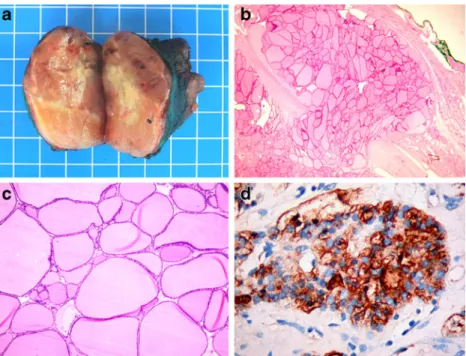
On gross section, the nodule measured 4×4×3.5 cm. It was encapsulated with a shiny colloidal appearance without calcification (Fig.1a). On histology, the nodule was encap-sulated, with variable capsule thickness. It was essentially macrofollicular (>200 μm) with a minor microfollicular component (Fig.1b). The cells were attenuated to cuboidal
with an eosinophilic cytoplasm and round hyperchromatic nuclei without any nuclear feature suspicious for papillary thyroid carcinoma (PTC) (Fig. 1c). No mitotic activity or necrosis was found. Focal transcapsular invasion was present (Fig. 1b), but no vascular invasion was found. Immunohistochemistry showed focal but strong HBME-1 staining essentially in the microfollicular areas (Fig. 1d) and absence of galectin-3 staining.
Comment
Here, we report the case of a minimally invasive follicular thyroid carcinoma (FTC) with a predominant macrofollicular growth pattern. Thyroid nodules with a macrofollicular architecture are considered for the most part as benign lesions and include nodular hyperplasia, adenomatous nodules, and some follicular adenomas. In contrast, FTCs are generally hypercellular and very few exhibit normofollicular or macrofollicular appearances [1]. There-fore, the index of suspicion should be raised whenever encountering a follicular neoplasm with a solid, trabecular, or microfollicular pattern of growth [1]. However, rare cases of thyroid malignant neoplasms with a macrofollicular pattern have been described such as the macrofollicular variant of PTC [2] and the even rarer macrofollicular variant of FTC [3]. Thus, lesions with a predominant macrofollicular component should also be assessed for nuclear features of PTC and for capsule and vascular invasion. Therefore, sampling the entire capsule is recommended.
Correspondingly, FNAB provides essential informa-tion regarding architectural and cytology aspects of the thyroid nodules and plays a key role as a screening test for thyroid follicular neoplasms [4, 5]. FTC classically shows microfollicular growth pattern and identification of this component is the key feature in order to classify these nodules as suspicious for a follicular neoplasm, even though C. De Vito
:
M. Pusztaszeri (*)Department of Clinical Pathology, Geneva University Hospital, 1 rue Michel-Servet,
1211 Geneva 14, Switzerland e-mail: [email protected] M. Bongiovanni
Institute of Pathology, Locarno, Switzerland F. Triponez
Department of Thoracic and Endocrine Surgery, Geneva University Hospital, Geneva, Switzerland
Endocr Pathol (2013) 24:167–168 DOI 10.1007/s12022-013-9241-3
the majority (>70 %) of these nodules turn out to be benign on surgical follow-up [4, 5]. Thus, cytological specimens of macrofollicular variant of FTC can be a pitfall and be misinterpreted as benign due to the lack of microfollicles [5]. Therefore, they may account for an undetermined fraction of the false-negative FNAB cases. However, some of these cases may fall into oblivion and never be detected as their malignant potential, if any, may never be apparent. Immuno-histochemistry (e.g., HBME-1, galectin-3) may support the diagnosis of malignancy for this rare variant of FTC with deceivably bland architectural and cytological features.
In conclusion, this case report illustrates a rare macro-follicular variant of FTC that can be misdiagnosed as a benign lesion, supports the need to thoroughly sample the nodule capsule, and assesses for vascular and capsular invasion also in macrofollicular patterned thyroid nodules.
References
1. Rosai JCM, DeLellis R: Atlas of Tumor Pathology: Tumors of the Thyroid Gland, series 3. Washington, Armed Forces Institute of Pathology, 1992.
2. Albores-Saavedra J, Housini I, Vuitch F, Snyder WH 3rd. Macrofollicular variant of papillary thyroid carcinoma with minor insular component. Cancer 80:1110–1116, 1997.
3. Bongiovanni M, Gremaud M, Moulin CS, Scheidegger C, Biton C, Clément S. Macrofollicular variant of follicular thyroid carcinoma: a clinical, cytologic, morphologic, and image analysis study of a unique case. Ann Diagn Pathol. 13:101–105, 2009.
4. Cibas ES, Ali SZ. The Bethesda System For Reporting Thyroid Cytopathology. Am J Clin Pathol. 132:658–665, 2009.
5. Maruta J, Hashimoto H, Suehisa Y, Yamashita H, Noguchi S, Aratake Y, Ohno E, Kobayashi TK. Improving the diagnostic accuracy of thyroid follicular neoplasms: cytological features in fine-needle aspiration cytology. Diagn Cytopathol. 39:28–34, 2011.
Fig. 1 a Macroscopical aspect of the macrofollicular variant of follicular thyroid carcinoma. b The tumor is encapsulated and shows a“mushroom-like” focus of transcapsular invasion into the adjacent thyroid parenchyma. c The tumor is composed mainly of macrofollicles lined with cuboidal or attenuated cells with bland nuclear features. d Tumor cells in the minor microfollicular areas are strongly immunopositive for HBME-1