HAL Id: hal-01614918
https://hal.sorbonne-universite.fr/hal-01614918
Submitted on 11 Oct 2017
HAL is a multi-disciplinary open access
archive for the deposit and dissemination of
sci-entific research documents, whether they are
pub-lished or not. The documents may come from
teaching and research institutions in France or
abroad, or from public or private research centers.
L’archive ouverte pluridisciplinaire HAL, est
destinée au dépôt et à la diffusion de documents
scientifiques de niveau recherche, publiés ou non,
émanant des établissements d’enseignement et de
recherche français ou étrangers, des laboratoires
publics ou privés.
Controlled arterial reflow after ischemia induces better
outcomes in the juvenile rat brain
Philippe Bonnin, Julien Pansiot, Elise Paven, Maxime Eloi, Sylvain
Renolleau, Olivier Baud, Pierre-Louis Leger, Christiane Charriaut-Marlangue
To cite this version:
Philippe Bonnin, Julien Pansiot, Elise Paven, Maxime Eloi, Sylvain Renolleau, et al..
Con-trolled arterial reflow after ischemia induces better outcomes in the juvenile rat brain. Journal
of Cerebral Blood Flow and Metabolism, Nature Publishing Group, 2017, 37 (9), pp.3091-3096.
�10.1177/0271678X17719614�. �hal-01614918�
Controlled arterial reflow after ischemia
induces better outcomes in the juvenile
rat brain
Philippe Bonnin
1,2, Julien Pansiot
1,3, Elise Paven
1,2,
Maxime Eloi
1,3, Sylvain Renolleau
4, Olivier Baud
1,3,
Pierre-Louis Leger
1,3,5,* and
Christiane Charriaut-Marlangue
1,3Abstract
Our objective was to determine whether controlled reflow on one side and/or the other side after bilateral carotid occlusion release could reduce cell death in focal ischemic P14 rats. Arterial blood flow was measured using ultrason-ography. Cell death, inflammation and nitrotyrosine were measured using immunofluorescence. When reflow was first induced in the contralateral side, we observed improved outcome markers compared with those when reflow was first induced in the ipsilateral side and/or simultaneous reflow was induced in both sides. Our data suggest that progressive rerouting of arterial flow through the circle of Willis toward the ischemic site reduced cell death.
Keywords
Ischemic stroke, macrocirculation, arterial re-flow, cell death
Introduction
Arterial ischemic stroke is increasingly recognized as a serious pediatric problem. The groups at risk of arterial ischemic stroke are newborns, especially full-term infants, and older children with sickle cell disease, or under specific conditions (surgery of congenital cardio-pathy, extracorporeal life supports), although 1/3 of childhood strokes remain cryptogenic.1
In the adult brain, restoration of blood flow (BF) to the ischemic territory is characterized by significant hyperemia within the penumbra that occurs immedi-ately after occlusion release.2,3In contrast, a progres-sive and incomplete reperfusion occurred in the cortical penumbra during early reflow in the neonatal P7 rat brain.4 In the juvenile P14 rat brain, rapid and com-plete reperfusion, without hyperemia was detected in the penumbra.5 In addition, juvenile rats responded
to ischemia by producing prostaglandins in a COX-2-and mPGES1-dependent manner.6
Whereas ischemic postconditioning (a series of rapid intermittent interruptions of BF at reflow) was demonstrated to be a harmless procedure that
attenuates cerebral BF (CBF) disturbances in adults, this paradigm did not ameliorate ischemic lesions in neonatal ischemic P7 rats, which suggests collateral support efficiency7 and a lower capacity to activate NO production in the immature brain (for review see Leger et al.8).
In the present study, we evaluated whether control-ling arterial reflow by gradual occlusion release allevi-ates damage using both imaging and molecular approaches.
1Universite´ Sorbonne Paris Cite´, Paris, France
2INSERM U965, Universite´ Denis Diderot, Physiologie Clinique –
Explorations Fonctionnelles, Hoˆpital Lariboisie`re, Paris, France
3PROTECT, INSERM U1141, Universite´ Denis Diderot, Paris, France 4Universite´ Rene´ Descartes, AP-HP, Hoˆpital Necker, PICU Paris, France 5
UPMC, AP-HP, Hoˆpital Armand Trousseau, PICU, Paris, France
Corresponding author:
Christiane Charriaut-Marlangue, INSERM UMR 1141, Hopital Robert Debre´, 48 bd Serurier, Paris 75019, France.
Email: [email protected] The last two authors share senior authorship.
Material and methods
All experiments of this study complied with the ethical guidelines of the Robert Debre ´ Hospital Research Council Review Board (A75-19-01), INSERM, and the ARRIVE guidelines (http://www.nc3rs.org/ ARRIVE) and were approved by the local ethic committee (Paris7, France). All experiments were per-formed in a blinded and randomized manner.
Ischemia was induced in Wistar P14 rats (Janvier, Le genest St-Isle, France, 31.2! 3.12 g, n ¼ 75) as previously reported.5 Briefly, thermoregulated (37.0! 0.5#C) and
anesthetized rats (induction 2%, maintenance 1.5% iso-flurane in air) were exposed to ipsilateral left middle cerebral artery (MCA) electrocoagulation (MCAo) com-bined with a transient (60 min) double occlusion of the common carotid arteries (CCAo). Carotid blood flow restoration was assessed by release of either (1) both clips at the same time (control group), (2) first clip on the right CCA, then 5 min after on the left CCA (R-L group), and conversely, (3) first clip on the left CCA, then 5 min after on the right CCA (L-R group).
Randomized allocation of animals
For each litter, the 10 pups were labeled from 1 to 10. The numbered pups ‘‘1, 3 and 6’’ a priori constituted the R-L group, the numbered pups ‘‘2, 5 and 9’’ con-stituted the control group, and the numbered pups ‘‘4, 7 and 10’’ constituted the L-R group. Then, each rando-mized animal was subjected to ischemia and reperfu-sion. The numbered pup 8 could replace an animal that died during anesthesia.
Sample size calculation
Assuming abeta risk of 0.2 and an alpha risk of 0.05, it was estimated (using the BiostaTGV software (https:// marne.u707.jussieu.fr/biostatgv)) that six animals in each group were needed to observe a significant differ-ence in gene and protein expression, and US imaging as previously reported.5,6As an example (mean 1¼ 13.5, mean 2¼ 9.8, common SD ¼ 2), four animals per group are necessary.
Inclusion/exclusion
All animals were included in this study (no mortality). Animals were then subjected to 2D color-coded ultrasound imaging.5 COXs and terminal synthesizing
enzymes genes were evaluated at 1 h after reflow. Protein expression6 (including cell death) were evalu-ated at 4 and/or 24 h after reflow. A detailed description of Materials and Methods can be found in the supple-mentary material.
Results
Blood flow redistribution according to occlusion
release protocols
The three groups of animals displayed similar mBFV under basal conditions in the three great arteries of the circle of Willis. At the end of ischemia, when both CCAs were occluded, mBFV in the basilar trunk (BT) increased in a similar manner in the three groups (Figure 1(a); p < 0.001 compared to basal values; Supplemental Figure 1), suggesting collateral supply.5 We also observed (for the three groups) residual mBFV in both intracranial carotid arteries (ICAs, Figure 1(b) and (c)) despite no visible flow in the CCA.
In the control group (simultaneous occlusion release in both CCAs), at 15 min after reflow, mBFV in the BT returned to basal values (11.1! 1.1 compared to 11.9 ! 1.7, respectively, NS), whereas reflow in both ICAs occurred 1 min after clip removal with a similar pattern in both the left and right ICAs, as previously reported.5
Fifteen minutes after re-flow, mBFV in the right and left ICA were not significantly different from basal values (Figure 1(b) and (c); Supplemental Figure 1).
In the R-L group, reflow in the right CCA was accompanied by a return to basal mBFV in the right ICA; reflow in the left CCA was accompanied with a return to basal mBFV in the left ICA, whereas mBFV in the right ICA and BT decreased (p < 0.01 vs. control). Fifteen minutes after reflow, mBFV in the BT (7.9! 1.8 cm/s) and right ICA (9.7 ! 1.5 cm/s) remained reduced (p < 0.01) compared with the basal values (10.6! 2.0 and 13.3 ! 3.4 cm/s, respectively Figure 1(b) and (c); Supplemental Figure 1). Mean BFV in the left ICA (10.7! 3.4 cm/s) decreased com-pared with the basal (14.9! 2.9 cm/s) values (p < 0.05). In the L-R group, reflow in the left CCA was accom-panied by a return to basal mBFV in the left ICA; reflow in the right CCA was accompanied by a return to basal mBFV in the right and left ICA and the BT. Fifteen minutes after reflow in the three great arteries, mBFV were similar to basal mBFV. No significant vari-ation in heart rates was detected between groups throughout the procedure.
COX-2 and mPGES1 gene and protein expression,
microglial activation and cell death
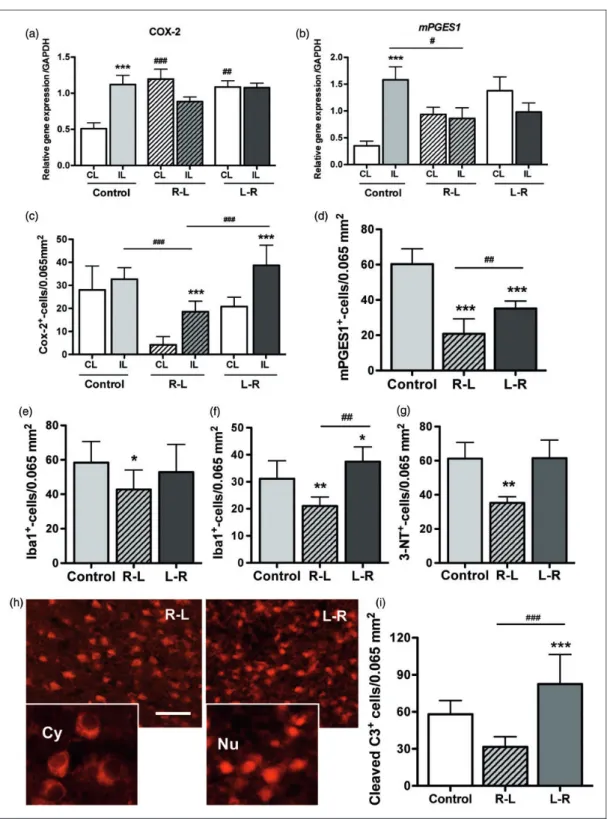
One hour after ischemia-reperfusion, whereas an increase in the COX-2 (p < 0.001) and mPGES1 (p < 0.001) genes was observed in the ipsilateral (IL) cortex in the control group,6 only a reduction in the mPGES1 (p < 0.05) gene was measured in the R-L group (Figure 2(a) and (b)).
Four hours after ischemia-reperfusion, the COX-2 and mPGES1 proteins in the IL cortex were reduced
(p < 0.001, Figure 2(c) and (d), Supplemental Figure 2), as well as microglial density (determined with Iba-1 immunostaining, Figure 2(e), Supplemental Figure 3), in the R-L group compared with those in the control
group. At 24 h after reperfusion, microglial density was reduced in the R-L group and increased in the L-R group (Figure 2(f), Supplemental Figure 3). The extent of protein nitrotyrosine formation (an index for reactive nitrogen species; Supplemental Figure 4) was higher in the control and L-R animals than that in R-L animals at 24 h after injury (Figure 2(g)). The density of cleaved-casp-3þ-cells (apoptotic marker) was increased in the L-R group (p < 0.001) compared with that in the control (p < 0.001) and R-L (p < 0.001) groups. Furthermore, the R-L animals displayed numerous cells with cytosolic cleaved-casp-3, whereas the L-R animals mostly displayed cells with nuclear cleaved-casp-3, suggesting completion of apoptosis in the latter group (Figure 2(g) and (h)) according to the pale lesion observed at 24 h after reflow (supplemental Figure 5).
Discussion
We here report that rerouting CBF by first inducing reflow in the CL side (R-L group) before reflow in the IL side may be beneficial by reducing inflammation, reactive nitrogen species and cell death.
To provide a better reperfusion in the ischemic pen-umbra, either thrombolysis or improvement of collateral supply has been proposed in the acute phase of ischemic stroke. Increased collateral recruitment can be obtained by the administration of drugs to increase vasodila-tion,9,10or by facilitating the synthesis of vasodilators6
leading to the rerouting of BF through the numerous native anastomoses in the cerebral vascular network. We have also demonstrated that an increase in collateral supply induced by exogenous NO-donors is beneficial during ischemia but deleterious during reflow due to an elevation in oxidative stress.10,11
During ischemia, both hemispheric arterial networks experience a decrease in vascular resistance mediated by the loss of the myogenic tone (deep decrease in intra-vascular pressure),12and the accumulation of metabolic
end-products as well as the initiation of anaerobic res-piration (CO2, glutamate, lactate, pyruvate, acidosis,
Ca2þ).13 The decrease in vascular resistance promotes the establishment of collateral recruitment after the changes in the arterial pressure gradient from the BT to the hemispheres (1) through the circle of Willis with perfusion of the two intracranial ICAs by the BT, through the posterior communicant arteries and the proximal segment of the posterior cerebral arteries (reverse-flow, the posterior cerebral artery is the first branch of the ICA in rodents)14; or (2) through the cortical arterial anastomoses that extend from the vas-cular supplies of the posterior and/or anterior cerebral arteries towards the vascular supply of the middle cere-bral artery, particularly in the ischemic penumbra.14 Figure 1. Blood flow redistribution in the three great arteries
of the circle of Willis after the transient CCA occlusion release protocols. (a–c) Mean BFV in the basilar trunk (BT, a), the right ICA (rICA, b), and left ICA (c) in the control (white bars, sim-ultaneous CCAs occlusion release, n¼ 6), R-L (gray bars, n ¼ 7), and L-R (black bars, n¼ 7) groups under basal conditions, at end of ischemia (Isch), after the first CCA release (R1), after the second CCA release (R2 – 5 min later), and after 15 min of re-flow. ***p < 0.001 (vs. basal);##p < 0.01,###p < 0.001 (control vs. R-L);zp < 0.05,zzzp < 0.001 (R-L vs. L-R).
Figure 2. COX-2 and mPGES-1 gene and protein expression, nitrotyrosine formation and cell death. (a–d) Quantification for the COX-2 and mPGES1 genes (a–b) and proteins (c–d) in the three groups of animals (n¼ 6 per group) in the left (IL) and right (CL) cortex. (e–f) Quantification of microglial cells (immunostained with Iba-1) in the IL cortex at 4 (e, n¼ 10) and 24 (f, in remote peri-infarct, n¼ 10) hours after reflow. (g) Quantification of 3-nitrotyrosine (3-NT) formation at 24 h after reflow. (h–i) Number of cleaved-caspase-3þ-cells in the ipsilateral peri-infarct in the three groups of animals at 24 h after reflow. CL: contralateral; IL: ipsilateral; Cy: cytoplasm; Nu: nuclear. *p < 0.05, **p < 0.01, and ***p < 0.001 vs. control.#p < 0.05,##p < 0.01,###p < 0.001 R-L group vs. L-R
During reflow, when the clamp on CCA one is released, arterial pressure and BF are instantaneously reestablished in a vasodilated vascular network. The vascular network requires a few seconds to control vas-cular tone and to restore adapted vasvas-cular resistance to the restored arterial pressure in order to control the local CBF. In the R-L group, arterial pressure and blood flow are reestablished in the right intracranial ICA first. The left ICA is then reperfused by a retro-grade flow coming from the confluence of both anterior cerebral arteries (azygos artery in rodents), allowing the progressive pressure loading and the lavage of the metabolic end-products, particularly in the ischemic penumbra. Local vascular tone/resistance can therefore be restored progressively without a strong increase in the local CBF. Resupplying the arteries after low blood flow involves a decrease in metabolic end-products such as CO2 and Hþ, with changes deep acidosis to mild
local acidosis that is known to be neuroprotective during post-conditioning.15Moreover, a mild hypoxic
exposure after severe hypoxia in adult rats constitutes a postconditioning mode that attenuates post-hypoxic neuronal injury and reduces brain edema with improve-ment in recovery after severe hypoxia.16This
postcon-ditioning mode enhances the expression of HIF-1-alpha and erythropoietin in surviving rats after severe hyp-oxia.16Here, 5 min after the right CCA clamp release and the consecutive release of the left CCA clamp, reflow is established in the ipsilateral vascular network with a previously restored vascular tone, avoiding any supplemental increase in local oxidative stress and inflammation. Altogether, these events may constitute ‘‘hypoxic post-conditioning’’, which has been shown to be neuroprotective when the duration of hypoxia is less than 10 min.15 In the L-R group, the arterial pressure
and blood flow are instantaneously restored in the intracranial left ICA vascular network, particularly in the cortical anastomoses and thus in the ischemic pen-umbra. Upon reperfusion, the sudden resupply of oxygen and nutriments re-energizes mitochondrial aer-obic respiration, resulting in a burst in reactive oxygen species production,17which leads to elevated local oxi-dative stress and the consecutive extension of the ische-mic lesion. Identical events occur in case of concomitant release of CCAs clamps. In addition, the development of high CBF velocities in severely asphyxiated infants has been reported to predict the development of severe hypoxic-ischemic encephalop-athy and poor prognosis.18
Compared with the control and L-R groups, our data show that the animals in the R-L group exhibit less prostaglandins synthesis, microglial activation, 3-nitrotyrosine formations, and cell death. In conclusion, the duration of arterial occlusion is of course mainly responsible of the lesion size; nevertheless, after an
embolic stroke, controlled mechanical ablation of the thrombus to ensure the progressive restoration of arter-ial pressure and blood supply could be beneficarter-ial and avoid the ‘‘blood tsunami’’ that leads to local oxidative stress and nitrogen species synthesis. Our work shows that a slowly progressive cerebral arterial-recanaliza-tion following stroke may induce better outcomes. However, additional studies in rodents are needed to define the optimal rate of reperfusion.
Funding
The author(s) received no financial support for the research, authorship, and/or publication of this article.
Declaration of conflicting interests
The author(s) declared no potential conflicts of interest with respect to the research, authorship, and/or publication of this article.
Author’s contributions
CCM and PLL designed the work; PB, JP, EP, ME, SR per-formed experiments; PB, PLL, OB and CCM analyzed data and wrote the manuscript.
References
1. Tsze DS and Valente JH. Pediatric stroke: a review. Emerg Med Int 2011; 2011: 734506.
2. Pinard E, Engrand N and Seylaz J. Dynamic cerebral microcirculatory changes in transient forebrain ischemia in rats: involvement of type i nitric oxide synthase. J Cereb Blood Flow Metab 2000; 20: 1648–1658.
3. Sutherland BA, Papadakis M, Chen RL, et al. Cerebral blood flow alteration in neuroprotection following cere-bral ischaemia. J Physiol 2011; 589: 4105–4114.
4. Leger PL, Bonnin P, Lacombe P, et al. Dynamic spatio-temporal imaging of early reflow in a neonatal rat stroke model. J Cereb Blood Flow Metab 2013; 33: 137–145. 5. Leger PL, Bonnin P, Moretti R, et al. Early recruitment of
cerebral microcirculation by neuronal nitric oxide synthase inhibition in a juvenile ischemic rat model. Cerebrovasc Dis 2016; 41: 40–49.
6. Leger PL, Pansiot J, Besson V, et al. Cyclooxygenase-2-derived prostaglandins mediate cerebral microcirculation in a juvenile ischemic rat model. Stroke 2016; 47: 3048–3052.
7. Leger PL, Bonnin P, Nguyen T, et al. Ischemic postcondi-tioning fails to protect against neonatal cerebral stroke. PloS One 2012; 7: e49695.
8. Leger PL, Bonnin P, Renolleau S, et al. Ischemic postcon-ditioning in cerebral ischemia: differences between the immature and mature brain? Intl J Dev Neurosci 2015; 45: 39–43.
9. Beretta S, Versace A, Carone D, et al. Cerebral collateral therapeutics in acute ischemic stroke: a randomized pre-clinical trial of four modulation strategies. J Cereb Blood Flow Metab. Epub ahead of print 23 January 2017. DOI: 10.1177/0271678X16688705.
10. Charriaut-Marlangue C, Bonnin P, Gharib A, et al. Inhaled nitric oxide reduces brain damage by collateral recruitment in a neonatal stroke model. Stroke 2012; 43: 3078–3084.
11. Charriaut-Marlangue C, Bonnin P, Pham H, et al. Nitric oxide signaling in the brain: a new target for inhaled nitric oxide? Ann Neurol 2013; 73: 442–448.
12. Harder DR, Narayanan J and Gebremedhin D. Pressure-induced myogenic tone and role of 20-hete in mediating autoregulation of cerebral blood flow. Am J Physiol Heart Circ Physiol 2011; 300: H1557–H1565.
13. Jin Z, Wu J and Yan LJ. Chemical conditioning as an approach to ischemic stroke tolerance: mitochondria as the target. Int J Mol Sci 2016; 17: 351.
14. Bonnin P, Leger PL, Deroide N, et al. Impact of intra-cranial blood-flow redistribution on stroke size during ischemia-reperfusion in 7-day-old rats. J Neurosci Meth 2011; 198: 103–109.
15. Fan YY, Hu WW, Nan F, et al. Postconditioning-induced neuroprotection, mechanisms and applications in cerebral ischemia. Neurochem Int 2017; 107: 43–56.
16. Rybnikova E, Vorobyev M, Pivina S, et al.
Postconditioning by mild hypoxic exposures reduces rat brain injury caused by severe hypoxia. Neurosci Lett 2012; 513: 100–105.
17. Chouchani ET, Pell VR, James AM, et al. A unifying mechanism for mitochondrial superoxide production during ischemia-reperfusion injury. Cell Metab 2016; 23: 254–263.
18. Ilves P, Lintrop M, Metsvaht T, et al. Cerebral blood-flow velocities in predicting outcome of asphyxiated new-born infants. Acta Pediatr 2004; 93: 523–528.