HAL Id: tel-01229540
https://tel.archives-ouvertes.fr/tel-01229540
Submitted on 16 Nov 2015
HAL is a multi-disciplinary open access archive for the deposit and dissemination of sci-entific research documents, whether they are pub-lished or not. The documents may come from teaching and research institutions in France or abroad, or from public or private research centers.
L’archive ouverte pluridisciplinaire HAL, est destinée au dépôt et à la diffusion de documents scientifiques de niveau recherche, publiés ou non, émanant des établissements d’enseignement et de recherche français ou étrangers, des laboratoires publics ou privés.
disability and other genetically heterogeneous
developmental disorders
Claire Redin
To cite this version:
Claire Redin. NGS-based approaches for the diagnosis of intellectual disability and other geneti-cally heterogeneous developmental disorders. Genomics [q-bio.GN]. Université de Strasbourg, 2014. English. �NNT : 2014STRAJ129�. �tel-01229540�
Présentée par:
Claire REDIN
Pour l’obtention du grade de:
Docteure de l’Université de Strasbourg
Discipline: Sciences du Vivant
Spécialité: Aspects Moléculaires et Cellulaires de la Biologie
THÈSE dirigée par:
Mr MANDEL Jean-Louis Professeur, Collège de France, Paris
co-encadrée par :
Mr MULLER Jean Maitre de conférences, Praticien Hospitalier, Strasbourg
Soutenue publiquement le 2 Mai 2014 MEMBRES DU JURY:
RAPPORTEURS:
Mr CHELLY Jamel Professeur, Praticien Hospitalier, Institut Cochin, Paris
Mr ROPERS Hans-Hilger Professeur, Max-Planck-Institute for Molecular Genetics, Berlin
EXAMINATEURS:
Mr VERLOES Alain Professeur, Praticien Hospitalier, Robert Debré, Paris
Mme PUJOL Aurora Chargée de recherches, Praticien Hospitalier, IDIBELL, Barcelona
NGS-based approaches for the diagnosis of intellectual
disability and other genetically heterogeneous
developmental disorders
Bardet-Biedl Syndrome & related ciliopathies,
leukodystrophies
Miguel de Unamuno
« La science consiste à passer d'un étonnement à un autre.»
Je tiens tout d’abord à remercier vivement mes rapporteurs, les Professeurs Jamel Chelly et Hans-Hilger Ropers pour avoir accepté de lire et juger mes travaux de thèse, contenus dans un manuscrit qui n’est - au final - pas aussi court que je ne l’aurais voulu… A Jamel particulièrement, un grand merci pour ton soutien à toute épreuve, notamment pour mon après-thèse. A Hans-Hilger, c’est un véritable honneur que vous ayez accepté de faire partie de ce jury, honneur auquel je n’aurais jamais songé lors de notre première rencontre il y a quatre ans à la conférence MR-NET d’Erlangen…
Je remercie également mes examinateurs, les Professeurs Alain Verloes et Aurora Pujol pour avoir également accepté de juger mes travaux. Vous n’avez pas la lourde tâche de lire en détail l’intégralité de ce dense manuscrit mais j’espère néanmoins que vous y trouverez des résultats intéressants, Alain sur les ciliopathies, Aurora sur les leucodystrophies. A Alain, c’est un très grand honneur pour moi que vous ayez consenti à cette tâche, vous qui faisiez partie des personnes pressenties pour la succession à la présidence de l’ASHG (j’avais bien sûr voté en votre faveur…). Aurora, c’était un véritable plaisir de collaborer avec toi au cours de cette dernière année sur le projet leucodystrophies, et j’espère vivement que nous serons amenées à collaborer à nouveau dans le futur (suite du projet leucos à Boston ? ;) ).
Je tiens ensuite à remercier mes deux co-directeurs de thèse (ou co-encadrants… ;) ), le Professeur Jean-Louis Mandel et le Docteur Jean Muller, pour ces quelques quatre années passées à vos côtés qui ont été un véritable plaisir. A Jean-Lou, que je ne remercierai jamais assez, pour tout ce que tu m’as apporté sur le plan scientifique, puits sans fond de connaissance, en génétique certes mais pas que ! Et pour tout le reste, parce que c’est tellement agréable de partager autre chose que de la science, et je garderai longtemps en tête ces moments de partage musicaux et artistiques. A Jean, je tiens à te remercier, juste une fois…. ;) Pour tout ce que tu m’as appris en bioinfo, pour tous ces bons moments passés ensemble, et pour toutes ces discussions animées de par nos deux caractères finalement assez trempés…
Au reste de notre (petite) équipe ‘Mandel’: Amélie, Angélique, Nicolas, et nos interims (Grace, Inès, Sébastien, …). C’était un véritable bonheur de travailler avec vous, j’espère que mon équipe d’accueil sera aussi chouette ! Amélie, tu m’as tellement appris en génétique, j’espère un jour avoir ton lot de connaissances… C’était un réel plaisir de travailler à tes côtés sur le projet DI. Petite mention spéciale aussi sur ‘notre’ étude de réévaluation des gènes de XLID dont l’idée nous est venue - rappelle-toi - une fin d’après-midi en terrasse, autour d’un verre de vin (alsacien bien sûr). A Angélique, qui m’a rejoint en milieu de parcours, sur un projet tellement difficile… Je te
humeur quotidienne et pour tous ces bons moments, merci d’avoir joué le rôle de maître de stage avec moi sur beaucoup de choses, et surtout merci de nous avoir entretenu gourmettement ces deux dernières années (et merci à Laurent, bien sûr !), nos pantalons s’en souviendront longtemps… :)
A notre frère adoptif de l’équipe Mandel, Ricardos, je te remercie pour ta gentillesse et ton inébranlable bonne humeur, et pour tous ces moments de fous rires passés ensemble notamment lors de notre première vraie rencontre, à Erlangen en 2010. J’espère que depuis, tu as pu retourner en Allemagne avec ta Modus ? ;) A Eric aussi bien sûr, notre second membre adoptif de l’équipe Mandel, qui s’occupe si bien de notre soutien psychologique en nous ramenant des chocolats à Pâques, des glaces l’été, et des fruits à toutes saisons.
Je tiens ensuite à remercier tous les autres membres du département de Neurogénétique et Médecine Translationnelle de l’IGBMC et du laboratoire de Bioinformatique et Génomique Intégrative, pour nos discussions, scientifiques et autres, et pour cette générale bonne entente qui règne au sein de ce département et des équipes. A Olga bien sûr car depuis ton départ les journées ne sont plus les mêmes, à Karim sans qui nos journées dénuées de quelques phrases sexistes ne seraient pas pareilles ;), à Brahim pour ta contribution à nos dodécalitres de café matinaux quotidiens, à Fabrice et Hubert pour nous obliger à aller prendre le soleil le midi de temps en temps, et à beaucoup d’autres que j’oublie.
Je tiens également à remercier tous les membres de la plateforme de séquençage de l’institut avec lesquels j’ai collaboré tout au long de ces quatre années de thèse, et qui est un peu comme ma famille d’accueil. A Bernard, le ‘chef’, pour ta patience à toute épreuve, nos discussions scientifiques mais aussi sportives, et surtout ton incroyable bonne humeur. A Muriel, Serge, et Claire la petite dernière, j’ai adoré travaillé avec vous… Parfois, dans les moments difficiles, j’avais juste envie de descendre et faire des manips à vos côtés tant votre bonne humeur est contagieuse. A Stéphanie et Michaël, nos bioinformaticiens hors-pairs, que nous avons tellement mis à contribution pour nos différents projets… Stéphanie, pour ta bonne humeur, et pour toutes ces heures passées avec toi à essayer de comprendre les données de séquençage de certains patients avec réarrangements complexes! Merci pour ton aide et pour tout ton travail, sans qui cette thèse n’aurait probablement pas pu être totalement ce qu’elle est.
Un grand merci à Mims, d’une efficacité et d’une patience exemplaires pour toutes nos requêtes quelles qu’elles soient au niveau administratif. Et merci pour ton invariable bonne humeur ! Un énorme merci également à Maïté, pour être tellement disponible dès que l’on a besoin d’un article introuvable ou d’une impression infaisable. Merci de nous faciliter à tous la tâche au quotidien.
Tout d’abord au sein de l’institut, avec la team des ‘na’, Ana, Dana, Helena, Heena, Lena, Serena sans qui ma première année de thèse à Strasbourg aurait été beaucoup plus difficile ; au groupe des ‘russes’ (même s’ils ne le sont pas tous) qui m’est devenu essentiel ces dernières années: Anna, Iskander, Irina, Karima, Katarzyna, Vladimir pour tous ces bons moments passés avec vous, notamment un certain week-end de Juillet qui restera longtemps dans mes souvenirs ; à mes compagnons d’OpenLab Morgan, Morgane, Patrick, Léa et Anaïs pour toutes ces journées passées en dehors de Strasbourg à initier les lycéens aux techniques de laboratoire, j’ai la tête pleine de souvenirs avec vous; à Jérôme, pour nos discussions interminables et enjouées détendues pendant les pauses cafés ; et enfin le meilleur pour la fin : à Amélie et Vincent, pour avoir partagé tant de soirées avec vous, tant de discussions aussi, merci d’avoir été là. Vous êtes certainement les Strasbourgeois qui vont le plus me manquer outre Atlantique (trouverons-nous de nouveaux partenaires de jeux aussi assidus ? ;) ).
Ensuite, aux autre amis de plus longue date, qui m’ont permis de me changer les idées en dehors de Strasbourg: à Alex, Amandine, Angélique, Aurélie, Clément, Fred, Kro, Laurianne, Perrine et Rémi, parce que vous comptez tous énormément pour moi.
A Nicolas, qui m’accompagne depuis maintenant quelques années et j’espère pour longtemps encore. Pour ton soutien incomparable et le bonheur au quotidien.
Enfin pour terminer, je tiens à remercier ma famille, également pour leur soutien et tous ces bons moments passés et à venir. A mes parents, mes sœurs Sarah et Mathilde, mes grands parents et à tous les autres, parce que j’ai une famille formidable.
L’intégralité du patrimoine génétique de chaque individu – ou génome – est stockée dans le noyau de chacune des cellules somatiques au sein de l’acide désoxyribonucléique (ADN). Une infime partie de l’ADN (seulement 1,5%) code pour des gènes qui seront alors transcrits en ARN messager (ARNm) puis décodés et traduits en protéines, responsables du phénotype ‘moléculaire’ de la cellule. Ce dernier conditionne le phénotype cellulaire à l’échelle de l’organe, qui à son tour déterminera le phénotype macroscopique d’un individu. La séquence de cet ADN est extrêmement conservée d’un individu à l’autre (à 99,9%) car soumise à une forte pression de sélection, mais rapportée à la taille conséquente du génome humain cela représente plusieurs millions de variations inter-individus d’impact variable sur le phénotype macroscopique unique de chacun.
La plupart de ces variations inter-individus est retrouvée dans l’ADN non codant (introns, microARNs, lincRNAs…), ayant des conséquences moindres (a minima indirectes) au niveau protéique, ou tout du moins difficilement interprétables à ce jour. Celles se trouvant dans les séquences codantes peuvent avoir divers effets plus ou moins dramatiques sur la traduction d’un gène en protéine, et le maintien de ses fonctions: production d’une protéine identique (changement silencieux), modification d’un ou plusieurs acides-aminés (faux-sens, insertions/délétions conservant le cadre de lecture), production d’une protéine tronquée (non-sens, variations d’épissage, petites insertions/délétions décalant le cadre de lecture, larges délétions/duplications/réarrangements d’un fragment d’ADN), ou bien absence de production de protéine (dégradation de l’ARNm portant un codon stop prématuré). Certaines de ces affections sont tolérées par l’organisme et sont donc d’ordre bénin pour l’individu (ex: expression de certains caractères phénotypiques, daltonisme…), d’autres entrainent un dysfonctionnement de la cellule et sont à l’origine de maladies génétiques plus ou moins sévères chez les individus.
Les maladies héréditaires mendéliennes (ou monogéniques) sont causées par une mutation (héritée ou apparue de novo dans une lignée germinale parentale) localisée dans un seul gène. A l’inverse, les maladies polygéniques et multifactorielles sont provoquées par la présence chez un même individu de mutations agissant en synergie dispersées dans plusieurs gènes, et par la contribution de certains facteurs environnementaux. On distingue les maladies monogéniques à transmission dominante (un seul des allèles parentaux porte une mutation), de celles à transmission récessive (les deux allèles du même gène portent une mutation). De même, pour les gènes situés sur les chromosomes sexuels, on distingue les maladies monogéniques à transmission dominante liée à l’X (presqu’autant de femmes atteintes que d’hommes), de celles à transmission récessive liée à l’X (plus d’hommes atteints que de femmes, car ils n’ont qu’un seul allèle pour les gènes du X). Enfin, il existe un dernier mode de transmission également appelé transmission maternelle, où la mutation contenue dans l’ADN mitochondrial est transmise à l’embryon exclusivement par l’ovocyte
et même gène (ex : drépanocytose, mutations retrouvées exclusivement dans le gène HBB). Pour les maladies monogéniques génétiquement hétérogènes, chez des individus présentant un même phénotype clinique, les mutations causales pourront être retrouvées dans un gène parmi un ensemble (plus ou moins grand) de gènes décrits comme impliqués dans la maladie (ex: dystrophie myotonique de type 2, mutations retrouvées soit dans le gène DMPK, soit dans le gène ZNF9 selon les patients).
Problématique
Cette hétérogénéité au niveau moléculaire rend la recherche exhaustive de mutations souvent très compliquée et la plupart du temps impossible en raison des coûts élevés des tests génétiques et du temps important de leur réalisation avec les stratégies dites ‘classiques’ (ex: séquençage Sanger). Ainsi, une très grande proportion de patients atteints de maladies génétiquement hétérogènes demeurent ainsi non diagnostiqués sur le plan moléculaire. Or l’obtention d’un diagnostic étiologique précis et précoce est primordial pour les familles afin d’être en mesure d’anticiper et de proposer une prise en charge et un traitement adaptés notamment chez le jeune enfant, et à plus long terme de développer des approches thérapeutiques personnalisées (thérapies gène/mutation spécifiques ?).
Mon projet principal de thèse a consisté donc - au travers de pathologies neurologiques de complexité croissante (nombre grandissant de gènes impliqués) - à établir et valider de nouvelles méthodes à visée diagnostique par les technologies de séquençage à haut-débit ciblé, afin de répondre à ce besoin.
Partie I – Mise au point du séquençage ciblé à haut débit sur une pathologie ‘peu’ complexe : le syndrome de Bardet-Biedl
Dans un premier temps, cette approche a été initiée et validée sur une pathologie ‘peu complexe’ (a minima moins hétérogène que la déficience intellectuelle): le Syndrome de Bardet-Biedl (BBS). BBS est une maladie monogénique récessive rare (estimée à 1/150’000) et relativement hétérogène aux niveaux clinique et génétique. En effet, à ce jour les mutations responsables de ce syndrome ont été retrouvées distribuées dans 19 gènes différents (BBS1-BBS19). Le phénotype clinique d’expression variable selon les individus se traduit par une rétinopathie pigmentaire, une malformation des extrémités (polydactylie), une obésité (souvent morbide), des anomalies rénales (kystes) et génitales (hypogonadisme), et une déficience cognitive légère à modérée. Des études fonctionnelles sur les protéines BBS et leurs mutations pathogènes ont démontré leur implication centrale dans le maintien structural et fonctionnel des cils primaires, et
un tableau clinique similaire chez les patients (syndrome d’Alström, de Joubert, Néphronopthise,…), compliquant considérablement la mise en place d’un diagnostic différentiel précis. En 2010, plus de 60% des patients avec ce syndrome restait sans diagnostic.
Pour cette étude, j’ai tout d’abord sélectionné 30 gènes d’intérêt, impliqués dans BBS ou d’autres syndromes avec phénotypes chevauchants. Puis, j’ai mis en place un protocole permettant de capturer spécifiquement les exons codants de ces gènes, et de les séquencer en parallèle chez un même patient par séquençage haut-débit. Cette approche permet également de séquencer de nombreux patients simultanément (jusqu’à 96 sur un même séquenceur Illumina, GAIIx).
Cette étude préliminaire sur 52 patients a permis d’identifier la mutation causale dans près de 70% d’entre eux (Redin C. et al., J. Med. Genet., 2012). De manière surprenante, nous avons identifié chez 12% des patients de larges délétions d’un ou plusieurs exons (à l’état hétérozygote ou homozygote) dans un gène BBS, à l’origine du phénotype observé. De tels évènements à l’état hétérozygote n’étaient auparavant pas détectables par les méthodes de séquençage Sanger classique, et étaient donc largement sous-estimés.
Parmi les gènes ciblés figure le gène ALMS1 impliqué dans le syndrome d’Alström (ALMS), qui de par sa grande taille n’était auparavant testé dans aucun laboratoire de diagnostic français, car trop coûteux. Grâce à notre approche, les patients présentant un phénotype évocateur de ce syndrome (similaire au syndrome de Bardet-Biedl, mais associant également une surdité) ont à présent la possibilité d’obtenir un diagnostic moléculaire. En particulier, notre étude souligne que le diagnostic différentiel entre BBS et ALMS est difficile, de par la présence de quelques patients initialement diagnostiqués BBS mais chez qui une mutation a finalement été identifiée dans le gène ALMS1, et inversement. Ceci est d’une importance majeure, puisque le développement de cardiomyopathies a été largement reporté chez de nombreux patients atteints d’ALMS (Marshall et al., 2007). Un diagnostic précoce chez ces patients est donc très important puisque permettant de surveiller et d’anticiper ces anomalies. Enfin, nous montrons également que pour les patients présentant un tableau clinique complet du syndrome de Bardet-Biedl le taux de diagnostic est supérieur à 81%, suggérant que parmi les 30% de patients ‘négatifs’ de notre cohorte, de nombreux ne sont peut-être pas de ‘vrais’ patients BBS.
Cette stratégie a depuis été transférée en routine au laboratoire de diagnostic de Strasbourg, où elle est proposée en test de routine depuis l’automne 2012 (plus de 170 patients supplémentaires ont ainsi été analysés).
Cette démarche ayant démontré sa pertinence pour le diagnostic d’une pathologie moins complexes (moins de gènes impliqués, expliquant cependant une plus grande proportion de patients), nous l’avons ensuite transposée et adaptée pour le diagnostic de la déficience intellectuelle (DI).
La déficience intellectuelle (DI, anciennement retard mental) constitue un enjeu de santé publique majeur, de par son incidence (environ 2%) et sa complexité, et représente un handicap souvent très lourd pour l’individu concerné et sa famille. Une proportion importante de ces déficiences intellectuelles a une origine génétique.
Les anomalies chromosomiques ou variants structuraux (délétions, duplications, inversions, translocations, etc) sont responsables de la déficience intellectuelle généralement associée à des malformations congénitales dans 10 à 15% des patients. La première cause de DI en France reste ainsi le syndrome de Down (ou trisomie 21). De nombreux autres syndromes délétionnels ou duplicationnels sont associés à des formes de DI à pénétrance partielle (i.e. peuvent être retrouvés chez des patients asymptomatiques), tels le 15q11-q13 ou 16p11.2.
En raison du biais de sexe conséquent observé dans la déficience intellectuelle (1.3-1.4 hommes atteints pour 1 femme) et suite à l’identification dans les années 60 de grandes familles avec ségrégation clairement liée à l’X, la recherche de gènes responsables s’est principalement concentrée - et ce jusqu'à récemment - sur les gènes du chromosome X, et donc responsables de déficience intellectuelle liée à l’X (XLDI, anciennement XLMR). A ce jour, et grâce aux efforts de larges consortiums internationaux, une centaine de gènes de XLDI a été identifiée (Gecz et al., 2009; Ropers, 2010; Lubs et al., 2012).
La recherche de gènes autosomiques associés à la déficience intellectuelle récessive s’est développée de façon plus récente. La méthode d’identification la plus évidente étant la recherche de régions d’homozygotie dans des familles consanguines couplée à des analyses de liaison, et suivies du séquençage systématique des gènes candidats. Cette approche a permis l'identification de >100 gènes de déficience intellectuelle autosomique récessive (PRSS12, CC2D1A, TUSC3, GRIK2, CACNA1G, KDM5A…) (Kaufman et al., 2010; Ropers, 2010; Kuss et al., 2011; Najmabadi et al., 2011; Musante and Ropers, 2014)
En raison de l'impact de la déficience intellectuelle sur l’aptitude à la reproduction, l’hypothèse d’un mode de transmission autosomal dominant familial avait longuement été délaissée, puisque supposé très rare pour des mutations pleinement pénétrantes. L’identification de syndromes associés à des microdélétions a alors prouvé que des mutations de novo pouvaient être une cause majeure dans les cas à ségrégation apparente "dominante". Ceci a depuis été largement confirmé par l’identification de nombreuses mutations ponctuelles tronquantes de novo reportées dans des cas
Bien que de nombreux gènes impliqués dans des formes monogéniques de déficience intellectuelle aient à ce jour été identifiés, le diagnostic de la cause génétique reste difficile. Pour les formes clairement syndromiques (Coffin-Lowry, Alpha-thalassémie,…), le séquençage Sanger du gène correspondant est proposé, tandis que pour les formes non syndromiques l’offre se réduit au test de l’X fragile et à la détection de CNVs pathogènes par CGH-array. Une vaste majorité (70%) des patients reste ainsi sans diagnostic moléculaire. L'explosion des technologies de séquençage à haut débit de ‘nouvelle-génération’ permet de pallier à ce manque, et de développer des stratégies de séquençage systématique et parallèle de tous les gènes de déficience intellectuelle connus.
Nous avons donc sélectionné un grand nombre de gènes décrits comme impliqués dans des formes monogéniques de déficience intellectuelle: une majorité dans des formes non-syndromiques, mais aussi dans quelques formes syndromiques, à condition que la déficience intellectuelle soit une composante majeure et constante du syndrome. Nous avons essayé d’être le plus exhaustif possible pour les gènes de DI liée à l’X (XLDI). Un total de 217 gènes ont ainsi été sélectionnés (99 du chromosome X et 116 des autosomes, impliqués dans des formes dominantes ou récessives). Ce set de gènes a été séquencé à ce jour chez un total de 106 patients adressés par 17 centres nationaux, présentant un retard de développement cognitif sans cause génétique préalablement identifiée (négatifs pour le test de l’X-fragile et l’analyse par CGH).
La majorité des mutations certainement causales identifiées (héritées ou de novo) conduisent à des protéines tronquées, et affectent des gènes d’XLDI (16/26) Ceci s’explique principalement par l’enrichissement majeur de notre cohorte en hommes, et d’autre part par notre exhaustivité des gènes de DI situés sur le chromosome X. Nous avons toutefois identifié également quelques mutations tronquantes de novo dans des gènes associés à des formes autosomiques dominantes de DI (RAI1, TCF4, DYRK1A). De façon surprenante, ces 1ersrésultats ne montrent aucune corrélation entre le degré de sévérité de la DI chez les patients et la détection d’une mutation causale: une proportion similaire de mutations est retrouvée chez les patients à DI légère par rapport à d’autres avec DI profonde. Cette approche nous a aussi permis de confirmer certains gènes pour lesquels l’implication dans la déficience intellectuelle n’avait jamais été répliquée (nouvelle mutation identifiée dans MAOA chez une famille avec un syndrome de Brunner, 20 ans après la 1ère
Brunner et al., 1993
étude ( ); nouvelle mutation identifiée dans NLGN3, etc…). Nous avons
également identifié des mutations dans des gènes syndromiques chez des patients avec un phénotype ‘atypique’, permettant ainsi de réviser et d’élargir les spectres phénotypiques associés à certains gènes (un jeune garçon sans phénotype musculaire avec une mutation tronquante dans DMD, un autre patient avec une mutation dans TCF4 sans la dysmorphie faciale typique du syndrome de Pitt-Hopkins, etc…).
qualité de vie future de ces patients. En effet, les individus atteints du syndrome de Pitt-Hopkins développent des épisodes d’hyperventilations nocturnes pouvant être fatales vers l’âge de 10-12 ans. Le patient avec une mutation tronquante retrouvée dans TCF4 étant beaucoup plus jeune, ce diagnostic génétique permettra d’anticiper et de surveiller cette manifestation. Pour le second patient avec mutation dans SLC2A1 (déficience en GLUT1), étant également assez jeune (7 ans), la mise en place d’un régime cétogène pourrait permettre d’améliorer (ou tout du moins de limiter l’aggravation de) sa condition. Pour le patient avec mutation dans MECP2 (impliqué dans le syndrome de Rett), suite au report de nombreux cas similaires avec dérégulations cardio-respiratoires (en plus des difficultés neurodéveloppementales) pouvant provoquer une mort subite (Weese-Mayer et al., 2006), cette condition pourra être étroitement contrôlée.
En parallèle de ce travail, nous avons conduit une ré-analyse exhaustive et systématique sur les mutations causales décrites dans les gènes d’XLDI. La présence de certaines de ces mutations à des fréquences conséquentes dans des populations ‘contrôles’ nous a conduit à émettre de sérieuses réserves quant à l’implication d’une douzaine de gènes publiés comme étant responsables - lorsque mutés - de déficience intellectuelle liée à l’X. Nous proposons ainsi une stratégie d’évaluation lors de l’identification d’une mutation candidate (Piton* A., Redin* C., Mandel JL., AJHG, 2013).
Partie III – Implémentation de l’approche de séquençage ciblé à haut débit pour le diagnostic des leucodystrophies
Au vu de l’efficacité de cette approche pour le diagnostic du Syndrome de Bardet-Biedl et autres ciliopathies associées et la déficience intellectuelle, nous l’avons testée pour le diagnostic des leucodystrophies, ou anomalies de la substance blanche.
Le terme leucodystrophies (LDs) regroupe un ensemble vaste et hétérogène de maladies monogéniques résultant d’anomalies du processus de myélinisation (formation et/ou maintien) dans le système nerveux central, provoqué par un dysfonctionnement des cellules gliales (notamment les oligodendrocytes). A ce jour, des mutations dans environ 50 gènes ont été reportées et identifiées comme à l’origine de différentes formes de LDs.
Dans certains cas, les données du patient (analyses IRM, traits phénotypiques, marqueurs biochimiques...) permettent d’orienter le clinicien vers un gène candidat, conduisant ainsi à un rendu diagnostic pour environ 35% d'entre eux. Cependant, des formes incomplètes ou atypiques de LDs peuvent rendre cette approche peu fructueuse. De plus, certaines formes cliniques démontrent une grande hétérogénéité génétique: pour des patients exhibant des manifestations cliniques très similaires, les mutations causales peuvent être retrouvées dans des gènes différents. Cette
Nous avons donc développé une approche de screening moléculaire par séquençage à haut débit ciblé, basé sur la capture et le séquençage des exons codants de 68 gènes impliqués dans des formes monogéniques de LDs et leucoencéphalopathies. Cette approche permet à la fois la détection de mutations ponctuelles, de petites insertions/délétions, mais également de délétions ou duplications d’exons ou d’un gène entier aussi bien à l’état homozygote/hémizygote qu’hétérozygote (comme démontré dans la partie précédente). Cette capacité de détection des CNVs est d’importance majeure, notamment dans la maladie de Pelizaeus-Merzbacher pour laquelle des duplications, triplications ou délétions entières de PLP1 sont fréquemment retrouvées.
Une preuve de concept a été effectuée sur 15 patients avec mutations connues, permettant de retrouver 96% des allèles mutés. L’ADN de 120 autres patients avec LDs d’origine génétique non déterminée a été séquencé via cette même approche, permettant d’identifier la mutation causale chez 20 patients (dans les gènes ALDH3A2, ABCD1, EIF2B2, EIF2B5, GFAP, POLR3A, POLR3B, POMGNT1, PLP1, RNASEH2B, RNASEH2C et SLC16A2). Des mutations potentiellement causales nécessitant des analyses complémentaires ont été identifiées chez dix patients. Enfin, chez cinq autres patients, nous avons identifié à l’état hétérozygote une mutation pathogénique (tronquante ou d’épissage) dans des gènes associés à des formes récessives de LD (ARSA, GBE1, PEX1, SAMHD1) sans qu’une deuxième mutation n’ait pu être détectée. Cette observation surprenante nécessite des investigations complémentaires et la confrontation avec les données cliniques de chaque patient. Il parait en effet peu probable que la totalité de ces mutations manquantes se situent dans des régions non codantes et soient donc non détectées par notre stratégie de capture d’exons. Parmi les hypothèses restantes on peut évoquer l’oligogénisme, ou encore la présence d’un variant hypomorphe sur le 2ème allèle dont la fréquence relativement élevée l'aurait fait paraître non pathogénique.
Avec un taux de mutations détectées de 17 à 25%, la stratégie par capture d’exons apparait timidement efficace pour le diagnostic des leucodystrophies. Une analyse approfondie de la clinique des patients négatifs comme l’âge de début de la maladie, la sévérité, le type de leucodystrophie (hypomyélinisante, démyelinisante,…), le nombre d'investigations génétiques et métaboliques effectuées sur les patients avant l’inclusion, etc… permettra d’établir si pour un profil particulier de patients, le séquençage d’un panel de gènes en particulier semble plus approprié que pour d’autres.
Conclusions & Perspectives
Grâce aux travaux menés au cours de ma thèse, les proportions conséquentes de mutations identifiées chez les patients pour chacune des pathologies cibles confirment la nécessité de l’implémentation en routine de cette approche ciblée comme nouvel outil de diagnostic pour les
certainement une réévaluation des gènes cibles à inclure dans la capture, ce qui permettrait d’augmenter le taux de mutations détectées chez les patients ‘tout-venants’.
Les résultats obtenus pour la déficience intellectuelle sont très prometteurs, avec 25% de résultats positifs (contre 16% pour l’X-fragile et la CGH-array cumulés). Le séquençage de 230 patients supplémentaires prévu prochainement permettra de mieux évaluer la contribution de chaque gène dans cette pathologie (d’autant plus important pour les gènes récemment identifiés), de réévaluer le spectre clinique associés à certains syndromes et enfin de préciser l’efficacité de cette méthode.
A l’échelle du patient, le rendu diagnostic permet dans certains cas de proposer des traitements pré ou post-symptomatiques, d’anticiper des symptômes d’apparition supposée tardive, et/ou tout simplement de poser un nom sur l’origine de la pathologie stoppant ainsi une errance diagnostique souvent compliquée pour le patient. Ces résultats permettent également d’envisager un conseil génétique pour la famille du patient en précisant le risque de récurrence ou non de la pathologie, de proposer un diagnostic préimplantatoire ou un diagnostic prénatal. Enfin, l’identification de l’anomalie moléculaire est un pré-requis obligatoire qui permettra au patient de bénéficier à l’avenir de la thérapie génique ciblée et/ou d’essais thérapeutiques.
Résumé de la thèse--- v
Table of contents ---xiii
List of Tables --- xvii
List of Figures --- xvii
List of appendices--- xxi
List of abbreviations--- xxi
GENERAL INTRODUCTION--- 1
I- From DNA alteration to disease: the basis of genetics --- 3
1. The Human genome --- 3
2. Genome organization and structure --- 3
3. Gene organization --- 4
4. Genetic variations inter-individuals --- 5
5. Discriminating benign variants vs disease-causing mutations --- 7
II- Classification of genetic disorders --- 9
1. Chromosomal diseases --- 9
2. Monogenic disorders --- 10
3. Polygenic/oligogenic and multifactorial disorders --- 12
III- Gene discovery and diagnostic methods--- 13
1. Gene mapping methods --- 13
2. Gene identification approaches --- 16
3. Diagnostic tools --- 17
IV. Next-generation sequencing technologies: a 2-in-1 tool? --- 21
1. General definitions--- 21
2. Next-generation technologies, or the 2ndgeneration of sequencers --- 22
3. The ‘newest’ generations: 3rdand 4thgenerations of sequencers --- 23
4. NGS applications in human genetics --- 26
V- Problematic, aims of the project --- 30
GLOBAL MATERIAL & METHODS--- 33
I- Targeted sequencing --- 35
1. Gene selection and design --- 35
2. DNA samples quality control --- 36
1. Raw bioinformatic pipeline--- 38
2. Copy number variants/Structural variants detection pipeline --- 39
3. Variant annotation --- 39
4. Bioinformatic tools and databases --- 40
III- Data analysis --- 46
1. Variant prioritization --- 46
2. Data visualization --- 47
IV- Validation, diagnosis --- 48
RESULTS - PART I TARGETED-SEQUENCING FOR THE DIAGNOSIS OF BARDET-BIEDL SYNDROME---- 49
I- Introduction - Cilia, this omnipresent organelle --- 51
1. Definition --- 51
2. Structure--- 51
3. Ciliary biogenesis --- 52
4. Cilia: preponderant modulators of key signaling pathways --- 54
5. Implication in disease: ‘ciliopathies’ --- 55
II- Bardet-Biedl Syndrome --- 60
1. Origins--- 60
2. Etiology --- 60
3. Physiopathology --- 60
4. Genetic heterogeneity --- 61
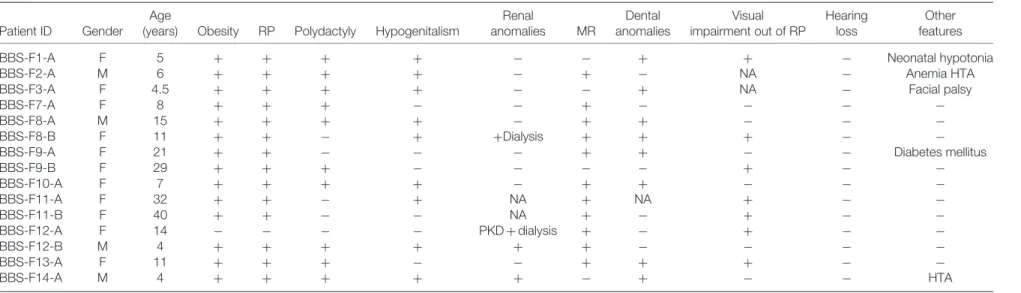
Clinical manifestations --- 63
5. Absence of genotype to phenotype correlations --- 64
6. Phenotypic overlap and differential diagnosis --- 65
7. Routine diagnostic offer for BBS patients --- 66
III- Results--- 69
Paper I: Targeted high-throughput sequencing for diagnosis of genetically heterogeneous diseases: efficient mutation detection in Bardet-Biedl and Alström syndromes. --- 69
Paper II: Clinical and genetic characterization of Bardet-Biedl syndrome in Tunisia: defining a strategy for molecular diagnosis.--- 71
Paper III:Mutations in SDCCAG8/NPHP10 Cause Bardet-Biedl Syndrome and Are Associated with Penetrant Renal Disease and Absent Polydactyly.--- 73
Paper IV:Mesoaxial polydactyly is a major feature in Bardet-Biedl syndrome patients with LZTFL1 (BBS17) mutations. --- 75
IV- Transfer in routine to Strasbourg diagnostic laboratory: a one-year retrospective experience--- 77
3. Overall diagnostic yield in ALMS and other patients--- 80
4. BBS and ALMS, true overlap or misdiagnosis? --- 80
5. Deciphering the patients with negative results --- 81
6. BBS genes load --- 82
7. Diagnosis of BBS in Strasbourg in the post-genomics era --- 84
V- Discussion --- 85
RESULTS - PART II TARGETED SEQUENCING FOR THE DIAGNOSIS OF INTELLECTUAL DISABILITY--- 89
I- Introduction --- 91 1. A bit of History… --- 91 2. Definition(s) --- 92 3. Classifications --- 94 4. Epidemiology--- 94 5. Etiology of ID --- 95
II- Genetics of intellectual disability--- 98
1. Chromosomal aberrations and copy number abnormalities --- 98
2. Monogenic forms of ID: XLID --- 100
3. Monogenic forms: autosomal dominant forms of ID --- 104
4. Monogenic forms: autosomal recessive forms of ID--- 105
5. Oligogenism, or the ‘multiple hits’ model--- 106
6. Emerging interconnected gene networks --- 106
III- Comorbidity: ID and associated clinical signs --- 108
1. Neuromuscular and neurologic manifestations --- 109
2. Cerebellar and structural brain abnormalities --- 110
3. Behavioral disorders--- 112
4. ID associated with metabolic disorders --- 113
IV. Diagnosis of ID in France --- 115
V- Results --- 117
Paper I: XLID-causing mutations and associated genes challenged in light of data from large-scale human exome sequencing. --- 117
Paper II: Efficient Strategy for the Molecular Diagnosis of Intellectual Disability using Targeted High Throughput Sequencing. --- 119
Paper III: 20 ans après: a second mutation in MAOA identified by targeted high-throughput sequencing in a family with altered behavior and cognition.--- 121
VI- Discussion--- 123
2. Definition of leukodystrophies --- 128 3. Genetics --- 129 4. Classification --- 129 5. Diagnosis of leukodystrophies --- 132 II- Results --- 136 1. Targeted genes --- 136 2. Cohort Description --- 138
3. Proof of principle yield--- 138
4. Diagnostic yield --- 140
5. Clinical findings --- 145
III- Discussion--- 147
GENERAL DISCUSSION--- 149
Table 1 (adapted from Thompson & Thompson, Genetics in Medicine): Incidence in fetuses and newborns of numerical and structural chromosomal abnormalities. ... 9 Table 2: Comparison and evolution of higher throughput DNA sequencing methods showing a large improvement between the first platforms and the newest, in terms of sequencing capacities,
throughput, speed and accuracy. ... 25 Table 3 (adapted from (Koboldt et al., 2013)): Impact of NGS technologies on unraveling the molecular basis of OMIM-listed disorders ... 26 Table 4: Studied pathologies and associated properties revealing and increasing degree of
complexity... 31 Table 5: Summary of annotation tool incorporated through VaRank, and associated threshold for considering a variant as potentially pathogenic. ... 44 Table 6 (from (Singla and Reiter, 2006)): Defects in ciliary functions cause several human diseases. ... 55 Table 7: BBS genes and associated method used for their identification... 62 Table 8: Beales’ criteria for the diagnosis of Bardet-Biedl Syndrome. From (Beales et al., 1999). . 63 Table 9: Mutation load of the different BBS genes and ALMS1 in our cohort of patients according to their compliance or not to Beales’ diagnostic criteria... 78 Table 10: Frequency of some clinical traits among patients with biallelic mutations detected in ALMS1... 81 Table 11: Comprehensive report of mutations in BBS genes reported in patients from the literature. ... 83 Table 12 (adapted from (Katz and Lazcano-Ponce, 2008)): Classification of different degrees of ID, with associated hallmarks depending on the age. ... 94 Table 13 (adapted from (Katz and Lazcano-Ponce, 2008)): Summarized proposed etiology of intellectual developmental disorders... 95 Table 14: List of most recurrent CNVs associated with ID showing the overlap with other
neuropsychiatric and neurologic disorders. ... 100 Table 15: Novel XLID genes, identified within the last two years (OMIM source). ... 102 Table 16 (adapted from (Petterson et al., 2007; Oeseburg et al., 2011)): Frequency of several co-morbid features in patients with ID... 108 Table 17 (adapted from (Kohlschutter and Eichler, 2011)): Classification of leukodystrophies based on MRI findings ... 131 Table 18 (adapted from (Kohlschutter and Eichler, 2011)): Metabolic, biochemical and other laboratory investigations that are useful to discriminate certain forms of leukodystrophies... 134
Table 20: Cohort description and diagnostic yield per category. ... 141 Table 21: Candidate variants detected by NGS in 122 patients with undetermined leukodystrophies. ... 143
Figure 1: Schematic structure of a human gene... 5 Figure 2 (Thompson & Thompson, Genetics in Medicine): Decisional tree to illustrate the possible mechanisms for a variant to cause a disease. ... 8 Figure 3 (from (Bowen et al., 2011)): Example of linkage analysis showing linkage evidence between the disease locus responsible for metachondromatosis and genetic markers located on chromosome 12. ... 14 Figure 4 (from (Williamson, 2006)): Karyotype analysis demonstrating an extra-copy of
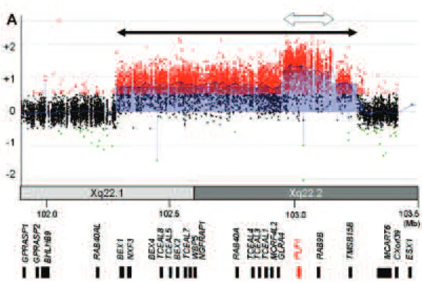
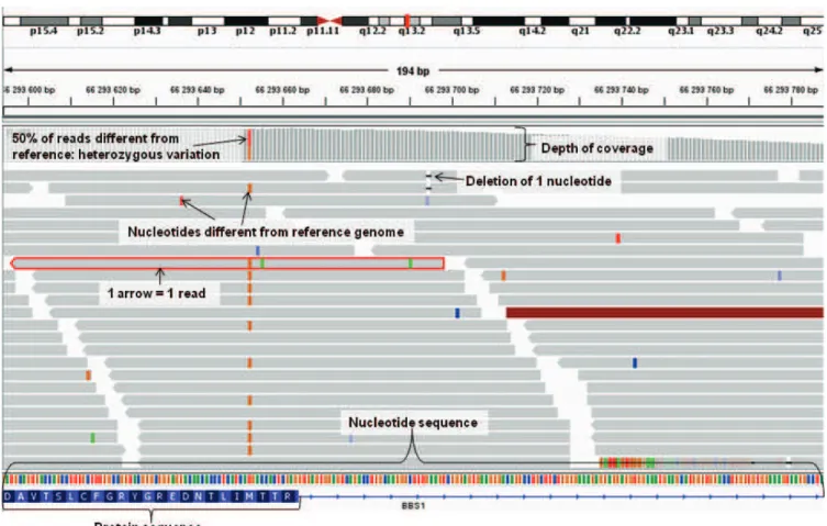
chromosome 21 in a male with Down syndrome... 17 Figure 5 (from (Chen et al., 2011)): FISH analysis using two probes of the X-chromosome (RP11-34P3 in Xq24 encompassing PLP1 in green, RP11-22P2 in Xq22 encompassing PGRMC1 in red) illustrating a tandem duplication on the X-chromosome that disrupts the NXF gene cluster in a woman... 18 Figure 6 (from (Shimojima et al., 2012)): Array-CGH analysis with abnormal patient/control signal ratio illustrating a duplication encompassing several genes of the X-chromosome (black arrow), and a triplication of PLP1 (white arrow) in a female with Pelizaeus-Merzbacher disease. ... 19 Figure 7: Visualization of high throughput sequencing data zoomed on BBS1 for one BBS patient, using integrative genome viewer (IGV; http://www.broadinstitute.org/igv/)... 21 Figure 8 (from Illumina sequencing introduction): Schematic overview of sample multiplexing prior to high throughput sequencing... 22 Figure 9: Evolution of complete published genomes, according to the three kingdoms of life ... 27 Figure 10 (from (Mertes et al., 2011)): Overview of the different targeted enrichment techniques. 28 Figure 11: Evolution of the number of studies using next-generation sequencing and exome
sequencing applications over time. ... 30 Figure 12: Improved version v2 of the bioinformatic pipeline for read mapping and variant calling (released in August, 2013) . ... 38 Figure 13: Principal component analysis performed on splicing predictions of 5 splicing prediction software in order to discriminate software with redundant results. ... 41 Figure 14: Visualization of sequencing data (zoomed on ALMS1, exon #8) in IGV for three different BBS patients... 47 Figure 15 (from (Bisgrove and Yost, 2006)): 9+0 and 9+2 axoneme structure. ... 52 Figure 16 (from (Hildebrandt et al., 2011)): Ciliary structure and components... 53 Figure 17 (from (Fliegauf et al., 2007)): Ciliary dysfunction in human disease: why are those organs/tissues affected?... 58 Figure 18: Distribution of ciliopathy genes among six major ciliopathy disorders, demonstrating extensive genetic overlap between such conditions... 64
dysfunction... 65 Figure 20: Decisional tree for the molecular diagnosis of BBS in Strasbourg until 2012... 67 Figure 21: Diagnostic yield in patients is highly correlated to their compliance with strict BBS diagnostic criteria... 77 Figure 22: Visualization in IGV of three patients heterozygous for the recurrent (c.1169T>G, p.Met390Arg) mutation in BBS1. Two of them also (upper panels) also carry the novel BBS1 recurrent mutation, detectable using targeted high-throughput sequencing. ... 79 Figure 23: Frequency of BBS clinical features among different classes of patients, depending on whether a mutation could be identified, and/or whether they comply with Beales’ diagnostic
criteria. ... 82 Figure 24: New decisional tree for the molecular diagnosis of BBS in Strasbourg incorporating the targeted NGS approach ... 84 Figure 25: Theoretical distribution of IQ scores, which follows the normal distribution, within the general population... 93 Figure 26 (adapted from (Baroff and Olley, 1999)): Familial correlations in general IQ depending on whether children are raised within the same environment or not. ... 96 Figure 27 (from (Tarjan et al., 1960)): Observed distribution of the IQ in the population as opposed to the theoretical normal distribution (Figure 25), showing an additional ‘bump’ curve of mean 35 primarily including individuals with ID of acquired or genetic etiology. ... 97 Figure 28 (adapted from (Lubs et al., 2012)): Chronological history and evolution of methods for XLID novel genes discovery... 101 Figure 29 (from (van Karnebeek and Stockler, 2012)): Summary of the 50 IEMs that can be
diagnosed using standard metabolic screening tests. ... 114 Figure 30 (from (Emery, 2010)): Schematic view of the oligodendrocytes maturation and initiation of the myelination process. ... 127 Figure 31 (from (Costello et al., 2009)): Decision tree for the diagnosis of leukodystrophies using MRI patterns. ... 133 Figure 32: Illustration of a heterozygous triplication of PLP1 in patient CRN163. ... 139 Figure 33: Illustration of a heterozygous deletion encompassing ARSA and MLC1 in patient
CRN147. ... 140 Figure 34: Illustration of the duplication of MBP in patient CRN199. ... 144
Appendix 1: DPY19L2 deletion as a major cause of globozoospermia... 157 Appendix 2: Globozoospermia is mainly due to DPY19L2 deletion via non-allelic homologous recombination involving two recombination hotspots... 159 Appendix 3: A mitochondrial pyruvate carrier required for pyruvate uptake in yeast, Drosophila, and humans. ... 161
ADHD: Attention Deficit and Hyperactivity Disorders
ALMS = Alström syndrome
ARID: Autosomal recessive Intellectual Disability
ASDs : Autism Spectrum Disorders ASS: Acceptor Splice Site
BBS: Bardet-Biedl Syndrome cDNA: complementary DNA
CGH: Comparative Genomics Hybridization CILD = primary CILliary Dyskinesia
CNS: Central Nervous System CNVs: Copy Number Variants ddNTPs: dideoxyribonucleotides DNA: DeoxyriboNucleic Acid dNTPs: deoxyribonucleotides
DSM-V: Diagnostic and Statistical Manual of Mental Disorders
DSS: Donor Splice Site
ENCODE project: ENCyclopedia Of DNA Elements
EVS: Exome Variant Server
FISH: Fluorescence In Situ Hybridization FPR: False-Positive Rate
Gb: Giga base pairs (=1x10⁹bp) gDNA: genomic DNA
GOLD: Genome OnLine Database
GWAS: GenomeWide Association Studies HGMD: Human Gene Mutation Database HGVS: Human Genome Variation Society Hh = Hedgehog
hr: hour
HSF: HumanSplicingFinder
HTS: High Throughput Sequencing
IBD: Identical By Descent
ICD-10 : International Classification of
Diseases, version 2010 ID: Intellectual Disability
IEMs: Inborn Errors of Metabolism IFT = Intraflagellar transport IGV: Integrative Genome Viewer indel: insertion/deletion
IQ: Intelligence quotient JBTS = Joubert syndrome kb: kilobase pairs (=1x10³bp) LCA = Leber Congenital Amaurosis
LINEs: Long Interspersed Nuclear Elements LOD: logarithm of odds
Mb: Megabase pairs (=1x10⁶bp)
MES: MaxEntScan
MIP: Molecular Inversion Probes
MKKS = McKusick Kaufmann syndrome MKS = Meckel syndrome
MRI: MagneticResonance Imaging mRNA: messenger RNA
mtDNA: mitochondrial DNA
MTOC = Microtubule organizing center NGS: Next-Generation Sequencing NPHP = Nephronophthisis
nt: nucleotide
PCP: Planar Cell Polarity
PCR: Polymerase Chain reaction PGM: Personal Genome Machine PKD = Polycystic Kidney Disease PKU: Phenylketonuria
PNS: Peripheral Nervous System qPCR: quantitative PCR
SINEs: Short Interspersed Nuclear Elements SLNS = Senior Loken syndrome
SNP: Single Nucleotide Polymorphism SNV: Single Nucleotide Variant
UTR: UnTranslated Region WES: Whole-Exome Sequencing WGS: Whole-Genome Sequencing XLID: X-linked Intellectual Disability
I- From DNA alteration to disease: the basis of genetics
1. The Human genome
The entire genetic heritage of each individual – otherwise referred as human genome – is stored mostly in the nucleus of each somatic cell within chemical molecules of deoxyribonucleic acid (gDNA, for genomic DNA). A tiny fraction is contained in the mitochondria (mtDNA, for mitochondrial DNA). These molecules of DNA store all the genetic information necessary to control all the steps of embryogenesis, development, homeostasis, metabolism, etc. Only a small proportion of genomic DNA (about 1.5%) encodes genes, which correspond to the units of genetic information. The human genome is assessed to contain about 21,000 genes (for a comparison, the mitochondrial chromosome contains only 37 genes; reviewed in (Frazer, 2012), linearly positioned along the chromosomes at precise loci. Genetic disorders are the consequences of alterations encoded in this molecule of DNA, and more precisely in its sequence. Our understanding of genetic disorders has tremendously improved with the completion of the Human Genome Project, reporting the entire sequence of a human genome in 2001 (Lander et al., 2001; Venter et al., 2001; International Human Genome Sequencing, 2004). Knowledge of the entire sequence provided researchers with a physical map to annotate genes and other biological elements onto a single reference. This allowed studying the human genome as a continuous entity rather than discretely, gene by gene. The draft of the human genome is continuously improved over the years thanks to patches and through novel releases (e.g. March, 2014: GRCh38 release, referred as hg38).
2. Genome organization and structure
All somatic cells of the body contain 23 pairs of chromosomes (22 autosomal, 1 sexual), one from each pair being inherited from one of the two parents. Germ cells only carry 23 chromosomes. Chromosomes are numbered according to their size that often but does not necessarily correlate to their gene contents. Genes are not distributed randomly along the chromosomes, but tend to cluster together. As a result, certain regions of chromosomes are highly gene-rich while other regions are called gene deserts, and certain chromosomes themselves are gene-rich as opposed to others.
Apart from genes, which as mentioned represent a very minor fraction, 5% of the genome – only –contains some regulatory elements that control gene expression, such as noncoding RNAs. The rest of the genome is mostly composed of repetitive sequences (over 50%; including satellite DNA, SINEs and LINES retrotransposons elements), whose role remains poorly understood but would supposedly be related to maintaining chromosomal structures. As for the genes, the repartition of those repetitive structures is not random, and they are clustered together along the chromosomes. Subsequently to the release of the human genome assembly, several projects that aimed at extensively annotating the human genome such as the Encyclopedia of DNA elements
(ENCODE) were launched. Launched in 2003, the ENCODE project aimed to identify all functional elements of the human genome and provide an annotated reference to the scientific community, after a pilot phase of annotating defined regions of the human genome as a training (Consortium et al., 2007; Weinstock, 2007). After years of intense labor, they proposed (among a plethora of other results) an extensive reference set of annotated genes and transcripts (GENCODE; (Derrien et al., 2012; Djebali et al., 2012; Harrow et al., 2012; Howald et al., 2012; Pei et al., 2012).
3. Gene organization
Genes are the units of the genetic information, which are mostly transcribed into mRNAs in the nucleus, translocated to the cytoplasm where they are translated into proteins, the molecules ultimately responsible for the ‘molecular’ phenotype of the cell. The genetic information is stored in the DNA via the genetic code, which from the sequence of nucleotides determines the sequence of aminoacids in a protein. Linear polypeptide chains originating from mRNA translation are not yet functional. Those premature proteins have to fold to acquire a specific three-dimensional structure, may assemble with other subunits to acquire a quaternary structure, may be chemically modified (e.g. addition of phosphate groups) and then are ready to operate. Most genes are capable of producing several associated transcripts by alternative splicing, generating a multitude of proteins. As a result, the 21,000 genes encode as many as a million of different proteins. However, not all genes code for proteins: the final product might be a non-coding RNA.
A human gene is generally composed of an alternation of exons and introns. Introns are portions of DNA that are initially transcribed into RNA, but are excised from the mature mRNA and are not represented into the final protein product. RNA splicing is a highly efficient process, guided by specific consensus sequences in the primary RNA transcript at the exon/intron junctions: 5’ donor splice site consensus sequence: AG«gt, 3’ donor splice site: an adenine branchpoint around 20 nucleotides before beginning of the exon, a polypyrimidine tract, and a consensus ag«G (Figure
1). At each extremity of the coding sequence (delimited by the methionine start codon and the stop
codon), the gene is flanked by 5’ and 3’ untranslated regions (5’UTR, 3’UTR). A promoter region is located at the 5’ end of each gene, which contains sequences regulating the time and place of gene expression. Other regulatory elements are dispersed throughout the gene (enhancers, silencers, etc), and can be located in the introns, at the 5’ or 3’ end of a gene but also at a considerable distance away. The 3’ end of a gene contains a signal sequence of AAUAAA tract, which promotes the termination of the mRNA and addition of the polyadenylation tail.
Figure 1:
UTR: untranslated region; DSS: donor splice site; ASS: acceptor splice site; Y: pyrimidines. Schematic structure of a human gene.
The correct expression of a gene and its associated protein production depends on a tightly orchestrated number of events, including gene dosage and structure, transcription, RNA splicing, mRNA stability, translation, structure, function and degradation of the protein product. Any genetic variation that would eventually impair one of these steps can have a dramatic effect and may lead to a disease phenotype. Those genetic variations include large structural variations, or punctual variations affecting protein-coding regions but also variations located in any of the essential regulatory elements (promoter, consensus splice sites and branching sites, regulatory sequences, polyA sequence signal, etc).
4. Genetic variations inter-individuals
The human genome is highly conserved (at 99.9%) from one individual to another mostly due to important selective pressure. However, due to the gigantic size of the diploid human genome (about 6x10⁹
In some disciplines, a mutation is defined as any change in the nucleotide sequence of a genome compared to the reference, devoid of any notion of pathogenicity. However, to avoid confusion and as recommended by HGVS (Human Genome Variation Society) guidelines, I will use the neutral terms ‘variation’ or ‘variant’ instead, and refer to ‘mutation’ for a genetic variation that causes a disease. Genetic variations can be classified into two main categories: the ones involving large fragments of DNA that can encompass up to entire chromosomes, and point variations that affect one or a few base pairs. Large structural variants arise either from impaired
base pairs) this represents millions of base pairs differing between individuals, which genetically determine macroscopic differences among individuals. A vast majority of those variation have no or benign effect on one’s phenotype (e.g. eye color, hair color, etc) while others are directly responsible for causing disease. Due to the minimal fraction of coding sequences, most of those variations are located in non-coding regions (introns, noncoding RNAs, repeat elements…) and are expected to have a minimal impact at the level of protein production, structure and function. Our actual understanding of non-coding regions being limited, no actual models hence no bioinformatics tools allow a precise interpretation of such non-coding variants apart from splicing predictions.
chromosomal segregation during cell division leading to an abnormal number of chromosomes, or spontaneous events during chromatid recombination leading to translocations, inversions, deletions, insertions, etc. Point variations arise mostly from DNA replication errors (frequency rate: 1x10⁻¹⁰
McCulloch and Kunkel, 2008
,
or less than one new mutation per cell division; ( )) or impaired DNA
repair, and are either spontaneous or fostered by the exposure to mutagen agents. The different types of variations affecting one or few nucleotides in a gene are detailed below.
Single nucleotide variants (SNVs)
The substitution from a base to another within the coding sequence can lead to a silent/synonymous change (no effect on the protein sequence, which are relatively frequent when the affected base is the third base of a codon), the replacement of one aminoacid by another referred to as missense variations (mostly when affecting the first or second bases of a codon), the replacement of one codon by a stop codon called nonsense variations, or the replacement of the key start or stop codons by another codon. Silent variations are usually benign since they do not affect the protein sequence, but they can be disruptive if affecting splicing (substitutions of nucleotides near/within splice sites or creating strong cryptic splice sites). Missense variations alter the sequence of the protein, and can be either benign or pathogenic depending on the importance of the affected residue with respect to structure or function of the protein. Nonsense variations incorporate a premature stop codon within the sequence, leading to an mRNA carrying a premature stop codon and that is highly vulnerable for nonsense mediated mRNA decay (reviewed in (Nicholson et al., 2010)). Either the protein is not translated at all due to the degradation of the mRNA, or it is translated under a truncated form that can be more or less stable and functional. Conversely, SNVs can destroy a termination codon and allow translation to continue resulting in a longer protein that may also be more or less stable and functional. Lastly, abolition of the start codon generally prevents any kind of translation resulting in the total absence of protein product.
Outside of the coding sequence, SNVs affecting the acceptor or donor splice sites, branching sites, promoter region or the polyadenylation sequence, or even creating competitor splice sites may result in aberrant amounts of mRNA products or altered RNA processing.
Short insertions/deletions (indels)
Gene variations include also the insertions or deletions of a few base pairs. In coding regions, these either conserve the reading frame if they are a multiple of three (in-frame variations), or cause a shift of the reading frame at the point of the insertion/deletion (frameshift variations). Some tools have been recently developed to predict their impact (e.g. (Hu and Ng, 2012)). In in-frame variations, the corresponding number of aminoacids will be inserted/deleted within the normal protein sequence, which can have variable impact on the protein structure and function.
Conversely, frameshift variations incorporate a number of abnormal aminoacids before a premature stop codon, which will have the same possible consequences as nonsense variations.
Dynamic mutations
Some variations are a bit more complex, and involve the amplification of a repeated region (generally trinucleotide tracts) during replication in the gametes. This amplification leads to an increased number of repeats (located or not within the coding sequence) that becomes toxic when bypassing a certain threshold. Within the coding sequence, this amplification result in an abnormally long protein, while within intronic or regulatory sequences it leads to altered transcription, mRNA processing or translation.
Polymorphisms vs rare variants
As mentioned, not all variations reported in an individual are necessarily pathogenic. Some are private or rare, others are relatively common in the general population. Variants with a frequency superior to 1% in the general population are generally called polymorphisms, the ones with a lower frequency are referred as rare variants. Polymorphisms can be of any of the previously discussed type, from SNVs, indels to structural variants. Polymorphisms are predominantly innocuous, as their high frequency makes them incompatible with a full penetrant role in rare diseases. They can nonetheless be associated to genetic risk or protective factors.
5. Discriminating benign variants vs disease-causing mutations
The entire challenge in human genetics – and even more with the arrival of the next-generation technologies flooding databases with variations of unknown significance – is to assess with the highest possible level of confidence whether a variant reported in a patient is responsible for the observed clinical features or not (summarized decisional tree in Figure 2).
First, the more the variants are supposed to affect the protein sequence, the more deleterious they shall be presumed. Frameshift, nonsense, start loss, and splice site variants along with large structural variants are the most likely to have an effect on the phenotype since a priori directly impacting on RNA processing and protein production.
Then, many additional keys can be gathered from in silico tools giving information on the conservation of the affected nucleotides but also of the affected aminoacid residue (if located within a coding sequence), the putative impact on the resulting protein structure (e.g. whether it is located within a protein domain, binding sites, etc), the putative effect on RNA splicing (abolition/creation
of splice sites), the pattern of gene expression, … All those are precious information that help predicting the functional impact of a variant (detailed in General methods, III).
Once a variant seems deleterious enough, co-segregation studies allow verifying whether the candidate variant segregates with the disease status in a family. The study of large cohorts of controls from similar geographical origin also ensures that the detected candidate is not a common variant in the population of concern. This should be computed carefully in relation to the disease of interest (e.g. prevalence, mode of inheritance, proposed penetrance, etc; see (Piton et al., 2013)). Once the variation becomes a stronger candidate, functional analyses of the variation can be performed at any protein/cellular/organism level to demonstrate the functional impact of the variation.
Figure 2 (Thompson & Thompson, Genetics in Medicine): Decisional tree to illustrate the possible mechanisms for a variant to cause a disease.
II- Classification of genetic disorders
The contribution of genetic factors in disease can be large or small, from causing the disease or modifying the course of the disease, to only predisposing to disease. Disorders caused entirely or partially by genetics factors are generally classified into several types: chromosomal disorders, monogenic or single gene disorders, and polygenic or multifactorial disorders.
1. Chromosomal diseases
In chromosomal diseases, structural defects from large chromosomal fragments to whole chromosomes cause the disease. The phenotype originates from an excess or deficiency in genes contained in the chromosomal abnormality. A single gene may be the driver or several genes can each contribute partially to the phenotype. Chromosomal diseases are rather common, with an assessed incidence of 0.7% in newborns (Table 1).
The most common type of chromosomal abnormalities is aneuploidy, which consists in either an extra copy or the absence of a chromosome. Trisomies are generally more viable than monosomies, since trisomy 13, 18, 21, X and Y are compatible with life as opposed to the single monosomy X, aka Turner syndrome. The only autosomal trisomies that are not lethal are the ones involving the three autosomes with the lowest number of genes. The most frequent chromosomal abnormality is the trisomy 21 or Down syndrome that has an assessed incidence of 0.1% (Weijerman and de Winter, 2010). The proposed mechanism for aneuploidies is a non-disjunction of chromosomal pairs during meiosis. If occurring during a mitotic division in the zygote, non-disjunction of a pair of chromosomes leads to mosaic aneuploidies. Aneuploidies are universally associated to physical and cognitive manifestations.
Abnormal Karyotype 1st Fetuses of mother
> 35 years trimester
abortuses Live births
Total incidence 1/2 1/50 1/160 Numerical abnormalities 96% 85% 60% Structural abnormalities 4% 15% 40% Balanced 0% 10% 30% Unbalanced 4% 5% 10%
Table 1 (adapted from Thompson & Thompson, Genetics in Medicine):
Other chromosomal abnormalities are structural defects involving chromosomal fragments that have been isolated from chromosome breakage, and which are eventually reconstituted in an abnormal combination. Chromosomal breakage occurs spontaneously at low frequency but can also be fostered by breaking reagents. The recombination between highly similar sequences (e.g.
Incidence in fetuses and newborns of numerical and structural chromosomal abnormalities.
pseudogenes, LINEs elements, low copy repeats, etc) or mispaired chromosomes may also provoke such fragility and lead to gene duplications or deletions. Like in numerical chromosomal abnormalities, structural chromosomal rearrangements can be mosaic.
Structural rearrangements can be either balanced (i.e. the final set of chromosomes has all chromosomal material, although displaced) or imbalanced (i.e. the final set of chromosomes has additional or missing chromosomal material). Unbalanced rearrangements include mostly large deletions (generally reflecting haploinsufficiency), duplications, unbalanced translocations, unbalanced inversions, and more rarely the formation of marker chromosomes (small supernumerary chromosomes), isochromosomes (chromosomes with a duplicated arm while the other is missing) or dicentric chromosomes (two arms of two separate chromosomes fused together). Unbalanced rearrangements are more likely to result in an abnormal phenotype since disrupting the normal balance of gene expression. During my thesis, I was involved in unraveling the molecular mechanism provoking an unbalanced-rearrangement responsible for globozoospermia ((Koscinski et al., 2011; Elinati et al., 2012); see Appendices 1 & 2).
Balanced rearrangements include paracentric and pericentric inversions and translocations. Carriers of balanced rearrangements generally do not have phenotypic manifestations since the full genetic material – although displaced – is present, unless the breakpoints disrupt the coding sequence of a particular gene.
Many copy number variants (CNVs) are rare private CNVs and one challenge is to evaluate their contribution in a particular phenotype. Indeed, although it is widely accepted that large structural variants are mostly disease causing, this is not the case for smaller CNVs that have a relatively high frequency within the general population (incidence of 1:8 for de novo microdeletions, and 1:50 for de novo microduplications; (Guilmatre et al., 2009)). The penetrance of some CNVs also appears to be highly variable (e.g. can be inherited from ‘healthy’ parents) and may lead to different phenotypic outcomes complicating the discrimination between risk factors and benign CNVs. One possibility to assess the contribution of a CNV in a phenotype is to compare their frequency in a huge cohort of controls, or to find the driver gene (or the disrupted gene in translocations) and identify additional patients with a similar phenotype carrying CNVs or point mutations affecting the same gene.
2. Monogenic disorders
Monogenic disorders are caused by mutations (inherited, or that appeared de novo in a parental germ line) located within a single gene, and are often called Mendelian disorders as their mode of inheritance follows Mendel’s laws. Although individually rare, they represent – as a group – a considerable proportion of all pediatric and adult disorders. As seen in the previous section, the mutation can be any of a single nucleotide variant (SNV), a small indel, or a submicroscopic CNV