Correction of Malunion after Pediatric
Supracondylar Elbow Fractures
Closing Wedge Osteotomy and External Fixation
Carol C. Hasler
1Abstract
Background: Malunion is the most common
complica-tion after pediatric supracondylar fractures. Cubitus varus may be cosmetically disturbing, consolidation in hyperextension restricts flexion. There is a considerable rate of complications for corrective osteotomies report-ed in the literature (incomplete correction, loss of cor-rection, and unsightly scars).
Patients and Methods: 13 patients (six boys and seven
girls), average age 11 years (5–23 years), underwent supracondylar closing wedge osteotomies and lateral external fixation. Cubitus varus deformity averaged 23° (15–50°). Four patients were treated by flexion
osteotomies, six by valgus osteotomies, and three by combined flexion-valgus osteotomies.
Results: Nine patients showed symmetric carrying
angles, three had 5° more valgus on the operated arm. One patient had 10° more varus which still resulted in a physiologic valgus. One patient had slight lateral bony prominence despite symmetric carrying angles. Three patients complaint about unsightly scars. All three had previous open reduction of their fractures with keloids. All patients had already reached a range of motion at the time of metal removal which was within 15° of the values at the time of the latest follow-up. Seven patients with significantly decreased elbow flexion regained an aver-age of 28° of flexion (20–35°). Two superficial pin track infections healed under oral antibiotics.
Conclusion: External fixation facilitates correction by
empirically searching for the desired carrying angle, cosmetic appearance, and function of the elbow. Func-tional aftertreatment shortens the recovery period. Metal removal is easy. Our experience confirms the excellent results in previous series on external fixators.
Key Words
Cubitus varus · Supracondylar fracture · Osteotomy · External fixation
Eur J Trauma 2003;29:309–15
DOI 10.1007/s00068-003-1325-1
Introduction
Malunion after pediatric supracondylar fractures is reported in recent papers to occur in up to 20% after closed or open reduction and internal fixation [1–3]. Cubitus varus, which never remodels with growth, or malunion in antecurvatum, which only remodels in younger children, accounts for this most common com-plication after pediatric elbow fractures. A patient’s desire for restoration may arise either from impaired cosmetic due to cubitus varus or from disabling lack of elbow flexion due to consolidation in hyperextension or from a combination of both (Figures 1 to 4). Various combinations of osteotomy techniques and fixation devices have been described. The specific supracondy-lar bony anatomy not only renders primary fracture treatment difficult but also represents an obstacle in achieving and maintaining correct alignment and satis-factory cosmetic appearance of the elbow. Incomplete correction of the deformity or loss of correction con-tributes significantly to poor results in up to 50% in ear-ly series [4–7]. Recent studies report higher success rates [8, 9].
This retrospective study reports our experience with supracondylar hinged and nonhinged closing wedge osteotomies followed by lateral external fixation.
1
Orthopedic Department, University Children’s Hospital, Basel, Switzerland.
Patients and Methods
During the period 1997–2002, 13 patients (six boys and seven girls) with an average age of 11 years (5–23 years) underwent supracondylar osteotomies and external fixa-tion for malunion at our institufixa-tion (Table 1). Five patients had injured their right elbow and eight patients their left elbow. Five patients had sustained a supra-condylar Gartland type II fracture with initial plaster treatment in four cases and closed reduction and plaster treatment in one. Seven patients had suffered from a supracondylar Gartland type III fracture with open reduction and K-wire fixation in all but one patient who was initially treated with closed reduction and K-wire fix-ation. One patient had sustained a displaced transcondy-lar fracture. One patient (case 1) had already been treat-ed with a corrective osteotomy and plate osteosynthesis for cubitus varus 8 years ago but complained of a dis-abling flexion deficit of his elbow.
The cubitus varus deformity averaged 23° (15–50°). The decision for a flexion osteotomy was made if the patient complained about the loss of function and spontaneous remodeling of the antecurvated distal humerus could not be expected (patient’s age > 7 years). The decision for a valgus osteotomy was always based on cosmetic impairments. There were no cases with elbow instability.
Four patients were treated by a flexion osteotomy, six by a valgus osteotomy, and three by a combined flex-ion-valgus osteotomy. The interval between trauma and
corrective osteotomy averaged 42 months (5–144 months). The average follow-up was 14 months (4 months to 6 years).
Preoperative and follow-up documentation includ-ed clinical assessment of elbow range of motion, stan-dard anteroposterior (AP) and lateral radiographs of the elbow and clinical photographs of both elbows’ flex-ion, extensflex-ion, both forearms’ pronation and supina-tion, and carrying angles of both elbows.
Surgical Technique
Operations were performed in supine position on an arm board with no tourniquet. Sterile draping of both arms was done in order to find the optimal amount of correction for a symmetric result. The position of the most distal pin was assessed under image intensifier
Figure 1. Patient no. 6;
10-year-old boy with a 20° cubitus varus 2 years after a supracondylar fracture of his left elbow.
Figure 3. Preoperative AP
radiograph. Cubitus varus. Open physis.
Figure 4. Preoperative lateral
radio-graph. Distal humerus malunited in extension.
Figure 2. 25° restriction of left elbow flexion due to malunion in
just proximal to the distal humeral growth plate in case of still open physis or in the distal humeral epiphysis in case of ceased growth. At the same time, the site of osteotomy (1 m proximal to the second pin) was deter-mined and skin marked with a sterile pen. The site of osteotomy was subperiosteally exposed then through a posterolateral skin before pin placement, which facili-tates the approach. In the case of a former open frac-ture reduction, that skin incision was used except when it was an anterior approach (case 2). Two 4-mm self-drilling and self-cutting pins were placed from the lat-eral site into the distal fragment. Two other pins were placed into the proximal fragment in an anteroposteri-or direction just distal to the anteroposteri-origin of the deltoid mus-cle to avoid the radial nerve. All pins were brought in through a 6- to 7-mm skin incision, which was spread by
a clamp, followed by introduction of a pin guide for soft tissue protection. After pin placement, two small blunt Hohmann retractors provided subperiosteal exposure of the osteotomy site, which was usually situated 1–2 cm proximal to the supracondylar region in case of still open physis or at the supracondylar region in case of already closed physis. A closing medially hinged valgus and/or posteriorly hinged flexion wedge osteotomy was per-formed with a saw under continuous irrigation. The low-er osteotomy was cut parallel to the elbow joint line. The external fixators (Hoffmann II compact®, Stryker How-medica Osteonics, Geneva, Switzerland) was mounted and fixed. The arm (carrying angle, lateral bump, range of motion for elbow extension and flexion, forearm prona-tion and supinaprona-tion) was compared to the contralateral arm. In case of a still disturbing lateral bump, the
Type of Prior Interval Age Removal OT Flexion- Varization Carrying Complica-fracture procedures to trauma (years) of metal extension (°) angles tions
(months) (weeks) (°) (°)
Preop. Postop. Injured Uninjured Preop. Postop.
1
Supra-condylar OT and plate OS 144 23 14 F 90-0-0 110-5-0 0 5 5 5 Anterior
for cubitus varus translation of
8 years ago the fragment
2 Supra- OR, K-wires, 17 12 10 F 90-0-10 120-10-0 0 5 5 5 condylar anterior approach
3 Supra- OR, K-wires,
condylar posterior approach 17 11 8 F 95-20-0 130-20-0 0 5 5 5 to open arthrolysis
4 Supra- OR, external fixa- 18 13 8 VF 95-0-0 125-5-0 20 –10 5 10 Superficial
condylar tion, lateral infection
approach 5 Supra- 18 9 8 F 115-0-20 140-0-5 0 10 10 10 condylar 6 Supra- 22 10 9 VF 115-0-25 140-0-5 15 –10 5 5 condylar 7 Supra- 72 10 8 V 140-0-10 125-0-0 30 –25 5 5 condylar 8 Supra- 50 7 9 V 140-0-20 140-0-15 25 –25 5 0 condylar 9 Supra- CR 5 5 9 VF 105-0-20 135-0-5 30 –20 10 10 condylar
10 Trans- OR, K-wires, lateral 50 6 8 V 120-0-0 140-0-5 50 –35 15 15 condylar Y approach
11 Supra- OR, K-wires, 30 8 8 V 125-0-0 125-0-0 20 –10 10 10 condylar lateral approach
12 Supra- CR, K-wires 48 12 10 V 150-0-10 150-0-0 20 –15 5 5 Superficial
condylar infection
13 Supra- OR, K-wires, lateral 60 15.5 8 V 140-0-5 140-0-5 25 –5 10 20 condylar approach
osteotomy was completed and the distal fragment medialized as much as needed. Derogation was only performed in case of impaired shoulder function com-pared to the contralateral side. The external fixators could be undone and refixed, until the desired cosmet-ic and functional result was achieved. Skin closure was achieved with continuous intracutaneous self-resorb-ing 3-0 suture. The incisions for the pins were left open, and sterile dressings were applied.
Aftertreatment
No physiotherapy was administered. The patients were encouraged to actively and passively move their elbow, shoulder, forearm, and wrist as soon and as much as tol-erated. Patients and parents were instructed on how to take care of the pin sites. Daily showers or baths were mandatory after healing of the skin incision, usually after day 10. Radiographs were taken postoperatively, after 4 and 8 weeks.
The external fixator was removed on an outpatient basis. The patient himself decided for or against anes-thesia.
Results Inpatient Days
Average 6.4 days (3–9 days).
Consolidation
Removal of metal was carried out on an average 9 weeks after osteotomy (range 8–14 weeks). There was one secondary anterior ad latus dislocation of the distal fragment without deterioration of the alignment (case 1), which led to delayed union of 14 weeks.
Cosmetic
Nine patients showed symmetric carrying angles, three had 5° more valgus on the operated arm. One patient (case 13) had 10° more varus which still resulted in a physiologic valgus of 10°, since the other elbow had a carrying angle of 20°.
One patient had lateral bony prominence despite perfect symmetric carrying angles. She had open physis and a relatively proximal osteotomy with subsequent lateral shift of the distal fragment as a consequence of valgization.
Three patients complained about unsightly scars. All three had previous open reduction of their fractures with scar excision at the time of osteotomy but with recurrence of their keloids.
Function
All patients had already reached a range of motion at the time of metal removal that was within 15° of the val-ues at the time of the latest follow-up. Seven patients with significantly decreased elbow flexion due to post-traumatic malunion in antecurvatum regained an aver-age of 28° of flexion (20–35°) but lost only averaver-aged 11.5° of extension (0–20°).
Complications
There were two superficial pin track infections which healed under a 7-day course of oral broad-spectrum antibiotics. No neurologic or vascular complications, pin loosening or refracture after metal removal were observed.
Discussion
Indication for Surgery
Cosmetically unaccepted cubitus varus is still the main indication for a corrective osteotomy after pediatric supracondylar fractures of the elbow. However, active athletes may also complain about specific disabilities [10]. An associated or isolated malunion in hyperexten-sion leads to a loss of flexion and may therefore con-tribute to the disability. Other clinical implications of cubitus varus such as rotatory instability of the elbow, dislocation or snapping of the triceps tendon, tardy ulnar nerve palsy, and posterior instability of the shoul-der are less frequent [11–14].
Timing of Surgery
Since cubitus varus deformities of the elbow have a low potential for remodeling, the decision and timing can be fully based on the patient’s and parents’ prefer-ences. In case of loss of flexion due to malunion in hyperextension, remodeling can be expected in patients below the age of 7 and with a deformity of < 20–30°. However, in older patients, early correction is warranted in case of restriction of daily activities or sports.
Results
In published studies, successful restoration of a symmet-ric carrying angle of the elbow in children with posttrau-matic cubitus varus did not lead to the desired cosmetic result in up to 60% of the patients [8, 9, 15–17]. Dissatis-fied patients usually complain about the persistence of a varus deformity, lateral bony prominence or an unsight-ly scar. A persisting cubitus varus and/or lateral
promi-nence may be due to the osteotomy tech-nique, excision of an inadequate wedge, or loss of fixation.
Osteotomy Technique. The most widely
used osteotomy, the French method with a lateral closing hinged wedge, shifts the distal fragment laterally. The latter is also wider than the proximal thus potentially contribut-ing to a lateral bulge. The younger the child, the more bony the remodeling will be to compensate for the initial mismatch in width [8, 17]. However, the more proximal the osteotomy is performed, the more lateral the condylar block will be shifted. With external fixation, mainly in patients with still open physis, the osteotomy is more proximal than the site of malunion and also more proximal compared to osteotomies where other implants like blade plates, staples or K-wires are used. One should consider this issue by medializing the distal fragment, if traditional hinged closing wedge osteotomy is not satis-factory. In our series, however, it was only
done in one patient and should have been done in another, who was not completely satisfied with the appearance of the elbow despite anatomic restoration of the carrying angle. In other series, simple hinged medial open or hinged lateral closing wedge osteo-tomies followed by external fixation yielded excellent results [18–20]. One-dimensional, two-dimensional or even three-dimensional correction is easily accom-plished with the external fixator which allows for empiric search of the optimal cosmetic appearance (Figures 5 and 6). The osteotomy can be securely held for intraoperative assessment of cosmetic in full exten-sion of the elbow. The ease of application and the inherent stability of external fixation avoid more com-plicated types of osteotomies like dome, arc or penta-lateral osteotomies, all designed to improve stability [21–24].
Inadequate Size of Wedge. Preoperative planning,
conscientious intraoperative judgment with the elbow fully extended and the forearm supinated, and clinical comparison to the contralateral elbow prevent this fail-ure.
Loss of Fixation. This is a potential hazard mainly if
simple K-wire fixation or mere plaster immobilization is used, but it is also observed in most studies with more rigid types of fixation [4, 6, 7, 10, 15, 25, 26]. In our series,
with full functional aftertreatment, one moderate sec-ondary anterior translation of the distal fragment occurred which did not interfere with the end result. Other publications on unilateral external fixation also reported maintenance or only insignificant loss of initial correction which underlines the stability of this con-struct [18, 19, 25].
Scars
Hypertrophic scars have a strong negative impact on the outcome of an operation which is almost exclusively done for cosmetic reasons [4, 6, 8, 10]. All three patients in our study with unsatisfying scars had preexisting keloids from previous open surgery. The risk of unsight-ly scars may be diminished by using previous incisions, by posterior or medial approaches to the distal humerus, by intracutaneous suturing techniques, and by postoperative skin and scar care. In case of external fix-ation, the incision at the pin sites should be excised and debrided at the time of frame removal, since they are always inflamed and contribute to hypertrophic scar-ring. Alternatively, the application of the frame and exposure of the humerus can be performed from the medial side to hide the scars. It requires transposition of the ulnar nerve and the acceptance of temporary less comfort caused by a medial fixator [18, 20].
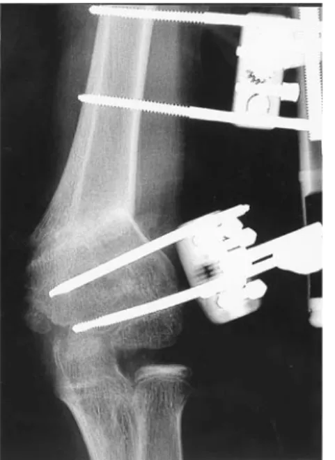
Figure 5. AP radiograph 6 weeks
postopera-tively. Lowest pin as close to the physis as possible. Consolidated closing wedge osteo-tomy with slight medialization of the distal fragment.
Figure 6. Lateral radiograph 6
weeks postoperatively. Flexion osteotomy corrected sagittal malunion.
Complications
Apart from unsatisfactory cosmetic results, various types of complications are reported. Transient peripher-al nerve pperipher-alsy may occur due to stretching by retractors or by indirect stretching of the soft tissues by the correc-tive maneuver itself after both open and closed osteotomies [19, 27]. Theoretically, pin placement for external fixation may damage a nerve. However, no such complication has been reported. It can be avoided by placing the pins in safe zones or by open exposure of the pin sites.
Infection is generally the most frequent reported complication of external fixation. Thorough instruction of the caregivers in pin site care is essential. The infec-tion rate in smaller series (five to six patients) of exter-nal fixation ranged from 0% to 40% [18, 25] and was 15% in our patients. All were superficial and controlled by short-term oral antibiotics.
Aftertreatment
Temporary postoperative immobilization in a posterior plaster slab in flexion or even in full extension is advocat-ed by most authors [9]. External fixation allows for imme-diate active and passive motion. There is no doubt that this functional aftertreatment with full use of the operat-ed elbow for daily activities enhances the patient’s com-fort and shortens the time of recovery despite avoidance of formal physiotherapy [28, 29]. All our patients had almost reached full range of motion already at the time of frame removal.
Removal of Metal
Unlike other implants, external fixation avoids a second operation for metal removal. External fixators could be removed after 9 weeks in this series comparable to oth-er studies with extoth-ernal fixation [18, 19].
Conclusion
External fixation of supracondylar osteotomies facilitates correction by empirically searching for the desired carry-ing angle, cosmetic appearance, and function of the elbow. It even gives opportunity for closed adjustment in case of unsatisfactory correction. Stability is provided even if there is only a small area of bony contact. Immedi-ate functional aftertreatment shortens the recovery peri-od. Metal removal is easy. Our experience in 13 patients confirms the excellent results in previous series of unilat-eral and ring fixators [18–20, 25, 28] (Figures 7 to 10).
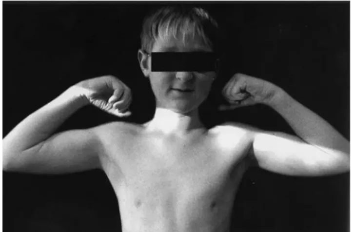
Figure 7. 1 year
postoperative-ly: symmetric carrying angles without lateral bony promi-nence.
Figure 8. 1 year postoperatively: symmetric full flexion.
Figure 9. AP radiograph
1 year postoperatively. Re-modeled osteotomy.
Figure 10. Lateral radiograph 1 year
postoperatively. Anatomic alignment of the distal humerus.
References
1. Davis RT, Gorczyca JT, Pugh K. Supracondylar fractures in chil-dren. Clin Orthop 2000;376:49–55.
2. Reitman RD, Waters P, Millis M. Open reduction and internal fix-ation for supracondylar humerus fractures in children. J Pediatr Orthop 2001;21:157–61.
3. Weinberg AM, Marzi I, Gunter SM, et al. Supracondylar humerus fracture in childhood – an efficacy study. Unfallchirurg 2002;105:208–16.
4. Ippolito E, Moneta MR, D’Arrigo C. Post-traumatic cubitus varus. J Bone Joint Surg Am 1990;72:757–65.
5. Labelle H, Bunnell WP, Duhaime M, et al. Cubitus varus deformity following supracondylar fractures of the humerus. J Pediatr Orthop 1982;2:539–44.
6. Oppenheim WL, Clader TJ, Smith C, et al. Supracondylar humeral osteotomy for traumatic childhood cubitus varus deformity. Clin Orthop 1984;188:34–9.
7. Rang M. Children’s fractures, 2nd edn. Philadelphia: Lippincott, 1983:167–9.
8. Barrett IR, Bellemore MC, Kwon YM. Cosmetic results of supra-condylar osteotomy for correction of cubitus varus. J Pediatr Orthop 1998;18:445–7.
9. Graham B, Tredwell SJ, Beauchamp RD, et al. Supracondylar osteotomy of the humerus for correction of cubitus varus. J Pedi-atr Orthop 1990;10:228–31.
10. Bellemore MC, Barrett IR, Middleton RWD, et al. Supracondylar osteotomy of the humerus for correction of cubitus varus. J Bone Joint Surg Br 1984;66:566–72.
11. Abe M, Ishizu T, Shirai H, et al. Tardy ulnar nerve palsy caused by cubitus varus deformity. J Hand Surg [Am] 1995;20:5–9. 12. Gurkan I, Bayrakci K, Tasbas B, et al. Posterior instability of the
shoulder after supracondylar fractures recovered with cubitus varus deformity. J Pediatr Orthop 2002;22:198–202.
13. O’Driscoll SW, Spinner RJ, McKee MD, et al. Tardy posterolateral rotatory instability of the elbow due to cubitus varus. J Bone Joint Surg Am 2001;83:1358–69.
14. Smith IJ, Williams CP. Failure of active extension after traumatic cubitus varus. A case report. J Bone Joint Surg Br 2002;84:1180–2. 15. Hernandez MA, Roach JW. Corrective osteotomy for cubitus
varus deformity. J Pediatr Orthop 1994;14:487–91.
16. Voss FR, Kasser JR, Trepman E, et al. Uniplanar supracondylar osteotomy with preset Kirschner wires for posttraumatic cubitus varus. J Pediatr Orthop 1994;14:471–8.
17. Wong HK, Lee EH, Balasubrmaniam P. The lateral condylar prominence. A complication of supracondylar osteotomy for cubitus varus. J Bone Joint Surg Br 1990:72:859–61.
18. Koch PP, Exner GU. Supracondylar medial open wedge osteoto-my with external fixation for cubitus varus deformity. J Pediatr Orthop B 2003;12:116–22.
19. Levine MJ, Horn BD, Pizzutillo PD. Treatment of posttraumatic cubitus varus in the pediatric population with humeral osteoto-my and external fixation. J Pediatr Orthop 1996;16:597–601. 20. Walsh SJ, Lamb GF, Barnes MJ, et al. Medial opening wedge
osteotomy with external fixation for correction of cubitus varus. Presented at the POSNA Meeting, Miami, 1995.
21. Kumar K, Sharma VK, Sharma R, et al. Correction of cubitus varus by French or dome osteotomy: a comparative study. J Trauma 2000;49:717–21.
22. Laupattarakasem W, Mahaisavariya B, Kowsuwon W, et al. Pen-talateral osteotomy for cubitus varus. Clinical experience of a new technique. J Bone Joint Surg Br 1989;71:667–70.
23. Matsushita T, Nagano A. Arc osteotomy of the humerus to cor-rect cubitus varus. Clin Orthop 1997;336:111–5.
24. Tien YC, Chih HW, Lin GT, et al. Dome corrective osteotomy for cubitus varus deformity. Clin Orthop 2000;380:158–66. 25. Jain AK, Dhammi IK, Arora A, et al. Cubitus varus: problem and
solution. Arch Orthop Trauma Surg 2000;120:420–5.
26. McCoy GF, Piggot J. Supracondylar osteotomy for cubitus varus. The value of the straight arm position. J Bone Joint Surg Br 1988;70:283–6.
27. King D, Secor C. Bow elbow (cubitus varus). J Bone Joint Surg Am 1951;33:572–6.
28. Karatosun V, Alekberov C, Alici E, et al. Treatment of cubitus varus using the Ilizarov technique of distraction osteogenesis. J Bone Joint Surg Br 2000;82:1030–3.
29. Song HR, Cho SH, Jeong ST, et al. Supracondylar osteotomy with Ilizarov fixation for elbow deformities in adults. J Bone Joint Surg Br 1997;79:748–52.
Address for Correspondence Carol C. Hasler
Orthopaedic Department University Children’s Hospital PO Box Römergasse 8 4005 Basel Switzerland Phone (+41 61) 685-5434, Fax -5006 e-mail: carolclaudius.hasler@ukbb.ch