HAL Id: hal-02345043
https://hal.archives-ouvertes.fr/hal-02345043
Submitted on 1 Dec 2020HAL is a multi-disciplinary open access archive for the deposit and dissemination of sci-entific research documents, whether they are pub-lished or not. The documents may come from teaching and research institutions in France or abroad, or from public or private research centers.
L’archive ouverte pluridisciplinaire HAL, est destinée au dépôt et à la diffusion de documents scientifiques de niveau recherche, publiés ou non, émanant des établissements d’enseignement et de recherche français ou étrangers, des laboratoires publics ou privés.
Physics-based oligomeric models of the yeast mitofusin
Fzo1 at the molecular scale in the context of membrane
docking
Astrid Brandner, Dario de Vecchis, Marc Baaden, Mickaël Cohen, Antoine
Taly
To cite this version:
Astrid Brandner, Dario de Vecchis, Marc Baaden, Mickaël Cohen, Antoine Taly. Physics-based oligomeric models of the yeast mitofusin Fzo1 at the molecular scale in the context of membrane docking. Mitochondrion, Elsevier, 2019, 49, pp.234-244. �10.1016/j.mito.2019.06.010�. �hal-02345043�
Title 1
Physics-based oligomeric models of the yeast mitofusin Fzo1 at
molecular-2
scale in the context of membrane docking.
3 4 5
Authors: 6
Astrid Brandner1*, Dario De Vecchis1*, Marc Baaden1, Mickael M. Cohen2# and Antoine Taly1# 7 8 * Contributed equally 9 # Correspondance 10 11 Affiliations: 12
Institut de Biologie Physico-Chimique, Centre National de la Recherche Scientifique, Paris, France. 13
1
Laboratoire de Biochimie Théorique, UPR 9080. 14
2
Laboratoire de Biologie Cellulaire et Moléculaire des Eucaryotes, Sorbonne Université, CNRS, 15
UMR 8226. 16
17
*Correspondence to: cohen@ibpc.fr or taly@ibpc.fr 18
ABSTRACT 19
20
Tethering and homotypic fusion of mitochondrial outer membranes is mediated by large GTPases 21
of the Dynamin-Related Proteins family called the mitofusins. The yeast mitofusin Fzo1 forms high 22
molecular weight complexes and its assembly during membrane fusion likely involves the 23
formation of high order complexes. Consistent with this possibility, mitofusins form oligomers in 24
both cis (on the same lipid bilayer) and trans to mediate membrane attachment and fusion. 25
Here, we rely on our recent Fzo1 model to investigate and discuss the formation of cis and trans 26
mitofusin oligomers. We have built 3 cis-assembly Fzo1 models that gave rise to 3 distinct trans-27
oligomeric models of mitofusin constructs. Each model involves two main components of mitofusin 28
oligomerization: the GTPase and the trunk domains. The oligomeric models proposed in this study 29
were further assessed for stability and dynamics in a membrane environment using a coarse-grained 30
molecular dynamics (MD) simulation approach. A narrow opening ‘head-to-head’ cis-31
oligomerization (via the GTPase domain) followed by the antiparallel ‘back-to-back’ trans-32
associations (via the trunk domain) appears to be in agreement with all the available experimental 33
data. More broadly, this study opens new possibilities to start exploring cis and trans conformations 34
for Fzo1 and mitofusins in general but also for other fusion-DRPs. 35
INTRODUCTION 36
37
Mitochondria are dynamic organelles organized as a cytoplasmic reticulum. Mitochondria 38
fuse their outer and inner membranes to form tubules. These mitochondrial tubules can interconnect 39
through fusion but can also fragment through fission of their membranes to yield a network with 40
remarkable plasticity. Together, fusion and fission thus regulate the whole morphology and 41
dynamics of the mitochondrial network, which makes these processes essential for maintenance of 42
mitochondrial integrity and consequently all mitochondrial functions (Friedman and Nunnari, 2014; 43
Shutt and McBride, 2013; Tilokani et al., 2018; Westermann, 2010). 44
From a mechanistic point of view, mitochondrial fission as well as fusion of outer and inner 45
membranes are all mediated by members of the Dynamin-Related Proteins family (Ramachandran, 46
2018). These large GTPases act as remodelers of intracellular lipid bilayers through two properties: 47
their capacity to bind biological membranes and their propensity to oligomerize into high order 48
macromolecular structures. Consistent with these features, fission DRPs are recruited to 49
mitochondrial outer membranes by specific adaptor proteins, where they auto-oligomerize upon 50
binding of GTP to assemble into macromolecular spirals that wrap around mitochondrial tubules 51
(Tilokani et al., 2018). Subsequent GTP hydrolysis induces conformational rearrangements of the 52
dynamins, which result in reduced diameter of the spirals and constriction of mitochondrial tubules, 53
followed by their separation. 54
Fusion-DRPs, on the other hand, are transmembrane proteins that promote homotypic 55
merging of the lipid bilayers in which they are inserted (Cohen and Tareste, 2018). The mitofusins 56
Mfn1 and Mfn2 (Fzo1 in yeast) fuse mitochondrial outer membranes whereas fusion between inner 57
membranes is mediated by Opa1 (Mgm1 in yeast). Another key member of fusion DRPs is the 58
Atlastin ATL-1 (Sey1 in yeast) that merges membranes from ER tubules. Mitofusins and Atlastins 59
have in common to auto-oligomerize in trans (from opposing lipid bilayers) and in a GTP binding 60
and hydrolysis-dependent manner to tether opposing lipid bilayers and promote their fusion (Cohen 61
and Tareste, 2018). Recent crystal structures of protein portions lacking the TM regions of ATL-1, 62
Sey1 and MFN1 further allow depicting the assembly of DRPs during membrane fusion as protein 63
dimers interacting in trans through their respective GTPase domains (Bian et al., 2011; Byrnes et 64
al., 2013; Cao et al., 2017; Moss et al., 2011; Yan et al., 2018; Yan et al., 2015). However, the yeast 65
mitofusin Fzo1 also forms high molecular weight complexes (Rapaport et al., 1998). In particular, 66
Fzo1 was shown to trigger the formation of a ring-shaped macromolecular complex during 67
mitochondrial docking (Brandt et al., 2016), suggesting that DRPs assembly during membrane 68
fusion might involve the formation of high order complexes rather than solely trans homodimers. 69
Consistent with this possibility, mitofusins form oligomers in both cis (on the same lipid 70
bilayer) and trans to mediate membrane attachment and fusion (Santel et al., 2003)(Ishihara et al., 71
2004; Griffin and Chan, 2006; Cao et al., 2017; Koshiba et al., 2004; Anton et al, 2011; Shutt et al., 72
2012). Besides the GTPase domain, mitofusins also include two heptad-repeat domains (HR1 and 73
HR2) that may get involved in homotypic interactions during mitochondrial tethering (Koshiba et 74
al., 2004; Griffin and Chan, 2006; De Vecchis et al., 2017). Whether GTPase and HR domain 75
interactions take place in cis or trans before or during mitochondrial tethering remains to be 76
investigated or confirmed (Franco et al., 2016; Koshiba et al., 2004). 77
Here, we rely on our recent Fzo1 model (De Vecchis et al., 2017) to investigate and discuss 78
distinct hypotheses for the formation of cis and trans mitofusin oligomers. Besides being the only 79
near full-length model as of today, it contains a membrane domain, which is a requisite for 80
investigating mitofusin orientations in a bilayer. We employed a modelling procedure that has been 81
guided by available experimental data from the literature. The models proposed in this study were 82
further assessed for stability and dynamics in a membrane environment using a coarse-grained 83
molecular dynamics (MD) simulation approach. This study opens new possibilities to start 84
exploring cis and trans conformations for Fzo1 and mitofusins in general but also for other fusion-85
DRPs. 86
87
88
METHODS 89
90
The modelling work presented here builds upon an experimentally validated model of the 91
monomeric unit, namely our previously published model of Fzo1 in a closed conformation (De 92
Vecchis et al., 2017). It is first used to generate a monomeric model of the open conformation. 93
Those two models are the basis for the construction of Fzo1 dimer models in cis and then tetramers, 94
via the dimerization in trans of Fzo1 cis-dimers. 95
96
Modelling the Fzo1 GTPase dimer construct 97
Two chains of the Fzo1 model in closed conformation (De Vecchis et al., 2017) were placed by 98
superimposing their GTPase domains onto that of BDLP in open conformation (PDB-id 2W6D, 99
Low et al., 2009). Then, only the coordinates of the two fragments that comprise the Fzo1 GTPase 100
domain (res 188-461) were retained to form the final GTPase domain dimer model. The loop 101
refinement tool implemented in MODELLER (Fiser et al., 2000) was used to remove a clash in both 102
chains involving an unresolved loop in the template 2J68 (Low and Löwe, 2006) (res 215-219). 103
Models were ranked according to the discrete optimized protein energy (DOPE) method (Shen and 104
Sali, 2006), selecting the best-scoring loop out of 20 candidates. 105
106
Modelling cis-dimer configurations 107
108
The Fzo1 head-to-head cis-dimer. Two Fzo1 chains in closed conformation (De Vecchis et al., 109
2017) were oriented facing each other within a compatible distance to accommodate two interacting 110
GTPase domains. Subsequently, the coordinates of residues 188-440 enclosed between hinges 2a, 111
2b (i.e. comprising the GTPase domain) were removed from both chains and replaced with the 112
GTPase dimer construct described above. The latter was manually positioned between the two 113
deleted chains resulting in the head-to-head interaction dimer (Fig. 2b). The backbone interruptions 114
were connected using the loop refinement tool implemented in MODELLER (Fiser et al., 2000) 115
using positions 185,188 and 436,445 as anchors. Solutions were ranked according to the DOPE 116
method (Shen and Sali, 2006), selecting the best-scoring loop out of 20 candidates. 117
118
The Fzo1 back-to-back cis-dimer. Two chains of the Fzo1 model in closed conformation (De 119
Vecchis et al., 2017) were manually oriented with respect to each other in order to generate the 120
back-to-back interaction (discussed in the text). In the resulting model system (Fig. 2c) the HR 121
domains face each other in a parallel fashion. 122
123
The Fzo1 open cis-dimer. The coordinates from BDLP in open conformation (PDB-id 2W6D, Low 124
et al., 2009) derived from the electron density map of native BDLP lipid tubes (accession code: 125
EMD-1589) were used as template to model Fzo1 in open conformation. Starting from our previous 126
Fzo1-BDLP target-template alignment (De Vecchis et al., 2017) and using an analogous approach 127
to the one described in Low et al., 2009, we introduced homologous chain breaks on the Fzo1 128
model (De Vecchis et al., 2017), resulting in five rigid blocks (Fig. 1a). Each fragment was 129
superposed to its corresponding fragment in 2W6D, in order to reconstitute the orientation found in 130
BDLP (Fig. 1). The MatchMaker tool from the UCSF Chimera software (Pettersen et al., 2004) was 131
used during this procedure. The loop refinement tool implemented in MODELLER (Fiser et al., 132
2000) enabled us to complete the model in the resulting backbone interruptions and to remove a 133
clash in both chains between the side chain of the Lys271 and the backbone of the Ala401 residues, 134
using positions 268 and 273 as anchors. Solutions were ranked according to the discrete optimized 135
protein energy (DOPE) method (Shen and Sali, 2006), selecting the best-scoring loop out of 10 136
models. 137
138
Modelling the trans-dimer configurations 139
140
The Fzo1 head-to-head trans tethered tetramer in open conformation. Two Fzo1 open cis-dimer 141
models obtained as described above were manually oriented to mimic the interactions in trans 142
towards their respective GTPase domains. In the resulting model system the two transmembrane 143
segments are located at opposite ends (Fig. 5a). 144
145
The Fzo1 head-to-head trans tethered tetramer (antiparallel). The transmembrane segment of two 146
Fzo1 head-to-head cis-dimers described above were manually oriented at opposite ends in order to 147
optimize the interaction between their respective HR domains oriented in an antiparallel fashion 148
(Fig. 5c). Note that this system, although antiparallel, could also be considered as back-to-back. 149
150
The Fzo1 head-to-head trans tethered tetramer (parallel). Two Fzo1 back-to-back cis-dimers 151
obtained as described above were initially positioned with the respective transmembrane segments 152
at opposite ends in order to mimic the supposed tethering process. Subsequently, the coordinates of 153
residues 101-491 and 816-855 enclosed between hinges 1a, 1b (i.e. comprising the GTPase domain 154
and the 3-helix bundle), were removed from the two resulting juxtaposing chains. Then, in a similar 155
way the GTPase dimer construct was built (see above), we superposed the GTPase domain alpha 156
carbons of two Fzo1 chains in closed conformation (De Vecchis et al., 2017) with the human 157
mitofusin dimer (PDB-Id: 5GOM, Cao et al., 2017). This choice was motivated and directly 158
inspired by the work from Gao and collaborators that proposed a possible Mfn1 trans cross 159
oligomer (Cao et al., 2017). From the resulting Fzo1 dimer, only the GTPase domain and the 3-160
helix bundle were selected and used to replace the aforementioned deleted portions, then generating 161
the trans head-to-head interaction (Fig. 5b). A clash in one chain (res 215-218) was removed from 162
the resulting Fzo1 dimer using the loop refinement tool implemented in MODELLER (Fiser et al., 163
2000). Models were ranked according to the discrete optimized protein energy (DOPE) method 164
(Shen and Sali, 2006), selecting the best-scoring loop out of 10 candidates. The same tool was used 165
to reconstitute the backbone interruptions. Positions 491,495 and 812,816 were selected as anchors. 166
The best-scoring loop was selected out of 10 candidates. 167
168
Molecular Dynamics. 169
System setup and parameters. 170
Topologies to run coarse-grained (CG) simulations were generated with the martinize tool choosing 171
the Martini v.2.1 force field with elastic network (de Jong et al., 2013; Monticelli et al 2008). The 172
force bond constant was set to 500 kJ mol-1 nm-2 with lower and upper elastic bond cutoffs of 0.5 173
and 0.9 nm respectively. Firstly, 5000 steps of steepest descent with position restraints for the 174
protein were run followed by 5000 steps without restraints. The obtained coordinates were inserted 175
in a POPC:POPE (1:1) membrane via the insane tool (Wassenaar et al., 2015), where the membrane 176
position was manually set up to match the reported transmembrane regions corresponding to 177
residues 706-726 and 737-757 according to UniProt numbering (De Vecchis et al., 2017). All 178
systems were fully solvated to mimic an environment of 150 mM of NaCl solution. See 179
Supplementary Table 1 for more details about each simulation setup. 180
All the final systems followed the same simulation protocol using the GROMACS 5.0.4 software 181
(Abraham et al., 2015). Further 5000 steps of steepest descent minimisation with position restraints 182
of 1000 kJ mol-1nm-2 in protein and lipids were followed by 5000 steps without position restraints. 183
Equilibration was performed in three stages, with timesteps of 20 fs. Firstly, 25000 steps of 184
equilibration were run at 310 K using the V-rescale thermostat (Bussi et al., 2007) and semi-185
isotropic pressure coupling via Berendsen barostat (Berendsen et al., 1984) with position restraints 186
of 1000 kJ mol-1 nm-2 for protein and lipids, followed by the same setup without position restraints. 187
Finally, the last equilibration step was run for 50000 steps with the V-rescale thermostat (coupling 188
constant tau_t = 1 ps) and semi-isotropic coupling with Parinello-Rahman barostat (Parinello and 189
Rahman, 1981) (coupling constant tau_p = 12 ps). Production runs were 1μs long for all the six 190
systems, following the same parameters as those used in the last equilibration setup. 191
192
Analysis. 193
Root mean square deviations were calculated in GROMACS 5.0.4 via the gmx rms tool considering 194
only backbone beads. Interfaces were obtained based on differences between solvent accessible 195
surface areas obtained with the gmx sas tool, using a probe radius of 0.2638 nm in agreement with 196
the CG water size. An interface between e.g protein A and B (IA/B) was calculated as the sum of the 197
surface areas of protein A and protein B in the interface, following the equation: IA/B= IA+IB= 198
(SASAA-SASAA_complex) + (SASAB-SASAB_complex) where SASAA is the solvent accessible surface 199
area calculated considering the protein A as isolated and SASAA_complex is the solvent accessible 200
surface area calculated for the protein A considering its solvent accessibility in the complex. 201
The distance in the membrane-normal Z axis between the phosphate beads from both bilayers 202
(oriented in the x,y plane) was measured in blocks (grid in x,y of size ~20 Å). Z coordinates of 203
atoms corresponding to each block were averaged and subsequently, the distance between the 204
average values of each block from the upper bilayer was subtracted from the lower one. Resulting 205
difference inter-membrane distances between the final and initial structure of the production run are 206
shown as a matrix. 207
208 209
RESULTS AND DISCUSSION 210
211
Putative models for Fzo1 cis-dimers 212
Mitochondrial tethering requires oligomerization of mitofusins in cis and then in trans (Anton et al., 213
2011; Fritz et al., 2001; Ishihara et al., 2004; Ishihara et al., 2003; Koshiba et al., 2004; Rapaport et 214
al., 1998; Santel et al., 2003). We thus reasoned that focusing on cis-oligomerization aspects in first 215
instance might facilitate investigating their general properties of assembly. In this regard, the 216
Bacterial Dynamin Like Protein from Nostoc punctiforme represents a starting point of choice. 217
BDLP was not only used as the template to generate our previous Fzo1 structural model (De 218
Vecchis et al., 2017) but this bacterial DRP also displays well established cis-oligomerization 219
properties (Low and Lowe, 2006; Low et al., 2009). Upon binding of non-hydrolysable analogues 220
of GTP, BDLP operates an extensive conformational change from a compact structure (that was 221
used as the template to generate the closed Fzo1 model, Fig. 1a) to an extended conformation. This 222
extended conformation allows insertion of BDLP into lipid bilayers through a membrane paddle 223
and favors its cis-oligomerization through GTPase domain interactions (PDB-id 2W6D, Low et al., 224
2009). The Fzo1 open-conformation model was obtained by superimposing our previously 225
published closed conformation model piecewise onto the open structure of BDLP (Fig. 1b; see 226
Methods for details). The final model of the Fzo1 dimer in open conformation was then superposed 227
with respect to the template structure 2W6D using the Cα atoms (Fig. 1c). The resulting structural 228
drift, measured as RMSD between target and template, is rather low at 0.63 Å, which underlines the 229
similarity of both molecular systems. 230
231 232
233
Figure 1. Key steps of the modelling workflow to build a model of Fzo1 in open conformation, 234
based on the BDLP structure. (a) The Fzo1 structural blocks delimited according to the putative
235
hinges proposed for BDLP are highlighted by different colors. The blocks are coloured in rainbow
236
from N-terminal (red) to C-terminal (blue). (b) Each block in Fzo1 (colored parts) was superposed
237
to its corresponding fragment in 2W6D (gray ribbon). Note the backbone interruptions in the
238
structure. (c) The final Fzo1 model in open conformation. Note that each GTPase domain (orange) 239
from both chains is in close contact with each other. The GDP nucleotide is shown in space-filling
240
representation. 241
242 243
The BDLP-like cis-oligomerization model (Fig. 2a) imposes an extensive conformational switch of
244
Fzo1 with an opening angle of about 180° between the four-helix bundle and the trunk of the
245
mitofusin. While a possible conformational reorganization has been experimentally documented in
246
this hinge region of Fzo1 (Cohen et al., 2011), a significantly lower angle of opening may not be
247
excluded. GTPase domain contacts observed for human mitofusin (Cao et al., 2017) provide a 248
template to form dimers with such a low opening angle (Fig. 2b). This assembly leads to a distinct 249
head-to-head orientation with notably narrow Fzo1 opening that differs from the significantly wide
250
Fzo1 opening seen in the BDLP-like cis-oligomerization model. 251
252
In either the wide or narrow head-to-head models, the GTPase domain interface favours cis-253
dimerization of Fzo1 after GTP binding. This contrasts with the current view that oligomerization 254
of mitofusins through their GTPase domain promotes their association in trans rather than cis (Cao 255
et al., 2017; Yan et al., 2018). In this latter scenario, cis-oligomerization should thus employ Fzo1 256
interaction regions distinct from the GTPase domain. Interestingly, in our recent Fzo1 model, HR2
257
is exposed to the solvent suggesting it could be available for putative hydrophobic interactions with
258
neighbouring Fzo1 molecules (De Vecchis et al., 2017). The coiled-coil structures would be 259
positioned back-to-back to yield a cis-dimer with GTPase domains available for interactions in 260
trans (Fig. 2c). This would notably be consistent with the crystal structure of the Mfn1 HR2 domain
261
(Koshiba et al., 2004) that supports the possibility that mitofusins could also associate through their 262
trunk region. However, in this crystal (PDB-id 1T3J), the interacting HR2 domains adopt an 263
antiparallel-orientation suggesting that the transmembrane segments of two mitofusin molecules 264
interacting through their trunks would be located on opposite membranes. 265
266 267 268
269
Figure 2. Putative Fzo1 models for interaction in cis configuration. a and b. Wide and narrow 270
head-to-head complexes, respectively, in which two Fzo1 molecules interact via their GTPase 271
domains, consistent with interactions observed for the bacterial BDLP (Low and Löwe, 2006; Low 272
et al., 2009) and human mitofusin (Cao et al., 2017). c. back-to-back complex for the closed model 273
where two Fzo1 molecules interact through their HR domains. The domains highlighted by color 274
are: violet, HRN; green, HR1; orange, HR2; red, GTPase and yellow, transmembrane. Phosphorus 275
atoms (blue) from lipid bilayer headgroups and GDP nucleotide are depicted in the space-filling 276 representation. 277 278 279 280
Figure 3. Lipid-protein and protein-protein interface evolution during CG MD simulations 281
for the dimeric complexes. A Lipid-protein interfaces for the three different studied cis-dimers. B 282
Protein-protein interfaces belonging to the three different cis-dimers. The colours in the curves 283
match the outline of the corresponding image in the inset (black: to-head narrow , red: head-284
to-head wide, cyan: back-to-back). The domains in the inset schemes are coloured in the same way 285
as Figure 2. 286
287 288
We have tested the robustness of the three cis-dimer models through coarse-grained molecular 289
dynamics (MD) simulations (see methods). The final conformations obtained for all three MD 290
simulations stay close to their respective starting models (Supplementary Fig. 1). This observation 291
suggests that the models are relatively stable, and therefore can all be considered as possible 292
molecular assemblies (see detailed analysis in supplementary material). Notably, the dynamics of 293
the closed back-to-back model shows a decrease in the membrane-protein interaction surface as 294
well as an increase in the protein-protein interface (Fig. 3, cyan curve). The latter is a consequence 295
of the augmented interaction of their transmembrane regions with each other that consequently 296
decreases the lipid-protein interface. At this point ouf our study, MD simulations alone do not 297
allow privileging the validity of any model over the others. For this reason we analyzed the 298
propensity of each cis-dimer to possibly associate with the mitochondrial outer membrane carrier 299
protein Ugo1 as cis-dimerization of Fzo1 has been suggested to involve the participation of Ugo1 300
(Anton et al., 2011). This 3-membrane spanning factor essential for outer membrane fusion 301
(Coonrod et al., 2007; Hoppins et al., 2009; Sesaki and Jensen, 2001, 2004; Wong et al., 2003) 302
interacts with Fzo1 through a region spanning residues 630-703 and 756-843 (Sesaki and Jensen, 303
2004) and may thus contribute deciphering the differential likelihood of the three cis-models. 304
Interestingly, it is obvious that in the wide and narrow head-to-head dimers, the region required for 305
potential interactions with Ugo1 is exposed and fully accessible (Fig. 4a, b). In contrast, this region 306
becomes partially masked and likely less accessible to potential Ugo1 molecules in the back-to-307
back model (Fig. 4c). If Ugo1 indeed participates in Fzo1 cis-dimers formation, these observations 308
would thus tend to favour head-to-head models as opposed to the back-to-back configuration. 309
310
311
Figure 4. Exposed Ugo1-interacting regions in Fzo1 putative cis-dimers. a, b and c. Final 312
snapshots from the simulations showing the Ugo1-interacting surfaces (yellow) for each of the cis-313
dimers. Each Fzo1 monomer is represented with a different colour (magenta or cyan) and the 314
membrane bilayer is depicted as a brown transparent surface. 315
316
Proposed models for Fzo1 trans-tethered oligomers 317
Based on the three cis-dimers of Fzo1 previously obtained, we aimed at modelling potential 318
mitofusin trans-oligomers. We took here in consideration the possibility that cis-dimers from one 319
membrane could engage in interactions with cis-dimers from an opposing membrane through the 320
availability of either their GTPase or trunk domains. Importantly, the average 8 nm distance 321
between outer membranes observed during Fzo1-mediated mitochondrial tethering (Brandt et al., 322
2016) provides an experimental estimation of the membrane separation that Fzo1 trans-oligomers 323
should favour. 324
325
Following GTP binding, wide opening of Fzo1 would take place and head-to-head cis-326
dimerization through GTPase domains would generate competency for trans-oligomerization. In 327
this configuration, trans interactions could take place through GTPase domains using an interface of 328
association distinct from that used during cis-dimerization (Fig. 5a). Such a surface of interaction 329
remains yet to be established experimentally. Moreover, the resulting Fzo1 trans-oligomer would 330
impose a tethering distance of 32 nm between the two juxtaposed outer-membranes, which is four 331
times higher than the average 8 nm distance experimentally observed (Brandt et al., 2016). 332
In the alternative configuration in which GTP binding would induce narrow opening of 333
Fzo1, the most exposed surface of trans-interaction between head-to-head cis-dimers would lie in 334
the trunk region. Anti-parallel associations reminiscent of those observed in the crystal structure of 335
the Mfn1 HR2 domain (Koshiba et al., 2004) would take place between dimers from opposing 336
membranes (Fig. 5c). This would impose a tethering distance of about 10 nm, which is compatible 337
with the cryo-ET experimental measures (Brandt et al., 2016). 338
In the context of the back-to-back HR-parallel cis-dimers (Fig. 2c) GTP binding would 339
induce the conformational switch of Fzo1, which would allow the GTPase domain to engage in 340
trans association with the GTPase domain of a back-to-back cis-dimer from an opposing membrane
341
(Fig. 5b). Although this Fzo1 trans-oligomer imposes a tethering distance of 9 nm which is 342
compatible with experimental observations, an extensive manipulation of the initial closed 343
conformation involving hinges 1a and 1b was required to generate the GTPase domain interface. 344
Without this modification; the starting orientation of the GTPase domain would prevent the 345
formation of the canonical G-interface observed in the dynamin superfamily (Daumke and Prafke, 346
2016) and Mfn1 dimers (Cao et al., 2017; Yan et al., 2018), (Supplementary Fig. 2). 347
348
349
Figure 5. Putative model complexes for Fzo1 trans-tethering interactions. a, the Fzo1 models in 350
open conformations, as suggested from analogy to the BDLP system, interacting through their 351
GTPase domains. b, the Fzo1 trans-tetramer in which the HRs are oriented in a parallel fashion and 352
the trans interaction occurs towards the GTPase domain. c, the Fzo1 trans-tetramer in closed 353
conformation. Here the interaction occurs towards the GTPase domain as well as through the 354
respective HR domains oriented in an antiparallel fashion. Outer membranes corresponding to two 355
different mitochondria are labelled as m1 and m2. The domains highlighted by color are: violet, 356
HRN; green, HR1; orange, HR2; red, GTPase and yellow, transmembrane. Phosphorus atoms 357
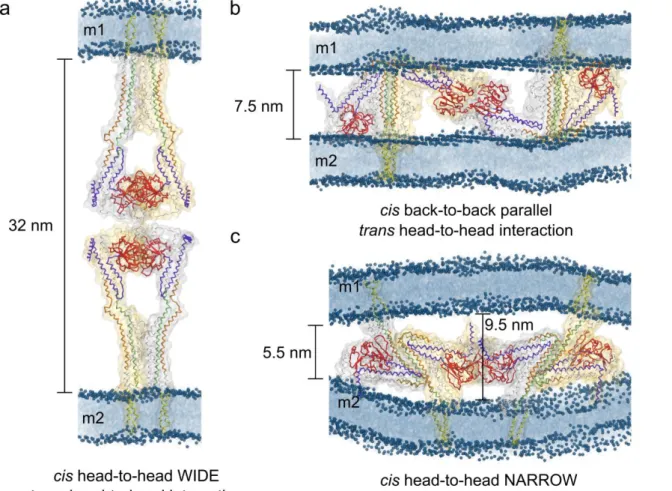
(blue) from lipid bilayer headgroups and GDP nucleotide are depicted in the space-filling 358 representation. 359 360 361 Trans-dimer dynamics. 362 363
The robustness of the tetramers has been tested through coarse-grained molecular dynamics 364
simulations. The final conformations obtained for all three MD simulations show a structural drift 365
with respect to their respective starting models that, although significant (Supplementary Table 2), 366
does not point to major issues in the structure (Fig. 6, 7). Thus, it is possible to infer that the models 367
are stable in a membrane environment, and can therefore be considered plausible (see detailed 368
analysis in supplementary material). When analysing the interface area between protein and lipids, 369
we detected that the head-to-head model with parallel HR interactions has a higher interaction 370
surface with the membrane compared to the other two tetramers (Fig 6a, red line). For this model, 371
we have also observed a jump in the protein-protein interface in one of the two back-to-back cis-372
dimers, as a consequence of an increased interaction between the transmembrane regions of each 373
monomer (Fig 6c, red curve). Interestingly, the head-to-head trans antiparallel model showed an 374
increase in membrane curvature during the simulation as a consequence of the strong interactions 375
between the protein terminal regions and lipids as well as a region spanning residues 380-386 in the 376
GTPase domain (Fig. 7c). Even taking in consideration that this model is missing the first 100 377
residues from the yeast mitofusin, the region including residues 380-386 appears to interact strongly 378
in the back-to-back cis-dimer as well, where the terminals do not interact with the membrane. An 379
analysis of the distance evolution between opposing membranes for all the tetrameric complexes 380
showed that globally it changed slightly during the MD run without affecting the comparison with 381
experiments. In particular, for the head-to-head trans anti-parallel interaction the initial distance 382
between the membranes was 10 nm, that value kept close only in the middle dimer (9.5 nm) after 1 383
μs, while at the extremes of the dimer the intermembrane distance stabilised at approximately 5.5 384
nm. This narrower value appeared as a consequence of the increased curvature in the membrane 385
caused by the strong protein interactions with the lipid (Fig. 6c, Supplementary Fig. 3). 386
387 388 389 390
391 392
Figure 6. Lipid-protein and protein-protein interface evolution during CG MD simulations 393
for the tetrameric complexes. A Lipid-protein interfaces for the three different studied tetrameric 394
complexes (black: head-to-head trans open, red: head-to-head trans parallel, cyan: back-to-back 395
trans antiparallel). B, C and D depict the evolution of the characteristic monomer-monomer
396
interfaces belonging to the trans-tetramer in open conformation, the trans-tetramer in which the 397
HRs are oriented in parallel and the trans-tetramer with HRs oriented in an antiparallel fashion. 398
Legends A, B, C and D represent each of the monomers in the complex, following the coloring of 399
image inset. 400
401
Figure 7. Final configurations of the putative model complexes for Fzo1 trans-tethering 402
interactions after relaxation through molecular dynamics simulations. a, the Fzo1 models in 403
open conformation, as suggested from the BDLP system analogy, interacting through their GTPase 404
domains. b, the Fzo1 trans-tetramer in which the HRs are oriented in a parallel fashion and the 405
trans interaction occurs towards the GTPase domain. c, the Fzo1 trans-tetramer in closed
406
conformation. Here the interaction occurs towards the GTPase domain as well as through the 407
respective HR domains oriented in an antiparallel fashion. In all figures the domains are colored: 408
violet, HRN; green, HR1; orange, HR2; red, GTPase and yellow, transmembrane. Phosphorus
409
atoms (dark blue) from the lipid headgroups of two mitochondria outer membranes (m1, m2) are 410
depicted as spheres, whereas the tails are highlighted as a blue shadow. Intermembrane distances 411
between the outer phosphate layer of the membranes are indicated. 412
413
CONCLUSION 414
415
In analogy to the macromolecular spirals formed during DRP-mediated membrane fission 416
(Jimah and Hinshaw, 2019), DRP-mediated membrane fusion may require the formation of macro-417
oligomers as observed with the Fzo1-dependent docking ring structure (Brandt et al., 2016). Such 418
assemblies require building blocks as previously shown for fission DRPs that associate with their 419
cofactors in the cytosol before recruitment to outer membranes and formation of the spirals (Koirala 420
et al., 2013; Lackner et al., 2009; Mears et al., 2011). In the context of mitofusins, these building 421
blocks would form on the same membrane imposing the assembly of cis-oligomers. 422
This rational led us to build 3 cis-assembly Fzo1 models that gave rise to 3 distinct trans-423
oligomeric models of mitofusin. Each model involves two main components of mitofusin auto-424
oligomerization: the GTPase domain interface (Cao et al., 2017; Yan et al., 2018) and the trunk 425
domain interface (Koshiba et al., 2004). A plethora of experimental data demonstrates that the 426
integrity of both regions is indeed essential for mitofusin-mediated mitochondrial fusion (Cohen et 427
al., 2011; Eura et al., 2003; Griffin and Chan, 2006; Hermann et al., 1998; Honda et al., 2005; 428
Koshiba et al., 2004; Rojo et al., 2002; Santel and Fuller, 2001). If considering BDLP, the GTPase 429
domain interface should trigger oligomerisation in cis of Fzo1 (Low et al., 2009). Nonetheless, this 430
interface is currently thought to trigger trans-oligomerization of mitofusins (Cao et al., 2017; Yan et 431
al., 2018). 432
Only one out of our three final trans models employs this strategy. In the back-to-back cis-433
dimers (with parallel-oriented HRs), two dimers from opposing membranes would engage in trans 434
interactions through their GTPase domains (Fig. 2c, and 5b). Yet, the generation of this trans-435
tetramer required an extensive rotation of the GTPase domains around the axis of their four helix 436
bundles to reconstitute the canonical G-interface where GTPase domains mirror each other in other 437
members of the dynamin superfamily (Cao et al., 2017; Daumke and Praefcke, 2016). Moreover, a 438
large area of the Ugo1 binding domain in each Fzo1 monomer was hindered by the HR interacting 439
interface required to form the back-to-back cis-dimers (Fig. 4c). This model thus accumulates 440
significant limitations upon confrontation to the experimental literature. 441
The two remaining models employ head-to-head cis-oligomerization properties. In this 442
configuration, GTP bound Fzo1 would undergo opening that could be narrow to wide. As seen for 443
the stabilization of GTP-bound fission DRPs with their cofactors (Lackner et al., 2009), this 444
conformational switch around the hinges 1a and 1b would be stabilized by concomitant binding of 445
Ugo1 through the available trunk and a neighbouring mitofusin molecule through the activated 446
GTPase domains. In both wide and narrow models the canonical G-interface is maintained. From 447
then, wide or narrow cis-oligomers would engage in trans-association with Fzo1 cis-complexes 448
from the opposing membrane. 449
The BDLP-like wide oligomers would associate in trans through a G-domain interface that 450
is distinct from that employed during cis-dimerization. To our knowledge, such an interface has 451
never been observed for other members of the dynamin superfamily. Moreover, these Fzo1 trans-452
oligomers would impose a tethering distance between outer membranes of ~ 32nm, which is 453
significantly larger than the distance observed experimentally between docked mitochondria (~ 8 454
nm, Brandt et al. 2016). Therefore, similar to the back-to-back system, the wide opening head-to 455
head model is thus difficult to reconcile with the literature. 456
In contrast, the narrow opening head-to-head mechanism employs a trans-oligomerization 457
strategy that turns out to fit particularly well with the past and current literature. The trunk region in 458
the cis-oligomers would expose a hydrophobic spine from the HR2 that would be available for 459
putative interactions (De Vecchis et al., 2017). These interactions would take place with an HR2 460
belonging to Fzo1 cis-oligomers from an opposing membrane in an anti-parallel fashion. This 461
would be consistent with anti-parallel HR2 interactions observed in 2004 for Mfn1 (Koshiba et al., 462
2004). Moreover, the tethering distance of 5.5 to 9.5 nm imposed by this Fzo1 oligomerization 463
model would agree with the ~ 8 nm separation between outer membranes observed experimentally 464
twelve years later (Brandt et al. 2016). 465
The narrow opening head-to-head cis-oligomerization followed by the antiparallel back-to-466
back trans-associations system thus allows speculating on the possible outcome of the fusion 467
complex. Following GTP hydrolysis, hinges 1a and 1b might restore the displacement of the 468
GTPase domain back to the trunk (i.e. a closed state). Similar to the dynamin BSE domain that may 469
transfer energy from the GTPase domain to the trunk during DRP-mediated fission (Chappie et al., 470
2010; Daumke and Praefcke, 2016), this movement could induce a sliding of interacting HRs with 471
respect to each other. This would contribute to further reducing the distance between outer 472
membranes to 2-3 nm as previously observed (Brandt et al. 2016). This cycle would reiterate 473
around this initial region of minimal contact to reach the docking stage characterized by an 474
extended area of membrane apposition delimited by the ring-shaped mitochondrial docking 475
complex (MDC). 476
It is tempting to propose that this MDC that stands in regions where the distance between outer 477
membranes reaches 6 to 8 nm (Brandt et al. 2016) corresponds to a macromolecular assembly of 478
Fzo1 oligomers reminiscent of those described in the present study. In addition, our models provide 479
enough detail to derive experimentally testable hypotheses for future work, for instance in terms of 480
possible crosslinks and surface-exposed residues. Conversely our models could be improved baased 481
on experimental observations. Regardless of these possibilities we hope that our hypotheses will 482
provide food for thoughts before elucidating the structure of full length mitofusins with their known 483
or yet to be described cofactors as well as their precise oligomerization properties. 484
485 486
BIBLIOGRAPHY 487
Abraham, M.J., Murtola, T., Schulz, R., Páll, S., Smith, J.C., Hess, B., and Lindahl, E. (2015). 488
GROMACS: High performance molecular simulations through multi-level parallelism from laptops 489
to supercomputers. SoftwareX 1–2, 19–25. 490
Anton, F., Fres, J.M., Schauss, A., Pinson, B., Praefcke, G.J., Langer, T., and Escobar-Henriques, 491
M. (2011). Ugo1 and Mdm30 act sequentially during Fzo1-mediated mitochondrial outer membrane 492
fusion. J Cell Sci 124, 1126-1135. 493
Berendsen, H.J.C., Postma, J.P.M., van Gunsteren, W.F., DiNola, A., and Haak, J.R. (1984). 494
Molecular dynamics with coupling to an external bath. J. Chem. Phys. 81, 3684–3690. 495
Bian, X., Klemm, R.W., Liu, T.Y., Zhang, M., Sun, S., Sui, X., Liu, X., Rapoport, T.A., and Hu, J. 496
(2011). Structures of the atlastin GTPase provide insight into homotypic fusion of endoplasmic 497
reticulum membranes. Proc Natl Acad Sci U S A 108, 3976-3981. 498
Brandt, T., Cavellini, L., Kuhlbrandt, W., and Cohen, M.M. (2016). A mitofusin-dependent docking 499
ring complex triggers mitochondrial fusion in vitro. Elife 5. 500
Bussi, G., Donadio, D., and Parrinello, M. (2007). Canonical sampling through velocity rescaling. J. 501
Chem. Phys. 126, 14101. 502
Byrnes, L.J., Singh, A., Szeto, K., Benvin, N.M., O'Donnell, J.P., Zipfel, W.R., and Sondermann, 503
H. (2013). Structural basis for conformational switching and GTP loading of the large G protein 504
atlastin. Embo J 32, 369-384. 505
Cao, Y.L., Meng, S., Chen, Y., Feng, J.X., Gu, D.D., Yu, B., Li, Y.J., Yang, J.Y., Liao, S., Chan, 506
D.C., et al. (2017). MFN1 structures reveal nucleotide-triggered dimerization critical for 507
mitochondrial fusion. Nature 542, 372-376. 508
Cohen, M.M., Amiott, E.A., Day, A.R., Leboucher, G.P., Pryce, E.N., Glickman, M.H., McCaffery, 509
J.M., Shaw, J.M., and Weissman, A.M. (2011). Sequential requirements for the GTPase domain of 510
the mitofusin Fzo1 and the ubiquitin ligase SCFMdm30 in mitochondrial outer membrane fusion. J 511
Cell Sci 124, 1403-1410. 512
Cohen, M.M., and Tareste, D. (2018). Recent insights into the structure and function of Mitofusins 513
in mitochondrial fusion. F1000Res 7. 514
Coonrod, E.M., Karren, M.A., and Shaw, J.M. (2007). Ugo1p is a multipass transmembrane protein 515
with a single carrier domain required for mitochondrial fusion. Traffic 8, 500-511. 516
De Vecchis, D., Cavellini, L., Baaden, M., Henin, J., Cohen, M.M., and Taly, A. (2017). A 517
membrane-inserted structural model of the yeast mitofusin Fzo1. Sci Rep 7, 10217. 518
de Jong, D.H., Singh, G., Bennett, W.F.D., Arnarez, C., Wassenaar, T.A., Schäfer, L. V, Periole, X., 519
Tieleman, D.P., and Marrink, S.J. (2013). Improved Parameters for the Martini Coarse-Grained 520
Protein Force Field. J. Chem. Theory Comput. 9, 687–697. 521
Eura, Y., Ishihara, N., Yokota, S., and Mihara, K. (2003). Two mitofusin proteins, mammalian 522
homologues of FZO, with distinct functions are both required for mitochondrial fusion. J Biochem 523
134, 333-344.
524
Fiser, A., Do, R.K., and Sali, A. (2000). Modeling of loops in protein structures. Protein Sci. 9, 525
1753–1773. 526
Friedman, J.R., and Nunnari, J. (2014). Mitochondrial form and function. Nature 505, 335-343. 527
Fritz, S., Rapaport, D., Klanner, E., Neupert, W., and Westermann, B. (2001). Connection of the 528
mitochondrial outer and inner membranes by Fzo1 is critical for organellar fusion. J Cell Biol 152, 529
683-692. 530
Griffin, E.E., and Chan, D.C. (2006). Domain interactions within Fzo1 oligomers are essential for 531
mitochondrial fusion. J Biol Chem 281, 16599-16606. 532
Hermann, G.J., Thatcher, J.W., Mills, J.P., Hales, K.G., Fuller, M.T., Nunnari, J., and Shaw, J.M. 533
(1998). Mitochondrial fusion in yeast requires the transmembrane GTPase Fzo1p. J Cell Biol 143, 534
359-373. 535
Honda, S., Aihara, T., Hontani, M., Okubo, K., and Hirose, S. (2005). Mutational analysis of action 536
of mitochondrial fusion factor mitofusin-2. J Cell Sci 118, 3153-3161. 537
Hoppins, S., Horner, J., Song, C., McCaffery, J.M., and Nunnari, J. (2009). Mitochondrial outer and 538
inner membrane fusion requires a modified carrier protein. J Cell Biol 184, 569-581. 539
Monticelli, L., Kandasamy, S.K., Periole, X., Larson, R.G., Tieleman, D.P., and Marrink, S.-J. 540
(2008). The MARTINI Coarse-Grained Force Field: Extension to Proteins. J. Chem. Theory 541
Comput. 4, 819–834. 542
Ishihara, N., Eura, Y., and Mihara, K. (2004). Mitofusin 1 and 2 play distinct roles in mitochondrial 543
fusion reactions via GTPase activity. J Cell Sci 117, 6535-6546. 544
Ishihara, N., Jofuku, A., Eura, Y., and Mihara, K. (2003). Regulation of mitochondrial morphology 545
by membrane potential, and DRP1-dependent division and FZO1-dependent fusion reaction in 546
mammalian cells. Biochem Biophys Res Commun 301, 891-898. 547
Jimah, J.R., and Hinshaw, J.E. (2019). Structural Insights into the Mechanism of Dynamin 548
Superfamily Proteins. Trends Cell Biol 29, 257-273. 549
Koirala, S., Guo, Q., Kalia, R., Bui, H.T., Eckert, D.M., Frost, A., and Shaw, J.M. (2013). 550
Interchangeable adaptors regulate mitochondrial dynamin assembly for membrane scission. Proc 551
Natl Acad Sci U S A 110, E1342-1351. 552
Koshiba, T., Detmer, S.A., Kaiser, J.T., Chen, H., McCaffery, J.M., and Chan, D.C. (2004). 553
Structural basis of mitochondrial tethering by mitofusin complexes. Science 305, 858-862. 554
Lackner, L.L., Horner, J.S., and Nunnari, J. (2009). Mechanistic analysis of a dynamin effector. 555
Science 325, 874-877. 556
Low, H.H., and Lowe, J. (2006). A bacterial dynamin-like protein. Nature 444, 766-769. 557
Low, H.H., Sachse, C., Amos, L.A., and Lowe, J. (2009). Structure of a Bacterial Dynamin-like 558
Protein Lipid Tube Provides a Mechanism For Assembly and Membrane Curving. Cell 139, 1342-559
1352. 560
Mears, J.A., Lackner, L.L., Fang, S., Ingerman, E., Nunnari, J., and Hinshaw, J.E. (2011). 561
Conformational changes in Dnm1 support a contractile mechanism for mitochondrial fission. Nat 562
Struct Mol Biol 18, 20-26. 563
Moss, T.J., Andreazza, C., Verma, A., Daga, A., and McNew, J.A. (2011). Membrane fusion by the 564
GTPase atlastin requires a conserved C-terminal cytoplasmic tail and dimerization through the 565
middle domain. Proc Natl Acad Sci U S A 108, 11133-11138. 566
Parrinello, M., and Rahman, A. (1981). Polymorphic transitions in single crystals: A new molecular 567
dynamics method. J. Appl. Phys. 52, 7182–7190. 568
Pettersen, E.F., Goddard, T.D., Huang, C.C., Couch, G.S., Greenblatt, D.M., Meng, E.C., and 569
Ferrin, T.E. (2004). UCSF Chimera--a visualization system for exploratory research and analysis. J. 570
Comput. Chem. 25, 1605–1612. 571
Ramachandran, R. (2018). Mitochondrial dynamics: The dynamin superfamily and execution by 572
collusion. Semin Cell Dev Biol 76, 201-212. 573
Rapaport, D., Brunner, M., Neupert, W., and Westermann, B. (1998). Fzo1p is a mitochondrial 574
outer membrane protein essential for the biogenesis of functional mitochondria in Saccharomyces 575
cerevisiae. J Biol Chem 273, 20150-20155. 576
Rojo, M., Legros, F., Chateau, D., and Lombes, A. (2002). Membrane topology and mitochondrial 577
targeting of mitofusins, ubiquitous mammalian homologs of the transmembrane GTPase Fzo. J Cell 578
Sci 115, 1663-1674. 579
Santel, A., Frank, S., Gaume, B., Herrler, M., Youle, R.J., and Fuller, M.T. (2003). Mitofusin-1 580
protein is a generally expressed mediator of mitochondrial fusion in mammalian cells. J Cell Sci 581
116, 2763-2774.
582
Santel, A., and Fuller, M.T. (2001). Control of mitochondrial morphology by a human mitofusin. J 583
Cell Sci 114, 867-874. 584
Sesaki, H., and Jensen, R.E. (2001). UGO1 encodes an outer membrane protein required for 585
mitochondrial fusion. J Cell Biol 152, 1123-1134. 586
Sesaki, H., and Jensen, R.E. (2004). Ugo1p links the Fzo1p and Mgm1p GTPases for mitochondrial 587
fusion. J Biol Chem 279, 28298-28303. 588
Shen, M.-Y., and Sali, A. (2006). Statistical potential for assessment and prediction of protein 589
structures. Protein Sci. 15, 2507–2524. 590
Shutt, T.E., and McBride, H.M. (2013). Staying cool in difficult times: mitochondrial dynamics, 591
quality control and the stress response. Biochim Biophys Acta 1833, 417-424. 592
Tilokani, L., Nagashima, S., Paupe, V., and Prudent, J. (2018). Mitochondrial dynamics: overview 593
of molecular mechanisms. Essays Biochem 62, 341-360. 594
Westermann, B. (2010). Mitochondrial fusion and fission in cell life and death. Nat Rev Mol Cell 595
Biol 11, 872-884. 596
Wong, E.D., Wagner, J.A., Scott, S.V., Okreglak, V., Holewinske, T.J., Cassidy-Stone, A., and 597
Nunnari, J. (2003). The intramitochondrial dynamin-related GTPase, Mgm1p, is a component of a 598
protein complex that mediates mitochondrial fusion. J Cell Biol 160, 303-311. 599
Yan, L., Qi, Y., Huang, X., Yu, C., Lan, L., Guo, X., Rao, Z., Hu, J., and Lou, Z. (2018). Structural 600
basis for GTP hydrolysis and conformational change of MFN1 in mediating membrane fusion. Nat 601
Struct Mol Biol 25, 233-243. 602
Yan, L., Sun, S., Wang, W., Shi, J., Hu, X., Wang, S., Su, D., Rao, Z., Hu, J., and Lou, Z. (2015). 603
Structures of the yeast dynamin-like GTPase Sey1p provide insight into homotypic ER fusion. J 604 Cell Biol 210, 961-972. 605 606 607 608 ACKNOWLEDGEMENTS 609 610
Funding for this work was provided by the French Agency for Research and “Initiative 611
d’Excellence” (ANR-11-LABX-0011, cluster of excellence LABEX Dynamo). This work was 612
performed using HPC resources from GENCI-CINES (grant number 2016-072292). Research in the 613
Cohen laboratory is supported by the ANR grant MOMIT (ANR-17-CE13-0026-01). 614
Supplementary: 615
616
Supplementary Table 1: Molecular dynamics simulation details for each system 617
Box size (x,y,z [nm])
Number of particles All Protein + lipid Dimers Head-to-head wide 25.1474 x 25.1474 x 28.9065 157017 27418 Head-to-head narrow 24.6357 x 24.6357 x 29.8118 156752 27058 Back-to-back 25.2349 x 25.2349 x 28.7671 157232 27298 Tetramers Head-to-head trans 29.4832 x 29.4832 x 67.4888 523921 78836
Head-to-head trans parallel 49.4896 x 39.5917 x 28.3762 497044 166292 Head-to-head trans antiparallel 30.4625 x 40.6166 x 32.2698 347636 101324
618 619
620
Supplementary Figure 1: Putative membrane-inserted Fzo1 models interacting in cis 621
configuration after molecular dynamics relaxation. a and b. Wide and narrow head-to-head 622
complexes, respectively. c. Back-to-back complex of the closed model. The domains are colored: 623
violet, HRN; green, HR1; orange, HR2; red, GTPase and yellow, transmembrane. Phosphorus
624
atoms (dark blue) from the lipid bilayer headgroups are depicted as spheres, whereas the bilayer is 625
highlighted as a blue shadow. 626
627
Supplementary Table 2: RMSD values averaged over the last 100 ns of simulation for each dimer 628
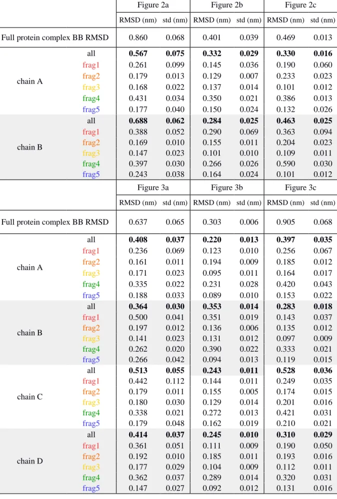
(Figure 2a, b, c) and tetramers (Figure 3a, b, c). Reported values include a full RMSD calculated 629
over all the backbone particles (BB) as well as by every chain and structural fragment separately. 630
Figure 2a Figure 2b Figure 2c
RMSD (nm) std (nm) RMSD (nm) std (nm) RMSD (nm) std (nm)
Full protein complex BB RMSD 0.860 0.068 0.401 0.039 0.469 0.013
chain A all 0.567 0.075 0.332 0.029 0.330 0.016 frag1 0.261 0.099 0.145 0.036 0.190 0.060 frag2 0.179 0.013 0.129 0.007 0.233 0.023 frag3 0.168 0.022 0.137 0.014 0.101 0.012 frag4 0.431 0.034 0.350 0.021 0.386 0.013 frag5 0.177 0.040 0.150 0.024 0.132 0.026 chain B all 0.688 0.062 0.284 0.025 0.463 0.025 frag1 0.388 0.052 0.290 0.069 0.363 0.094 frag2 0.169 0.010 0.155 0.011 0.204 0.023 frag3 0.147 0.023 0.101 0.010 0.109 0.011 frag4 0.397 0.030 0.266 0.026 0.590 0.030 frag5 0.243 0.038 0.164 0.024 0.101 0.012
Figure 3a Figure 3b Figure 3c
RMSD (nm) std (nm) RMSD (nm) std (nm) RMSD (nm) std (nm)
Full protein complex BB RMSD 0.637 0.065 0.303 0.006 0.905 0.068
chain A all 0.408 0.037 0.220 0.013 0.397 0.035 frag1 0.236 0.069 0.123 0.010 0.256 0.067 frag2 0.161 0.011 0.194 0.009 0.185 0.012 frag3 0.171 0.023 0.095 0.011 0.164 0.017 frag4 0.335 0.022 0.231 0.028 0.420 0.043 frag5 0.188 0.033 0.089 0.010 0.153 0.022 chain B all 0.364 0.030 0.353 0.014 0.283 0.018 frag1 0.500 0.041 0.351 0.019 0.143 0.037 frag2 0.197 0.012 0.136 0.006 0.135 0.012 frag3 0.141 0.023 0.131 0.012 0.097 0.009 frag4 0.262 0.020 0.390 0.022 0.333 0.021 frag5 0.266 0.042 0.094 0.013 0.119 0.015 chain C all 0.513 0.055 0.243 0.011 0.528 0.036 frag1 0.442 0.112 0.144 0.011 0.249 0.035 frag2 0.179 0.011 0.155 0.005 0.174 0.015 frag3 0.180 0.030 0.129 0.014 0.201 0.016 frag4 0.338 0.021 0.272 0.013 0.421 0.031 frag5 0.179 0.048 0.162 0.019 0.210 0.021 chain D all 0.414 0.037 0.245 0.010 0.310 0.029 frag1 0.361 0.051 0.111 0.009 0.190 0.050 frag2 0.192 0.010 0.185 0.011 0.193 0.016 frag3 0.177 0.029 0.104 0.009 0.112 0.011 frag4 0.362 0.037 0.289 0.014 0.320 0.031 frag5 0.147 0.027 0.092 0.012 0.131 0.016 631 632 633 634
635
Supplementary Figure 2. a. The Fzo1 GTPase dimer construct in which the canonical G-interface 636
commonly observed in the dynamin superfamily (Daumke and Prafke, 2016) is reconstituted. b. 637
The back-to-back HR-parallel trans-tetramer during the modeling procedure (see Method). The 638
Figure shows how the two GTPase domains that interact in trans share an apparent incorrect 639
orientation (compared with a). Without an extensive conformational rearrangement that would 640
involve both hinges 1a and 1b, the formation of the canonical G-interface observed in the dynamin 641
superfamily (Daumke and Prafke, 2016) would be impeded. In particular, the dotted line should be 642
located on the same side. The domains are: violet, HRN; green, HR1; orange, HR2; red, GTPase 643
and yellow, transmembrane. Phosphorus atoms (blue) from lipid bilayer headgroups and GDP 644
nucleotide are depicted in the space-filled representation. 645
646
647 648
Supplementary Figure 3: Inter-bilayer difference distance matrix (Å). Left panel shows the 649
final difference in distances compared to the initial structure, red regions correspond to regions that 650
got closer in space compared to the initial structure of the production run whereas blue regions 651
represent areas that moved further apart. The right panel shows a rotation of 90 degrees around the 652
y-axis to visualize the position of the complex. 653
654 655 656 657