HAL Id: inserm-01154785
https://www.hal.inserm.fr/inserm-01154785
Submitted on 23 May 2015HAL is a multi-disciplinary open access
archive for the deposit and dissemination of sci-entific research documents, whether they are pub-lished or not. The documents may come from teaching and research institutions in France or abroad, or from public or private research centers.
L’archive ouverte pluridisciplinaire HAL, est destinée au dépôt et à la diffusion de documents scientifiques de niveau recherche, publiés ou non, émanant des établissements d’enseignement et de recherche français ou étrangers, des laboratoires publics ou privés.
Distributed under a Creative Commons Attribution| 4.0 International License
4D cone-beam computed tomography using
motion-aware regularization
Cyril Mory, Simon Rit
To cite this version:
Cyril Mory, Simon Rit. 4D cone-beam computed tomography using motion-aware regularization. Journées RITS 2015, Mar 2015, Dourdan, France. pp 118-119. �inserm-01154785�
Actes des Journées Recherche en Imagerie et Technologies pour la Santé - RITS 2015 118
4D cone-beam computed tomography using motion-aware regularization
Cyril Mory
1∗, Simon Rit
21 iMagX project, ICTEAM Institute, Universit´e catholique de Louvain, Louvain-la-Neuve, Belgium
and Universit´e de Lyon, CREATIS ; CNRS UMR5220 ; Inserm U1044 ; INSA-Lyon ; Universit´e Lyon 1 ; Centre L´eon B´erard, France
2 Universit´e de Lyon, CREATIS ; CNRS UMR5220 ; Inserm U1044 ; INSA-Lyon ; Universit´e Lyon 1 ; Centre L´eon B´erard, France ∗ Corresponding author, [email protected].
Abstract - In Image-Guided RadioTherapy (IGRT) of lung tumors, patients undergo a 4D CT, on the basis of which their treatment is planned. It is implicitely as-sumed that their breathing motion will not change much throughout the treatment. At the beginning of a treat-ment fraction, a Cone Beam CT (CBCT) acquisition is performed, and used to re-position the patient. Obtaining a 4D reconstruction from this cone beam data would al-low the therapists to check whether the breathing motion of the day still matches that of the planning CT, and if not, take appropriate corrective actions. But 4D tomog-raphy from a single cone beam CT acquisition implies a severe lack of projection data, and efficient methods have only started to appear during the last few years. Some perform regularization along time to explicitely enforce similarity between consecutive frames, which consider-ably improves image quality. In IGRT, breathing motion can be estimated on the 4D planning CT, and used to re-fine the 4D CBCT reconstruction results. We describe a 4D CBCT reconstruction method that combines regular-ization techniques with the use of motion information. Index Terms - Image Processing, Radiotherapy, X-Ray imaging
I. INTRODUCTION
In image-guided radiotherapy (IGRT) of lung tumors, breathing motion affects the image guidance. On a treat-ment day, physicians currently have no reliable method to make sure that the patient’s breathing motion is the same as in the planning CT. This task requires a 4D reconstruction of the cone beam CT data, but most reconstruction meth-ods currently available suffer major drawbacks: static re-construction techniques, like FDK [1] or SART [2], gen-erate images almost free of streaks, but in 3D, not 4D, and in which the moving structures are strongly blurred. Their respiration-correlated counterparts generate severely degraded 4D images with strong streak artifacts, unless the acquisition time is substantially increased [3]. Advanced methods have been developed to achieve streak-free and blur-free 4D reconstructions. Mostly, they are based either on motion compensation [4, 5] or on regularization using some a priori information [6, 7]. Recently, mixed methods have been proposed, which combine both approaches [8].
We describe a new mixed 4D reconstruction method that combines regularization techniques with the use of motion information, and show its results on real patient data.
II. MATERIALS AND METHODS
Our method, coined Motion-Compensated RecOnstruc-tiOn using Spatial and TEmporal Regularization (MC-ROOSTER), assumes that a rough segmentation of the pa-tient is available. Movement is expected to occur only in-side the segmented region. It also requires a 4D Deforma-tion Vector Field (DVF) describing the patient’s breathing motion. The algorithm consists in iteratively enforcing five different constraints in an alternating manner:
• MinimizeP a quadratic data-attachment term
α kRαSαf − pαk 2
2, with α the projection’s
in-dex, f a 4D sequence of volumes, Rα the forward
projection operator for the projection with index α, Sα a linear interpolator which, from the 4D
sequence f, estimates the 3D volume through which projection α has been acquired, and pαthe measured
projection with index α. Minimization is performed by conjugate gradient
• Enforce positivity
• Average along time outside the segmentation, remov-ing motion everywhere but in the segmented region, yielding fAveraged
• Perform spatial Total Variation (TV) denoising on each 3D volume of f independently, i.e. solve fSpace = arg min
f kf − fAveragedk 2 2 +
γSpaceT VSpace(f )
• Perform temporal TV denoising along curved tra-jectories determined by the 4D DVF, i.e. compute fDenoised = W−1Dtime(W fSpace), with Dtime the
total variation denoising along time operator (similar to spatial TV denoising), W the trilinear interpola-tion warping operator applying the DVF’s mointerpola-tion, and W−1its inverse
This constitutes one iteration of the main loop, the output of which is fed back to the conjugate gradient minimizer for the next iteration.
119 Actes des Journées Recherche en Imagerie et Technologies pour la Santé - RITS 2015
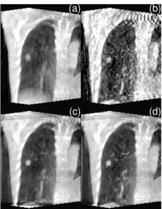
Figure 1: Coronal slice of patient with lung tumor, in end-inhale position (a) Static FDK (b) Respiration-correlated FDK (c) ROOSTER with null DVF (d) MC-ROOSTER with DVF estimated on the planning CT
III. RESULTS
A CBCT acquisition performed on a patient was recon-structed with static 3D FDK, respiration-correlated FDK, and MC-ROOSTER with either a null DVF or the one es-timated on the 4D planning CT. Estimation was performed using a method that allows sliding motion at the border be-tween the lungs and the chest wall [9]. The size of the re-constructed sequence of volumes was set to 145⇥185⇥245 voxels, with isotropic voxels of size 1.5 mm in each dimen-sion. The results are displayed in Figure 1. The static FDK reconstruction is very blurry, and the respiration-correlated one is degraded by strong streak artifacts (although they do not appear as streaks with this cut plane). Without mo-tion informamo-tion (sub-figure (c)), MC-ROOSTER already provides a usable 4D reconstruction: the small structures are partially blurred out, but the tumor is correctly recon-structed. Making use of relevant motion information (sub-figure (d)) dramatically increases the preservation of small structures.
IV. DISCUSSION-CONCLUSION
This paper shows that MC-ROOSTER can be employed for 4D CBCT of the lungs. Motion information can be used to
improve the reconstruction result, but it also works without any a priori information on motion.
We have shown that high-quality 4D CBCT of the lungs from a single rotation is possible, and that a method based on regularization can be considerably improved by making the regularization along time “motion-aware”.
REFERENCES
[1] L. A. Feldkamp, L. C. Davis, and J. W. Kress, “Practi-cal cone-beam algorithm,” Journal of the Opti“Practi-cal Soci-ety of America A, vol. 1, no. 6, pp. 612–619, Jun. 1984. [2] A. H. Andersen and A. C. Kak, “Simultaneous alge-braic reconstruction technique (SART): a superior im-plementation of the art algorithm,” Ultrasonic Imag-ing, vol. 6, no. 1, pp. 81–94, Jan. 1984.
[3] J.-J. Sonke, L. Zijp, P. Remeijer, and M. van Herk, “Respiratory correlated cone beam CT,” Medical Physics, vol. 32, no. 4, p. 1176, 2005.
[4] S. Rit, J. W. H. Wolthaus, M. van Herk, and J.-J. Sonke, “On-the-fly motion-compensated cone-beam CT using an a priori model of the respiratory motion,” Medical physics, vol. 36, no. 6, pp. 2283–2296, Jun. 2009. [5] J. Wang and X. Gu, “High-quality four-dimensional
cone-beam CT by deforming prior images,” Physics in Medicine and Biology, vol. 58, no. 2, p. 231, Jan. 2013. [6] F. Bergner, T. Berkus, M. Oelhafen, P. Kunz, T. Pan, R. Grimmer, L. Ritschl, and M. Kachelrie, “An inves-tigation of 4d cone-beam CT algorithms for slowly ro-tating scanners,” Medical Physics, vol. 37, no. 9, p. 5044, 2010.
[7] C. Mory, V. Auvray, B. Zhang, M. Grass, D. Schfer, S. J. Chen, J. D. Carroll, S. Rit, F. Peyrin, P. Douek, and L. Boussel, “Cardiac c-arm computed tomography us-ing a 3d + time ROI reconstruction method with spatial and temporal regularization,” Medical physics, vol. 41, no. 2, p. 021903, Feb. 2014.
[8] J. Wang and X. Gu, “Simultaneous motion estimation and image reconstruction (SMEIR) for 4d cone-beam CT,” Medical Physics, vol. 40, no. 10, p. 101912, Oct. 2013.
[9] V. Delmon, S. Rit, R. Pinho, and D. Sarrut, “Regis-tration of sliding objects using direction dependent b-splines decomposition,” Physics in Medicine and Biol-ogy, vol. 58, no. 5, p. 1303, Mar. 2013.