HAL Id: hal-02550612
https://hal.umontpellier.fr/hal-02550612
Submitted on 22 Apr 2020
HAL is a multi-disciplinary open access archive for the deposit and dissemination of sci-entific research documents, whether they are pub-lished or not. The documents may come from teaching and research institutions in France or abroad, or from public or private research centers.
L’archive ouverte pluridisciplinaire HAL, est destinée au dépôt et à la diffusion de documents scientifiques de niveau recherche, publiés ou non, émanant des établissements d’enseignement et de recherche français ou étrangers, des laboratoires publics ou privés.
Tomography Study of 239 Patients
Boris Jung, Stéphanie Nougaret, Gerald Chanques, Grégoire Mercier, Moussa
Cisse, Sophie Aufort, Benoit Gallix, Djillali Annane, Samir Jaber
To cite this version:
Boris Jung, Stéphanie Nougaret, Gerald Chanques, Grégoire Mercier, Moussa Cisse, et al.. The Absence of Adrenal Gland Enlargement during Septic Shock Predicts Mortality. A Computed Tomography Study of 239 Patients. Anesthesiology, Lippincott, Williams & Wilkins, 2011, �10.1097/ALN.0b013e318225cfd7�. �hal-02550612�
CRITICAL CARE MEDICINE
The Absence of Adrenal Gland Enlargement during
Septic Shock Predicts Mortality
A Computed Tomography Study of 239 Patients
Boris Jung, M.D., Ph.D.,* Stephanie Nougaret, M.D.,† Ge´rald Chanques, M.D., Ph.D.,‡
Gregoire Mercier, M.D.,§ Moussa Cisse, M.D.,‡ Sophie Aufort, M.D.,! Benoit Gallix, M.D., Ph.D.,# Djillali Annane, M.D., Ph.D.,** Samir Jaber, M.D., Ph.D.††
ABSTRACT
Background: Assessment and management of septic shock
associated adrenal function remain controversial. The aim of this study was to explore the prognostic value of adrenal gland volume in adults with septic shock.
Methods: A short cosyntropin test and determination of
adrenal volume by computed tomography were performed within 48 h of shock in patients with septic shock (n ! 184) and in 2 control groups: 40 ambulatory patients and 15
nonseptic critically ill patients. The primary endpoint was intensive care unit mortality.
Results: At intensive care unit discharge, 59 patients with
septic shock died. Adrenal volume was 12.5 cm3[95% CI, 11.3–13.3] and 8 cm3[95% CI, 6.8 –10.1] in the nonseptic group (P " 0.05 with both septic cohorts) and 7.2 cm3 [95%CI, 6.3– 8.5] in the ambulatory patient group (P " 0.05 in patients with septic shock). In patients with septic shock, adrenal volume less than 10 cm3was associated with higher 28-day mortality rates with an area under the receiver operating curve of 0.84 [95% CI, 0.78 – 0.89]. Adrenal vol-ume above 10 cm3was an independent predictor of intensive care unit survival (hazard ratio ! 0.014; 95% CI [0.004 – 0.335]).
Conclusion: A total adrenal gland volume less than 10 cm3 during septic shock was associated in univariate and multi-variate analysis with mortality at day 28 in patients with septic shock. Whether adrenal gland volume can be a surro-gate of adrenal gland function and used to guide
hydrocor-* Assistant Professor of Anesthesiology and Critical Care, Saint Eloi University Hospital, Centre Hospitalier Universitaire Montpellier, Mont-pellier, France. † Research Fellow of Radiology, Department of Abdom-inal Imaging, Saint Eloi University Hospital. ‡ Staff Intensivist, Anes-thesia and Critical Care Department, Saint Eloi University Hospital. § Assistant Professor of Medical Statistics, Department of Medical Sta-tistics, Arnaud de Villeneuve University Hospital, Centre Hospitalier Universitaire Montpellier.! Staff Radiologist, Saint Eloi University Hos-pital, Centre Hospitalier Universitaire Montpellier. # Professor of Radi-ology and Chairman, Department of Abdominal Imaging, Saint Eloi University Hospital, Centre Hospitalier Universitaire Montpellier. ** Professor of Critical Care, Chairman, Department of Critical Care Medicine, Raymond Poincare´ University Hospital, Paris V University, Paris-Ouest Faculty of Medicine, Garches, France. †† Professor of An-esthesiology and Critical Care and Chairman, Department of Anesthe-siology and Critical Care and Equipe soutenue par la Re´gion et l’Institut National de la Sante´ et de la Recherche Me´dicale 25, Saint Eloi Univer-sity Hospital, Centre Hospitalier Universitaire Montpellier.
Received from the Intensive Care Unit, Anesthesia and Critical Care Department, Saint Eloi University Hospital, Centre Hospitalier Universitaire Montpellier, Montpellier, France. Submitted for publication October 17, 2010. Accepted for publication March 25, 2011. Support was provided solely from institutional and/or departmental sources. Presented in part at the 2009 XXXth Annual Meeting of the European Society of Intensive Care and Emergency Medicine (ISICEM), Brussels, Belgium, March 9–12, 2009. All authors contributed to the report. Current Controlled Trials Identifier: ISRCTN79359473.
Address correspondence to Dr. Jaber: Intensive Care Unit, An-esthesia and Critical Care Department, Saint Eloi Teaching Hospital, University Montpellier 1; 80 avenue Augustin Fliche, 34295 Mont-pellier CEDEX 5, France. s-jaber@chu-montMont-pellier.fr. This article may be accessed for personal use at no charge through the Journal Web site, www.anesthesiology.org.
Copyright © 2011, the American Society of Anesthesiologists, Inc. Lippincott Williams & Wilkins. Anesthesiology 2011; 115:334–43
What We Already Know about This Topic
• The use of low dose steroids in patients with septic shock appears to improve mortality in subgroups of patients, but identification of patients who will benefit is difficult
What This Article Tells Us That Is New
• Computed tomography identification of adrenal gland volume may be helpful in identifying septic patients who might benefit from steroid therapy
! This article is featured in “This Month in Anesthesiology.” Please see this issue of ANESTHESIOLOGY, page 9A.
" This article is accompanied by an Editorial View. Please see: Eikermann M, Schmidt U: Does adrenal size matter? ANESTHESIOLOGY2011; 115:223–5.
tisone therapy in septic shock patients needs to be further investigated.
B
UNDLE therapy of septic shock, a disease with a mor-tality rate ranging from 30 to 50%,1–5 may include low-dose steroid.6 –11 Indeed, independent factors of poor outcome include hypothalamic–pituitary–adrenal axis im-pairment resulting in critical illness-related corticosteroid in-sufficiency.8,9,11–16Several studies have reported that daily treatment of certain patient subgroups presenting with septic shock with a prolonged (5–7 days) low dose (200 –300 mg) of hydrocortisone offers potential benefits of reduced inten-sive care unit (ICU) length of stay or mortality.6,8,13,14,17 However, one multiple center study failed to confirm the effect of such treatment on mortality18and the recent inter-national consensus conference on critical illness-related cor-ticosteroid insufficiency management in the ICU setting rec-ommended considering low-dose steroid therapy only for vasopressor-dependent patients with septic shock.15Key pa-tient selection criteria may include diagnosis of impaired hypothalamic–pituitary–adrenal axis or a poor response to endogenous steroids.10,11However, currently there is no ab-solute plasma cortisol concentration that distinguishes ade-quate and inadeade-quate adrenal response.11,12,15More sophis-ticated tests are not routinely performed and are therefore not recommended to guide treatment.11,15In a pilot study using computed tomography (CT), we previously reported an increased adrenal gland volume, unrelated to adrenal hemorrhage, in patients with septic shock.19,20The aim of the current study was to describe adrenal gland volume and its accuracy as a prognostic value in septic shock.
We hypothesized that the absence of adrenal gland volume enlargement could be associated with mortality in patients with septic shock.
Materials and Methods
Study Design
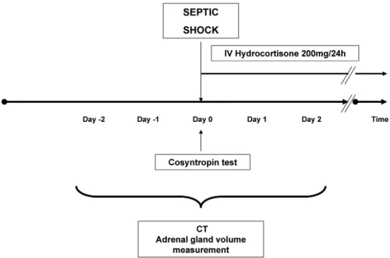
This study was conducted from January 2005 to January 2009 in a 16-bed medical–surgical ICU. In accordance with French law, informed consent was not mandatory, given that this observational study did not modify current diagnostic or therapeutic strategies. The study design is represented in figure 1. Our study followed the Standards for Reporting Diagnostic Accuracy (STARD) recommen-dations concerning the report of studies of diagnostic ac-curacy.21,22This study was approved by the Montpellier University Hospital Institutional Review Board, Mont-pellier, France.
Study Population Septic Shock
Consecutive patients were included if they presented with septic shock23and had been explored, within the first 48 h of shock, with an abdominal CT and measurements of basal and response plasma cortisol concentrations, before and after a short cosyntropin test with 250 !g cosyntropin intrave-nous injection (fig. 1). In our ICU, it is part of the standard of care to order a body CT scan in the early phase of septic shock to rule out any surgical or interventional radiology treatment for source control but also to assess the initial pattern of an acute lung injury. Thus, more than 70% of the
Fig. 1.Design of the study. Cosyntropin test was done at the time of the shock onset. Hydrocortisone treatment was started once the test had been done. To be included in the study, patients needed to be explored with an abdominal computed tomography (CT) scan within the first 48 h of the shock onset.
CT scans performed combine both lung and abdominal CT. CT may be delayed or not performed when the cause of sepsis is obvious and/or its risks overwhelm its benefits. In this study, patients younger than 18 yr, who are pregnant, with pituitary or adrenal disease, or with a history of steroid use were excluded.
During the study period, septic shock management fol-lowed the 2004 Surviving Sepsis Campaign guidelines.4,5All patients were treated with intravenous hydrocortisone, 50 mg four times a day, during 5–7 days regardless of the result of the cosyntropin stimulation test.2
Control Subjects
Patients without sepsis admitted to the ICU (ICU nonseptic group, n ! 15) and 40 consecutive ambulatory patients who had a normal virtual colonoscopy served as control subjects (control group, n ! 40).
Measurements and Clinical Evaluation
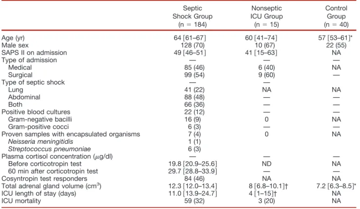
We collected data for sex, age, date, reason for ICU admis-sion, and Simplified Acute Physiology Score II24for all ICU patients (tables 1 and 2).
Laboratory Variables
No laboratory variables were collected in the control group. In the ICU groups, cortisol was assayed at baseline and 60
min after cosyntropin stimulation. Plasma cortisol concen-tration was measured using immunoradiology assay (SP2100, Beckmancoulter SAS, Roissy, France). Response to cosyntropin stimulation was considered to be positive if T60-T0 (#) was above 9 !g/dl.8,9In the septic shock group, we also recorded serum protein and albumin concentrations and the results of the blood cultures as well as plasma creat-inine, bilirubin, lactate, platelets, leukocytes, hemoglobin, and procalcitonin (tables 1, 2).
CT Scan of the Adrenal Glands
Semiautomatic measurement of the adrenal glands by CT scan was performed as previously described by our group in a pilot study.20The measurement technique was reproducible as we reported in the pilot study a concordance correlation coefficient between the radiologist and the intensivist of 0.87 (95% CI, [0.76 – 0.93]) for the total gland volume measure-ment. The adrenal contour was semiautomatically traced by one radiologist (SN), who was unaware of the clinical status of the subjects, every 3-mm section. The radiologist traced the contour twice for each patient in random order. The scanner software automatically calculated the adrenal vol-ume by summing the area on each slice. Adrenal gland volume between control and septic shock groups were compared.
Table 1. Characteristics of the 239 Studied Patients (184 Septic Shock, 15 Nonseptic ICU, and 40 Control Patients) Septic Shock Group (n ! 184) Nonseptic ICU Group (n ! 15) Control Group (n ! 40) Age (yr) 64 $61–67% 60 $41–74% 57 $53–61%* Male sex 128 (70) 10 (67) 22 (55) SAPS II on admission 49 $46–51% 41 $15–63% NA Type of admission — — — Medical 85 (46) 6 (40) NA Surgical 99 (54) 9 (60) —
Type of septic shock — —
Lung 41 (22) NA NA
Abdominal 88 (48) — —
Both 66 (36) — —
Positive blood cultures 22 (12) — —
Gram-negative bacilli 16 (9) 0 NA
Gram-positive cocci 6 (3) — —
Proven samples with encapsulated organisms 7 (4) 0 NA
Neisseria meningitidis 1 (1)
Streptococcus pneumoniae 6 (3)
Plasma cortisol concentration (!g/dl) — — — Before corticotropin test 19.8 $20.9–25.6% ND NA 60 min after corticotropin test 29.7 $28.8–33.9% — — Cosyntropin test responders 84 (46) NA NA Total adrenal gland volume (cm3) 12.3 $12.0–13.4% 8 $6.8–10.1%† 7.2 $6.3–8.5%*
ICU length of stay (days) 11.0 $13.9–24.7% 4 $1–15%† NA
ICU mortality 59 (32) 3 (20) NA
Data are median and 95% confidence interval or n (%). Cosyntropin test responders were patients with a # plasma cortisol concentration 60 min after a short corticotropin test of more than 9 !g/dl. (To convert values for cortisol to nM/l, multiply by 27.6.) *P " 0.05 between septic shock and control groups. †P " 0.05 between septic shock and nonseptic ICU groups.
ICU ! intensive care unit; NA ! not applicable; ND ! not done; SAPS II ! Simplified Acute Physiology Score.
Adrenal Gland Volume and Outcome in Septic Shock
Study Outcomes
The main endpoint was mortality in the ICU in relation to adrenal gland volume. The secondary endpoints were to an-alyze total adrenal gland volume in relation to septic shock, and to the cosyntropin stimulation test results.
Assumptions for the sample size calculation were based on previous studies on septic shock performed in our ICU.25 Assuming a 40% mortality in the ICU in the group with adrenal gland volume above 10 cm3during septic shock, we calculated that 154 patients would need to be studied to detect a 50% absolute increase in mortality if the adrenal gland volume was less than 10 cm3; with 80% statistical power and a one-sided " value of 0.05.
Statistical Analysis
Data are represented as mean & SD or median and 95% CI. Continuous data were compared with Student t test or Mann-Whitney U test when appropriate. Categoric data were analyzed with chi-square and Fisher exact test. The Pearson correlation coefficient was used to assess the corre-lation between the cosyntropin test results and adrenal gland volume measurement. Sensitivity and specificity of adrenal volume and basal/response plasma cortisol concentration curves to predict mortality were assessed using receiver oper-ating characteristics (ROC) curves and compared with the Mann-Whitney U test. The cutoff point for ROC was
pre-determined by the statistics software to automatically mini-mize the mathematic distance between the ROC curve and the ideal point as a method to minimize misclassification of patients.21 As previously reported, we also determined two cutoff points to detect volume, which predicts mortality with the best sensitivity and the best specificity.21Survival at day 28 was estimated by the Kaplan–Meier method and com-pared between groups with the log-rank test. A theory-driven logistic regression model was also developed in three steps. First, we identified all variables previously reported as asso-ciated with mortality in septic shock. Then, we assessed the
Fig. 2.Flow chart of the study. ACTH ! adrenocorticotropic hormone; CT ! computed tomography; ICU ! intensive care unit.
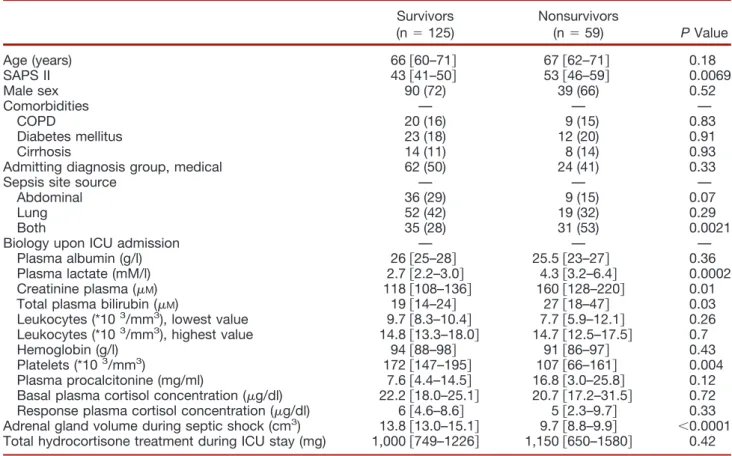
Table 2. Characteristics of the 184 Septic Shock Patients
Survivors (n ! 125) Nonsurvivors (n ! 59) P Value Age (years) 66 $60–71% 67 $62–71% 0.18 SAPS II 43 $41–50% 53 $46–59% 0.0069 Male sex 90 (72) 39 (66) 0.52 Comorbidities — — — COPD 20 (16) 9 (15) 0.83 Diabetes mellitus 23 (18) 12 (20) 0.91 Cirrhosis 14 (11) 8 (14) 0.93
Admitting diagnosis group, medical 62 (50) 24 (41) 0.33
Sepsis site source — — —
Abdominal 36 (29) 9 (15) 0.07
Lung 52 (42) 19 (32) 0.29
Both 35 (28) 31 (53) 0.0021
Biology upon ICU admission — — —
Plasma albumin (g/l) 26 $25–28% 25.5 $23–27% 0.36 Plasma lactate (mM/l) 2.7 $2.2–3.0% 4.3 $3.2–6.4% 0.0002 Creatinine plasma (!M) 118 $108–136% 160 $128–220% 0.01
Total plasma bilirubin (!M) 19 $14–24% 27 $18–47% 0.03
Leukocytes (*103/mm3), lowest value 9.7 $8.3–10.4% 7.7 $5.9–12.1% 0.26
Leukocytes (*103/mm3), highest value 14.8 $13.3–18.0% 14.7 $12.5–17.5% 0.7
Hemoglobin (g/l) 94 $88–98% 91 $86–97% 0.43 Platelets (*103/mm3) 172 $147–195% 107 $66–161% 0.004
Plasma procalcitonine (mg/ml) 7.6 $4.4–14.5% 16.8 $3.0–25.8% 0.12 Basal plasma cortisol concentration (!g/dl) 22.2 $18.0–25.1% 20.7 $17.2–31.5% 0.72 Response plasma cortisol concentration (!g/dl) 6 $4.6–8.6% 5 $2.3–9.7% 0.33 Adrenal gland volume during septic shock (cm3) 13.8 $13.0–15.1% 9.7 $8.8–9.9% "0.0001
Total hydrocortisone treatment during ICU stay (mg) 1,000 $749–1226% 1,150 $650–1580% 0.42 Data are median and 95% CI or n (%).
colinearity among the candidate variables and selected the more clinically relevant variable in case of high correlation. Finally, we constructed a full multivariable model forcing all selected predictors (age, sex, Simplified Acute Physiology Score II, comorbidities, plasma creatinine and lactate, basal and random plasma cortisol concentration) and adding the total adrenal gland volume during septic shock. Two-way multiplicative interactions were systematically evaluated. The statistical significance of predictors was assessed using bootstrap resampling with replacement (200 samples). Odds ratio of dying was calculated along with 95% CI. All values were two-tailed and P " 0.05 was considered significant. Statistical analysis was performed with SAS v9 (Cary, NC) and checked by an independent statistician (GM).
Results
Patients
During the study period, among 1,462 patients admitted to the ICU, 470 presented with septic shock. Among these 470, 266 either did not have a cosyntropin test or CT scan or had CT but not in the first 48 hours of septic shock. Thus, 184 consecutive patients meeting the inclusion criteria were ana-lyzed (fig. 2). Table 1 shows the main characteristics of the patients with septic shock (n ! 184), the ICU patients with-out sepsis (ICU nonseptic group, n ! 15) and the control
Fig. 3. Individual adrenal gland volume values of the 16 patients explored with an adrenal computed tomography scan before, during, and after resolution of sepsis. Although total adrenal gland volume increased more than 50% in each patient during sepsis (P " 0.001), it returned near to its
baseline value after sepsis resolution.
Fig. 4. Total adrenal gland volume variation expressed in percentage for the 100 patients with septic shock who were evaluated with a computed tomography scan before and during septic shock. Total adrenal gland volume during septic shock compared with volume measured before septic shock increased more in survivors than in nonsurvivors ('33% & 24
vs. ' 8% & 7, respectively, P " 0.0001).
Fig. 5. Receiver operating characteristic (ROC) curves be-tween adrenal gland volume and mortality in the 184 patients with septic shock. Best cutoff value was 10 cm3 and was
associated with a sensitivity of 70% [95% CI, 56 – 81%], a specificity of 92% [95% CI, 86 –96%], a positive predictive value of 82%, an accuracy of 84% and an area under the ROC curve of 0.84 [95% CI, 0.78 – 0.89].
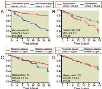
Fig. 6.Survival curves of patients according to total adrenal gland volume (# or more than 10 cm3) (A), basal (" or $ 32
!g/dl) (B), response plasma cortisol concentration (# or more
than 2.7!g/dl) (C) and response plasma cortisol
concentra-tion (" or $ 9!g/dl)8(D) in the validation 184 septic shock
patients (n ! 184). (A), Twenty-two of the 139 patients with total adrenal gland volume of 10 cm3or more died at day 28
compared with 37 of the 45 with total adrenal gland volume less than 10 cm3;P " 0.01. (To convert values for cortisol to
nM/l, multiply by 27.6).
Adrenal Gland Volume and Outcome in Septic Shock
group (n ! 40). Table 2 shows the comparison between survivors and nonsurvivors in the 184 patients with septic shock.
Adrenal Gland Volume in Control, Nonseptic, and Septic Shock ICU Groups
In the septic shock group, CT was performed 1 day [95% CI, 1–2 days] after the cosyntropin test, which was performed in the first 12 hours [95% CI, 8 to 16 h] after the administra-tion of vasopressors for septic shock.
Total adrenal gland volume of the 184 patients in the septic shock group (12.5 cm3[95% CI, 11.3–13.3])was sig-nificantly higher in comparison with the 15 patients in the nonseptic ICU group (8.0 cm3[95% CI, 6.8 –10.1]; P " 0.0001 for both comparison), and with the 40 patients in the control group (7.2 cm3[95% CI, 6.3– 8.5], P " 0.0001 for both comparison). There was no significant difference in total adrenal gland volume between the nonseptic ICU and control groups (table 1).
Among the 184 patients with septic shock who had a CT scan and cosyntropin test, 100 were also explored before
septic shock and had a medical checkup that could be con-sidered as a baseline value (time before septic shock onset: 123 days [95% CI, 26 – 618 days]). During septic shock, adrenal gland volume increased in comparison with this baseline value (12.4 cm3[95% CI, 11.3–13.3] vs. 8.8 cm3 [95% CI, 7.8 –9.6]; P " 0.0001).
Furthermore among those 100 patients, 16 survivors were explored with a third CT scan after ICU discharge (time after ICU discharge: 100 days [95% CI, 44 –214 days]) at the referent physician’s discretion for a systematic follow-up or for any reason other than sepsis. Interestingly, in those 16 survivors adrenal gland volume returned to baseline value (fig. 3).
Adrenal Gland Volume and Cosyntropin Test Results
There was no evidence for a correlation between adrenal gland volume measurement during septic shock and baseline cortisol concentration (r ! 0.09; [CI 95%, (0.11– 0.29],
P ! 0.36) and a weak correlation with the # cortisol (r !
0.34 [CI 95%, 0.15– 0.51], P " 0.001).
Adrenal Gland Volume, Cosyntropin Test Response and Outcome
In septic shock, total adrenal gland volume during shock was 13.8 cm3[95% CI, 13.0 –15.1] in the 125 survivors com-pared with 9.7 cm3[95% CI, 8.8 –9.9] (P " 0.0001) in the 59 nonsurvivors. In the 100 patients who had a CT scan before and during septic shock, adrenal glands volume in-creased more during shock in the survivors ('33 & 24%) than in the non survivors ('8 & 7%) (P " 0.0001) (fig. 4). In the 184 patients with septic shock, ROC analysis iden-tified a total adrenal gland volume of 10 cm3 as the best predictor of death with a sensitivity of 70% [95% CI, 56 – 81], a specificity of 92% [95% CI, 86 –96], a positive pre-dictive value of 82%, an accuracy of 84%, and an area under the ROC curve of 0.84 [95% CI, 0.78 – 0.89] (fig. 5). Using this threshold, the likelihood ratio of death in the ICU was 8.7 times greater if the total adrenal gland volume was less
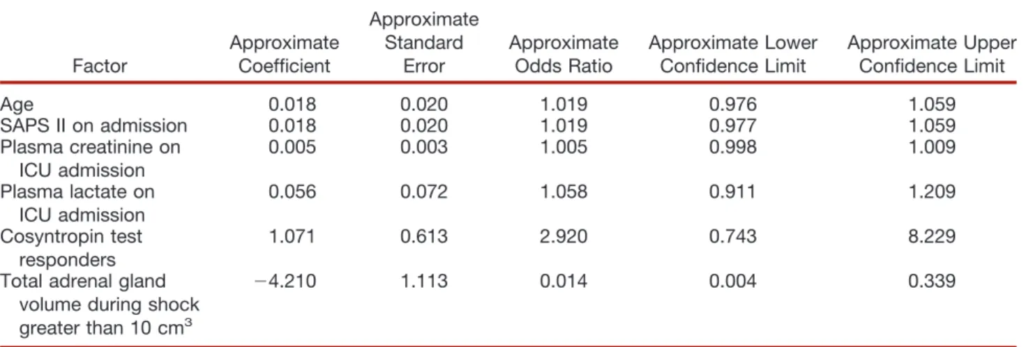
Table 3. Multivariate Logistic Regression Analysis of Risk Factors for Mortality in the 184 Septic Shock Patients
Factor Approximate Coefficient Approximate Standard Error Approximate Odds Ratio Approximate Lower Confidence Limit Approximate Upper Confidence Limit Age 0.018 0.020 1.019 0.976 1.059 SAPS II on admission 0.018 0.020 1.019 0.977 1.059 Plasma creatinine on ICU admission 0.005 0.003 1.005 0.998 1.009 Plasma lactate on ICU admission 0.056 0.072 1.058 0.911 1.209 Cosyntropin test responders 1.071 0.613 2.920 0.743 8.229 Total adrenal gland
volume during shock greater than 10 cm3
(4.210 1.113 0.014 0.004 0.339
ICU ! intensive care unit; SAPS II ! Simplified Acute Physiology Score.
Table 4. Mortality Comparisons in the 184 Septic Shock Patients by Combining Adrenal Gland Volume and Plasma Response Cortisol
Day 28 Mortality, n (%) Adrenal Gland Volume )10 cm3 (n ! 135) Adrenal Gland Volume #10 cm3 (n ! 49) P Value Plasma response cortisol )9 !g/dl 9/66 (14) 14/18 (78) "0.001 Plasma response cortisol #9!g/dl 7/69 (10) 21/31 (68) "0.001
P comparisons between adrenal gland volume higher and less
than 10 cm3. To convert values for cortisol to nM/l, multiply by
than 10 cm3. To better describe the ROC analysis, we also determined the cutoff value that resulted in the best sensitiv-ity (15 cm3allowing a sensitivity of 98% [95% CI, 91–99] but a specificity of 41% [95% CI, 32–50]) and in the best specificity (7.3 cm3allowing a specificity of 98% [95% CI, 93–99] but a sensitivity of 25% [95% CI, 15–38]).
The best cutoff value for baseline plasma cortisol concen-tration was 32 !g/dl with a sensitivity of 33% [95% CI, 20 – 47], a specificity of 79% [95% CI, 70 – 86] and an area
under the ROC curve of 0.52 [95% CI, 0.44 – 0.59] and 2.7 !g/dl for response cortisol concentration with a sensitivity of 44% [95% CI, 30 – 60], a specificity of 73% [95% CI, 63– 82] and an area under the ROC curve of 0.55 [95% CI, 0.46 – 0.63].
The area under the ROC curves was significantly higher for total adrenal gland volume than basal cortisol concentra-tion (P " 0.001) and than peak cortisol concentraconcentra-tion (P " 0.001). Figure 6 shows the survival curves in the sepsis vali-dation for total adrenal gland volume (# or more than 10 cm3) (fig. 6A), basal plasma cortisol concentration (# or more than 32 !g/dl) (fig. 6B), and response cortisol concen-tration (# or more than 2.7 !g/dl) (fig. 6C) and # cortisol (# or more than 9 !g/dl)9(fig. 6D). Total adrenal gland volume was strongly associated with 28-day survival (hazard ratio 5.0 [95% CI, 4.3–14.1]; P " 0.0001) (fig. 6A). Al-though Simplified Acute Physiology Score II, lactate, plate-lets, plasma creatinine, and total bilirubin concentration were associated with mortality on univariate analysis (table 2), multivariate analysis showed that adrenal gland volume was the sole independent risk factor of mortality at day 28 (table 3). Interestingly, 68% of the patients with # cortisol less than 9 !g/dl and a total adrenal gland volume of less than 10 cm3died before day 28, whereas only 14% of the patients with a total adrenal gland volume higher than 10 cm3and a # cortisol higher than 9 !g/dl died before day 28 (P " 0.0001) (table 4). Four representative patients are presented in figures 7 and 8.
In a multiple regression model including sex, age, Simpli-fied Acute Physiology Score upon ICU admission, admission category, sepsis source, and total hydrocortisone dose as in-dependent data and adrenal gland volume as the in-dependent data; male sex and total hydrocortisone dose were statistically associated with adrenal gland volume (table 5). There were 3 of 15 deaths (20%) at 28 days in the nonseptic ICU patient group. In this group, adrenal gland volume was not associ-ated with 28-day mortality.
Discussion
This study shows that, in septic shock, total adrenal gland volume measured by CT scan was an independent prognos-tic factor for 28-day mortality. Adrenal gland volume was nearly doubled in septic shock groups in comparison with the nonseptic ambulatory group, and was increased by 35% in comparison with the nonseptic ICU group. Second, in hydrocortisone–treated septic shock, the cosyntropin test re-sult was poorly correlated with adrenal gland volume. Fi-nally, by multivariate analysis, we report for the first time that total adrenal gland volume less than 10 cm3 during septic shock may represent a mortality risk factor.
Adrenal glands volume was evaluated with a semiauto-mated organ volumetric technique by a radiologist (SN) by CT and dedicated software (Myrian!, Intrasense, Montpellier, France). This software is widely available
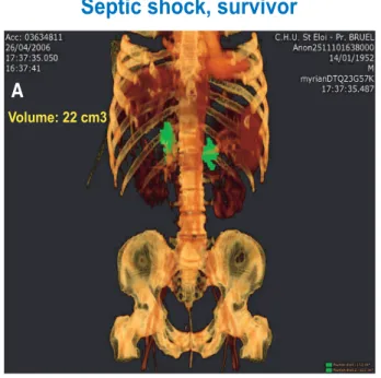
Volume: 22 cm3
Volume: 9.7 cm3
Septic shock, survivor
Septic shock, non survivor
A
B
Fig. 7. Representative patients included in this study with three-dimensional computed tomography reconstruction. (A) A patient who survived. His total adrenal gland volume
was 22 cm3, measured with a semiautomated, dedicated
software for volumetry measurement (Myrian", Intrasense, Montpellier, France). (B) A patient who died. His total adrenal
gland volume was 9.7 cm3.
Adrenal Gland Volume and Outcome in Septic Shock
and is certified for organ volume measurement. The use of semiautomated organ volumetry technique has become part of a routine process in radiology for various situa-tions, such as liver or gastric surgery,26but volumetry has been poorly studied in intensive care medicine27–29and was used only once for adrenal gland volume measure-ment, in a pilot study.20Adrenal gland volume has been previously shown to increase in stress conditions such as depression30and Cushing disease.31
In this study, adrenal gland volume was not correlated with basal cortisol but showed some correlation with cortisol response to cosyntropin. These findings suggested that mor-phologic assessment of the adrenal glands may provide addi-tional information on adrenal function to improve the diag-nosis of critical illness-related corticosteroid insufficiency, which remains challenging.11,12,15,32 In practice, patients with adrenal gland volume less than 10 cm3had higher risk of death (fig. 5 and 6). In the current study, every patient
Septic shock, survivor
Septic shock, non survivor
ICU patient, non septic
Control patient
A
B
C
D
E
F
Volume: 22 cm3 Volume: 9.7 cm3 Volume: 6.9 cm3 Volume: 7.3 cm3Fig. 8.Representative patients included in this study. (A and B) A patient who survived septic shock. His total adrenal gland
volume was 22 cm3. (C and D) A patient who died of septic shock. His total adrenal gland volume was 9.7 cm3. A control patient
(E) was explored by virtual colonoscopy as well as a nonseptic intensive care unit (ICU) patient (F). Total adrenal gland volume
with septic shock was treated with hydrocortisone. Multiple regression showed that a higher total dose of hydrocortisone was associated with a smaller adrenal gland (table 5). More than a very unlikely direct effect of a very short hydrocorti-sone treatment on adrenal gland,33this association may be interpreted as the sicker the patients, the longer they were treated with hydrocortisone and the smaller the adrenal gland volume.
The mechanisms leading to the increased volume did not include adrenal gland hemorrhage or necrosis. Furthermore, there was no sign of excess fluid loading between septic shock groups on CT (such as ascitis or bowel edema) and it is then unlikely that adrenal gland enlargement could be related to a gland edema. Interestingly, in 16 patients who had been evaluated before, during, and after recovery of sepsis, we were able to demonstrate that the morphologic changes in adrenal glands were reversible (fig. 3).
This study has some limitations. Although the external validity of this study may be limited because of its single center and observational design, we included all consecutive patients in whom a CT and a cosyntropin test were pre-scribed within the 48 h of septic shock onset, representing 40% of patients with septic shock (fig. 2). All patients with septic shock received hydrocortisone as part of routine man-agement. Therefore, we could not analyze the effect of this treatment on both adrenal morphology and patient outcome or speculate on the adrenal gland volume in patients with septic shock who were not treated with hydrocortisone. In this first study showing the association between adrenal gland volume and mortality, we did not include patients who did not have septic shock or those acutely treated with ste-roids, which could represent a limitation to our study. Fi-nally, although we clearly demonstrated a link between ad-renal gland volume and mortality, our study was not designed to explain the potential pathophysiologic pathways. However, according to the gland density profile similar to normal parenchyma, the radiologist did not observe any sign of gland edema, hemorrhage or necrosis.
In conclusion, our findings show that in septic shock, CT of the adrenal gland may contribute to assess adrenal func-tion and a total adrenal gland volume less than 10 cm3may identify high-risk patients. Further studies may evaluate the adrenal gland volume in association with the cosyntropin stimulation test in other populations at high risk of critical illness-related corticosteroid insufficiency such as brain
dead34or multiple trauma patients35and whether assessment of adrenal gland morphology on CT may identify patients likely to respond to corticosteroids.
The authors thank Christian Bonnel, M.D., Medical Coordinator, Intrasense Company, Montpellier, France, for the technological sup-port of CT volumetry (Myrian® Software, Montpellier, France) and Julie Carr, M.D., Fellow, Anesthesia and Critical Care Department, Saint Eloi University Hospital, Centre Hospitalier Universitaire Mont-pellier, MontMont-pellier, France, for her English editing.
References
1. Annane D, Bellissant E, Cavaillon JM: Septic shock. Lancet 2005; 365:63–78
2. Dellinger RP, Levy MM, Carlet JM, Bion J, Parker MM, Jae-schke R, Reinhart K, Angus DC, Brun-Buisson C, Beale R, Calandra T, Dhainaut JF, Gerlach H, Harvey M, Marini JJ, Marshall J, Ranieri M, Ramsay G, Sevransky J, Thompson BT, Townsend S, Vender JS, Zimmerman JL, Vincent JL: Surviving Sepsis Campaign: International guidelines for management of severe sepsis and septic shock: 2008. Crit Care Med 2008; 36:296 –327
3. Menon K, Ward RE, Lawson ML, Gaboury I, Hutchison JS, He´bert PC, Canadian Critical Care Trials Group: A prospec-tive multicenter study of adrenal function in critically ill children. Am J Respir Crit Care Med 2010; 182:246 –51 4. Bone RC, Balk RA, Cerra FB, Dellinger RP, Fein AM, Knaus
WA, Schein RM, Sibbald WJ: Definitions for sepsis and organ failure and guidelines for the use of innovative therapies in sepsis: The ACCP/SCCM Consensus Conference Committee. American College of Chest Physicians/Society of Critical Care Medicine 1992. Chest 2009; 136:e28
5. Wheeler AP: Recent developments in the diagnosis and man-agement of severe sepsis. Chest 2007; 132:1967–76 6. Annane D, Bellissant E, Bollaert PE, Briegel J, Confalonieri M,
De Gaudio R, Keh D, Kupfer Y, Oppert M, Meduri GU: Corticosteroids in the treatment of severe sepsis and septic shock in adults: A systematic review. JAMA 2009; 301: 2362–75
7. COIITSS Study Investigators, Annane D, Cariou A, Maxime V, Azoulay E, D’honneur G, Timsit JF, Cohen Y, Wolf M, Far-toukh M, Adrie C, Santre´ C, Bollaert PE, Mathonet A, Ama-thieu R, Tabah A, Clec’h C, Mayaux J, Lejeune J, Chevret S: Corticosteroid treatment and intensive insulin therapy for septic shock in adults: A randomized controlled trial. JAMA 2010; 303:341– 8
8. Annane D, Se´bille V, Charpentier C, Bollaert PE, Franc¸ois B, Korach JM, Capellier G, Cohen Y, Azoulay E, Troche´ G, Chaumet-Riffaud P, Chaumet-Riffaut P, Bellissant E: Effect of treatment with low doses of hydrocortisone and fludrocor-tisone on mortality in patients with septic shock. JAMA 2002; 288:862–71
9. Annane D, Se´bille V, Troche´ G, Raphae¨l JC, Gajdos P, Bellis-sant E: A 3-level prognostic classification in septic shock based on cortisol levels and cortisol response to corticotro-pin. Jama 2000; 283:1038 – 45
10. Jaeschke R, Angus DC: Living with uncertainty in the inten-sive care unit: Should patients with sepsis be treated with steroids? JAMA 2009; 301:2388 –90
11. Marik PE: Critical illness-related corticosteroid insufficiency. Chest 2009; 135:181–93
12. Annane D, Maxime V, Ibrahim F, Alvarez JC, Abe E, Boudou P: Diagnosis of adrenal insufficiency in severe sepsis and septic shock. Am J Respir Crit Care Med 2006; 174:1319 –26 13. Bollaert PE, Charpentier C, Levy B, Debouverie M, Audibert G, Larcan A: Reversal of late septic shock with supraphysi-ologic doses of hydrocortisone. Crit Care Med 1998; 26: 645–50
Table 5. Multiple Regression Analysis Examining Adrenal Gland Volume as the Independent Variable
Factor Coefficient
Standard
Error P Value
Total hydrocortisone dose (0.0014 0.00067 0.04 Male 3.37 0.97 "0.01 Other variables entered in the model were age, medicalversus
surgical, site of sepsis (lung, abdominal, other), and SAPS II (Simplified Acute Physiology Score).
Adrenal Gland Volume and Outcome in Septic Shock
14. Briegel J, Forst H, Haller M, Schelling G, Kilger E, Kuprat G, Hemmer B, Hummel T, Lenhart A, Heyduck M, Stoll C, Peter K: Stress doses of hydrocortisone reverse hyperdynamic sep-tic shock: A prospective, randomized, double-blind, single-center study. Crit Care Med 1999; 27:723–32
15. Marik PE, Pastores SM, Annane D, Meduri GU, Sprung CL, Arlt W, Keh D, Briegel J, Beishuizen A, Dimopoulou I, Tsa-garakis S, Singer M, Chrousos GP, Zaloga G, Bokhari F, Vogeser M, American College of Critical Care Medicine: Recommendations for the diagnosis and management of cor-ticosteroid insufficiency in critically ill adult patients: Con-sensus statements from an international task force by the American College of Critical Care Medicine. Crit Care Med 2008; 36:1937– 49
16. Rivers EP, Gaspari M, Saad GA, Mlynarek M, Fath J, Horst HM, Wortsman J: Adrenal insufficiency in high-risk surgical ICU patients. Chest 2001; 119:889 –96
17. Briegel J, Sprung CL, Annane D, Singer M, Keh D, Moreno R, Mo¨hnle P, Weiss Y, Avidan A, Brunkhorst FM, Fiedler F, Vogeser M, CORTICUS Study Group: Multicenter comparison of cortisol as measured by different methods in samples of patients with septic shock. Intensive Care Med 2009; 35: 2151– 6
18. Sprung CL, Annane D, Keh D, Moreno R, Singer M, Freivogel K, Weiss YG, Benbenishty J, Kalenka A, Forst H, Laterre PF, Reinhart K, Cuthbertson BH, Payen D, Briegel J, CORTICUS Study Group: Hydrocortisone therapy for patients with sep-tic shock. N Engl J Med 2008; 358:111–24
19. Chanques G, Annane D, Jaber S, Gallix B: Enlarged adrenals during septic shock. Intensive Care Med 2007; 33:1671–2 20. Nougaret S, Jung B, Aufort S, Chanques G, Jaber S, Gallix B:
Adrenal gland volume measurement in septic shock and control patients: A pilot study. Eur Radiol 2010; 20:2348 –57 21. Ray P, Le Manach Y, Riou B, Houle TT: Statistical evaluation
of a biomarker. ANESTHESIOLOGY2010; 112:1204 –10 22. Bossuyt PM, Reitsma JB, Bruns DE, Gatsonis CA, Glasziou PP,
Irwig LM, Lijmer JG, Moher D, Rennie D, de Vet HC, Stan-dards for Reporting of Diagnostic Accuracy: Towards com-plete and accurate reporting of studies of diagnostic accu-racy: The STARD Initiative. Ann Intern Med 2003; 138:40 – 4 23. Bone RC, Balk RA, Cerra FB, Dellinger RP, Fein AM, Knaus WA, Schein RM, Sibbald WJ: Definitions for sepsis and organ failure and guidelines for the use of innovative therapies in sepsis: The ACCP/SCCM Consensus Conference Committee. American College of Chest Physicians/Society of Critical Care Medicine. Chest 1992; 101:1644 –55
24. Le Gall JR, Lemeshow S, Saulnier F: A new Simplified Acute
Physiology Score (SAPS II) based on a European/North Amer-ican multicenter study. Jama 1993; 270:2957– 63
25. Lefrant JY, Muller L, Raillard A, Jung B, Beaudroit L, Favier L, Masson B, Dingemans G, The´venot F, Selcer D, Jonquet O, Capdevila X, Fabbro-Peray P, Jaber S, Sepsi d’Oc Study Group in the AzuRe´a Group: Reduction of the severe sepsis or septic shock associated mortality by reinforcement of the recommendations bundle: A multicenter study. Ann Fr Anesth Reanim 2010; 29:621– 8
26. Parrish FJ: Volume CT: State-of-the-art reporting. AJR Am J Roentgenol 2007; 189:528 –34
27. Constantin JM, Grasso S, Chanques G, Aufort S, Futier E, Sebbane M, Jung B, Gallix B, Bazin JE, Rouby JJ, Jaber S: Lung morphology predicts response to recruitment maneuver in patients with acute respiratory distress syndrome. Crit Care Med 2010; 38:1108 –17
28. Malbouisson LM, Muller JC, Constantin JM, Lu Q, Puybasset L, Rouby JJ, CT Scan ARDS Study Group: Computed tomog-raphy assessment of positive end-expiratory pressure-in-duced alveolar recruitment in patients with acute respiratory distress syndrome. Am J Respir Crit Care Med 2001; 163: 1444 –50
29. Terragni PP, Rosboch G, Tealdi A, Corno E, Menaldo E, Davini O, Gandini G, Herrmann P, Mascia L, Quintel M, Slutsky AS, Gattinoni L, Ranieri VM: Tidal hyperinflation during low tidal volume ventilation in acute respiratory dis-tress syndrome. Am J Respir Crit Care Med 2007; 175:160 – 6 30. Amsterdam JD, Marinelli DL, Arger P, Winokur A: Assessment of adrenal gland volume by computed tomography in de-pressed patients and healthy volunteers: A pilot study. Psy-chiatry Res 1987; 21:189 –97
31. Pojunas KW, Daniels DL, Williams AL, Thorsen MK, Haugh-ton VM: Pituitary and adrenal CT of Cushing syndrome. AJR Am J Roentgenol 1986; 146:1235– 8
32. Hamrahian AH, Oseni TS, Arafah BM: Measurements of serum free cortisol in critically ill patients. N Engl J Med 2004; 350:1629 –38
33. Arlt W, Allolio B: Adrenal insufficiency. Lancet 2003; 361: 1881–93
34. Nicolas-Robin A, Barouk JD, Amour J, Coriat P, Riou B, Langeron O: Hydrocortisone supplementation enhances he-modynamic stability in brain-dead patients. ANESTHESIOLOGY
2010;112: 1204 –10
35. Hoen S, Asehnoune K, Brailly-Tabard S, Mazoit JX, Benhamou D, Moine P, Edouard AR: Cortisol response to corticotropin stimulation in trauma patients: Influence of hemorrhagic shock. ANESTHESIOLOGY2002; 97:807–13