UPDATE / MISE AU POINT NEUROLOGIE-URGENCES
Hyperbaric Emergencies and Decompression Illness
Urgences hyperbares et maladie de décompression
C.-M. Muth · P. Radermacher
Received: 13 April 2015; Accepted: 04 June 2015 © SRLF et Lavoisier SAS 2015
Abstract Decompression illness (DCI) is the most important emergency resulting from exposure to hyperbaric environ-ments. Immediate diagnosis and management mostly allow for complete recovery without sequelae. Emergency and critical care physicians need to be aware of the possible symptoms, since there are no DCI-specific symptoms, and the clinical presentation can simulate a variety of other pathologies. Initial treatment of choice is the administration of pure oxygen together with adjunctive measures such as fluid expansion and eventually symptomatic medication, followed by hyperbaric oxygen (HBO) treatment. Patient management under hyperbaric conditions requires taking into account the physics of hyperbarism to avoid any procedure-related pitfalls.
Keywords Decompression illness · Pulmonary barotrauma · Arterial gas embolism · Decompression sickness ·
Hyperbaric oxygen therapy · Diving
Résumé La maladie de décompression représente l’urgence la plus importante liée à l’exposition au milieu hyperbare. En général, le diagnostic immédiat, ainsi que la prise en charge rapide permettent au malade de récupérer sans aucune séquelle. L’urgentiste et le réanimateur doivent connaître les signes cliniques possibles, car il n’y en a aucun de spé-cifique, et les signes cliniques peuvent être trompeurs. La prise en charge initiale comprend l’administration de l’oxy-gène pur, l’expansion volémique, un traitement réanimatoire symptomatique, ainsi qu’éventuellement une oxygénothéra-pie hyperbare (OHB). Pour l’OHB, le médecin doit connaî-tre les aspects pratiques et les gestes imposés par les lois
physiques de l’hyperbarie, afin d’éviter des complications liées au milieu hyperbare.
Mots clés Maladie de décompression · Surpression pulmonaire · Embolie gazeuse artérielle · Oxygénothérapie hyperbare · Plongée
Introduction
The most important hyperbaric emergency, Decompression illness (DCI), occurs as a result of decrease in ambient pres-sure in persons that have been exposed to an elevated ambient pressure such as compressed air workers and divers. DCI encompasses both decompression sickness (DCS), which is caused by tissue bubble formation due to super-saturation of inert gases, and arterial gas embolism (AGE) caused by entry of gas into blood vessels during a rapid decompression due to pulmonary gas trapping and alveolar rupture. Therefore the common characteristic is tissue damage by excess gas during and after decompression. The most severe clinical expression corresponds to severe neurological symptoms, requiring both intensive care and hyperbaric O2(HBO) [1–3]. DCI treatment
is still empiric, as there are no randomized controlled trials on the best treatment protocols. Nevertheless, albeit evidence-based-medicine standards are missing, there is an interna-tional consensus on the current treatment guidelines [3–5].
Mechanisms of decompression illness
The large majority of DCI cases are related to diving with self compressed underwater breathing apparatus (“scuba”), since this leisure activity has become very popular. During diving and compressed air working, body tissues saturate with N2(or any other inert gas used as a component of the
breathing gas mixture, e.g. He), at the elevated ambient pres-sure. This is according to Henry’s law, which describes the proportional relationship between the amount of physically
C.-M. Muth (*)
Sektion Notfallmedizin, Klinik für Anästhesiologie, Universitätsklinikum Universitätsklinikum, Prittwitzstraße 43, 89075 Ulm, Germany e-mail : [email protected] P. Radermacher
Institut für Anästhesiologische Pathophysiologie und Verfahrensentwicklung, Universitätsklinikum, Ulm, Germany DOI 10.1007/s13546-015-1091-1
dissolved gas in a liquid and the partial pressure of that gas above the liquid (Table 1).
The decrease in ambient pressure during decompression leads to a decrease of the partial pressure of the gases of the breathing gas and the additional inert gas is eliminated from the tissues. With rapid decompression the decrease in ambi-ent pressure may exceed the elimination rate of N2, resulting
in tissue super-saturation and, as a consequence, in the for-mation of free gas bubbles.
Inert gas bubbles precipitate DCS, but the disorder is not simply related to the presence of such bubbles. Indeed, it is known that in many divers bubble formation will not lead to symptoms. Normally, and if the amount of such bubbles do not exceed a critical number, the venous system transports these bubbles to the lungs, where they are eliminated. DCS arises when gas bubbles cause mechanical tissue compres-sion or venous embolization. Finally, paradoxical gas embo-lism may occur through trans-pulmonary passage of venous gas bubbles or via extra- or, to a smaller portion, intrapul-monary right-to-left shunts. A patent foramen ovale (PFO) is the most common pathway in divers [6].
A prerequisite for arterial gas embolism is the entry of gas into the pulmonary veins or the arteries of the systemic circulation. Mechanisms include the overexpansion of the lung through decompression barotrauma, and paradoxical embolism through the presence of a PFO as stated above.
In pulmonary barotrauma the volume of an enclosed gas will increase according to Boyle’s law as ambient pressure
decreases. When expanding, intrapulmonary gas, which was inhaled under higher pressure, is not adequately exhaled dur-ing decompression, and airway over-distension can cause pulmonary barotrauma. Depending on the site of tissue rup-ture, gas may track along the peri-vascular sheaths and cause mediastinal emphysema or pneumothorax. Gas may also pass into the pulmonary vessels with subsequent arterial gas embolism into the cerebral or, even, in rare cases, into the coronary circulation.
Physiopathology
Initial changes mainly are due to mechanical effects of the bubble. Gas bubbles can result in tissue ischaemia caused by vascular obstruction or tissue compression due to the expanding volume of the bubbles. Inflammatory processes and tissue damage lead to oedema. Consequently, increased diffusion distances impeding gas elimination aggravate the damage. This largely depends on the kinetics of the gas con-tained within the bubble and the size and location of the embolus. Interactions between the blood-gas interface and the endothelium result in further tissue damage, mediated by activation of complement, platelets and neutrophils. These secondary effects trigger an inflammatory cascade ultimately causing endothelial damage with capillary leakage, fluid loss from the intravascular space, and haemoconcentration.
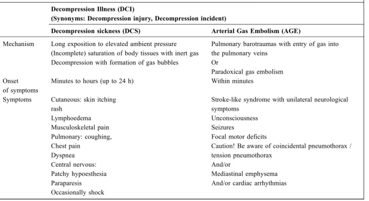
Table 1 Mechanisms and Symptoms of DCI Decompression Illness (DCI)
(Synonyms: Decompression injury, Decompression incident)
Decompression sickness (DCS) Arterial Gas Embolism (AGE) Mechanism Long exposition to elevated ambient pressure
(Incomplete) saturation of body tissues with inert gas Decompression with formation of gas bubbles
Pulmonary barotraumas with entry of gas into the pulmonary veins
Or
Paradoxical gas embolism Onset
of symptoms
Minutes to hours (up to 24 h) Within minutes
Symptoms Cutaneous: skin itching rash Lymphoedema Musculoskeletal pain Pulmonary: coughing, Chest pain Dyspnea Central nervous: Patchy hypoesthesia Paraparesis Occasionally shock
Stroke-like syndrome with unilateral neurological symptoms
Unconsciousness Seizures
Focal motor deficits
Caution! Be aware of coincidental pneumothorax / tension pneumothorax
And/or
Mediastinal emphysema And/or cardiac arrhythmias
Clinical manifestations and presentation
As stated earlier, the large majority of DCI occurs during scuba diving. During a rapid ascent with large pressure changes, arterial gas embolism from pulmonary barotrauma is a possible complication accounting for recreational scuba-diving fatalities [2]. Severe DCS is most common after short, deep scuba-dives or multiple dives over several days but may occur after any decompression when there is a signifi-cant venous gas load, especially in the presence of right-to-left shunts [7]. A further decrease of ambient pressure, as it occurs during long distance commercial flights after diving also increases the risk for DCS [8].
The symptoms of DCS can vary in a wide range from mild to severe, ranging from skin itching, rash and lymphoe-dema and vague constitutional symptoms or musculoskeletal pain to patchy hypoesthesia and paraparesis, or even shock and cardiopulmonary arrest. The central nervous nitrogen saturation and elimination kinetics and its limited ischemia tolerance favor the development of neurological symptoms. Sensory symptoms including numbness, tingling, paraesthe-siae, and abnormal sensation are more common than severe neurological symptoms, that typically develop progres-sively, beginning with mild paraesthesia, followed by regional numbness, weakness and, occasionally, paresis of the affected limbs [1]. Symptoms usually occur within hours after decompression but may also present immedi-ately. Respiratory DCS presenting with coughing, chest pain, dyspnea, and haemoptysis may occur when a high venous gas load overwhelms the pulmonary bubble filter. Inner ear DCS presents as vertigo, tinnitus and hearing loss [9]. In contrast to DCS, arterial gas embolism typically pre-sents as a stroke-like syndrome with unilateral neurologi-cal symptoms, depending on the affected areas of the brain. Cognitive symptoms and unconsciousness are most frequently observed. Seizures, focal motor deficits, visual disturbance and sensory changes are also common [10]. Bubbles may, however, occlude any artery, including the coronary or skeletal muscle vessels. Spinal cord lesions with sensory or motor paraplegia are more likely to result from DCS. Arterial gas embolism and DCS can be discrimi-nated according to the onset of symptoms, with gas embo-lism predominantly developing within a few minutes after or even during decompression. Nevertheless, symptoms of arterial gas embolism may be indistinguishable from DCS or even non-diving related disorders. Finally, severe neuro-logical DCS may be superimposed on gas embolism.
Diagnostic evaluation
The outcome of especially severe DCI largely depends on the time to treatment, so definitive therapy should start as early as
possible. Furthermore, no specific tests are available [11]. With respect to this the term DCI is useful, as it allows assign-ing a diagnosis without differentiatassign-ing between arterial gas embolism and DCS. History and physical as well as orientat-ing neurological examination are mandatory for the initial assessment. The onset of the patient’s complaints related to a decompression, including information on time to onset of symptoms, is crucial. In hospital, laboratory investigations are useful to evaluate haemoconcentration and dehydration that will occur as a result of the pathophysiological processes and serum creatine kinase activity may serve as a marker of the severity of arterial gas embolism. A chest X-ray allows eval-uating the presence of a pneumothorax, which also can be the result of a pulmonary barotrauma. Because of the severe danger of an evolving tension pneumothorax during decom-pression from hyperbaric oxygen therapy, a drainage prior to recompression is mandatory [12].
Treatment of DCI
As stated above, treatment for DCI relies on empirical evi-dence and for obvious reasons, no controlled prospective studies in humans are available comparing treatment with “no treatment” [4,5]. It is international consensus that treat-ment has to start as early as possible with the aim of a rapid elimination of the dissolved gas and the correction of hyp-oxia, which is best achieved by hyperbaric oxygen therapy [1,3,12] but can start with normobaric oxygen administered in a concentration as high as available (Table 2).
Emergency Treatment
The protection and maintenance of vital functions is the pri-mary goal. If necessary, cardio-pulmonary resuscitation has to be performed. Indeed the venous but also the primary arterial gas embolism may lead to serious impairment of the cardio-vascular system.
Furthermore, empirical data and animal studies show that early normobaric hyperoxia with inspiratory O2
concentra-tions close to 100% improves clinical outcome, because it prevents further inert gas uptake and increases the diffusion gradient of inert gas from the bubble into the tissue. There-fore 100% O2 should be administered via a tightly fitting
face mask, either from a demand-valve regulator or by a closed-circuit apparatus. For somnolent or comatose patients endotracheal intubation should be performed to maintain adequate oxygenation and ventilation.
In addition, fluid resuscitation is useful to counteract hae-moconcentration and dehydration [13]. Divers are prone to dehydration because of fluid loss through respiration and increased diuresis during the scuba dive, due to an increase in intrathoracic blood volume, induced by the immersion.
Intravascular accumulation of gas bubbles and subsequent endothelial damage with capillary leakage aggravate dehy-dration. Depending on the patient’s level of consciousness, mild DCI may be managed by oral fluids; otherwise, intra-venous administration is recommended [10,12]. Adequate fluid resuscitation will allow continued inert gas washout from tissues by maintening the microvascular flow.
Patients with DCI should be kept supine. Temperature control of the patient is necessary as hypothermia impairs tissue nitrogen elimination due to peripheral vasconstriction. Frequently, patients require transport from remote locations. Since altitude exposure may worsen the symptoms due to additional N2release and bubble growth under reduced
pres-sure, transport should take place in airplanes maintaining sea level cabin pressure. Ground or helicopter transportation at a flight level as low as considered“safe” by the pilot are pref-erable, with hyperbaric oxygen therapy given at the nearest location possible.
Definitive Treatment
The definitive treatment currently is a recompression with hyperbaric oxygen in a pressure chamber. The expected
effects of recompression are: reduction of the volume of gas bubbles, with restoration of tissue structure and blood flow; reabsorption of bubbles inert gas; increase oxygen delivery to the tissues.
Recompression reduces excess intracorporeal gas and increases the driving force for its return into solution. How-ever, recompression per se causes only limited bubble shrink-age, especially as gas emboli do not maintain a spherical shape when entrapped in vessels. Hyperbaric oxygen therapy accelerates gas elimination, both by raising the ambient pres-sure and by creating systemic hyperoxia. Hyperbaric oxygen therapy requires patient placement in a pressure chamber with a treatment pressure at 2-3 times sea level while breathing 100% O2. This usually results in an increase of the arterial
PO2up to about 260 to 270 kPa and an amount of O2
physi-cally dissolved in the blood of approximately 60 to 70 ml/L. The improved outcome of DCI after hyperbaric oxygen treat-ment results from its physiological effects, i.e. an increased diffusion gradient for O2into the gas bubble and for N2out of
the bubble, improved tissue O2delivery, hyperoxic
vasocon-striction, and inhibition of ß2-integrin-dependent neutrophil
adherence.
It is mandatory to start the treatment as early as possible, as recovery from DCI is seen to be inversely related to the time of initiation of hyperbaric oxygen therapy [11]. Most improvement occurs when treatment is started within min-utes, although improvement was still observed when treat-ment began hours later or in rare cases even a day later. Repetitive recompression treatments should be considered as long as there is clinical improvement, but the more hyper-baric treatments are needed to relieve symptoms, the less likely they are to be effective.
The most common treatment algorithm is the so called “U.S. Navy treatment Table 6” that comprises cycles of 100% O2breathing at 18 m sea water (0.28 MPa) and 9 m
sea water (0.19 MPa) with a total recompression time of approximately 4 hours 45 minutes [1,14]. There is an ongo-ing discussion on the best recompression regimen, and vari-ous other treatment algorithms are discussed, e.g. recom-pression to 50 m sea water while breathing O2-enriched
gas mixtures using N2(“nitrox”) or He (“heliox”) as carrier
gas has been recommended for arterial gas embolism and severe cases of DCS [14]. However, no clear advantage has been shown yet.
Adjunctive Measures
There are scarce data on the efficacy of pharmacologic inter-ventions [4,5]. Nevertheless, aspirin is frequently used for its analgesic, anti-inflammatory and anti-platetet aggregating properties [15]. Anticoagulants have been advocated to counteract haemoconcentration and coagulopathy. Low-dose heparin or low-molecular-weight heparin may be
Table 2 Treatment recommendations for DCI Initial treatment: Patient History
Physical examination Flat supine position CPR, if neccessary
Insufflation of 100% inspired oxygen Intubation, when unconscious Intravenous line
Infusion therapy
If pneumothorax is present: Insertion of a chest tube
If general seizures occur (AGE): Diazepam up to 30 mg or midazolam up to 10 mg; if no success: Barbiturates!
HBO-T as soon as possible ! In hospital treatment Continuing of insufflation of 100% inspired oxygen Foley catheter Renal protection
Exclusion of pneumothorax or Insertion of a chest tube, if pneumothorax is present and was not performed at scene
If patient is intubated: perform myringotomia in both ears HBO-T as soon as possible ! Definite
treatment
given in patients with leg weakness due to DCI as a prophy-laxis against deep vein thrombosis [12]. Corticosteroids have been recommended for arterial gas embolism to coun-teract brain oedema. However, cerebral arterial gas embo-lism provokes cytotoxic brain oedema, which, in general, is unresponsive to corticosteroids. Again, no study to date is available. The non-steroidal anti-inflammatory drug tenoxicam reduced the number of recompressions required [4], but further studies are needed. In an animal study lido-caine improved neuronal recovery after cerebral air embo-lism and one single trial showed cerebral protection in car-diac surgery, with bolus lidocaine administration (1.5 mg/ kg) and maintaining a therapeutic concentration thereafter, but it should kept in mind that an overdose may result in severe neurological and cardiac side effects.
Intensive care in the hyperbaric environment
The management of critically ill patients in a hyperbaric chamber differs largely from the emergency department or the intensive care unit. The narrow space, noise, decreased ambient lighting and altered sound transmission impair clini-cal observation and give limited access [12,16]. For ECG and invasive blood pressure monitoring, electrical connections are necessary across the chamber wall to the outside physiologi-cal monitor [12,16]. While transcutaneous pulse oximetry is of only limited value due to hyperoxia, transcutaneous PO2
sensors may provide information on adequate tissue oxygen-ation. Intravenous lines should be placed prior to hyperbaric oxygen treatment since the hyperbaric chamber environment complicates insertions. As stated above, any untreated pneu-mothorax contraindicates hyperbaric exposure, since it will result in a life-threatening tension pneumothorax during decompression due to gas expansion [12,16]. Therefore, a chest tube must be inserted, and consequently the appropriate instruments must be readily available inside the chamber. During hyperbaric therapy chest tubes should be removed from vacuum drainage and a Heimlich valve inserted [16].
Cardiopulmonary resuscitation can be performed during hyperbaric therapy, but the medical staff is exposed to increased tissue N2uptake resulting in an increased risk of
the occurrence of decompression problems, when the cham-ber pressure is decreased. Cardiac defibrillation is accom-plished with a significant risk of catastrophic fire due to the elevated PO2of the pressurized chamber atmosphere. It
is therefore strongly recommended to avoid electric defibril-lation inside the chamber and slowly decompress with the attendants breathing 100% O2 from 9 m sea water until
reaching surface pressure [12,16].
For fluid administration by gravity, plastic containers should be used and have to be vented. The use of glass bot-tles can result in massive gas embolism during decompres-sion. Flow-controlled automatic infusion pumps can be used
when equipped with a battery as a power supply, but may show substantial variations in performance and accuracy under hyperbaric conditions [17].
Patients should be sedated and, if indicated, intubated before therapy starts and the chamber pressurized. Total intravenous anesthesia is the method of choice. Air has to be evacuated from the endotracheal cuff prior to hyperbaric exposure, and the cuff must be filled with an equivalent amount of liquid (e.g. distilled water) to achieve an appro-priate seal [12,16]. The endotracheal tube must be tightly secured and stabilized in place with documentation of its depth and auscultation of bilateral breath sounds. Unin-tended tube displacement may cause bronchial obstruction and subsequent over-inflation of the unvented part of the lung with consecutive barotrauma and/or a sudden drop in blood pressure due to decreased venous return [12,16].
Intubated patients sometimes need myringotomy or, in case of repeated hyperbaric treatments, tympanostomy, as they are unable to actively equilibrate their middle ear by the Valsalva manoeuvre. Patients with a nasal endotracheal tube can suffer from barotrauma of the sinuses during compression [16].
Ventilated patients will frequently require deep sedation or even muscle relaxation because of limited options in terms of ventilation mode. A pressure-controlled ventilatory mode is preferable when controlled ventilation is used to avoid over-estimation of tidal volume and minute ventilation (most ventilators use mass-flow measurements); a higher level of inspiratory pressure support is often needed to com-pensate for increased work of breathing resulting from the compression-related rise in gas density [16,18–20].
Conclusion
Decompression illness is an emergency situation resulting from a decompression after an exposition to hyperbaric envi-ronment, as it can be observed in divers. Clinical manifesta-tions are decompression sickness and arterial gas embolism. The rapid recognition of the related symptoms to evoke the diagnosis in a compatible context is of great importance in order to start the treatment as soon as possible. This treat-ment is not well codified but relies on non specific intensive care measures and on recompression using hyperbaric oxy-gen therapy, which needs to organize the transport of the patients in centers equipped with recompression chambers. Link of interest: None.
References
1. Vann RD, Butler FK, Mitchell SJ, Moon RE (2010) Decompres-sion Illness. Lancet 377:153–67
2. Tetzlaff K, Reuter M, Leplow B, et al (1997) Risk factors for pulmonary barotrauma in divers. Chest 112:654–9
3. Vann RD, Butler FK, Mitchell SJ, Moon RE (2011) Decompres-sion illness. Lancet 377:153–64
5. Bennett MH, Lehm JP, Mitchell SJ, Wasiak J (2012) Recompres-sion and adjunctive therapy for decompresRecompres-sion illness. Cochrane Database of Systematic Reviews 5:CD005277
4. Bennett MH, Lehm JP, Mitchell SJ, Wasiak J (2010) Recompres-sion and adjunctive therapy for decompresRecompres-sion illness: a systematic review of randomized controlled trials. Anesth Analg 111:757–62 6. Gempp E, Blatteau JE, Stephant E, Louge P (2009) Relation
between right-to-left shunts and spinal cord decompression sick-ness in divers. Int J Sports Med 30:150–3
7. Wilmshurst P, Bryson P (2000) Relationship between the clinical features of neurological decompression illness and its causes. Clin Sci 99:65–75
8. Freiburger JJ, Denoble PJ, Pieper CF, et al (2002) The relative risk of decompression sickness during and after air travel follow-ing divfollow-ing. Aviat Space Environ Med 73:980–4
9. Klingmann C (2012) Inner ear decompression sickness in compressed-air diving. Undersea Hyperberb Med 39:589–94 10. Muth CM, Shank ES (2000) Gas embolism. N Engl J Med
342:476–82
11. Ball R (1993) Effect of severity, time to recompression with oxy-gen, and re-treatment on outcome in forty-nine cases of spinal cord decompression sickness. Undersea Hyperb Med 20:133–45
12. Tetzlaff K, Shank ES, Muth CM (2003) Evaluation and manage-ment of decompression illness – An intensivists’ perspective. Intensive Care Med 29:2128–36
13. Boussuges A, Blanc P, Molenat F, et al (1996) Haemoconcentra-tion in neurological decompression illness. Int J Sports Med 17:351–5
14. Antonelli C, Franchi F, Della Marta ME, et al (2009) Guiding principles in choosing a therapeutic table for DCI hyperbaric therapy. Minerva Anesthesiol 75:151–61
15. Bessereau J, Coulange M, Genotelle N, et al (2008) Place de l ’as-pirine dans le traitement médicamenteux de l’accident de désa-turation. Thérapie 63:419–23
16. Muth CM, Radermacher P, Shank ES (2002) When HBO meets the ICU– intensive care patients in the hyperbaric environment. In Bakker DJ, Cramer FS (eds) Hyperbaric Surgery, pp 111–58 (Flagstaff: Best Publishing)
17. Lavon H, Shupak A, Tal D, et al (2002) Performance of infusion pumps during hyperbaric conditions. Anesthesiology 96:849–54 18. Blanch PB, Desaultes DA, Gallagher TJ (1991) Deviations in
function of mechanical ventilators during hyperbaric compres-sion. Respiratory Care 36:808–14
19. Lefebvre JC, Lyazidi A, Parceiro M, et al (2012) Bench testing of a new hyperbaric chamber ventilator at different atmospheric pressures. Intensive Care Med 38:1400–4
20. Stahl W, Radermacher P, Calzia E (2000) Functioning of ICU ven-tilators under hyperbaric conditions— comparison of volume — and pressure-controlled modes. Intensive Care Med 26:442–8