Partial block in B lymphocyte development
at the transition into the pre-B cell receptor
stage in V
pre-B1
-deficient mice
Annica Mårtensson, Yair Argon
1, Fritz Melchers
2, Jeanne L. Dul
1and
Inga-Lill Mårtensson
Immunology Unit, Department of Cell and Molecular Biology, University of Lund, Box 7031, 220 07 Lund, Sweden
1Department of Pathology and the Committee on Immunology, University of Chicago, IL 60637, USA 2Basel Institute for Immunology, Basel, Switzerland
Keywords: deficiency,λ5, pre-B cell receptor, surrogate light chain, Vpre-B
Abstract
The surrogate light chain (SL) is composed of two polypeptides, Vpre-Bandλ5. In large pre-BII cells the SL chain associates with Igµheavy chain (µH) to form the pre-B cell receptor (pre-BCR). In mice there are two Vpre-Bgenes which are 98% identical within the coding regions. The two genes are co-expressed at the RNA level and encode functional proteins that can assemble withλ5. However, it is not known whether both gene products serve the same functionin vivo. Here we have established mice that lack the Vpre-B1gene (VpreB1–/–), but still express the Vpre-B2gene, both as RNA and protein. In Vpre-B1–/–mice, the bone marrow cellularity and the percentage of B220F cells is normal. However, among the B220Fcells, the percentage of pre-BI cells is increased, and the percentage of pre-BII and immature B cells is slightly decreased, suggesting that the lack of Vpre-B1causes a partial block at the transition from pre-BI to pre-BII cells, i.e. into the pre-BCR stage. The number of cells that produce a functional pre-BCR is thus lower, but the cells that reach this stage are normal as they can be expanded by proliferation and then differentiate into more mature cells. The spleens of Vpre-B1homozygous mutant mice show normal numbers of B and T lymphocytes. Moreover, the Ig loci are allelicly excluded and the homozygous mutant mice respond with normal levels of antigen-specific antibodies to T-dependent antigens. These results
demonstrate that Vpre-B2alone is capable of supporting B lymphocyte development in the bone marrow and can give rise to immuno-competent cells in the periphery.
Introduction
Theλ5 and Vpre-Bgenes are expressed early in B lymphocyte
development, i.e. in pro-B/pre-BI and large pre-BII cells (1– 6). Together they encode the surrogate light chain (SL) chain, which has distinct homologies to conventional Ig chains (7). In pro-/pre-BI cells the SL chain associates with a complex of glycoproteins; however, its function at this stage of develop-ment is not yet clear (8). In the large pre-BII cell stage the SL chain associates with the Igµheavy (µH) chain and forms the pre-B cell receptor (pre-BCR) (9,10). Experiments in which either the transmembrane part ofµH or theλ5 gene has been disrupted show that the pre-BCR affects the expansion of large pre-BII cells into more mature B cell compartments (11,12). In mice, theλ5 chain is encoded by a single gene, whereas
Correspondence to: I.-L. Mårtensson, The Babraham Institute, Cambridge CB2 4AT, UK
Transmitting editor: K. Takatsu Received25 May 1998,accepted24 November 1998
the Vpre-B chain is encoded by two genes (3,13). The two
Vpre-Bgenes are almost identical; only 9 bp difference in the
coding regions causing a difference of 4 amino acids between the two polypeptides (3,7). On the mRNA level, expression of the Vpre-B1gene has been detected in all BI and
pre-BII bone marrow cells, while Vpre-B2was only found in some
of the same pre-BI and pre-BII cells. This, however, might be due to a difference in detection levels (6). Both genes encode functional proteins that can assemble withλ5 when expressed in COS cells (6). Due to the high degree of homology it is not known if both Vpre-Bproteins are present in BI and
pre-BII cells. In this study we analyze how the lack of a functional Vpre-B1gene affects B lymphocyte development.
pre-B1
Fig. 1.Homologous recombination of the Vpre-B1loci. (A) Configuration
of the Vpre-B1/λ5 genomic locus before (top) and after (bottom)
homologous recombination with the targeting vector (middle). Homologous integration was detected using EcoRI-digested DNA and a probe positioned 39 outside the vector. The targeted locus would give rise to a band of 7.2 kb in size instead of the 9.6 kb non-targeted allele. Restriction enzyme sites: K,KpnI; M,MboI; X,XbaI; R,EcoRI; Pv,PvuII; ‘Pv’, destroyedPvuII site; Ps,PstI; B,BamHI. (B) Southern blot to distinguish the targeted locus. The genomic DNAs were digested withEcoRI. Lanes 1–8; the E14 embryonic stem cell line, the positive Y2 clone, mouse #129 (1/1), mouse #128 (1/–), mouse #127 (–/–), mouse #120 (1/1), mouse #119 (1/–), mouse #118 (–/–). Mice #127–129 are littermates as are #118–120.
Methods
DNA constructs
All DNA constructs were made using conventional DNA techniques (14). The Vpre-B1targeting construct was made in
several steps using subcloned fragments from the phage 7pB12 (13). The final construct contained the following in 59 to 39 order (Fig. 1): a 3.2 kb XbaI–PvuII fragment from the region 59 of exon I was followed by the neo gene from pMC1neopolyA. Downstream of neo is an additional 46 bp (fromPvuII to PstI) also located in the 59 untranslated region of Vpre-B1. The rest of exon I and most of exon II were removed
by deleting the fragment between thisPstI site and the PstI site within exon II. The C-terminal 109 bp of exon II is present, as well as the 2.1 kb of 39 sequence up to the XbaI site. This construct therefore contains the sequence for 36 out of 142 amino acids encoded by the Vpre-B1 gene. The thymidine
kinase gene was inserted downstream of the Vpre-B1sequence
(but was not taken advantage of). Embryonic stem cells and transfections
The procedure has been described before (12). Briefly, the embryonic stem cell line E14 was grown in Iscove’s medium supplemented with 2-mercaptoethanol, MEM, antibiotics, LIF (0.1%) and 15% FSC (Boehringer). The LIF was harvested from CHO-LIF cells. Tissue culture plates were gelatinized with 0.1% gelatine after which a layer of G418-resistant embryonic fibroblasts was plated. The fibroblasts were irradi-ated with 3000 rad before adding the embryonic stem cells. The embryonic stem cells were used at 107cells per
transfec-tion event. The cells were transfected by electroporatransfec-tion and after ~36 h selected in 0.2 mg (active) G418/ml. Resistant colonies were picked, expanded and frozen. Parallel cultures, in the absence of feeder cells, were set up for preparation of genomic DNA.
Screening of positive embryonic stem cell clones
Genomic DNA from individual clones were screened by Southern blotting. The DNA was digested with KpnI. The probe used was derived from a region outside the construct. Positive colonies were confirmed by digestion with EcoRI using a probe not contained in the targeting construct (Fig. 1; probe, 1.9 kbBamHI–KpnI fragment). Clone Y2, which was later used to establish mice, was further confirmed by several other restriction enzyme digests.
Mice
The Y2 clone was used to establish chimeric mice, using blastocysts from C57Bl/6. The male chimeras were bred with C57Bl/6 females, and heterozygous and homozygous mice were established. After this the mice were interbred. The C57Bl/6 mice were obtained from Bomholtgaard (Ry, Denmark).
Screening tail DNA for genotype
Tail DNA was prepared as described (12). To distinguish between wild-type, heterozygous and homozygous mice we used Southern blotting as described above or a PCR assay in which a mixture of 3 primers (VpB1 sense, VpB1 antisense andneo sense) were used. The thermal cycling profile was: 94°C, 20 s; 62°C, 20 s; 72°C, 30 s for 30 cycles. Under these conditions the wild-type allele gives rise to a 430 bp fragment and the disrupted allele yields a 230 bp fragment.
Oligonucleotides
The oligonucleotides used were as follows. neo sense: 59-AAGGCACCTTGGAGGTAATGCAT-39. HGPRT sense: GCTG-GTGAAAAGGACCTCT. HGPRT anti-sense: 5 9-CACAGGACT-AGAACACCTGC. The Vpre-B1, Vpre-B2 and λ5 primers have
been described before (6). RNA isolation and RT-PCR
Total RNA from bone marrow cells was isolated using Ultraspec and conditions according to the manufacturer (Ultraspec; Biotec). First-strand cDNAs were synthesized from 1µg total RNA by reverse transcription with random primers and these cDNAs were used as templates in PCR reactions
pre-B1
Fig. 2.Lack of Vpre-B1expression in bone marrow cells of Vpre-B1–/–
mice. Bone marrow cells from the mice in Fig. 1 were used for RNA preparations. An RT-PCR assay, that distinguishes Vpre-B1from Vpre-B2
message, was used to analyze for Vpre-B gene expression (6).
The lower band represents RNA (cDNA) while the upper band corresponds to genomic DNA (87 bp larger). The presence of λ5 message was also tested. HGPRT was used as a control for the amount and quality of the cDNA. Lanes 1, 5, 9 and 13: negative control (–); lanes 2, 6, 10 and 14: mouse #118 (–/–); lanes 3, 7, 11 and 15: mouse #119 (1/–); lanes 4, 8, 12 and 16: mouse #120 (1/1).
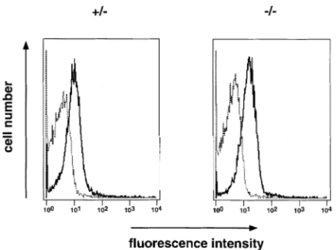
Fig. 3.In vitrocultured pre-B cells express Vpre-B2on the cell surface.
Total bone marrow from heterozygous and homozygous 7-week-old littermates were cultured on ST2 cells in the presence of IL-7 (15). After 6 days cells were stained with antibodies recognizing CD45R and Vpre-B(VP245) or an isotype-matched control antibody (rat IgG2a)
and analyzed by FACS. In the lymphocyte gate 99% of the cells expressed the CD45R marker. The data are presented as a histogram showing cell number and fluorescence intensities of the isotype control (dashed line) and VP245 (solid line) antibody stainings.
(14) using a DNA thermal cycler (Perkin-Elmer, Foster City, CA) with Taq polymerase from Gibco/BRL (Bethesda, MD). Differential amplification of specific Vpre-B1 and Vpre-B2
tran-scripts was performed as described (6). Samples were also assayed for level of HGPRT transcripts, as a control for the quality and quantity of RNA. Alsoλ5 message was analyzed to confirm that it was not affected by the Vpre-B1 deletion or
by the insertion of theneo-resistance gene. Theλ5 message was detected as described before (6).
Pre-B cell cultures
Pre-B cells from bone marrow of the indicated mice (Fig. 3) were cultured for 6–7 days as described (15). Briefly, ~25– 503103total bone marrow cells were plated in 24-well plates
onγ-irradiated ST2 stromal cells in the presence of 100 U IL-7/ml (16). The cells were cultured in IMDM-supplemented with 2-mercaptoethanol, antibiotics and 5% heat-inactivated FCS.
Flow cytometric analysis
Single-cell suspensions from bone marrow were prepared by flushing out cells from femurs with ice-cold staining buffer (HBSS containing 3% FCS). Spleen, lymph node and thymus cells in single-cell suspensions were stained with the anti-bodies described below and then analyzed by flow cytometry (FACS) using a FACSort (Becton Dickinson). The cells were gated on the extended lymphocyte gate and then analyzed for reactivity with mAb as described (17,18). Antibodies recognizing the following markers were used: CD45R (B220, FITC-conjugated from PharMingen), CD43, CD25 and IgM (biotinylated from PharMingen), c-kit (19) and Vpre-B(VP245)
(8) (the latter antibodies were biotinylated by us), CD4 [phyco-erythrin (PE)-conjugated from PharMingen], CD4 (GK1.5, FITC-conjugated by us), CD5 (biotinylated from PharMingen), CD8 (PE-conjugated from PharMingen), CD19 (biotinylated from PharMingen) and IgD (1.19, biotinylated by us). Biotinyl-ated antibodies were revealed by PE–streptavidin (Southern Biotechnology Associates, Birmingham, AL). For the Ig allelic exclusion analysis antibodies recognizing IgMa(RS3.1,
FITC-conjugated by us) and IgMb (PE-conjugated from Phar-Mingen) allotypes were used.
Immunizations and ELISA
Wild-type, heterozygous and homozygous mice (four to five mice in each group) were immunized i.p. with 100µg of the T-dependent antigen phenyl-oxazolone coupled to ovalbumin as alum precipitate. Two weeks later sera were collected and analyzed for the presence of hapten-specific (plates coated with phenyl-oxazolone coupled to BSA) or carrier-specific (ovalbumin) antibodies in ELISA.
Results
Establishment of Vpre-B1-deficient mice
To analyze the role of Vpre-B1and Vpre-B2in B cell development we set out to establish mice lacking the germline Vpre-B1gene.
Comparing the restriction enzyme maps of the two Vpre-B genes shows that the homology between the two extends no further than a few hundred base pairs 59 and 39 of the coding regions (6). Therefore, a vector that would disrupt the Vpre-B1
gene would not be expected to integrate at the Vpre-B2locus
at this frequency. A vector to replace the Vpre-B1gene with
the gene encoding neomycin resistance was constructed as shown in Fig. 1(A). Embryonic stem cells were transfected and G418-resistant clones screened for homologous recomb-ination. One clone, Y2, out of 352 analyzed was found to carry a disrupted Vpre-B1gene on one allele and was therefore
used to establish mice homozygous for the deletion in the Vpre-B1gene (VpB1–/–). A Southern blot is shown in Fig. 1(B)
in which EcoRI-digested DNA from several sources was analyzed by hybridization with the 1.9 kb probe in Fig. 1(A). The E14 embryonic stem cell line shows one band of ~9.6 kb, while the Y2 clone shows two bands, one of which is 9.6 kb and the other is 7.2 kb. The latter band corresponds to the disrupted Vpre-B1allele. In the same figure is shown DNA from individual mice, some of which are wild-type (1/1), heterozygous (1/–) and homozygous (–/–). Thus, the Y2
pre-B1
embryonic stem cell clone gave rise to mice with a deletion in the Vpre-B1gene.
Presence of Vpre-B2but not Vpre-B1RNA
To confirm that the mutation abolished expression of Vpre-B1
transcripts, bone marrow RNA was prepared from the mice analyzed in Fig. 1. The RNAs were analyzed in a RT-PCR-based assay which distinguishes between Vpre-B1 and V pre-B2 mRNA (6). As seen in Fig. 2, Vpre-B1 wild-type (1/1),
heterozygous (1/–) and homozygous mutant (–/–) mice all made Vpre-B2 mRNA. However, Vpre-B1 transcripts could not
be detected in Vpre-B1–/–mice, while they were found in both
the wild-type and heterozygous mutant mice, as expected. PCR of Vpre-B1or Vpre-B2produced two bands, the larger of
which corresponds to either genomic DNA or unspliced RNA (the primers span the 87 bp intron). Note that Vpre-B1–/–mice
also lacked this band when Vpre-B1, but not Vpre-B2expression
was examined (cf. Fig. 2, lanes 10 and 14). Since the Vpre-B1
sense primer is situated in the region that was deleted upon homologous recombination, this provided additional evidence that this part of the Vpre-B1gene had been deleted. The same cDNA preparations were also analyzed for the presence of
λ5 (and HGPRT) message and similar levels were detected
in all mice. This demonstrates that expression of theλ5 gene was not affected by the Vpre-B1deletion or by the insertion of
theneo resistance gene into the Vpre-B1locus which is located
4–5 kb upstream of theλ5 gene. Thus, the targeting vector disrupted the Vpre-B1gene and prevented its expression but
did not alter expression ofλ5 or Vpre-B2.
The Vpre-B2gene encodes a functional protein
Earlier experiments showed that both Vpre-B1and Vpre-B2could
associate withλ5 to form a SL chain (6). Since the Vpre-B2, but not the Vpre-B1, gene was expressed in the mutant mice
we investigated if Vpre-B2 was present on the cell surface of bone marrow-derived pre-B cells culturedin vitro (15). After 6 days in culture on stromal cells, in the presence of IL-7, the cells were analyzed by FACS after staining for expression of B220 (CD45R) versus an antibody (VP245) that recognizes both the Vpre-B1 and Vpre-B2proteins or an isotype-matched
control antibody (6,8). The cells in the lymphocyte gate were large and 99% stained positive for B220 (data not shown). Figure 3 shows that the VP245 antibody recognized a cell surface bound Vpre-Bprotein in cells from both Vpre-B1 hetero-zygous and homohetero-zygous mutant mice which, in the latter case, corresponds to only Vpre-B2. Thus, as suggested earlier
pre-B1
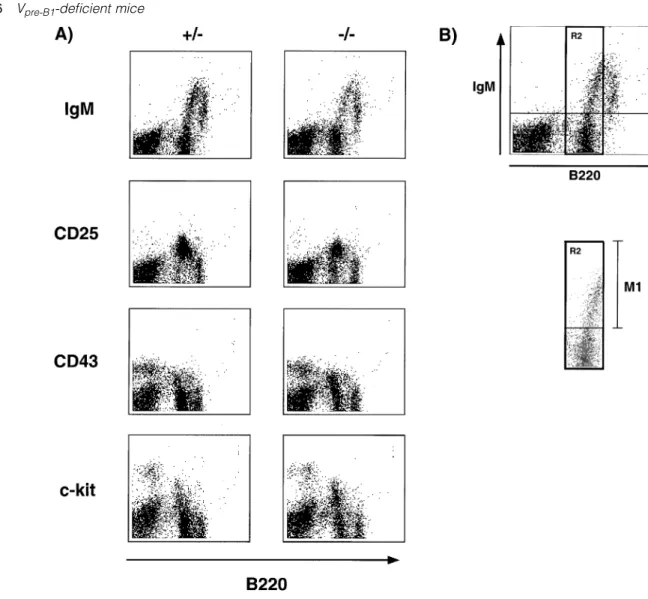
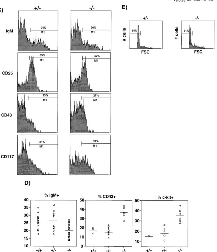
Fig. 4.Partial block in B cell development at the transition into the pre-BCR stage. (A) Expression of CD45R versus IgM, CD25 (IL2-Rα), CD43 and CD117 (c-kit) respectively on bone marrow cells from Vpre-B1heterozygous (1/–) and homozygous (–/–) mutant mice at ~8 weeks of age.
Shown is the staining pattern after gating on lymphoid cells. (B) Expression of CD45R versus IgM. The B220lo/intcells were selected as gate
R2 and the percentage of IgM1cells within this gate determined; M1. (C) The cells in (A) were analyzed by gate R2 (B220lo/int) to determine
M1. The data are displayed as histograms showing the M1 values, i.e. percentage of cells in gate R2 being positive for IgM, CD25, CD43 or CD117. (D) Between one and 10 mice per group (in several separate experiments) were analyzed as in (C). The M1 values for IgM, CD43 and CD117 are shown. Student’st-test on the decrease in percentage of IgM1cells gave aP, 0.1, i.e. highly significant. The CD117 staining has been confirmed with more wild-type and homozygous mutant mice using another CD117 antibody. (E) The CD251cells in (A) were gated and the percentage of small (FSC) cells determined.
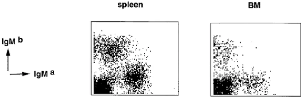
pre-B1
Fig. 5.Allelic exclusion of the IgH loci. Cells from spleen (left panel) and bone marrow (right panel) were stained with markers specific for IgMaversus IgMballotype. Cells from V
pre-B1homozygous mutant, Ig allotype heterozygous, mice are shown.
(6), the Vpre-B2gene encodes a functional protein that can be
expressed on the cell surface of pre-B cells. Partial block at the transition into the pre-BCR stage
To determine whether the failure to express Vpre-B1 had any
effect on B cell development we first analyzed the spleen of Vpre-B1–/–mice. Here, we found that the total number of cells,
and the percentages of cells expressing B220, IgM, CD8 and CD4 were about normal, suggesting that the peripheral B and T cell compartments were not affected (data not shown). We then analyzed B220 and IgM expressing B cells in lymph nodes which showed the same pattern in all three genotypes (data not shown). Lastly, analysis of thymus demonstrated a normal pattern of CD4 and CD8 positive cells (data not shown). Thus, spleen, lymph node and thymus in the Vpre-B1
-deficient mice were normal in terms of B and T lymphocytes. Since SL chain is expressed and functions specifically at early stages of B cell development, bone marrow cells were analyzed by different means. The cells were counted and the number of nucleated cells in homozygous mutant mice was found to be similar to that of wild-type and heterozygous mutant mice. Thereafter cells were analyzed for expression of B220 in combination with either IgM, CD25, CD43 or CD117 (c-kit). The differential expression of these markers is characteristic of their differentiation stage (17,18). The percentage of cells expressing B220 was found to be ~50% in all three genotypes. As shown in Fig. 4(A), the homozygous mutant mice stained positive for B220 in combination with all the above-mentioned markers. However, the percentage of cells in the various compartments differed in Vpre-B1–/– mice
as compared to heterozygous and wild-type mice: the B2201 c-kit1 and B2201 CD431 compartments were increased, while the B2201CD251and B2201IgM1compartments were slightly decreased.
To more closely analyze these differences, bone marrow cells that expressed low to intermediate levels of B220 (B220lo/int) were analyzed further (B220highcells are recirculat-ing cells). The B220lo/intcells were gated (Fig. 4B, gate R2)
and the percentage of cells within this gate that expressed the above markers determined (Fig. 4B, M1). As shown in the histograms in Fig. 4(C) and the summary in Fig. 4(D), the percentage of B220lo/intIgM1cells was slightly decreased in
Vpre-B1–/–mice. The same result was also observed for B220lo/ intCD251cells (Fig. 4C and data not shown). In contrast, the
percentage of B220lo/intCD1171 cells was ~2-fold higher in
homozygous as compared to heterozygous mutant mice. This was also the case for B220lo/intCD431 cells. A summary of
several experiments, which also included wild-type mice, is shown in Fig. 4(D). Since the number of bone marrow cells in all three genotypes was about the same, the increase in the percentage of B220lo/int CD1171 cells represented an
increase in the actual number of B220lo/intCD1171cells. The
size distribution among the cells that expressed either CD117, CD43, CD25 or IgM was similar among the three genotypes, suggesting that the lack of Vpre-B1did not affect the expression
of these markersper se (Fig. 4E and data not shown). Thus, the number of bone marrow cells expressing B220lo/int in combination with CD117 and CD43 was enriched for by a factor of 2, while B220lo/intCD251 and B220lo/int IgM1 cells were each decreased by ~20% in Vpre-B1–/–mice.
Pre-BI cells express B220, CD117 and CD43 but not CD25; pre-BII cells are B2201CD251but lack c-kit and CD43. The CD25 marker is not found on more differentiated cells, i.e. immature and mature B cells (18). Large pre-BII cells that express a functional pre-BCR are expanded by proliferation and, upon differentiation, give rise to small pre-BII cells. Among the B220lo/int CD251 cells ~20 and 80% constituted
large and small cells respectively in all three genotypes (Fig. 4E and data not shown). This suggested that cells with a functional pre-BCR were expanded also in the marrow of Vpre-B1 homozygous mutant mice. Thus, bone marrow from
Vpre-B1–/–mice exhibited an enrichment in pre-BI cells (B220lo/ int CD1171 CD431) and a reduction in large pre-BII cells
(B220lo/intCD251) suggesting that the lack of V
pre-B1affected
B lymphocyte development at the transition into the pre-BCR stage.
Ig allelic exclusion in VpreB1-deficient mice
To determine if the Vpre-B2-containing pre-BCR was functional in terms of allelic exclusion of the Ig loci, we analyzed spleen and bone marrow cells by FACS for the presence of both Ig allotypes in Iga/Igb heterozygous mice. As shown in Fig. 5, .98% of all sIgM1 B cells of bone marrow and of spleen
from VpreB1–/–Ig allotype heterozygous mice expressed either
one or the other, but not both Ig allotypes on one cell. Thus, a defective Vpre-B1gene did not affect Ig allelic exclusion. Normal immune response against T-dependent antigens We next examined the ability of Vpre-B1-deficient mice to mount
pre-B1
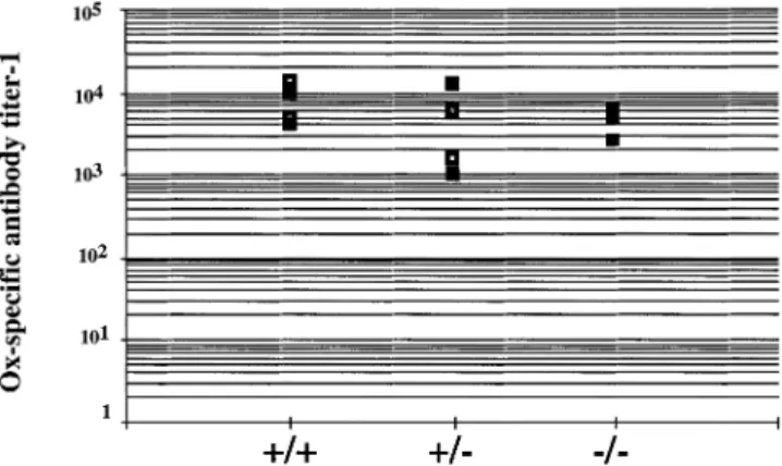
Fig. 6.Normal immune response to a T-dependent antigen. Vpre-B1
wild-type (1/1), heterozygous (1/–) and homozygous (–/–) mutant mice were immunized (i.p.) with 100µg alum precipitate of the T-dependent antigen phenyl-oxazolone coupled to ovalbumin as a carrier. Two weeks later sera were taken and analyzed for hapten (Ox)-specific antibodies in ELISA. Each group contained four or five mice. Shown is the titer of each serum at 50% of the plateau level. Sera from non-immunized mice:,1.
hapten phenyl-oxazolone coupled to the carrier ovalbumin. Two weeks after immunization, serum were tested in ELISA for the presence of hapten- and carrier-specific antibodies. As shown in Fig. 6, all mice showed a good response to the hapten, independent of the status of the Vpre-B1 loci. The
response to the ovalbumin carrier was also normal in all mice (data not shown). Therefore, the absence of Vpre-B1 did not
alter the immune response to T-dependent antigens.
Discussion
Our aim here was to evaluate the role in B lymphocyte development of one of the two Vpre-B genes present in the
mouse genome, i.e. the Vpre-B1gene. We find that disruption
of the Vpre-B1gene causes a partial block in B cell development at the transition into the pre-BCR stage as defined by an increase in the number of pre-BI cells and a slight decrease in the number of large pre-BII cells. However, the number of mature B cells in the periphery was not affected. Moreover, allelic exclusion of IgH gene expression, in the central and peripheral B cell compartments, is functional as are T cell-dependent immune responses. This demonstrates that both Vpre-B gene products are functional and involved in the
formation of the pre-BCR. It also shows that Vpre-B2alone is sufficient, but not as efficient as the two genes together, in supporting B lymphocyte development.
We showed earlier that the expression pattern at the RNA level of the two Vpre-Bgenes (andλ5) is similar in fetal liver
cells and in pro-B/pre-BI and large pre-BII bone marrow cells. Single-cell RT-PCR of the latter two populations detected Vpre-B1(andλ5) mRNA in all cells, but Vpre-B2transcripts were
found in only a portion (30%) of the same cells (6). This discrepancy was proposed to be due to a difference in sensitivity of the primer pairs used in the assays, a hypothesis which is supported by our results herein: the number of large pre-BII cells in Vpre-B1–/–mice is too high to be derived from
only one-third of the normal numbers of pre-BI cells. In
transfection experiments, both Vpre-B gene products have
been shown to assemble with theλ5 protein. However, it is not known if they do so in vivo since there is no antibody described that distinguishes the two Vpre-B proteins (6,20). Our results here demonstrate that the Vpre-B2 gene product
can be incorporated in both the pro-B and pre-B cell receptors of pro-B/pre-BI and large pre-BII bone marrow cells. Further-more, Vpre-B2can function in all steps of development,
especi-ally in the pre-BCR-dependent proliferative expansion of large pre-BII cells. If the pre-BCR is also involved in allelic exclusion of the IgH chain gene expression, then Vpre-B2 in pre-BCR
can also do that.
The importance of the membrane form of theµH chain and theλ5 gene product at the pre-BCR stage was demonstrated earlier in mice lacking the respective gene product (11,12). Here we show that also the Vpre-B1gene product is important
at the pre-BCR stage since the lack of Vpre-B1 causes an
enrichment of cells at the pre-BI stage, i.e. a partial block at the transition into the pre-BCR stage. It has recently been shown that not all IgµH chains made from productively VDJH
-rearranged IgH loci can pair with the SL chain (21). Cells expressing such non-pairingµH chains cannot form a pre-BCR and do not participate in the proliferative expansion of large pre-BII cells. Hence, cells expressing theseµH chains do not appear in the subsequent pre-B and B cell repertoires. Some of the inability to pair may well be due to a structural incompatibility of a VH domain in interactions with Vpre-B. If
so, then pairing to Vpre-B1and Vpre-B2may differ since some
of the nucleotide differences between the two Vpre-Bgenes
encode amino acid replacements. The VH repertoires of
normal and Vpre-B1–/–mice may, therefore, differ. This will be
investigated in future experiments.
The cells that progress through the pre-BCR stage in Vpre-B1–/–mice seem to be normal in that they are expanded
by proliferation, and the number of more mature cells is just slightly decreased as compared to heterozygous mutant and wild-type mice. This did not significantly affect the number of B cells detected in the periphery. This is in agreement with experiments that showed that a minor decrease in the production of pre-B cells (here pre-BII) in the marrow did not affect the number of B cells in the periphery, while a major decrease in B cells did (22). Such a minor decrease in pre-B cells could be envisioned as the situation in Vpre-B1-deficient
mice while a major (20 to 40-fold) reduction in pre-B(II) cells is compatible with that found in mice lacking λ5 that also have a significant decrease in the number of peripheral B cells. Our data herein shows that mice expressing only one Vpre-B gene (Vpre-B2) are immunocompetent and give rise to both
hapten- and carrier-specific antibodies when challenged with T cell-dependent antigens. Thus, in mice, as is the case in humans, one Vpre-Bgene is sufficient, if not as efficient.
Acknowledgements
We thank Genetics Institute for LIF-producing CHO cells, Drs H. Karasuyama for the gift of the mAb VP245, and Dr F. Ivars for reading and discussing the manuscript. We also thank MaryAnn Sa¨llstro¨m (Lund) and Urs Mueller (Basel) for blastocyst injections. This work was supported by a fellowship from Pharmacia-Upjohn to A. M., by the Swedish Medical Society (A. M.), by the Swedish Medical Research Council, the Kungliga Fysiografiska Society, the O¨ sterlund,
pre-B1
the Kocks, the Crafoord, the A.-G. Crafoord and the Zoe´ga Founda-tions (I.-L. M), by the Arthritis Foundation (J. D.), and by NIH grant AI-30178 (Y. A.). The Basel Institute for Immunology was founded and is supported by Hoffman-LaRoche Inc.
Abbreviations
µH Igµheavy chain pre-BCR pre-B cell receptor SL surrogate light chain
VpreB1–/– Vpre-B1homozygous mutant mice
References
1 Sakaguchi, N. and Melchers, F. 1986. λ5, a new light-chain-related locus selectively expressed in pre-B lymphocytes. Nature324:579.
2 Sakaguchi, N., Berger, C. N. and Melchers, F. 1986. Isolation of a cDNA copy of an RNA species expressed in murine pre-B cells.EMBO J.5:2139.
3 Kudo, A. and Melchers, F. 1987. A second gene, Vpre-B in the
lambda 5 locus of the mouse, which appears to be selectively expressed in pre-B lymphocytes.EMBO J.6:2267.
4 Kudo, A., Thalmann, P., Sakaguchi, N., Davidson, W. F., Pierce, J. H., Kearney, J. F., Reth, M., Rolink, A. and Melchers, F. 1992. The expression of the mouse Vpre-B/λ5locus.Int. Immunol.4:831.
5 Karasuyama, H., Rolink, A., Shinkai, Y., Young, F., Alt, F. W. and Melchers, F. 1994. The expression of Vpre-B/λ5 surrogate light
chain in early bone marrow precursor B cells of normal and B cell-deficient mutant mice.Cell77:133.
6 Dul, J., Argon, Y., Winkler, T., ten Boekel, E., Melchers, F. and Mårtensson, I.-L. 1996. The murine Vpre-B1and Vpre-B2genes both
encode a protein of the surrogate light chain and are co-expressed during B cell development.Eur. J. Immunol.26:906.
7 Melchers, F., Karasuyama, H., Haasner, D., Bauer, S., Kudo, A., Sakaguchi, N., Jameson, B. and Rolink, A. 1993. The surrogate light chain in B-cell development.Immunol. Today14:60. 8 Karasuyama, H., Rolink, A. and Melchers, F. 1993. A complex of
glycoproteins is associated with Vpre-B/λ5 surrogate light chain
on the surface ofµheavy chain-negative early precursor B cell lines.J. Exp. Med.178:469.
9 Pillai, S. and Baltimore, D. 1987. Formation of disulphide-linked
µ2ω2 tetramers in pre-B cells by the 18 Kω-immunoglobulin light chain.Nature329:172.
10 Pillai, S. and Baltimore, D. 1988. The omega and iota surrogate immunoglobulin light chains. Curr. Top. Microbiol. Immunol. 137:136.
11 Kitamura, D., Roes, J., Ku¨hn, R. and Rajewsky, K. 1991. A B cell-deficient mouse by targeted disruption of the membrane exon of the immunoglobulinµchain gene.Nature350:423.
12 Kitamura, D., Kudo, A., Schaal, S., Mu¨ller, W., Melchers, F. and Rajewsky, K. 1992. A critical role of λ5 protein in B cell development.Cell69:823.
13 Kudo, A., Sakaguchi, N. and Melchers, F. 1987. Organization of the murine Ig-related λ5 gene transcribed selectively in pre-B lymphocytes.EMBO J.6:103.
14 Sambrook, J., Fritsch, E. F. and Maniatis, T. 1989. Molecular Cloning: A Laboratory Manual, 2nd edn. Cold Spring Harbor Laboratory Press, Cold Spring Harbor, NY.
15 Rolink, A., Kudo, A., Karasuyama, H., Kikuchi, Y. and Melchers, F. 1991. Long-term proliferating early pre B cell lines and clones with the potential to develop surface Ig-positive, mitogen reactive B cellsin vitroandin vivo.EMBO J.10:327.
16 Karasuyama, H. and Melchers, F. 1988. Establishment of mouse cell lines which constituvely secrete large quantities of interleukin 2, 3, 4 or 5, using a modified cDNA expression vectors.Eur. J. Immunol.18:97.
17 Grawunder, U., Leu, T. M. J., Schatz, D. G., Werner, A., Rolink, A. G., Melchers, F. and Winkler, T. H. 1995. Downregulation of RAG1 and RAG2 gene expression in pre-B cells after functional immunoglobulin heavy chain rearrangement.Immunity3:601. 18 Rolink, A., Grawunder, U., Winkler, T. H., Karasuyama, H. and
Melchers, F. 1994. IL-2 receptorαchain (CD25, TAC) expression defines a crucial stage in pre-B cell development.Int. Immunol. 6:1257.
19 Ogawa, M., Matsuzaki, Y., Nishikawa, S., Hayashi, S. I., Kunisada, T., Sudo, T., Kina, T., Nakauchi, H. and Nishikawa, S. I. 1991. Expression and function of c-kit in hemopoietic precursor cells.J. Exp. Med.174:63.
20 Karasuyama, H., Kudo, A. and Melchers, F. 1990. The proteins encoded by Vpre-Bandλ5 pre-B cell-specific genes can associate
with each other andµheavy chains.J. Exp. Med.172:969. 21 ten Bochel, E., Melchers, F. and Rolink, A. 1997. Changes in the
VH gene repertoire of developing precursor B lymphocytes in mouse bone marrow mediated by the pre-B cell receptor. Immunity7:357.
22 Agene`s, F., Rosado, M. M. and Freitas, A. A. 1997. Independent homeostatic regulation of B cell compartments.Eur. J. Immunol. 27:1801.