Thymocytes can tolerize thymocytes by
clonal deletion in vitro
Hanspeter Pircher, Klaus-Peter Muller
1, Bruno A. Kyewski
1, and Hans
Hengartner
Institute of Experimental Immunology, Department of Pathology, University of Zurich, Schmelzbergstrasse 12, 8091 Zurich, Switzerland
'Institute for Immunology and Genetics, German Cancer Research Center, Heidelberg, Germany Key words: apoptosis, TCR transgenic mice, thymocytes
Abstract
Clonal deletion of thymocytes bearing TCR for self antigens is one major mechanism of T cell tolerance induction. Peptide antigen-induced deletion of thymocytes from a/3 TCR transgenic mice has been studied using single cell suspension cultures. The results show that antigen-presenting immature CD4+CD8+ thymocytes can tolerize antigen-reactive immature thymocytes in vitro by programmed cell death (apoptosis) 6 - 8 h after antigen exposure. Antigen-induced apoptosis of immature thymocytes was inhibited by antibodies specific for the a/3 TCR, CD3, CD8, and LFA-1 molecules. This implies that clonal elimination of self-reactive CD4+CD8+ thymocytes does not depend on specialized deleting cell types in the thymus and occurs
whenever the TCR of immature thymocytes bind antigen fragments presented by MHC molecules. Introduction
During T cell development the TCR repertoire is shaped in the thymus by positive and negative selection events. It is now well established that clonal deletion of self-reactive T cells is the major mechanism of T cell tolerance induction for thymic self antigens. This has been demonstrated in normal mice for superantigens (1 - 3 ) and in a/3 TCR transgenic mice for conventional MHC-restricted peptide antigens (4-6).
Clonal deletion is thought to occur via antigen-induced programmed cell death (apoptosis) of autoreactive thymocytes. Anti-CD3 treatment of fetal thymic organ culture has been proposed to be an in vitro model of negative selection (7,8). This view has been recently challenged by two studies which show that thymocytes which are resistant to anti-CD3-induced apoptosis are still susceptible to superantigen-induced deletion in vivo (9,10).
To learn more about the molecular and cellular requirement for tolerance induction by deletion, an assay system, originally described by Swat et al. (11,12), has been utilized which allows the study of antigen-induced deletion of thymocytes in single cell suspension cultures in vitro. This approach allows the definition of the minimal cellular requirement for clonal deletion and the identification of cell surface molecules involved in this process.
Methods
Mice
The TCR transgenic mice (line 327) have been previously described (6). Mice 6 - 1 2 weeks of age were used.
Thymocyte cultures
Single cell suspensions of thymocytes were prepared by squeezing the whole thymus through a wire screen. Clumps were allowed to settle and then discarded. The cells were > 9 9 % Thy-1+, < 0 . 1 % M a c - 1+, <0.7% Mac-2 + , and <0.4% lgM + .
After washing, thymocytes (5 x 10⁶ cells/ml) were cultured
(37°C, 5% CO2) in Iscove's modified Dulbecco's medium
(IMDM) supplemented with 15% FCS in 24-well Costar tissue culture plates (1 ml/well) for 14 h if not otherwise indicated. The lymphocytic choriomeningitis virus (LCMV) glycoprotein aa33-42 (KAVYNFATCG) (13) peptide was used at 30>M. Lymph node cells (2 x 10⁶ cells/well) from a/3 H-2b TCR
tran-sgenic mice (>7O°/o CD8+) were added to the indicated
cultures. FACS-sorted CD4+CD8+ thymocytes (>99.8% pure)
were cultured (5 x 10⁶ cells/ml) in 96-well tissue culture plates
(200 /il/well) in the presence or absence of 30 /*M LCMV pep-tide for 14 h. Afterwards, the cells were restained with anti-CD4 and -CD8 mAb.
Inhibition of antigen-induced deletion
Thymocytes were cultured in the presence of the supernatants of the following B cell hybridomas: 53-6.72, anti-CD8 (14); GK1.5, anti-CD4 (15); H129.37, anti-LFA-1-a (16); KT3, anti-CD3 (17); B20.1, anti-TCR Va2 (18); KJ16, anti-TCR VS8 (19), J11d and
B2A2, anti-HSA (20,21); KM202, anti-CD44 (22); and M1/9 and 23G, anti-CD45 (23,24). Per cent deletion of CD4KshCD8h:sh
thymocytes was calculated by:
Correspondence to: H. Pircher
°/oCD4h'9hCD8h'9h thymocytes with LCMV peptide
100 x 1
-%CD4hi9hCD8hi9h thymocytes without LCMV peptide
Flow cytometry analysis
Thymocytes were double stained with anti-CD4 - phycoerythrin (PE) and anti-CD8-(luorescein isothiocyanate (FITC) (Becton Dickinson, Mountain View, CA) mAb. CD4/CD8 expression of the anti-CE4 or anti-CD8 treated cultures was determined by res-taining with CD4 (15) or CD8 (14) mAb and goat anti-rat I g G - F I T C (TAGO, Burlingame, CA), followed by CD8-biotin-avidin-PE or CD4-PE (Becton Dickinson) respectively. Thymocytes were analyzed on an EPICS profile analyzer (Coulter, Hialeah, FL) with four logarithmic scales. Data were only collected from viable (i.e. propidium iodide negative) cells gated by a combination of forward light scatter (FS) and 90° side scatter (SS).
Results and discussion
The molecular and cellular requirements for clonal elimination of self-reactive CD4+CD8+ thymocytes from a/3 TCR transgenic
mice by apoptosis was examined in single cell suspension cultures. These mice express an LCMV/H-2Db-specific TCR
(Va2A/,j8.1) on most thymocytes (70-80%) and peripheral
CD8+ T cells (6). Apoptosis of immature CD4 + CD8+
thymocytes was analyzed by flow cytometry based on the fact that thymocytes undergoing programmed cell death express reduced levels of CD4 and CD8 molecules (11). Thus, the degree of cell death among cultured CD4+CD8+ thymocytes can be
monitored by the shift from a CD4hi9hCD8hi9h to a CD4lowCD8low
phenotype (11,12). When thymocytes from TCR transgenic H-2b
mice were cultured in the presence of the LCMV peptide (glycoprotein aa33 - 42) recognized by the transgenic TCR, the population of immature thymocytes expressing CD4 and CD8 at a high level (CD4h'9hCD8h'9h) gradually decreased and a
distinct apoptotic cell population bearing lower levels of CD4 and CD8 (CD4'°WCD8|OW) appeared (Fig. 1A). The disappearance of
CD4hishCD8hi9h thymocytes was MHC-restricted (Fig. 1B, top)
and antigen-specific (Fig. 2, top), and was not observed in LCMV peptide-treated thymocyte cultures from non-transgenic H-2b
mice where only a minute fraction [<1/105 (25)] of T cells bear
an LCMV-specific TCR (Fig. 1B). Apoptosis of CD4+CD8 +
thymocytes was not caused by direct cell lysis mediated by
mature thymocytes because co-culture of normal H-2b
LCMV peptide Medium Control
Oh
2 h
4 h
6 h
8 h
10 100 1 10 100CD4
100 10 100 10 ioo ooio Q
100 10 1 100 10 1B LCMV peptide Medium Control
H-2b H-2d t g+ H-2D H-2D t g+ T cells 29% 64% 75% 51% 56% 65% 76% 54% 100 10 1 100 10 • 00 100 U 10 100 10 1 10 100 1 10 100
CD4
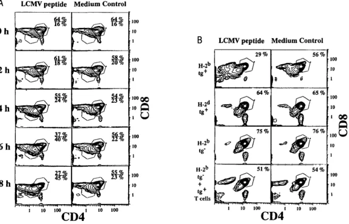
Fig. 1. Peptide antigen-induced apoptosis in thymocyte cultures. (A) Thymocytes from TCR transgenic mice (H-2b) were cultured for the indicated
time in the presence (left) or absence (right) of the LCMV peptide (GP aa33 - 42) and subsequently analyzed for expression of CD4 and CD8 molecules by flow cytometry. The percentages given in each plot indicate the relative number of CD4h'9hCD8h'sh and CD4kw'CD8lov" cells in the indicated gate.
Most (70 - 80%) of the total input cell numbers were recovered as trypan blue negative thymocytes after 8 h in peptide-treated and control cultures. (B) Thymocytes from the indicated mice were cultured for 14 h with or without the LCMV peptide and analyzed for CD4 and CD8 expression. In the bottom row, peripheral CD8+ T cells from TCR transgenic mice (H-2b) were added to thymocyte cultures from normal H-2b mice. The percentage
given in each plot indicates the relative number of CD4™ghCD8h'9h thymocytes. Most (60 - 70%) of the total input cell numbers were recovered as
trypan blue negative thymocytes. In LCMV peptide-treated H-2b transgenic cultures, only 30 - 40% of the total input cells were recovered as trypan
LCMV GP LCMVGP LCMV NP aa 33-42 aa 32-42 aa 118-132
100 30 10 3 100 30 10 3 100 30 10 3
8
anti-CD8 anti-CD4 anti-LFA-1
anti-CD3 anti-TCR anti-TCR
V a 2 V | 3 8
dilution of mAb
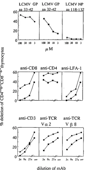
Fig. 2. Inhibition of LCMV peptide-induced deletion of CD4hiBhCD8hiBh
thymocytes with mAb. Apoptosis was induced either with the LCMV glycoprotein a a 3 3 - 4 2 (100 jiM • ) or with the suboptimal a a 3 2 - 4 2 (100/»M • ) peptide.
Medium Control LCMV peptide
^ i . V , r ; . j ' ^ - . : \ - . ; ' j •••Tii; '••/••-.•"••'' • • t , ' . / . \ ;•«->, ••.^/•;.-.-~-/;1-:'.- • . •V . i.* • j • - . . . • • • .-i
•-. .-*.* •
• c , - • %Fig. 3. Peptide antigen-induced cell aggregation. Thymocytes from TCR
transgenic H-2b mice were incubated at 37°C in the absence (left) or
presence (right) of LCMV peptide for 3 h. Afterwards, the cell suspension was mounted on a glass slide, air dried, and stained using M a y - G r u n w a l d - G i e m s a eosine-methylene blue solution. Magnification: top panel x 2 1 ; bottom panel x105.
thymocytes and mature transgenic CD8+ T cells, in the
presence of LCMV peptide, did not reduce the number of normal CD4hishCD8hi9h thymocytes (Fig. 1B, bottom). In addition,
unprimed TCR transgenic thymocytes and peripheral T cells did not show any cytolytic activity when tested on LCMV peptide-coated target cells in a 51Cr release assay (not shown) and
elimination of mature thymocytes by cell sorting did not abolish apoptosis of CD4+CD8+ thymocytes (see below).
Monoclonal antibodies were tested for their ability to interfere with LCMV peptide-induced apoptosis: mAb specific for transgenic TCR a and & chains, CD3, CD8, and LFA-1 molecules inhibited antigen-induced elimination of CD4hishCD8hiflh
thymocytes in a dose-dependent manner (Fig. 2). Inhibition was more pronounced when a suboptimally antigenic LCMV peptide (glycoprotein aa32-42) was used (Fig. 2, top). Antibodies specific for other thymocyte antigens, such as the CD44 arid CD45 molecules, and the heat stable antigen (HSA), did not block apoptosis, whereas mAb H141-51 (26), specific for the LCMV peptide-presenting MHC class I molecules H-2Db, were found
to be inhibitory (data not shown). Neonatal anti-CD4 antibody treatment has been shown to prevent Mis- (27) and l-E-mediated (28) deletion of self-reactive T cells in vivo.
During the course of these experiments it was noted that the addition of LCMV peptide to thymocyte cultures from TCR transgenic H-2b mice induced a transient cell aggregation
2 - 3 h after peptide addition. These aggregates often appeared
LCMV peptide Medium Control
TCR t g+ H-2b TCR tgH H-2d 36.1 % 1.4% 7.7% 1.4% 100 10 100 10 1
8
TCR tg + H-2d 60Fig. 4 . CD4+CD8+ thymocytes induce antigen-specific apoptosis of
C D 4+C D 8+ thymocytes. Fluorescence data were collected only from
'viable' cells with high FS and low SS (population A; bottom) which did not stain with propidium iodide. Cells from population B were mostly propidium iodide positive.
as characteristic thymocyte chains (Fig. 3). A similar effect was not observed when LCMV peptide-treated thymocyte cultures from normal H-2b or TCR transgenic H-2d mice were examined
(not shown). This observation suggests that LCMV peptide-coated thymocytes may act as antigen-presenting cells and therefore induce visible cell -cell binding via the LCMV-specific transgenic TCR present on most thymocytes. Alternatively, it is possible that antigen-induced TCR cross-linking on thymocytes stimulates adhesiveness through LFA-1 molecules as shown for mature T cells (29).
To formally demonstrate that immature CD4+CD8+ thymocytes
are able to present the antigenic LCMV peptide to themselves and thereby induce apoptosis, CD4+CD8+ thymocytes from TCR
transgenic mice were purified by cell sorting, cultured in the presence or absence of the LCMV peptide for 14 h, restained with CD4 and CD8 mAb, and analyzed by flow cytometry. Thymocytes from TCR transgenic H-2d mice served as a
specificity control because H-2d MHC molecules do not present
the LCMV peptide to the transgenic TCR (not shown). LCMV peptide-induced apoptosis occurred only in thymocyte cultures derived from transgenic H-2b but not from H-2d mice, as judged
by the disappearance of CD4hishCD8h'9h cells and the
appearance of the CD4lowCD8low population (Fig. 4, top).
Fluorescence data were collected only from 'viable' cells which did not stain with propidium iodide.
In addition, plots of FS and 90° SS of the recovered cells revealed an increase (34 to 59%) of the cell population with low FS and high SS (population B in Fig. 4, bottom) in LCMV peptide-treated thymocyte cultures derived from TCR transgenic H-2b
mice. Most of these cells were stained by propidium iodide, indicating permeable cytoplasmic membranes (not shown). A similar effect was not observed in LCMV peptide-treated thymocyte cultures derived from TCR transgenic H-2d mice (Fig.
4, bottom). These results show that the addition of the LCMV peptide to purified CD4 + CD8+ thymocytes from TCR
transgenic H-2b mice also specifically decrease cell viability from
66 to 4 1 % (Fig. 4, bottom).
The possibility that the observed antigen-induced apoptosis of immature thymocytes in these experiments was a result of the few contaminating cells in the purified CD4+CD8+ population
cannot be completely ruled out. However, it is considered unlikely that the <0.2% contaminating cells in the preparations were able to induce CD4/CD8 down-regulation in 30% of 'viable' thymocytes and to decrease the viability of the recovered cells from 61 to 4 1 % in this in vitro culture system within 14 h. In addition, the extent of deletion of purified CD4+CD8 +
thymocytes (Fig. 4) was comparable to the results obtained with total thymocyte preparations (Fig. 1).
FACS-purified double negative (CD4~CD8-) thymocytes injected into the thymus of irradiated host mice have been shown to induce T cell tolerance to allogeneic MHC class I antigens (30). The results of this study directly demonstrate that CD4+CD8+
thymocytes induce clonal deletion of self-reactive immature CD4 + CD8+ thymocytes by apoptosis in vitro. This
com-plements a recent report by Swat et al. (12) which revealed that adherent cell preparations from thymus and spleen are able to induce antigen-specific clonal deletion in vitro. Mice, neonatally infected with LCMV, delete LCMV-specific CD8+ T cells in the
thymus (6). Because LCMV does not infect thymocytes, clonal
deletion was induced in this system either by infected thymic epithelia or bone marrow-derived macrophages/dendritic cells. In this study the LCMV peptide has been used as a model an-tigen for self determinants expressed or passively acquired (31,32) by thymocytes. This approach allowed the demonstra-tion that tolerance inducdemonstra-tion, via antigen-triggered self destruc-tion of CD4+CD8+ thymocytes, only requires the presence of
the appropriately processed self peptides presented by MHC molecules.
Acknowledgements
This work was supported by SNF grants to H. P. and H. H. In addition H. P. is supported by the Stiftung Prof. Dr. M. Cloetta. We thank Eva Niederer and Marc Condrau for their patience and expertise in flow cytometry and Alana Althage and Rolf M. Zinkernagel for reviewing this manuscript. Abbreviations FITC FS HSA IMDM LCMV PE SS References fluorescein isothiocyanate forward light scatter heat stable antigen
Iscove's modified Dulbecco's medium lymphocytic choriomeningitis virus phycoerythrin
side scatter
1 Kappler, J. W., Roehm, N., and Marrack, P. 1987. T cell tolerance by clonal elimination in the thymus. Cell 49:273.
2 Kappler, J. W., Staerz, U. D., White, J., and Marrack, P. 1988. Self tolerance eliminates T cells specific for Mis-modified products of the major histocompatibility complex. Nature 332:35.
3 MacDonald, H. R., Schneider, R., Lees, R. K., Howe, R. C , Acha-Orbea, H., Festenstein, H., Zinkernagel, R. M., and Hengartner, H. 1988. T cell receptor V/3 use predicts reactivity and tolerance to Mlsa
-encoded antigens. Nature 332:40.
4 Kisielow, P., Bluthmann, H., Staerz, U. D., Steinmetz, M., and von Boehmer, H. 1988. Tolerance in T cell receptor transgenic mice involves deletion of nonmature C D 4+8+ thymocytes. Nature
333:742.
5 Sha, W. C , Nelson, C. A., Newberry, R. D., Kranz, D. M., Russell, J. H., and Loh, D. Y. 1988. Positive and negative selection of an antigen receptor on T cells in transgenic mice. Nature 336:73. 6 Pircher, H. P., Burki, K., Lang, R., Hengartner, H., and Zinkernagel,
R. 1989. Tolerance induction in double specific T-cell receptor tran-sgenic mice varies with antigen. Nature 342:559.
7 Smith, C. A., Williams, G. T., Kingston, R., Jenkinson, E. J., and Owen, J. J. T. 1990. Antibodies to CD3/T-cell receptor complex induce death by apoptosis in immature T cells in thymic cultures. Nature 337:181. 8 Finkel, T. H., Cambier, J. C , Kubo, R. T., Born, W. K., Marrack, P., and Kappler, J. W. 1989. The thymus has two functionally distinct populations of immature alpha beta* T cells: one population is deleted by ligation of alpha beta TCR. Cell 58:1047.
9 Sentman, C. L., Shutter, J. R., Hockenbery, D., Kanagawa, O., and Korsmeyer, S. J. 1991. bcl-2 inhibits multiple forms of apoptosis but not negative selection in thymocytes. Cell 67:879.
10 Strasser, A., Harris, A. W., and Cory, S. 1991. bcl-2 transgene inhibits T cell death and perturbs thymic self-censorship. Cell 67:889. 11 Swat, W., Ignatowicz, L., and Kisielow, P. 1991. Detection of apoptosis
of immature C D 4+8+ thymocytes by flow cytometry. J. Immunol.
Methods 137:79.
12 Swat, W., Ignatowicz, L, von Boehmer, H., and Kisielow, P. 1991. Clonal deletion of immature C D 4+8+ thymocytes in suspension
culture by extrathymic antigen-presenting cells. Nature 351:150. 13 Pircher, H. P., Moskophidis, D., Rohrer, U., Burki, K., Hengartner,
H., and Zinkernagel, R. M. 1990. Viral escape by selection of cyto-toxic T cell-resistant virus variants in vivo. Nature 346:629. 14 Ledbetter, J. A. and Herzenberg, L. A. 1979. Xenogeneic monoclonal
antibodies to mouse lymphoid differentiation antigens. Immunol. Rev. 47:63.
15 Dialynas, D. P., Wilde, D. B., Marrack, P., Pierres, A., Wall, K. A., Havran, W., Often, G., Loken, M. R., Pierres, M., Kappler, J. W., and Fitch, F. W. 1983. Characterization of the murine antigenic determinant, designated L3T4a, recognized by monoclonal antibody GK1.5: expression of L3T4a by functional T cell clones appears to cor-relate primarily with class II MHC antigen reactivity. Immunol. Rev. 74:29. 16 Pierres, M., Goridis, C , and Golstein, P. 1982. Inhibition of murine
T cell-mediated cytolysis and T cell proliferation by a rat monoclonal antibody immunoprecipitating two lymphoid cell surface polypeptides of 94000 and 180000 molecular weight. Eur. J. Immunol. 12:60. 17 Tomonari, K. 1988. A rat antibody against a structure functionally
related to the mouse T-cell receptor/T3 complex. Immunogenetics 28:455.
18 Pircher, H. P., Rebai, N., Groettrup, M., Gregoire, C , Speiser, D. E., Patt Happ, M., Palmer, E. .Zinkernagel, R. M., Hengartner, G., and Malissen, B. 1992. Preferential positive selection of Va2+ CD8+ T
cells in mouse strains expressing both H-2k and T cell receptor Vaa
haplotypes: determination with a V«2 specific monoclonal antibody. Eur. J. Immunol. 22:399.
19 Haskins, K., Hannum.'C, White, J., Roehm, N., Kubo, R., Kappler, J. W., and Marrack, P. 1984. The antigen-specific, major histocom-patibility complex-restricted receptor on T cells. VI. An antibody to a receptor allotype. J. Exp. Med. 160:452.
20 Bruce, J., Symington, F. W., Mckearn, T. J., and Sprent, J. 1981. A monoclonal antibody discriminating between subsets of T and B cells. J. Immunol. 127:2496.
21 Scollay, R., Bartlett, P., and Shortman, K. 1984. Tcell development in the adult murine thymus: changes in the expression of the surface antigens Ly-2, L3T4 and B2A2 during development from early precursor cells to emigrants. Immunol. Rev. 82:79.
22 Miyake, K., Medina, K. L, Hayashi, S.-l., Ono, S., Hamaoka, T., and
Kincade, P. W. 1990. Monoclonal antibodies to Pgp-1/CD44 block lympho-hemopoiesis in long-term bone marrow cultures. J. Exp. Med. 171:477.
23 Springer, T., Galfre, G., Sprecher, D. S., and Milstein.C. 1978. Monoclonal xenogeneic antibodies to murine cell surface antigens: identification of novel leukocyte differentiation antigens. Eur. J. Immuol. 8:539.
24 Birkeland, M. L, Johnson, P., Trowbridge, I. S., and Pure,' E. 1989. Changes in CD45 isoform expression accompany antigen-induced murine T-cell activation. Proc. Natl Acad. Sci. USA 86:6734. 25 Moskophidis, D., Assmann Wischer, U., Simon, M. M., and Lehmann
Grube, F. 1987. The immune response of the mouse to lymphocyte choriomeningitis virus. V. High numbers of cytolytic T lymphocytes are generated in the spleen during acute infection. Eur. J. Immunol. 17:937.
26 Lemke, H., Hammerling, G. H., and Hammerling, U. 1979. Fine specificity analysis with monoclonal antibodies of antigens controlled by the major histocompatibility complex and by the Qa/TL region in mice. Immunol. Rev. 47:175.
27 MacDonald, H. R., Hengartner, H., and Pedrazzini, T. 1988. Intrathymic deletion of self-reactive cells prevented by neonatal anti-CD4 antibody treatment. Nature 335:174.
28 Fowlkes, B. J., Schwartz, R. H., and Pardoll, D. N. 1988. Deletion of self-reactive thymocytes occurs at a C D 4+8+ precursor stage.
Nature 334:620.
29 Dustin, M. L. and Springer, T A. 1989. T-cell receptor cross-linking transiently stimulates adhesiveness through LFA-1. Nature 341:619. 30 Shimonkevitz, R. P. and Bevan, M. J. 1988. Split tolerance induced by the intrathymic adoptive transfer of thymocyte stem cells. J. Exp. Med. 168:143.
31 Sharrow, S. O., Mathieson, B. J., and Singer, A. 1961. Cell surface appearance of unexpected host MHC determinants on thymocytes from radiation bone marrow chimeras. J. Immunol. 1981:1327. 32 Shores, E. W., Sharrow, S. 0., and Singer, A. 1991. Presence of CD4
and CD8 determinants on CD4~CD8~ murine thymocytes: passive acquisition of CD8 accessory molecules. Eur. J. Immunol. 21:973.