HAL Id: hal-02901589
https://hal.archives-ouvertes.fr/hal-02901589
Submitted on 17 Jul 2020
HAL is a multi-disciplinary open access
archive for the deposit and dissemination of
sci-entific research documents, whether they are
pub-lished or not. The documents may come from
teaching and research institutions in France or
abroad, or from public or private research centers.
L’archive ouverte pluridisciplinaire HAL, est
destinée au dépôt et à la diffusion de documents
scientifiques de niveau recherche, publiés ou non,
émanant des établissements d’enseignement et de
recherche français ou étrangers, des laboratoires
publics ou privés.
Development of ultrasensitive Doppler imaging method
for the surgical management of open-brain tumors
Chloé Barthélémy, Elise Khoury, Steve Beuve, Ilyess Zemmoura, Jean-Luc
Gennisson, Adrian Basarab, Denis Kouamé, Jean-Pierre Reménieras
To cite this version:
Chloé Barthélémy, Elise Khoury, Steve Beuve, Ilyess Zemmoura, Jean-Luc Gennisson, et al..
De-velopment of ultrasensitive Doppler imaging method for the surgical management of open-brain
tu-mors. IEEE International Ultrasonics Symposium (IUS 2019), Oct 2019, Glasgow, United Kingdom.
pp.1429-1432, �10.1109/ULTSYM.2019.8925955�. �hal-02901589�
Official URL
https://doi.org/10.1109/ULTSYM.2019.8925955
Any correspondence concerning this service should be sent
to the repository administrator:
tech-oatao@listes-diff.inp-toulouse.fr
This is an author’s version published in:
http://oatao.univ-toulouse.fr/26248
Open Archive Toulouse Archive Ouverte
OATAO is an open access repository that collects the work of Toulouse
researchers and makes it freely available over the web where possible
To cite this version: Barthélémy, Chloé and Khoury, Elise and
Beuve, Steve and Zemmoura, Ilyess and Gennisson, Jean-Luc and
Basarab, Adrian and Kouamé, Denis and Reménieras, Jean-Pierre
Development of ultrasensitive Doppler imaging method for the
surgical management of open-brain tumors.
(2019) In: IEEE
International Ultrasonics Symposium (IUS 2019), 6 October 2019 - 9
October 2019 (Glasgow, United Kingdom)
Development of ultrasensitive Doppler imaging
method for the surgical management of open-brain
tumors
C. Barthélémy1, E. Koury1, S. Beuve1, I. Zemmoura1, JL. Gennisson2, A. Bassarab3, D. Kouamé3, JP. Remeniéras1
1UMR 1253, iBrain, Université de Tours, Inserm, Tours, France.
2IR4M, CNRS UMR8081, Université Paris Sud, CEA Service Hospitalier Frédéric Joliot, France. 3IRIT, Université de Toulouse, France.
Abstract—Gliomas are infiltrating tumors in the healthy brain parenchyma with no clear boundaries and can be located near or within "functional" brain zones driving as an example, motor skills, sensitivity, cognition or vision. Two conflicting objectives must be achieved during cerebral glioma surgery: (1) to obtain a tumor excision as complete as possible, the oncological prognosis being improved by surgery; and (2) to limit the risk of definitive neurological deficit by respecting the brain areas infiltrated by the tumor remaining functional. The objective of our work is to develop an intraoperative biomecanical analysis and a micro vascularization imaging method and validate the interest of these techniques for the diagnosis of tumor neo-angiogenesis and ultimately to target the surgical procedure during surgery.
Index Terms—Brain, cerebral tumors, viscoelasticity, neo-angiogenosis, blood flow, sensitive Doppler, robust PCA, opti-mization.
I. INTRODUCTION
Gliomas are tumors infiltrating healthy brain tissue and can be located near or in functional areas of the brain. Currently, the tumor is detected before surgery by morphological and vascular MRI. A neuronavigation system makes it possible to locate the position of this tumor in 3D thanks to a spatial positioning system linked to a stereotactic helmet. This allows the surgeon to precisely define the skull area to be opened at the front of the tumor. When the skull is opened, the "brain shift" phenomenon shifts the position of the tumor relative to preoperative MRI images and prohibits the use of neuronavigation for tumor resection. Thus, intraoperative ultrasound echography currently remains the real-time imaging method routinely used in the neurosurgery unit to delimit the area infiltrated by the tumor in the healthy brain. Our project aims to evaluate the interest of US elastography and intraoperative sensitive Doppler in the surgical management of brain tumors.
II. MATERIALS ANDMETHODS
The French research ethics board approved our experimental protocol (CPPIDF1-2018-ND42-cat2. ELASTOGLI project). The brain tumor removal procedure takes place at the Regional University Hospital Bretonneaux of Tours, Department of
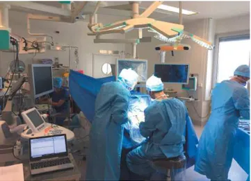
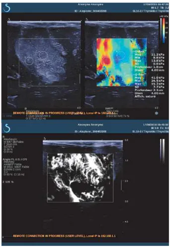
Neurosurgery. Fig. 1 shows the operating room during the surgical operation. We can see the Brainlab NeuroNavigation system, the SuperSonic Imagine Aixplorer ultrasound scanner and the computer with the SonicLab V12 research pack to upload the sequences. The surgeon places the conventionnal SL10-2 ultrasound probe, composed by 192 elements on the area of the brain to be studied. Different strategies are used during the surgery. Firstly, we record the in vivo Bmode image, the Young modulus image and the AngioPlus power Doppler image with the commercial mode of the Aixplorer. Fig. 2 depicts the B-mode images (upper left) of a heterogeneous glioma. SWE imaging show a Young modulus of 11.2KPa in the center of the glioma and 41kPa in the left part of the glioma. Power Doppler imaging shows the glioma perfusion. We can clearly see the details of the tumor vasculature and a small area at the bottom of the image where the tumor is necrotic. This real-time commercial imaging mode allows us to select precisely the region of interest in the tumor for more advanced analysis using the research sequence.
Fig. 1. Operating room of the Neurosurgery department Tours hospital. We identify the Brainlab neuronavigation system, the US Aixplorer imaging system and the computer to upload the research sequences.
Fig. 2. B-mode, Young modulus and Power Doppler AngioPlus images of a heterogeneous cerebral glioma obtained in vivo with the Aixplorer commercial mode
II.A. Shear Wave Spectroscopy (SWS)
We want to assess the frequency dependence of the shear wave speed inside a small region of the glioma. The SWE sequence is upload on the Aixplorer with the SonicLab software. The SWS experiment is based on two steps, the generation of the shear waves and ultrafast imaging of their propagation. For a detailed description of basic principles and phase estimation algorithm, the reader can refer to [1]. Data are transfered on a computer for off line beamforming and signal processing. Fig. 3 visualizes the Shear wave propagation induced inside the cerebral glioma by the US radiation force nonlinear effect. SW are presented at three different times (t1=0.29msec, t2=0.96msec and t3=1.7msec) superposed to the Bmode image at the same position than in Fig. 2. The Bmode image is obtained from the SWE sequence and has not the same quality than in Fig. 2. A small rectangle shows the region of interest. Axial lignes between 18.9mm and 19.8mm are summed and the SW is analysed from 20mm to 27mm in the lateral direction (corresponding to the rectangle in Fig. 3). SW elastogram is shown in Fig. 4. From a Voigt model, the elastic modulus µ = 2.2kP a(E = 6.6kP a) and the shear viscous modulus is η= 0.7P a.s
Fig. 3. Shear wave propagation induced inside the cerebral glioma by the US radiation force nonlinear effect. SW are presented at three different times (t1=0.29msec, t2=0.96msec and t3=1.7msec) superposed to the Bmode image at the same position than in Fig. 2
Fig. 4. Elastogram in Time t and space x domain and the SW dispersion in function of the frequency in the [131-568]Hz bandwidth. From a Voigt model we obtain µ = 2.2kP a(E = 6.6kP a) and the shear viscous modulus is η= 0.7P a.s
II.B. Perfusion and tissue/noise filtering A. RPCA Theory
Recently, adaptive spatiotemporal singular values decom-position (SVD) clutter filtering was proposed to extract the blood part of the scattered US echos. This made it possible to quantify the small variations in blood flow related to neural activation and made possible to carry out the first brain activation mapping in vivo [2]. Through this decomposition, the filtering strategy is based on empirical thresholding the correlation of spatial singular vectors magnitude. This leads to the adaptive separation of the sub-spaces corresponding to respectively the tissues and the blood flow [3]. This correlation matrix have different shape depending on whether it is in vitro (rather square blood sub-space) or in vivo (rather elliptical blood sub-space) data. Moreover, the decorrelation of the singular spatial vectors corresponding to blood is link to the magnitude of the blood flow. These vectors decorrelate faster when the flow is fast. This further modifies the shape of this matrix. This shape must have an impact on the relevance of the thresholds, especially in the case where these sub-spaces have slowly surface variation (low 2D gradients). The choice of the optimal threshold is thus not obvious in most of practical applications. For this study, this SVD method will be considered as the reference method.
An alternative solution consists in using robust principal component analysis (RPCA) techniques [4]–[7]. We denote by S ∈ CNzNx×Nt the Casorati matrix obtained from 3D
IQ complex (number) Doppler data, recorded via ultrafast imaging, with depth Nz, probe width Nxand acquisition time
Alternatively, S can be modelled as:
S= T + B + N , (1) with T ∈ CNzNx×Nt the tissue, B ∈ CNzNx×Nt the blood,
and N ∈ CNzNx×Nt the noise matrices. Assuming that
the blood (i.e., the flow) is sparse and the tissue weakly changes or moves over time, reasonable assumptions in most of practical applications, it is thus possible to estimate B from S. Classically, sparsity is catched in a tractable manner by the l1-norm and weak changes or high correlation by the nuclear
norm||.||∗. This results in solving an inverse problem in which
the estimation of B and T , say ( ˆB, ˆT), can be obtained by minimizing the following function:
( ˆB, ˆT) = arg min
B,T||S − B − T || 2
F + λ||B||1+ ||T ||∗ (2)
where||.||2
F is the Frobenus norm. λ >0 is a hyper-parameter
to be tuned, balancing the trade-off between the sparsity of the blood and low-rankness of the tissues. To solve the convex optimization problem above, many algorithms exist in the literature, such as the one proposed in [5]. This method is referred to as robust principal component analysis (RPCA). A comprehensive review and analysis of this kind of methods can be found in [8]. Some of these techniques have been revisited in ultrasound imaging by Bayat et al. [9] who solved this problem, assuming that B is sparse in the Fourier domain, or by Sathyanarayna et al. [10] who investigated the same problem via a sparse coding through expressing B in a specific dictionary. A detailed decription of the algorithm for solving (2) can be find in [11]. Our method does not explicitly take into account the limited resolution of Doppler data caused by the system impulse response (i.e., the PSF). Very recent work of [11] proposed a joint method coupling deconvolution process and RPCA inverse problem for filtering. This method is not addressed in this work .
B. Experimental glioma perfusion
A sequence consisting of 3 tilted plane waves [-5°, 0°, +5°] obtained at a frame rate of 3 kHz during 1 s was uploaded in the Aixplorer. After compounding, the resulting image frame rate is PRF=1kHz. Acquired data were exported for off-line processing. Note that the thresholds used within SVD were manually tuned to their best values. Fig. 6 shows the Power Doppler summed from the 1000 filtered complex IQ frames. Our perfusion results are not unfortunately obtained during the same acquisition than the SWE part of this paper. These data on Fig. 6 have been obtained from a previous brain surgery. The tumor is situated from the distance 5mm to 30mm depth. Some fine vascularisation is visible on the upper part of the tumor which is not very vascularized. A central region is necrotic between 17mm and 23mm with a circular shape. On the lower part of the tummor, we can see that this Power Doppler image shows a fine vascularization. The image has the same dynamic (-50dB) for comparison. RPCA image seems little more noisier than the SVD method on the lower region of the image.
Fig. 5. Sensitive Doppler sequence consisting in 3 tilted US plane waves [-5°, 0°, +5°] obtained at a frame rate of 3 kHz during 1 s. After compounding, the resulting image frame rate is PRF=1kHz. 1000 images are acquired, i.e a time acquisition of 1sec
(a) Adaptive spatiotemporal SVD clutter filtering method
(b) Robust principal component analysis (RPCA) clutter filtering method
Fig. 6. Comparison of adaptive spatiotemporal SVD method and robust principal component analysis (RPCA) method for ultrafast US clutter filtering. Experimental Power Doppler imaging obtained in vivo on a human cerebral glioma.
III. CONCLUSION
We present the research that we are conducting on tumors in vivo during surgery. We propose to characterize the vis-coelastic behavior of the a small tumor ROI through a Voigt model applied on the SW speed dispersion curve in function of the frequency. The tumor is heterogeneous in terms of Bmode and elasticity image. We will correlate these data with the grade of the tumor obtained by histology. The SWE sequence associated with our phase estimation algorithm is well applied to this problem. The dispersion curve is of good quality in our analysis bandwidth. For perfusion, the sensitive sequence with 1000 temporal resulting beamformed IQ data give enough sensibility to visualize the small vessels inside the tumors. Two methods for blood and tissue-sub-space separation were compared in this work. SVD is a straightforward and easy-to-implement method, based on the correlation of spatial singular vectors magnitude. However, it requires a manual tuning of the two thresholds needed to separate tissue, blood and noise components, to visualize only the blood flow. From this perspective, SVD may not be reproducible from one operator to another. The selection of blood directly depends on the selection of these thresholds, without any further control on the result. RPCA algorithm is more complex than classical SVD, It estimates the two main components, tissue and blood, by exploiting their low-rank or sparse properties, leading to an automatic method without the need of manually tuning the parameters. And for the same sequences, we find that RPCA yields the same result if there is no change in the parameters λ and µ. Moreover, it has parameters on which one can play to optimize the result. This method is therefore more advantageous and easier to handle and we are still in the process of improving this method. We are currently conducting in vitro flow phantom tests to accurately quantify the quality of the different filtering methods we are developing.
REFERENCES
[1] T. Deffieux, G. Montaldo, and Fink. Tanter, M, “Shear wave spec-troscopy for in vivo quantification of human soft tissues visco-elasticity,” IEEE Transactions on Medical Imaging, vol. 28, no. 3, pp. 313–, Mar. 2009.
[2] C. Demené, T. Deffieux, M. Pernot, B. F. Osmanski, V. Biran, J. L. Gennisson, L. A. Sieu, A. Bergel, S. Franqui, J. M. Correas, I. Cohen, O. Baud, and M. Tanter, “Spatiotemporal clutter filtering of ultrafast ultrasound data highly increases doppler and fultrasound sensitivity,” IEEE Transactions on Medical Imaging, vol. 34, no. 11, pp. 2271–2285, Nov. 2015.
[3] J. Baranger, B. Arnal, F. Perren, O. Baud, M. Tanter, and C. Demené, “Adaptative spatiotemporal svd clutter filtering for ultrafast doppler imaging using similarity of spatial singular vectors,” IEEE Transactions on Medical Imaging, vol. 37, no. 7, pp. 1574–1586, July 2018. [4] R. A. Maronna, “Robust m-estimators of multivariate location and
scatter,” Ann. Stat., vol. 4, no. 1, pp. 51–67, 1976.
[5] J. Wright, A. Ganesh, S. Rao, Y. Peng, and Y. Ma, “Robust principal component analysis: Exact recovery of corrupted low-rank matrices via convex optimization,” Proc. Neural Inf. Process. Syst, pp. 1–9, 2009. [6] F. Torre and M. Black, “Robust principal component analysis for
computer vision,” Proc. Int. Conf. Comput. Vis., pp. 362–369, 2001. [7] S. J. Devlin, R. Gnanadesikan, and J. R. Kettenring, “Robust estimation
of dispersion matrices and principal components,” J. Amer. Stat. Assoc., vol. 76, no. 374, pp. 354–362, 1981.
[8] Thierry Bouwmans, Andrews Sobral, Sajid Javed, Soon Ki Jung, and El-hadi Zahzah, “Decomposition into low-rank plus additive matrices for background/foreground separation: A review for a comparative evaluation with a large-scale dataset,” Computer Science Review, vol. 23.
[9] M. Bayat and M. Fatemi, “Concurrent clutter and noise suppression via low rank plus sparse optimization for non-contrast ultrasound flow doppler processing in microvasculature,” in IEEE International Conference on Acoustics, Speech and Signal Processing ICASSP’2018, Calgary, Canada, 2018.
[10] S. G. Sathyanarayna, S. T. Acton, and J. A. Hossack, “Suppression of clutter by rank adaptive reweighted sparse coding,” in IEEE International Ultrasonics Symposium IUS’2017, Washington DC, USA, 2017.
[11] H. Shen, C.Barthelemy, E.Khoury, I.Zemmoura, JP.Remeniéras, A.Basarab, and D.Kouamé, “High-resolution and high-sensitivity blood flow estimation using optimization approaches with application to vas-cularization imaging,” in IEEE International Ultrasonics Symposium IUS’2019, Glasgow, UK, Oct. 2019.
![Fig. 5. Sensitive Doppler sequence consisting in 3 tilted US plane waves [-5°, 0°, +5°] obtained at a frame rate of 3 kHz during 1 s](https://thumb-eu.123doks.com/thumbv2/123doknet/12898142.371175/5.892.454.831.132.294/sensitive-doppler-sequence-consisting-tilted-plane-waves-obtained.webp)
