HAL Id: hal-01291681
https://hal.sorbonne-universite.fr/hal-01291681
Submitted on 21 Mar 2016HAL is a multi-disciplinary open access
archive for the deposit and dissemination of sci-entific research documents, whether they are pub-lished or not. The documents may come from teaching and research institutions in France or abroad, or from public or private research centers.
L’archive ouverte pluridisciplinaire HAL, est destinée au dépôt et à la diffusion de documents scientifiques de niveau recherche, publiés ou non, émanant des établissements d’enseignement et de recherche français ou étrangers, des laboratoires publics ou privés.
Distributed under a Creative Commons Attribution| 4.0 International License
Use of Prokaryotic Two-component and Metazoan-type
Tyrosine Kinase Signaling
Pauline Schaap, Israel Barrantes, Pat Minx, Narie Sasaki, Roger W.
Anderson, Marianne Bénard, Kyle K. Biggar, Nicolas E. Buchler, Ralf
Bundschuh, Xiao Chen, et al.
To cite this version:
Pauline Schaap, Israel Barrantes, Pat Minx, Narie Sasaki, Roger W. Anderson, et al.. The Physarum polycephalum Genome Reveals Extensive Use of Prokaryotic Two-component and Metazoan-type Ty-rosine Kinase Signaling. Genome Biology and Evolution, Society for Molecular Biology and Evolution, 2016, 8 (1), pp.109-125. �10.1093/gbe/evv237�. �hal-01291681�
& & &
Main Manuscript
The Physarum polycephalum Genome Reveals Extensive
Use of Prokaryotic Two-component and Metazoan-type
Tyrosine Kinase Signaling
Pauline Schaap1, Israel Barrantes2, Pat Minx3, Narie Sasaki4, Roger W. Anderson5, Marianne Bénard6, Kyle K. Biggar7, Nicolas E. Buchler8, Ralf Bundschuh9, Xiao Chen10, Catrina Fronick3, Lucinda Fulton3, Georg Golderer11, Niels Jahn12, Volker Knoop13, Laura F. Landweber10, Chrystelle Maric14, Dennis Miller15, Angelika A. Noegel16, Rob Peace17, Gérard Pierron14, Taeko Sasaki4, Mareike Schallenberg-Rüdinger13, Michael Schleicher18, Reema Singh1, Thomas Spaller19, Kenneth B. Storey17, Takamasa Suzuki20, Chad Tomlinson3, John J. Tyson21, Wesley C. Warren3, Ernst R. Werner11, Gabriele Werner-Felmayer11, Richard K. Wilson3, Thomas Winckler19, Jonatha M. Gott22, Gernot Glöckner16,23*, Wolfgang Marwan2*
*Senior authors, listed in alphabetical order
© The Author(s) 2015. Published by Oxford University Press on behalf of the Society for Molecular Biology and Evolution. This is an Open Access article distributed under the terms of the Creative Commons Attribution Non-Commercial License (http://creativecommons.org/licenses/by-nc/4.0/), which permits non-commercial re-use, distribution, and reproduction in any medium, provided the original work is properly cited. For commercial re-use, please contact
journals.permissions@oup.com
at UPMC on March 21, 2016
http://gbe.oxfordjournals.org/
&
Author details
1College of Life Sciences, University of Dundee, DD15EH, UK. 2Magdeburg Centre for Systems Biology and Institute for Biology, University of Magdeburg,
Pfälzerstrasse 5, 39106 Magdeburg, Germany. 3The Genome Institute, Washington University School of Medicine, St Louis, MO 63108, USA. 4Department of Biological Sciences, Graduate School of Science, Nagoya University, Furocho, Chikusaku, Nagoya, 464-8602 Aichi, Japan. 5Department of Molecular Biology and
Biotechnology, University of Sheffield, Firth Court, Western Bank, Sheffield S10 2TN, UK. 6UPMC Univ Paris 06, Institut de Biologie Paris-Seine (IBPS), CNRS UMR-7622, F-75005, Paris, France. 7Biochemistry Department, Schulich School of Medicine and Dentistry, Western University, London, ON, N6A 5C1, Canada. 8Center for Genomic and Computational Biology, Department of Biology, and Department of Physics, Duke University, Durham, NC 27710, USA. 9Department of Physics, Department of Chemistry & Biochemistry, Division of Hematology, Department of Internal Medicine, Center for RNA Biology, The Ohio State University, 191 West Woodruff Avenue, Columbus, OH 43210-1117, USA. 10Department of Ecology & Evolutionary Biology, Princeton University, Princeton, NJ 08544, USA. 11Biological Chemistry, Biocenter, Innsbruck Medical University, 6020 Innsbruck, Austria. 12Genome Analysis, Leibniz Institute on Aging - Fritz Lipmann Institute (FLI), Beutenbergstraße 11, 07745 Jena, Germany. 13IZMB - Institut für Zelluläre und Molekulare Botanik, Universität Bonn, Kirschallee 1, 53115 Bonn, Germany. 14Institut Jacques Monod, CNRS UMR7592,
at UPMC on March 21, 2016
http://gbe.oxfordjournals.org/
& !
Université Paris Diderot Paris7, 75013 Paris, France.15The University of Texas at Dallas, Biological Sciences, FO 31, 800 West Campbell Road, Richardson, TX 75080, USA. 16 Institute for Biochemistry I, Medical Faculty, University of Cologne, Joseph-Stelzmann-Straße 52, 50931 Cologne, Germany. 17Carleton University, 1125 Colonel By Drive, Ottawa, Ontario K1S 5B6, Canada. 18Institute for Anatomy III / Cell Biology, BioMedCenter, Ludwig-Maximilians-Universität, Grosshaderner Str. 9, 82152 Planegg-Martinsried, Germany. 19Institut für Pharmazie, Friedrich-Schiller-Universität Jena, Semmelweisstraße 10, 07743 Jena, Germany. 20Department of Biological Sciences, Graduate School of Science and JST ERATO Higashiyama Live-holonics Project, Nagoya University, Furocho, Chikusaku, Nagoya, 464-8602 Aichi, Japan. 21Department of Biological Sciences, Virginia Polytechnic Institute and State University, Blacksburg, VA 24061, USA. 22Center for RNA Molecular Biology, Case Western Reserve University, School of Medicine, 10900 Euclid Ave, Cleveland, OH 44106-4960, USA. 23Leibniz Institute of Freshwater Ecology and Inland Fisheries (IGB), Müggelseedamm 301, 12587 Berlin.
at UPMC on March 21, 2016
http://gbe.oxfordjournals.org/
&
Abstract
Physarum polycephalum is a well-studied microbial eukaryote with unique
experimental attributes relative to the other experimental model organisms. It has a sophisticated life cycle with several distinct stages including amoebal, flagellated, and plasmodial cells. It is unusual in switching between open and closed mitosis according to specific life cycle stages. Here we present the analysis of the genome of this enigmatic and important model organism and compare it with closely related species. The genome is littered with simple and complex repeats and the coding regions are frequently interrupted by introns with a mean size of 100 bases.
Complemented with extensive transcriptome data, we define ca. 31,000 gene loci, providing unexpected insights into early eukaryote evolution. We describe extensive use of histidine kinase based two-component systems and tyrosine kinase signaling, the presence of bacterial and plant type photoreceptors (phytochromes,
cryptochrome, and phototropin) and of plant-type pentatricopeptide repeat proteins, as well as metabolic pathways, and a cell cycle control system typically found in more complex eukaryotes. Our analysis characterizes P. polycephalum as a prototypical eukaryote with features attributed to the last common ancestor of Amorphea, i.e. the Amoebozoa and Opisthokonts. Specifically, the presence of tyrosine kinases in Acanthamoeba and Physarum as representatives of two distantly related subdivisions of Amoebozoa argues against the later emergence of tyrosine kinase signaling in the opisthokont lineage and also against the acquisition by horizontal gene transfer.
at UPMC on March 21, 2016
http://gbe.oxfordjournals.org/
& & !
Key words: Amoebozoa, tyrosine kinase receptor, two-component system,
signaling, phytochrome
Introduction
P. polycephalum belongs to the Amoebozoa, the sister group to the Opisthokonts
(i.e. fungi and animals) (Cavalier-Smith 2003) which both together form the supergroup Amorphea (Adl, et al. 2012). In the course of its complex life cycle (Fig. 1A), P. polycephalum is able to differentiate into various specialized cell types depending on environmental conditions (Burland, et al. 1993). Macroscopic
multinucleate plasmodial cells contain a naturally synchronous replicating and differentiating population of nuclei and can grow to tens or even hundreds of cm in size. Individual pieces cut off from a single plasmodium maintain synchrony,
providing unique experimental options for single cell biology. The occurrence of open and closed mitosis in amoebae and plasmodia, respectively, is another well-known aspect of its rich cell biology (Aldrich and Daniel 1982; Dove, et al. 1986; Sauer 1982). Accordingly, P. polycephalum has served as a classical model organism in cell and developmental biology since the early 1960s and has been used extensively to study cell cycle regulation, cell differentiation (amoeba-flagellate transition,
spherulation, sporulation), cell fusion, DNA replication, developmental gene expression, histone modification, sensing and response (e.g. chemotaxis) (for reviews see (Aldrich and Daniel 1982; Burland, et al. 1993; Dove, et al. 1986; Sauer 1982)). More recently, P. polycephalum plasmodia have been used for studies
ranging from cell biology and biophysics to soft matter computing for path finding, the biosensory control of robots, or the generation of music (Braund and Miranda 2014;
at UPMC on March 21, 2016
http://gbe.oxfordjournals.org/
& Tero, et al. 2010; Tsuda, et al. 2007). Plasmodial cells also contain thousands of mitochondria, providing the means to study the highly unusual form of insertional RNA editing used to generate functional mitochondrial transcripts.
From the evolutionary biology point of view, P. polycephalum is interesting because of its placement into the Amoebozoa, where the genomes of several
distantly related representatives (A. castellanii, E. histolytica, etc.) are already known (Fig. 1B). The model organism most closely related to P. polycephalum for which a genome sequence has previously been determined is Dictyostelium discoideum (Eichinger, et al. 2005). The two model species differ in several important aspects. D.
discoideum is a cellular slime mold where differentiation occurs in developmentally
interacting cells while in the acellular slime mold P. polycephalum, a single, multinucleate plasmodial cell differentiates in a manner reminiscent of Drosophila embryos or other developing syncytia. A comparison of P. polycephalum and D.
discoideum may help to elucidate the different evolutionary trajectories the members
of the two branches, cellular and acellular slime molds, have taken in establishing sophisticated life cycles that are so different.
The established lab strains of D. discoideum do not form viable progeny after mating, which hinders genetic experiments, whereas mating of strains and
segregation of meiotic progeny in P. polycephalum is under ready experimental control. The classical genetics are well-established in P. polycephalum and the possibility to experimentally switch the life cycle between haploid and diploid stages (Fig. 1A) is very useful for the generation and analysis of mutants (Dee 1987, 1982). Amoebal strains derived from different natural plasmodial isolates, such as Wis-1 and Wis-2, (see Appendix A of Supplementary Methods, Results and Discussion) allow forward genetic approaches by mapping genes identified in phenotypic screens.
at UPMC on March 21, 2016
http://gbe.oxfordjournals.org/
& & !
Mendelian analysis based on the co-segregation of phenotype and single nucleotide polymorphisms (SNPs) (Schedl and Dove 1982) can be performed by sequencing pools of cDNAs of phenotype-carrying segregants (Barrantes and Marwan,
unpublished results). These high throughput methods combined with Mendelian genetics mitigate current difficulties of reproducibly making transgenic lines. Methods for knocking down gene expression have recently been developed for P.
polycephalum (Haindl and Holler 2005; Itoh, et al. 2011), opening up additional
experimental avenues. Here, we present an overview of the whole genome of P.
polycephalum, focussing on some of its peculiar features such as its surprisingly
extended signaling system.
Materials and Methods
Assembly
Combined sequence reads from different libraries (including 3 kb and 8 kb mate pairs) were assembled using the Newbler software version 2.6 (Margulies et al 2005), and the Newbler contigs were then assembled into scaffolds using Bambus version 2.3 (Pop et al 2004), and a custom transcriptome-guided scaffolding algorithm. The 10.0 draft assembly is comprised of 69,687 scaffolds with an N50 scaffold length of 54,474kb and an N50 contig length of 2.2kb. The assembled coverage is 54.6X, and the assembly spans over 220Mb. We removed from the
at UPMC on March 21, 2016
http://gbe.oxfordjournals.org/
& assembly all contaminating sequences, trimmed vectors (X), and ambiguous bases (N). Additionally, shorter contigs (≤200bp) were removed prior to public release. The draft genome sequence of P. polycephalum was deposited in the
DDBJ/EMBL/GenBank database (Accession Number ATCM00000000.3). The genome data can also be downloaded from http://www.physarum-blast.ovgu.de/.
Generation of a reference transcriptome
All transcript data were assembled according to their sample origin since previous analyses showed that genes have a wealth of introns and thus alternative splicing could play a role (Glöckner, et al. 2008). This initial analysis yielded 733,443 potential transcripts.
After clustering with usearch (Edgar 2010) we obtained 31,770 clusters each represented by the largest sequence in a cluster. The mean length of transcripts in the thus constructed reference transcriptome is 1264 bases. These transcripts were mapped onto the genome using blat (Kent 2002). Only 485 transcripts could not be mapped to the genome sequence, of which only 90 have a blast score of 250 or higher in a search against the reference sequence database (NCBI July 2013). Fifteen of these 90 appear to be of P. polycephalum mitochondrial origin and a further 13 are certainly contaminants from the sequencing process since they have identity values higher than 85 % to some bacteria and mouse sequences. Thus, only a minor fraction of transcripts is not represented by the assembled genome. Manual inspection of some transcript/genome alignments revealed that transcripts frequently span gaps in the scaffolds which account for missing small exons. Inability to
assemble genome junctions at these positions is likely caused by the low complexity of intron sequences and the small size of the missing exons. Since contiguity of a
at UPMC on March 21, 2016
http://gbe.oxfordjournals.org/
& !
sequence is required for accurate gene prediction, we relied heavily on the transcript data for annotation and analysis. The Transcriptome Reference Assembly has been deposited at DDBJ/EMBL/GenBank under the accession GDRG00000000. The version described in this paper is the first version, GDRG01000000. All transcriptome sequence data and files containing the gene loci-transcript conversion, the transcript map, and the automatic annotation of the reference transcriptome can be
downloaded from http://www.physarum-blast.ovgu.de/.
Gene prediction and construction of gene loci
For an ab initio gene prediction we used Augustus (Stanke, et al. 2004). The transcriptome data were used to train this program according to the manual and to provide splice information for the algorithm. The program itself was run with default values. This way we predicted 47779 protein coding genes. We noticed, however, that the fragmented nature of the genome sequence and missing contiguity within scaffolds led to an overprediction. To get a better estimate of the number of protein coding genes in the P. polycephalum genome we tried to combine gene prediction and reference transcriptome for the definition of gene loci. To this end we mapped the reference transcriptome data to the genome. We then fused predicted genes and reference transcripts to gene loci if predicted genes were covered by the same
reference transcript and/or if predicted genes were present in the near vicinity (<50 bases apart) and at least one of them was covered or partially covered by a transcript sequence. Predicted genes without transcript coverage and no transcript in the near vicinity (< 50 bp apart) were counted as independent gene loci. In this way we defined 34438 gene loci, of which 17280 are not supported by transcript data (Table 1).
at UPMC on March 21, 2016
http://gbe.oxfordjournals.org/
& &
Small RNAs
For the definition of small RNAs we used RNAseq transcript data from a previous experiment (Bundschuh, et al. 2011). Since this specific experiment was performed on mitochondrial preparations, we only expect the more abundant nuclear small RNAs to be observed. However, the absence of poly-A selection and the use of a small RNA kit in library preparation promised a view orthogonal to traditional RNA-Seq approaches.
For detailed information on strains used, preparation of nucleic acids, and bioinformatic methods see Supplementary File 1.
Results and Discussion
Genome properties and reference transcriptome
The P. polycephalum genome proved to be highly recalcitrant for assembly owing to abundant and frequently long (mononucleotide or pyrimidine-rich) repeat stretches present in intergenic regions or within its numerous and highly variable intron sequences. Construction of gene models therefore heavily relied on
accompanying transcriptome data (see Materials and Methods).
The genetic material used for sequencing the P. polycephalum genome was derived from axenic, haploid amoebal cells (strain LU352). The assembly of 454 and paired end Illumina reads with an estimated coverage of 50x (calculating with a
at UPMC on March 21, 2016
http://gbe.oxfordjournals.org/
& & !
genome size of 250 Mb) resulted in scaffolds covering 182 Mb. However, assembling the genome has been challenging due to extremely long stretches of di-, tri-, and tetranucleotide repeats and large homopolymeric tracts, which likely lead to polymerase slippage and premature polymerase termination in vitro. The
unresolvable sequence patches cause innumerable gaps in the final sequence, resulting in a mean scaffold size of only slightly larger than 2 kb. PacBio sequencing yielded only minimal improvement. We therefore tested whether our assembly captures most of the coding information of the genome by investigating the primary metabolism capacity encoded by the assembly. We found that all expected pathways were entirely present and therefore conclude that the current assembly is complete enough for further analysis.
Gene content and introns
To further evaluate gene content and completeness, we generated transcriptome data sets from multiple life cycle stages and constructed a reference transcriptome (see Materials and Methods).
We next trained the gene prediction program augustus (Stanke, et al. 2004) with sequences derived from mapped transcripts. Gene predictions using the splicing information from all transcripts initially yielded more than 47,000 gene models (Table 1). However, since in the current assembly some shorter introns and exons are missing in scaffolds due to simple repeat sequences at both ends, we used the reference transcripts together with the gene predictions to define gene loci.
Transcripts and predicted genes overlapping each other or being in close vicinity on the assembly (in the range of an intron length) were fused to a common gene locus. A total of 34,438 gene loci were defined in this way (Table 1), half of which are
at UPMC on March 21, 2016
http://gbe.oxfordjournals.org/
& represented by transcripts (17,158). The gene loci with corresponding transcripts have a mean length of 1951 bases, while those without transcript support have a mean length of only 579 bases. This suggests that the majority of unsupported gene loci may represent false positive predictions, which is fairly typical of highly
fragmented genome assemblies.
There are scores of complex repeats in the genome, which contains a minimum of 484 integrase domains (PF00665) and 1014 reverse transcriptase domains (PF07727 and PF00078). This greatly exceeds the number present in other Amoebozoa species such as A. castellanii and D. disoideum, which contain only 38 and 118 reverse transcriptase domains, respectively. In total, P. polycephalum
complex repetitive elements contribute approximately 3 Mb or 1.3 % to the size of the genome (Table S4). Most P. polycephalum genes contain multiple introns, many of which are composed of extended repeats flanked by splicing signals. The median size of the 160,988 introns confirmed by transcript data is 231 bases, and the total number of predicted introns is 676,387. Thus, the mean number of confirmed introns per gene is approximately 5. If only the genes with transcript data are counted, the number of introns per gene increases to well above 9. This intron frequency is higher than the estimated number of introns in the last common ancestor of eukaryotes (Zhou, et al. 2014). Thus, P. polycephalum has likely acquired introns during its species history.
Domain analysis
Using iprscan (Zdobnov and Apweiler 2001) we defined the domain content for each gene locus. A comparison with A. castellanii and D. discoideum showed that P.
polycephalum has the highest number of domain hits of the three organisms,
at UPMC on March 21, 2016
http://gbe.oxfordjournals.org/
& & !
reflecting the larger genome and the higher gene count. In terms of distinct domains,
A. castellanii and P. polycephalum are similar, whereas fewer types of domains have
been identified in D. discoideum, suggesting domain loss in the social amoebae (Table 2). Further analysis of domains revealed that 304 domains were present at least once in the genomes of P. polycephalum and A. castellanii, but absent from D.
discoideum (Table S2). Among these are a considerable number of domains
associated with signaling functions. Domains enriched compared to those of A.
castellanii and D. discoideum include transposon domains and, again, domains
associated with signaling functions (Table S3).
Non-coding RNAs
Small non-coding RNAs perform important functions in all organisms, but can be difficult to annotate de novo due to their short length and their poorly-defined
sequence patterns. However, data obtained in a previous mitochondrial RNA-Seq experiment allowed us to annotate a number of small non-coding P. polycephalum RNAs encoded in the nuclear genome. This resulted in the confirmation of many known small P. polycephalum RNAs (Supplemental Spreadsheet 1, 1st sheet). In addition, we found 24 novel transfer RNAs, the spliceosomal U2 and U6 RNAs, 30 box C/D snoRNAs, 9 novel lncRNAs, which clustered into three groups by sequence similarity, as well as 24 RNAs shorter than 200 nucleotides clustering into three groups and 28 singleton RNAs shorter than 200 nucleotides (Supplementary Spreadsheet 1, 2nd sheet). These latter RNAs and the box C/D snoRNAs have no detectable sequence similarity with any sequence in the non-redundant nucleotide database.
at UPMC on March 21, 2016
http://gbe.oxfordjournals.org/
We ran tRNAscan-SE (PMID: 9023104) with default parameters on the genome, which resulted in 319 putative tRNA loci and 248 unique putative tRNA sequences (Supplementary Spreadsheet 1, 3rd sheet). These included all but one (tRNA-Tyr1 in Supplementary Spreadsheet 1, 2nd sheet) of the 56 new tRNAs identified from the sequencing experiment as well as all previously annotated P. polycephalum tRNAs with the exception of tRNA-Lys1, tRNA-Ser2, and tRNA-Asp1. A total of 201 putative tRNA loci with 156 unique putative tRNA sequences were novel. These included tRNAs for all 20 amino acids as well as 6 putative tRNA-SeC loci (each with a unique sequence) and four putative tRNA loci of undetermined type (each with a unique sequence).
PPR proteins
The P. polycephalum genome is particularly rich in genes encoding
pentatricopeptide repeat (PPR) proteins. PPR proteins are sequence-specific RNA-binding proteins that act in diverse processes of RNA maturation, mainly in
mitochondria and chloroplasts (Schmitz-Linneweber and Small 2008). The PPR motif is a loosely conserved 35 aa motif identifying individual ribonucleotides on a one-repeat-per-nucleotide basis (Barkan, et al. 2012). As in other taxa, the P.
polycephalum proteins contain PPR arrays of variable sizes ranging from only a few
up to more than 20 tandem PPRs in individual proteins. With some 100 PPR protein genes, P. polycephalum features much more than tenfold the number found in other slime mold genomes such as Polysphondylium pallidum or D. discoideum. In fact, the set of PPR proteins present in P. polycephalum is significantly larger than in other
at UPMC on March 21, 2016
http://gbe.oxfordjournals.org/
& Amoebozoa and, more broadly, within the Amorphea super-domain at large. Indeed, larger numbers of PPR proteins have hitherto only been observed in Dinoflagellates and in land plants, where large families of PPR proteins were initially discovered (Small and Peeters 2000). Two observations are particularly intriguing: In contrast to other Amorphea, P. polycephalum has a highly derived mitochondrial genome, the transcripts of which are affected by abundant and diverse types of RNA editing, including alterations of transcript sequences by nucleotide insertions and base conversions (Bundschuh, et al. 2011; Mahendran, et al. 1991; Takano, et al. 2001). We speculate that some of the PPR proteins in P. polycephalum take part in these processes. Most notably, 16 of the P. polycephalum PPR proteins are “plant-like” in carrying a carboxyterminal DYW domain with cytidine deaminase similarity (Fig. S21) (Schallenberg-Rüdinger, et al. 2013). Several such DYW-type PPR proteins have been characterized as site-specific C-to-U editing factors in plants. Intriguingly, the first discovery of PPR proteins with a DYW domain outside of land plants in the genome of Naegleria gruberi led to the subsequent discovery of C-to-U editing in the mitochondria of this heterolobosean protist (Knoop and Rüdinger 2010; Rüdinger, et al. 2011). As in plants and Naegleria, the residues likely coordinating a zinc ion for deaminase activity are highly conserved in the P. polycephalum DYW domains. Given that in its natural environment there is direct physical contact with the slime mold growing on decaying plant materials, horizontal gene transfer (HGT) from plants may be a possible source of the P. polycephalum PPR genes. However, no evidence for particularly high similarity of any Physarum PPR protein to a homologue in
another taxon indicating a recent HGT has been found. The DYW-type PPR protein families of Physarum polycephalum, Naegleria gruberi and the moss Physcomitrella
patens, for example, cluster taxon-wise without evidence for recent HGT (Fig. S21C).
at UPMC on March 21, 2016
http://gbe.oxfordjournals.org/
& Moreover, whereas plant DYW-type PPR proteins feature characteristic “PLS-type” PPR arrays with alternating long (L) and short (S) variants of the classic (P) PPRs, most Physarum DYW-type homologues display “LS”-type repeats (Fig. S21A).
Metabolism and the cytoskeleton
Pteridine metabolism
Pteridines comprise a group of molecules that contain pteridine (pyrimido [4,5-b] pyrazine), a bicyclic ring system, as a common structural element. Pteridines (e.g. folate, biopterin, riboflavin etc.) function as important cofactors (or their precursors) of various enzymatic reactions. Pterines are synthesized from the common precursor GTP (guanosine 5’ triphosphate).
Analysis of the P. polycephalum genome for pteridine metabolic enzymes
revealed that it is unusual in encoding enzymes for all common pteridine biosynthetic pathways (for the biosynthesis of molybdopterin, tetrahydrofolate,
tetrahydrobiopterin, 7-aminomethyl deazaguanine, and riboflavin) and, in addition, the enzymes alkylglycerol monooxygenase and nitric oxide synthases. Alkylglycerol monooxygenase and nitric oxide synthase, in its full length form with oxygenase and reductase domains, are regularly found only in animals. Nitric oxide synthases are required for sporulation in P. polycephalum by acting via cGMP signaling (Golderer, et al. 2001). This link between animal-type NO synthases and cyclic nucleotide signaling, which is widely used in P. polycephalum (see Section Signaling), is
noteworthy and to date is unique in unicellular eukaryotes. This suggests an ancient origin of this typically animal-type signaling enzyme and its signal transduction mechanism via cGMP.
at UPMC on March 21, 2016
http://gbe.oxfordjournals.org/
& !
Another unique finding is that riboflavin biosynthetic enzymes are encoded as two tri-functional reading frames (Fig. S10) rather than by separate cistrons, a feature thus far not observed for sequences from any other species represented in Genbank.
P. polycephalum is much more diverse than D. discoideum in its pteridine
metabolic equipment. It contains enzymes for the biosynthesis of 7-aminomethyl-7-deaza guanine (preQ1), a precursor for the special tRNA base queuosine (Q), as well as for the biosynthesis of riboflavin. The reading frames for nitric oxide synthases found in P. polycephalum do not occur in the D. discoideum genome. Furthermore, another surprising feature is the comparatively frequent presence of homologous enzymes that catalyze the same enzymatic reaction, whereas most species including
D. discoideum use only one. These homologs share in the mean 72 % sequence
similarity, have the motifs required for enzymatic activity conserved, are expressed and are therefore expected to be enzymatically active. This assumption has been experimentally confirmed for the two inducible nitric oxide synthase homologs NOS2a and NOS2b (Messner, et al. 2009). The reasons for or advantages of this apparent redundancy are currently unknown. On the other hand, D. discoideum produces an additional unique pteridine, dictyopterin, an isomer of biopterin (Klein, et al. 1990) that is missing in P. polycephalum.
For the 24 genes analyzed in Table S5 encoding for 26 enzymes, closest
orthologs for twelve were found in Amoebozoa (eight of these in Dictyosteliida), six in bacteria, four in animals, three in Capsaspora owczarzaki, an Opisthokonta of
uncertain placing, and one in red algae (Table S5).
Shikimate pathway
at UPMC on March 21, 2016
http://gbe.oxfordjournals.org/
& The P. polycephalum genome also contains genes encoding enzymes for
complete shikimate and aromatic amino acid synthesis pathways, which are not present in either the social amoebae or Entamoebae (Richards, et al. 2006). In fungi, alveolates, and oomycetes a pentafunctional polypeptide is formed (AROM). This organization is also observed in P. polycephalum (gene locus 13138), strongly suggesting that this is the ancestral state of the shikimate pathway protein domain structure. In contrast, it was recently shown that A. castellanii has an unusual
arrangement of such pathway genes (Henriquez, et al. 2015), which might represent a later rearrangement within the Amoebozoa clade. Like all other free living
Amoebozoa so far, P. polycephalum contains a phosphoenolpyruvate carboxylase (gene_locus_38419), which is primarily found in bacteria and plants. The purpose of this enzyme could be to supply the TCA cycle with C4 bodies when they are required for amino acid biosynthesis.
Actin cytoskeleton and motor domains
The very intriguing oscillatory cytoplasmic streaming in P. polycephalum triggered extensive studies on the cytoskeleton. It was first reported by Noburo Kamiya (Kamiya 1940), i.e. long before actin and myosin had been identified as driving forces in muscle and non-muscle cells. At up to 1350 µm/sec, the rhythmic streaming in different developmental stages of P. polycephalum is among the fastest intracellular motilities known to date (Kamiya 1959; Wohlfarth-Bottermann 1979). For comparison: an average kinesin moves with a velocity of about 0.6 µm/sec along a microtubular track.
at UPMC on March 21, 2016
http://gbe.oxfordjournals.org/
& & !
In the current study many major families of actin-binding proteins have been found (Supplemental Spreadsheet 2). The profilins as members of G-actin sequestering proteins, fragmin as representative of the Ca2+-dependent F-actin severing proteins, the heterodimeric F-actin capping proteins (formerly 'Physarum beta-actinin'), F-actin crosslinking proteins like alpha-actinin and filamin, myosins as major motor proteins, and formin-like proteins with FH1 and FH2 domains are
present in comparable numbers as in D. discoideum and more complex organisms. Even peculiar actin isoforms like D. discoideum filactin, an actin with a large N-terminal extension (Joseph, et al. 2008) could be identified in the P. polycephalum genome.
A seemingly unique and exciting feature of the actin cytokskeleton in P.
polycephalum, however, is the presence of the actin-fragmin kinase (Contig 47652).
Fragmin, homologous to severin in D. discoideum and gelsolin in more complex organisms (Yin, et al. 1990), was first described by the Hatano group (Hasegawa, et al. 1980) and its 1:1 complex with actin was further investigated by Vandekerckhove and colleagues (Eichinger, et al. 1996; Gettemans, et al. 1992). The latter group isolated a kinase which phosphorylated actin, but only in the 1:1 actin-fragmin complex. Upon phosphorylation the activity of the actin-fragmin complex changed to an F-actin capping function and was, therefore, a putative regulator of fast changes in local viscoelasticities in the F-actin network, a prerequisite for cytoplasmic
streaming. Surprisingly, the protein sequence of the purified protein did not show any of the well known protein kinase domains (Hanks and Hunter 1995) and it could not be excluded that a minor contamination was responsible for actin phosphorylation. Only the crystallization of the protein finally showed that it was in fact a kinase whose structure was in its catalytic domain nearly identical to protein kinase A (Steinbacher,
at UPMC on March 21, 2016
http://gbe.oxfordjournals.org/
& & et al. 1999). These data lead to major conclusions: The actin-fragmin kinase is clearly an example for convergent evolution. There is no common ancestor as in the case of divergent evolution of conventional protein kinases. Thus, the evolutionary pressure is on the structure of the molecule in determining its function. There is no need to keep a distinct amino acid sequence with a certain arrangement of protein domains, just structure/function relationships count. Consequently, this type of actin phosphorylation in P. polycephalum is possibly not unique as suggested above. One cannot exclude convergent evolution of kinases with similar substrate specificity also in other organisms as these molecules may have completely unrelated sequences and are not easily detectable by sequence similarities. Perhaps most importantly: this example demonstrates the limitations of any whole genome approach. In the end only biochemistry and molecular cell biology will cross the border from 'gene products of unknown function' to proteins with known activities.
P. polycephalum possesses a wealth of motor domains. We searched all defined
gene loci for the presence of such domains. With this initial search we found 43 gene loci with a kinesin domain (PF00225), 23 gene loci with a dynein heavy chain domain (PF03028), and 53 gene loci with myosin motors (PF00063). Since the genome assembly is fragmented and thus gene loci likely do not always represent a whole gene, we further investigated these loci making use of the reference transcriptome library. This approach enabled us to define eight complete dyneins, one N-terminal half, and two N-terminal fragments. Ten further fragments of varying length show homology to the middle and C-terminal parts of dyneins. Thus, the P. polycephalum genome likely encodes at least eleven dynein proteins. This number is in the same range as in other species.
at UPMC on March 21, 2016
http://gbe.oxfordjournals.org/
& & !
With the same approach we defined fifteen complete or nearly complete myosin proteins and sixteen fragments of varying lengths. Five of the full length genes and seven of the fragments have the highest similarity to class VII unconventional myosins indicating an amplification of this family in P. polycephalum.
Kinesins have a wider length range than the other motor proteins and the regions outside the motor domain are highly variable. This makes it impossible to distinguish between full length genes and fragments as with the other motor domain proteins. Matching possible kinesin gene loci with transcript data yielded 39 defined loci. Three of these are small without transcript data. The mean length of the others is well
above 700 amino acids. Thus we conclude, that at least 36 kinesin genes reside in the P. polycephalum genome. Strikingly, most of the gene loci we defined here are represented in our reference transcriptome library indicating their expression.
Dynein domains and, much more pronounced, kinesin domains are
overrepresented in P. polycephalum as compared to two other organisms within the Amoebozoa, D. discoideum and A. castellanii (Table S3). The enrichment of dynein domains is likely due to the fact that P. polycephalum possesses flagella. It was shown previously that a plasmodial species, Reticulomyxa filosa, has amplified its complement of kinesin domain bearing genes, which may be associated with the increased requirements of intracellular transport in a huge cell (Glöckner, et al. 2014). We see this gene family expansion also in P. polycephalum, hinting at a common prerequisite for acquiring a plasmodial life stage.
at UPMC on March 21, 2016
http://gbe.oxfordjournals.org/
& &
Cell cycle regulation, cellular signalling, and photoreceptors
The detection of external stimuli and the processing of these stimuli into an appropriate response is an essential property of all living organisms. For a free-living, motile, phagocytotic, unicellular organism like P. polycephalum, physiologically
relevant stimuli may include environmental factors such as light, humidity,
temperature, pH, osmolarity, mechanical stress, or chemical signals released by mates, prey, predators, competitors or symbionts. As a true free-living species P.
polycephalum should have retained most, if not all, components of such signaling
systems inherited from the last common ancestor (LCA) of Amoebozoa. Since
Opisthokonta and Amoebozoa form together the Amorphea (Adl, et al. 2012), it might even be possible to trace inventions thought to have appeared only in Metazoa back to this LCA.
In eukaryotes including mammalian cells, much of the sensory input directly or indirectly feeds into the control of the mitotic or meiotic cell cycle as part of the sensory control of developmental decisions or programs. Therefore genes encoding potential cell cycle regulatory proteins were also characterized.
Key cell cycle regulators are conserved between Amoebozoa and Metazoa
Building on early, pioneering studies of eukaryotic cell cycle regulation in P.
polycephalum (Rusch, et al. 1966; Sachsenmaier, et al. 1972; Solnica-Krezel, et al.
1991), the availability of its genome sequence now provides unique opportunities to study the functional dynamics of the regulation of mitosis, meiosis, and the
coregulation of cell cycle and cell fate decision. By analysing macroscopic samples
at UPMC on March 21, 2016
http://gbe.oxfordjournals.org/
& !
taken at regular time intervals from a single plasmodial cell at subsequent points of a time series one can exploit the remarkable natural mitotic synchrony of plasmodial nuclei (see Supplementary Results and Discussion) and resolve time-dependent regulatory events within individual cells at transcriptome and proteome-wide scales. In this context the current finding that the control system of the mitotic cycle of P.
polycephalum is typical of most eukaryotes, including plants and animals while
different to that of the yeasts (Fig. 2) makes an important point.
Much of our knowledge of the molecular mechanisms controlling progression through the eukaryotic cell cycle has come from studies of model organisms, notably Fungi (budding yeast and fission yeast) and Metazoa (frog and fruit fly embryos and mammalian cell lines). Although there are deep functional similarities across all these organisms, suggesting a “universal” control mechanism for eukaryotes (Nurse 1990; Nurse 2002), there are differences in the regulators that control the G1/S transition in yeast cells (“start”) and animal cells (“restriction point”). The key decision point for proliferation and development in animal cells occurs in G1 phase. Recent reviews have suggested that the metazoan regulatory proteins E2F, Rb and Cyclin D may constitute the primitive G1/S control system found in most eukaryotes (Cross, et al. 2011; Doonan and Kitsios 2009; Harashima, et al. 2013). In support of this
hypothesis, key cell cycle regulators appear to be strikingly conserved across Amoebozoa including P. polycephalum. The ancestral G1/S regulators were
seemingly displaced in a fungal ancestor of the yeasts by unrelated but functionally analogous proteins (SBF, Whi5 and Cln3) (Medina et al., unpublished results) (Fig. 2). Thus, P. polycephalum offers an attractive model and alternative to yeasts for studying the functional dynamics of a mammalian cell type mitotic cycle.
at UPMC on March 21, 2016
http://gbe.oxfordjournals.org/
& &
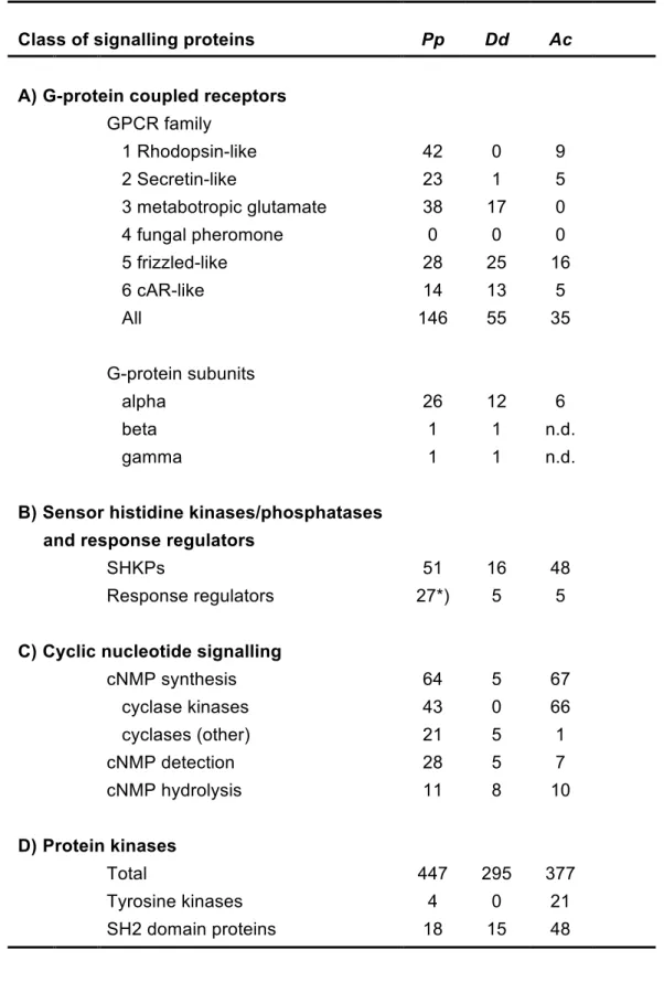
G-protein coupled receptors and heterotrimeric G-proteins
The G-protein coupled receptors (GPCRs) are a large and diverse family of transmembrane proteins in metazoa and other eukaryotes that detect a broad range of physical and chemical stimuli. Based on sequence similarity GPCRs are
subdivided into six families, where members of each family detect different sets of ligands. We found at least 146 GPCRs, with representatives from all families except family 4, the fungal pheromone receptors (Fig. S13). The largest number of GPCRs (42) are present in family 1, the rhodopsin-like receptors, which apart from light receptors also contains receptors for cytokines and neuropeptides. The number of GPCRs in P. polycephalum is considerably larger than that for other Amoebozoa (Table 3A), such as D. discoideum with 55 GPCRs (Heidel, et al. 2011) and A.
castellanii with just 35 (Clarke, et al. 2013).
The number of heterotrimeric G-proteins, which are activated by GPCRs as the next step in the signal processing cascade, is also large with 26 G-alpha subunits (Fig. S14), as compared to 12 in D. discoideum and six in A. castellanii. Thus, these gene families have been considerably expanded, possibly to cope with a wealth of different environmental conditions.
Blue light photoreceptors and phytochromes
There are distinct classes of chromoproteins that act as photoreceptors in both prokaryotes and eukaryotes in sensing the visible part of the spectrum including near UV: cryptochromes, LOV-domain photoreceptors, rhodopsins, and phytochromes (Heintzen 2012). Photoreceptor domains that are light-sensitive through covalently or non-covalently bound chromophores are often attached to signaling domains that relay the photosensory output to the cellular signaling network. While
at UPMC on March 21, 2016
http://gbe.oxfordjournals.org/
& & !
photobiochemical and physiological functions of quite a number of photoreceptors are well-studied in bacteria, plants, fungi, and animals, this is not the case for the members of the amoebozoa clade.
At least two photoreceptors, a phytochrome-like and a blue light photoreceptor act synergistically in controlling sporulation in P. polycephalum in response to far-red and to blue light (Lamparter and Marwan 2001; Starostzik and Marwan 1995a;
Starostzik and Marwan 1995b) (for details see Supplementary Methods, Results and Discussion), but these receptors had not been identified at the molecular level. Five members of three classes of putative photoreceptors are expressed by P.
polycephalum: one cryptochrome and one photolyase, one LOV-domain
photoreceptor, and two phytochromes (Fig. 3).
Cryptochrome and Photolyase. Phypoly-transcript_04617 (cryA,
cryptochrome-like photoreceptor; gene locus 1148) encodes a protein with highest similarity to chicken cryptochrome-2 and other related animal cryptochromes. Downstream of the DNA-photolyase and FAD-binding domain of cryA is a short C-terminal part of
approximately 100 amino acids encoded in the genomic sequence that is missing in Phypoly-transcript_04513 (gene locus 3553 and 3554) which shares greatest
sequence similarity with DNA-photolyases and is designated as plyA (for
photolyase A type photoreceptor) and is presumably a DNA-photolyase rather than a sensory blue light photoreceptor. The physiological role of both plyA and cryA in P.
polycephalum however remains to be established.
LOV-domain photoreceptor. The Phypoly-transcript_01902 (gene locus 13045)
encodes a protein with a domain architecture composed of RasGAP, PB1, PAS_9, SAM, and PB1 superfamily domains (Fig. 3). BLAST analysis revealed the highest similarity to the phototropin-2 photoreceptor from Arabidopsis thaliana (P93025) and
at UPMC on March 21, 2016
http://gbe.oxfordjournals.org/
& & to other LOV domain-carrying phototropins from plants and bacteria as well as to the fungal white collar 1 (WC1) blue light photoreceptor from Neurospora. The PAS_9 domain of the predicted P. polycephalum LovA protein contains the highly conserved NCRFLQ motif with the cysteine residue serving as a potential chromophore binding site embedded in additonal highly conserved residues (Fig. S18) that are involved in chromophore binding and crucial for the photochemical properties of phototropin-type photoreceptors (Heintzen 2012). The domain composition of the predicted protein and the highly conserved residues for chromophore binding in the PAS_9 domain suggests that LovA may act as an unconventional blue light photoreceptor that integrates, modulates, and/or relays multiple signals and might even bind DNA through its PAS_9 domain.
Phytochromes. Originally considered to be plant-specific photoreceptors,
phytochromes regulate many aspects of metabolism, motility, gene regulation and development (Heintzen 2012). Even before prokaryotic genome projects uncovered the early evolutionary origin of phytochromes, phytochrome-like photoreceptors were discovered in Aspergillus (Mooney and Yager 1990) and P. polycephalum (Lamparter and Marwan 2001; Starostzik and Marwan 1995b). Two phytochrome genes, phyA and phyB, are expressed in P. polycephalum, partially encoded by transcripts 20261 (gene locus 28349) and 03416 (gene locus 5996), respectively. Although the
sequence identity between PhyA and PhyB is only 33.8 %, the two proteins share a similar domain architecture with the phytochrome-type PAS_2, GAF, PHY domain arrangement at the N-terminus and a C-terminal hybrid kinase-like part, composed of HisK, HATPase, and REC domains (Fig. 3). A BLAST search of the PAS_2, GAF, PHY domain portion of the proteins against the UniProt database revealed closest similarity to Nostoc PHYA (Q9LCC2) and four other bacterial phytochromes followed
at UPMC on March 21, 2016
http://gbe.oxfordjournals.org/
& !
by plant phytochromes. Sequence alignment with the most similar phytochromes suggests that the chromophore binding site may be a cysteine close to the N-terminus that is conserved between P. polycephalum PhyA and PhyB and various bacterial phytochromes, while the cysteine of the CHxxYxxNMG motif that serves as a chromophore binding site in plant phytochromes is replaced by valine in the two P.
polycephalum proteins (Fig. S20). Whether the photochemistry of the two
phytochromes is identical or one of them may be specifically synthesized in the dark in its far-red light absorbing Pfr form (Lamparter and Marwan 2001) to trigger
sporulation (see Supplementary Information) remains to be established. The deletion upstream of the conserved PASDIPPQARRL motif in PhyA as compared to PhyB could conceivably cause an according functional difference.
Sensor histidine kinases/phosphatases
Other important receptors for external stimuli are the sensor histidine
kinases/phosphatases (SHKPs), which are very abundant in prokaryotes and also present in significant numbers in plants, fungi and Amoebozoa, but not in metazoa (Wolanin, et al. 2002). Sensor histidine kinases/phosphatases initiate forward or reverse relay, respectively, of a phosphoryl group to/from a conserved aspartate in a response regulator. The response regulator subsequently regulates the activity of an effector, such as an enzyme or transcription factor. The P. polycephalum genome contains ~51 SHKPs with a large variety of different functional domain architectures (Fig. 4A). Many of these SHKPs contain small molecule or light sensing domains, such as the GAF, PAS/PAC, or phytochrome domains, which are likely to detect stimuli and activate phosphotransfer, while others contain protein kinase or AAA-ATPase domains. These domains could be downstream effectors, which are
at UPMC on March 21, 2016
http://gbe.oxfordjournals.org/
&
regulated by phosphorelay. The number of SHKPs in P. polycephalum is about equal to that in A. castellanii and three times larger than that in D. discoideum (Table 3B) but there are up to five times more response regulators in P. polycephalum than in either D. discoideum or A. castellanii.
While most P. polycephalum response regulators have no additional features, two have leucine-rich repeats and two have a phosphodiesterase domain and are closely related to the D. discoideum cAMP phosphodiesterase RegA (Fig. 4B). In
Dictyostelids RegA is a negative regulator of encystation and sporulation, which both
require activation of PKA by cAMP (Du, et al. 2014; Shaulsky, et al. 1998). In P.
polycephalum, sporulation is induced by light and this induction was shown to be
mediated by a phytochrome-type receptor (Starostzik and Marwan 1995b). The light-sensing part of this photoreceptor is likely to be the phytochrome domain that is present in two P. polycephalum SHKPs (see below; Fig. 4A), which suggests that the stimuli that control sporulation in P. polycephalum also act on intracellular cAMP levels by regulating RegA activity.
Cyclic nucleotide signalling
The cyclic nucleotides cAMP, and to a lesser extent cGMP, are widely used as intracellular second messengers for a broad range of external stimuli in all domains of life. cAMP is particularly important for Dictyostelid development, where it regulates encystation, progression through multicellular development, maturation of spore and stalk cells, and maintenance of spore and cyst dormancy by acting on PKA (Schaap 2011). In addition, Dictyostelids also use cAMP extracellularly as a chemoattractant to organize fruiting body morphogenesis in all species, and aggregation in a subset
at UPMC on March 21, 2016
http://gbe.oxfordjournals.org/
& !
that contains the model organism D. discoideum (Alvarez-Curto, et al. 2005). In contrast, there are a few documented roles for cAMP or cGMP in P. polycephalum. cGMP plays a role in the induction of sporulation in P. polycephalum (Golderer et al., 2001) and there is sporadic evidence of roles for cAMP in cell division, motility and gravity sensing (Block, et al. 1998; Kuehn 1972; Matveeva, et al. 2012).
To evaluate the prevalence of cAMP and cGMP signaling in P. polycephalum, we investigated the presence of the cyclases, binding proteins, and phosphodiesterases that respectively synthesize, detect, or degrade cyclic nucleotides.
To our surprise the number of cyclase and cyclic nucleotide binding proteins in P.
polycephalum by far surpasses that in D. discoideum. Firstly, P. polycephalum has
64 nucleotidyl cyclases against 5 in D. discoideum. A. castellanii has even more (67), but 66 of these cyclases belong to a massively amplified family of transmembrane proteins that contain a cyclase domain, flanked by two protein kinase domains. P.
polycephalum has a family of 43 of those proteins, suggesting a common
requirement for amplification of these cyclases in some Amoebozoa. P.
polycephalum has in addition 21 cyclases with a highly variable functional domain
architecture (Fig. S16). This set is dominated by proteins with 2 sets of 6
transmembrane domains that flank 2 cyclase domains. This is the configuration of most mammalian adenylate cyclases and of D. discoideum ACA and GCA. In addition there are cyclases with GAF and PAS/PAC domains and with domains involved in either actin remodeling, ubiquitination or calcium export. P. polycephalum also has a close homolog of D. discoideum SGC, a guanylate cyclase that is involved in chemotaxis, and it has a close homolog of D. discoideum and A. castellanii AcrA, an adenylate cyclase that is essential for spore maturation in D. discoideum (Loomis 2014).
at UPMC on March 21, 2016
http://gbe.oxfordjournals.org/
& The number of cyclic nucleotide phosphodiesterases in P. polycephalum is about equal to that in D. discoideum and A. castellanii (Fig. S15). However, the number of proteins predicted to be capable of binding cAMP or cGMP is truly astonishing and exceeds the numbers in D. discoideum and A. castellanii by five and four fold, respectively (Table 3). While the majority of these proteins only have cNMP binding domain(s), there are also homologs of the D. discoideum cGMP binding proteins GbpC and GbpD, which additionally have protein kinase, RasGEF and DEP domains (Fig. S17). There is also a homolog of D. discoideum PdeE, in which an intrinsic Lactamase_B domain acts as a cAMP phosphodiesterase. In addition, there are putative cNMP binding proteins with additional RhoGEF, VWA and P2X receptor domains that are not present in other organisms. The multitude and variety of cNMP binding domains and nucleotidyl cyclases in P. polycephalum indicates that cyclic nucleotide signalling is likely to play a very dominant role in its physiology and development.
Protein kinases
The protein kinases that modify the function of downstream effector proteins by phosphorylation of serine/threonine (S/T) or tyrosine (Y) residues are the most common intermediates of signal processing cascades. Humans have 518 protein kinases and even yeast has over a hundred (Hanks 2003). Inspection of the P.
polycephalum transcriptome revealed the presence of a total of 447 proteins with
S/T, Y or S/T/Y (dual specificity) kinase domains (see Supplemental Methods,
Results, and Discussion and Table 3 for further details). This is 1.5 and 1.2 fold more than in D. discoideum and A. castellanii, respectively, and therefore a more modest increase than observed for the other signal transduction proteins described above.
at UPMC on March 21, 2016
http://gbe.oxfordjournals.org/
& & !
The initial analysis based on Interpro identifiers for substrate specificity revealed the presence of 29 tyrosine kinases, but a more in depth comparison with sequence signatures of validated tyrosine kinases revealed that only four of those contain all essential residues for tyrosine substrate recognition (Fig. 5). Three of these proteins contain a single transmembrane domain that provides them with structural similarity to metazoan receptor tyrosine kinases (RTKs), which are the targets for a broad range of peptide growth factors, hormones and cytokines. In metazoa, tyrosine phosporylated substrates become binding sites for SH2 domain proteins, which further transduce the response (Liu and Nash 2012). P. polycephalum has 18 SH2 domain proteins (Fig. 6), of which three are similar to D. discoideum and metazoan STAT transcription factors, while D. discoideum and A. castellanii have 15 and 48 SH2 domain proteins, respectively.
Tyrosine kinases play very dominant roles in metazoan embryogenesis and adult physiology, and were until recently considered to be a hallmark of metazoan
evolution because they were present only in metazoa and one of its unicellular allies, a choanoflagellate (King and Carroll 2001) and absent from yeast, plants and D.
discoideum (Lim and Pawson 2010). This notion was overturned by the identification
of some tyrosine kinases outside of Opisthokonta (Suga, et al. 2012) and the presence of 21 tyrosine kinases in A. castellanii (Clarke, et al. 2013), a member of Lobosa, one of the two major subdivisions of Amoebozoa. P. polycephalum and D.
discoideum reside in the other subdivision Conosa (Schilde and Schaap 2013). The
presence of tyrosine kinases in both subdivisions of Amoebozoa, in their sister group Opisthokonta, and in other eukaryote divisions indicates that tyrosine kinases were likely present in the last common ancestor to all eukaryotes and were selectively lost from many phyla. The more widely distributed tyrosine kinase like enzymes, e.g. D.
at UPMC on March 21, 2016
http://gbe.oxfordjournals.org/
& &
discoideum Pyk2, which can also phosphorylate SH2 domain tyrosines (Araki, et al.
2014), has probably taken over their role in these phyla.
Conclusions
P. polycephalum has for long been a classic model organism in cell biology. The
accessibility of both genome sequence and extensive transcriptome data sets strongly enhances its usefulness as a model system. The availability of the
transcriptome of amoebae and plasmodia at different stages of development now permits reverse genetic approaches through identifying mutants in genes of interest in mutant libraries obtained by chemical mutagenesis of amoebal cells. In addition, the transcriptome sequences provide the basis for quantitative proteomic approaches by mass spectrometry. Our analysis highlights a wealth of interesting molecular features and will help in enabling work at the molecular level in combining the well-established classic genetics with second generation sequencing approaches. The results of the P. polycephalum sequencing project described here provide interesting information with respect to the evolution of signaling systems in the
Amorphea branch of the eukaryotes. Virtually all aspects discussed here characterize
P. polycephalum as an organism with higher molecular complexity than other
sequenced Amoebozoa. This becomes most obvious for cellular signalling through the extensive use of two component signaling systems in parallel with receptor tyrosine kinase signal transduction. Other important aspects such as cell cycle regulation, cytoskeletal motor proteins, and the enzymes of the pteridine metabolism point in the same direction. Together with recent findings on A. castellanii (Clarke, et
at UPMC on March 21, 2016
http://gbe.oxfordjournals.org/
& & !
al. 2013), our results indicate that the molecular evolution of these features, notably signaling through receptor tyrosine kinases which has previously been considered a hallmark of multicellularity of animals, is deeply rooted in the Amorphea and have been secondarily lost in other amoebozoa like D. discoideum and in the fungi. Another interesting feature of P. polycephalum is the occurrence of photoreceptors: two bacterial-type phytochromes, not found in other Amoebozoa, a phototropin-like LOV domain blue light photoreceptor, and a cryptochrome. In conclusion, among the unicellular genetic model organisms P. polycephalum displays many features of animal cells and, with respect to its molecular complexity, the cross-talk of signaling molecules, and the resulting dynamic behavior. As such, it is anticipated that it will serve as an interesting model system contributing complementary information for the study of mammalian and other animal cells. In combination with its ability to form multinucleate giant plasmodia, this provides unique options for investigating fundamental biological processes in individual cells.
Supplementary Material
Supplementary File 1: Supplementary Methods, Results and Discussion Supplementary File 2: Supplementary Spreadsheets
at UPMC on March 21, 2016
http://gbe.oxfordjournals.org/
&
Acknowledgments
Funding for the sequence characterization of the P. polycephalum genome was provided by National Institutes of Health (NIH) grant HG003079 to RKW, The
Genome Institute at Washington University School of Medicine. PS received funding from the Wellcome Trust and the BBSRC. IB was supported by the Excellence Initiative of Saxony-Anhalt through the International Max Planck Research School, Magdeburg. WM received funding from the German Federal Ministry of Education and Research (BMBF) via the FORSYS program. Pteridine work was funded by the Austrian Science Fund (FWF), P22406 to ERW. JMG was funded through (NIH GM54663, NSF MCB-1243687).
at UPMC on March 21, 2016
http://gbe.oxfordjournals.org/
& !
Literature Cited
Adl SM, et al. 2012. The revised classification of eukaryotes. Journal of Eukaryotic Microbiology 59: 429-493.
Aldrich HC, Daniel JW. 1982. Cell Biology of Physarum and Didymium. In. New York: Adademic Press.
Alvarez-Curto E, et al. 2005. Evolutionary origin of cAMP-based chemoattraction in the social amoebae. Proc. Natl. Acad. Sci. USA 102: 6385-6390.
Araki T, et al. 2014. Two Dictyostelium tyrosine kinase-like kinases function in parallel, stress-induced STAT activation pathways. Mol Biol Cell 25: 3222-3233. Barkan A, et al. 2012. A combinatorial amino acid code for RNA recognition by
pentatricopeptide repeat proteins. PLoS genetics 8: e1002910.
Block I, Rabien H, Ivanova K 1998. Involvement of the second messenger cAMP in gravity-signal transduction in Physarum. Adv Space Res 21: 1311-1314.
Braund E, Miranda ER editors. 9th Conference on Interdisciplinary Musicology – CIM14. papers2://publication/uuid/AC77D621-D506-4D95-A026-AC65533A364F; 2014 Mar 12: Berlin.
Bundschuh R, Altmüller J, Becker C, Nürnberg P, Gott JM 2011. Complete
characterization of the edited transcriptome of the mitochondrion of Physarum
polycephalum using deep sequencing of RNA. Nucleic Acids Research 39:
6044-6055.
Burland TG, Solnica-Krezel L, Bailey J, Cunningham DB, Dove WF 1993. Patterns of inheritance, development and the mitotic cycle in the protist Physarum
polycephalum. Adv. Microb. Physiol. 35: 1-69.
Cavalier-Smith T 2003. Protist phylogeny and the high-level classification of Protozoa. European Journal of Protistology 39: 338-348.
Clarke M, et al. 2013. Genome of Acanthamoeba castellanii highlights extensive lateral gene transfer and early evolution of tyrosine kinase signaling. Genome Biol 14: R11.
Cross FR, Buchler NE, Skotheim JM 2011. Evolution of networks and sequences in eukaryotic cell cycle control. Philosophical transactions of the Royal Society of London. Series B, Biological sciences 366: 3532-3544.
Dee J 1987. Genes and development in Physarum. Trends Genet. 3: 208-213. Dee J. 1982. Genetics of Physarum polycephalum. In: Aldrich HC, Daniel JW,
editors. Cell Biology of Physarum and Didymium: Academic Press. p. 211-251. Doonan JH, Kitsios G 2009. Functional evolution of cyclin-dependent kinases.
Molecular Biotechnology 42: 14-29.
at UPMC on March 21, 2016
http://gbe.oxfordjournals.org/
& & Dove WF, Dee J, Hatano S, Haugli FB, Wohlfarth-Bottermann K-E. 1986. The
Molecular Biology of Physarum polycephalum. In. New York: Plenum Press. p. 253-269.
Du Q, et al. 2014. The cyclic AMP phosphodiesterase RegA critically regulates encystation in social and pathogenic amoebas. Cellular Signalling 26: 453-459. Edgar RC 2010. Search and clustering orders of magnitude faster than BLAST.
Bioinformatics (Oxford, England) 26: 2460-2461.
Eichinger L, Bomblies L, Vandekerckhove J, Schleicher M, Gettemans J 1996. A novel type of protein kinase phosphorylates actin in the actin–fragmin complex. EMBO J. 15: 5547–5556.
Eichinger L, et al. 2005. The genome of the social amoeba Dictyostelium discoideum. Nature 435: 43-57.
Gettemans J, De Ville Y, Vandekerckhove J, Waelkens E 1992. Physarum actin is phosphorylated as the actin–fragmin complex at residues Thr203 and Thr202 by a specific 80 kDa kinase. EMBO J. 11: 3185–3191.
Glöckner G, Golderer G, Werner-Felmayer G, Meyer S, Marwan W 2008. A first glimpse at the transcriptome of Physarum polycephalum. BMC Genomics 9: 6. Glöckner G, et al. 2014. The genome of the foraminiferan Reticulomyxa filosa. Curr
Biol 24: 11 - 18.
Golderer G, Werner ER, Leitner S, Gröbner P, Werner-Felmayer G 2001. Nitric oxide synthase is induced in sporulation of Physarum polycephalum. Genes Devel. 15: 1299-1309.
Haindl M, Holler E 2005. Use of the giant multinucleate plasmodium of Physarum
polycephalum to study RNA interference in the myxomycete. Analytical
biochemistry 342: 194-199.
Hanks SK 2003. Genomic analysis of the eukaryotic protein kinase superfamily: a perspective. Genome Biology 4: 111.
Hanks SK, Hunter T 1995. The eukaryotic protein kinase superfamily: kinase (catalytic) domain structure and classification. FASEB J. 9: 576–596. Harashima H, Dissmeyer N, Schnittger A 2013. Cell cycle control across the
eukaryotic kingdom. Trends in Cell Biology 23: 345-356.
Hasegawa T, Takahashi S, Hayashi H, Hatano S 1980. Fragmin: a calcium ion sensitive regulatory factor on the formation of actin filaments. Biochemistry 19: 2677-2683.
Heidel A, et al. 2011. Phylogeny-wide analysis of social amoeba genomes highlights ancient origins for complex intercellular communication. Genome Res.: 1882-1891.
Heintzen C 2012. Plant and fungal photopigments. Wiley Interdisciplinary Reviews: Membrane Transport and Signaling 1: 411-432.
Henriquez FL, et al. 2015. The Acanthamoeba Shikimate Pathway has a Unique Molecular Arrangement and is Essential for Aromatic Amino Acid Biosynthesis. Protist 166: 93-105.
at UPMC on March 21, 2016
http://gbe.oxfordjournals.org/
& & !
Itoh K, et al. 2011. DNA packaging proteins Glom and Glom2 coordinately organize the mitochondrial nucleoid of Physarum polycephalum. Mitochondrion 11: 575-586.
Joseph J, et al. 2008. The actinome of Dictyostelium discoideum in comparison to actins and actin-related proteins from other organisms. PLoS ONE 3: e2654. Kamiya N 1940. The control of protoplasmic streaming. Science (New York, NY) 92:
462-463.
Kamiya N. 1959. Protoplasmic streaming. Wien: Springer-Verlag.
Kent JW 2002. BLAT – The BLAST like alignment tool. Genome Research 12: 656-664.
King N, Carroll S 2001. A receptor tyrosine kinase from choanoflagellates: molecular insights into early animal evolution. Proc Natl Acad Sci USA 98: 15032-15037. Klein R, Thiery R, Tatischeff I 1990. Dictyopterin,
6-(D-threo-1,2-dihydroxypropyl)-pterin, a new natural isomer of L-biopterin. Isolation from vegetative cells of
Dictyostelium discoideum and identification. Eur J Biochem 187: 665-669.
Knoop V, Rüdinger M 2010. DYW-type PPR proteins in a heterolobosean protist: plant RNA editing factors involved in an ancient horizontal gene transfer? FEBS letters 584: 4287-4291.
Kuehn GD 1972. Cell cycle variation in cyclic adenosine 3',5'-monophosphate-dependent inhibition of protein kinase from Physarum polycephalum. Biochem Biophys Res Commun 49: 414-419.
Lamparter T, Marwan W 2001. Spectroscopic detection of a phytochrome-like photoreceptor in the Myxomycete Physarum polycephalum and the kinetic mechanism for the photocontrol of sporulation by Pfr. Photochem. Photobiol. 73: 697-702.
Lim WA, Pawson T 2010. Phosphotyrosine signaling: evolving a new cellular communication system. Cell 142: 661-667.
Liu BA, Nash PD 2012. Evolution of SH2 domains and phosphotyrosine signalling networks. Philosophical Transactions of the Royal Society B: Biological Sciences 367: 2556-2573.
Loomis WF 2014. Cell signaling during development of Dictyostelium. Developmental Biology 391: 1-16.
Mahendran R, Spottswood MR, Miller DL 1991. RNA editing by cytidine insertion in mitochondria of Physarum polycephalum. Nature 349: 434-438.
Marchler-Bauer A, et al. 2015. CDD: NCBI's conserved domain database. Nucleic Acids Research 43.
Matveeva NB, Teplov VA, Nezvetskii AR, Orlova TG, Beilina SI 2012. Involvement of cyclic adenosine monophosphate in the control of motile behavior of Physarum
polycephalum plasmodium. Biofizika 57: 832-839.
Messner S, et al. 2009. Physarum nitric oxide synthases: Genomic structures and enzymology of recombinant proteins. Biochem. J. 418: 691-700.
at UPMC on March 21, 2016
http://gbe.oxfordjournals.org/