HAL Id: inserm-02438743
https://www.hal.inserm.fr/inserm-02438743
Submitted on 14 Jan 2020
HAL is a multi-disciplinary open access
archive for the deposit and dissemination of
sci-entific research documents, whether they are
pub-lished or not. The documents may come from
teaching and research institutions in France or
abroad, or from public or private research centers.
L’archive ouverte pluridisciplinaire HAL, est
destinée au dépôt et à la diffusion de documents
scientifiques de niveau recherche, publiés ou non,
émanant des établissements d’enseignement et de
recherche français ou étrangers, des laboratoires
publics ou privés.
Distributed under a Creative Commons Attribution - NoDerivatives| 4.0 International
License
Nathalie Gaspar, Angela Di Giannatale, Birgit Geoerger, Françoise Rédini,
Nadège Corradini, Natacha Enz-Werle, Franck Tirode, Perrine Marec-Bérard,
Jean-Claude Gentet, Valérie Laurence, et al.
To cite this version:
Nathalie Gaspar, Angela Di Giannatale, Birgit Geoerger, Françoise Rédini, Nadège Corradini, et al..
Bone Sarcomas: From Biology to Targeted Therapies. Sarcoma, Hindawi Publishing Corporation,
2012, 2012, pp.1-18. �10.1155/2012/301975�. �inserm-02438743�
Volume 2012, Article ID 301975,18pages doi:10.1155/2012/301975
Review Article
Bone Sarcomas: From Biology to Targeted Therapies
Nathalie Gaspar,
1Angela Di Giannatale,
1Birgit Geoerger,
1Franc¸oise Redini,
2Nad`ege Corradini,
3Natacha Enz-Werle,
4Franck Tirode,
5Perrine Marec-Berard,
6Jean-Claude Gentet,
7Val´erie Laurence,
8Sophie Piperno-Neumann,
8Odile Oberlin,
1and Laurence Brugieres
11Department of Oncology for Children and Adolescents, Institut Gustave Roussy, 114 rue Edouard Vaillant, 94805 Villejuif Cedex, France
2Inserm U957-EA 3822, Facult´e de M´edecine, 1 rue Gaston Veil, 44035 Nantes Cedex 1, France 3Oncopediatric Departement, CHU de Nantes, Boulevard Jacques Monod, 44093 Nantes, France
4Pediatric Onco-Hematology Department, EA 4438UdS, CHRU Strasbourg, Avenue Moli`ere, 67000 Strasbourg, France 5Unit´e 830 INSERM, Institut Curie, Centre de Recherche, 26 rue d’Ulm, 75248 Paris Cedex 05, France
6Oncopediatric Departement, Centre L´eon B´erard, 28 rue Laennec, 69008 Lyon, France
7Oncopediatric Departement, Hˆopital La Timone, 264 rue Saint Pierre, 13385 Marseille Cedex 5, France 8Department of Medical Oncology, Institut Curie, 26 rue d’Ulm, 75248 Paris Cedex 05, France
Correspondence should be addressed to Nathalie Gaspar,[email protected] Received 1 July 2012; Accepted 10 October 2012
Academic Editor: R. Pollock
Copyright © 2012 Nathalie Gaspar et al. This is an open access article distributed under the Creative Commons Attribution License, which permits unrestricted use, distribution, and reproduction in any medium, provided the original work is properly cited.
Primary malignant bone tumours, osteosarcomas, and Ewing sarcomas are rare diseases which occur mainly in adolescents and young adults. With the current therapies, some patients remain very difficult to treat, such as tumour with poor histological response to preoperative CT (or large initial tumour volume for Ewing sarcomas not operated), patients with multiple metastases at or those who relapsed. In order to develop new therapies against these rare tumours, we need to unveil the key driving factors and molecular abnormalities behind the malignant characteristics and to broaden our understanding of the phenomena sustaining the metastatic phenotype and treatment resistance in these tumours. In this paper, starting with the biology of these tumours, we will discuss potential therapeutic targets aimed at increasing local tumour control, limiting metastatic spread, and finally improving patient survival.
1. Introduction
Primary bone sarcomas, osteosarcomas (OS), and Ewing sarcomas (EW) are diseases occurring mainly in adolescent and young adults and account for around 15% of child-hood/adolescent cancers. First-line therapeutic strategies in these diseases consisted in chemotherapy (CT) before and after local treatment (including dose CT for high-risk EW [1]) and a local treatment by surgery but also or only by radiotherapy in EW. Some patients remain very difficult to treat, such as tumour with poor histological response to preoperative CT (or large initial tumour vol-ume for EW not operated) [2, 3], patients with multiple metastases at diagnostic [1, 4], or those who relapsed [5].
In order to develop new therapies against these diseases we need to unveil the founder molecular abnormalities underlying the malignant characteristics and to broaden our understanding of the phenomena sustaining the metastatic phenotype and treatment resistance in these tumours. Both diseases are sustained by different biology abnormalities but also share some common characteristics (angiogenesis, etc.). The main objective of this paper is to discuss potential therapeutic targets aimed at increasing local control of the primary tumour, limiting metastatic spread, and finally improving patient survival. We then review preclinical data and both paediatric and adult trials performed or ongoing and choose to present them by pathway rather than by tumour.Table 1and Figures1and2present the same data by tumour type.
Ta b le 1: M olecular targets ac co rding to malig n ant char act er istics and cur re nt de ve lo pment o f target ed ther apies in o st eosar comas and E w ing sar comas. T argets A gents Clinical de ve lo pment in d iff er ent tumour ty pes Ost eosar co ma E w ing sar coma E W S-FLI1 inhibition A n tisense oligon u cl eotide, antisense RN A, siRN A P Mi th ra m yc in I p ed/II ad ong o ing , NCT01610570 YK-4-279 P ET -743 (t ra bect edin; Y o ndelis ∗) I ped/II ad I p ed/II ped Cell gr o w th Inhibition GFR inhibit ors IGFR inhibit ors R 1507; SCH 717454; CP -751871; IMC-A12 I p ed II p ed (E W :10–15% objecti ve re sponse ra te ) mT OR inhibit ors E ver olim u s (RAD001, A finit or ∗) I/II p ed I p ed Te m si ro li m u s (To ri se l ∗) I/II p ed I/II ped Ridafor olim u s I p ed ongoing ,N CT01431547 al l solid tumours II/III ad II/III ad C o mbination E W S-FLI1 antisense oligon u cl eotide + m T O R inhibit or / P Rapam ycin + ir inot ecan (RAP IRI) I p ed ongoing ,N CT01282697 al l solid tumours PP C ixutum u mab + te msir olim u s II ped/II ad ongoing (NCT01614795)/ II ped/II ad ongoing (NCT01614795) Ridafor olim u s + dalotuzumab I p ed ongoing ,N CT01431547 al l solid tumours Ip ed / M u ltitarget inhibit ors Im atinib mesylat e, G li ve c ∗(PDFGR, c-KIT ,BCR -ABL) P II p ed Im atinib + ifosfamide I ped / Dasatinib ,S pr yc el ∗(S rc ,B CR -ABL) P I ped Cell cy cl e inhibit ors CDK inhibit ors S CH 727965 (dinacic lib) I ad / R exin-G / A u ro ra A inhibit ors P MLN8237 I/II ped o ngoing , NCT01154816/NCT00739427 P Solid tumours or le uk emia P A T 9283 I p ed ongoing , NCT00985868/NCT01431664 P Solid tumours/le u ke mia P PLK1 inhibit or ,B I 2536 P / MDM2 inhibit ors, n u tlin-3 I ad o ngoing ,N CT01462175 solid tumours P
Ta b le 1: C o ntin ued. T argets A gents Clinical de ve lo pment in d iff er ent tumour ty pes Ost eosar co ma E w ing sar coma A n gi ogenesis inhibition Sor afenib ,N ex av ar ∗(Raf, c-KIT ,PD GFR, VEGF) II ad I p ed o ngoing ,N CT01518413 Sunitinib ,S ut ent ∗(Flt3, c-KIT ,PD GFR, VEGF) I ped I ped P az o panib (V EGFR1–3, PD GFR α/ β,c -K it ) P Ip ed P az o panib + to p ot ecan I p ed ongoing ,N CT00326664 CNS tumours PP P az o panib + ev er olim u s I ad o ngoing ,N CT01430572 solid tumours // Cedir anib ,A ZD2171 (V EGFR) III p ed/ad I p ed Be vacizumab A vastin ∗NCT00667342 tr ial I/II p ed I/II p ed Be vacizumab + vincr istine/t o p ot ecan/cy clo phosphamide / II p ed ongoing ,N CT00516295 III ped/ad ongoing , NCT00667342 / Be vacizumab + CT 1st-line ra nd omised stud y, co mbination CT R esistanc e to ce ll death Ap o p to si s BCL2 inhibit ors, na vi to cl ax (AB T -263) / P TRAIL inhibit ors PP SMA C mimetic, L C L161 I ad o ngoing ,N CT01098838 solid tumours PP P A RP inhibit ors /P A u to phag y A ntisense oligon u cl eotide of X-link ed IAP P P T elomer ase acti vi ty T elomer ase inhibit or ,TMPyP4 P P Inhibition o f m etastatic phenot yp e In vasion Gamma secr etase (NO CTH Inhibit ors), M K-0752 I ad/p ed le u ke mia P P M ig ration M ET/ALK inhibit ors (C riz otinib) II ped > 15 y o ngoing ,N CT00939770, solid tumours P/ R esistanc e to anoikis G SK3beta inhibit ors (Wnt p ath w ay acti vation) I ad o ngoing ,N CT01457417, NCT00741377 m yeloma P/ Chemotactism CX CR4 inhibit ors (pler ixafor) I p ed ongoing ,N CT01319864 le uk emia/MDS PP A dhesion In te gr in inhibit ors, cileng itide E MD121974 I p ed ongoing ,N CT01165333 CNS P /
Ta b le 1: C o ntin ued. T argets A gents Clinical de ve lo pment in d iff er ent tumour ty pes Ost eosar co ma E w ing sar coma M o dulat ion ant itumour imm une re sponse INF α III ped/ad (EURAMOS I tr ial, re sults await ed) / 1st-line ra nd omised stud y, co mbination CT L -MTP -P E, mifam u rt ide M EP A C T ∗ III p ed/ad (INT -0133 tr ial closed) / Inhaled sarg ramostim (r h GM-CSF) II ad ongoing , NCT00066365: Pu lmonar y relapses / Celec o xib ,CO X2 inhibit ors / P A n ti-GD2 antibodies (c h 14.18) I/II ped, ne ur oblast oma I ped P Bone micr oen vir onment III p ed/ad OS2006 tr ial ongoing ,N CT00470223 III p ed/ad p ro to co l ew ing 2008 and EE2012 Z oledr onic acid, Z o meta ∗ 1st-line ra nd omised stud y, co mbination CT 1st-line ra nd omised stud y, co mbination CT F o r localised E W + good hist olog ical response Denosumab (A c anti-RANKL) II ad/ped > 12 ans o ngoing , NCT00680992 GCT PP Samar ium II ad / Other p ath w ay s H edgehog inhibit ors Smo inhibit or LDE225 (ongoing) I p ed ongoing ,N CT01125800 solid tumours P/I p ed, N CT01125800 P HD A C inhibit ors V or inostat, valpr oic acid, FK228 P/I p ed I p ed HSP90 inhibit ors 17-AA G Ip ed Ip ed CNS: ce nt ra l n er vo us syst em; G FR: gr o w th fact o r rec ept o r; P :pr ec linical studies; I: phase I tr ial; II: phase II tr ial; III: phase III tr ial; ped: paed iat ric; ad: adult; C T :c h emother ap y; C GT :g iant ce ll tumour .
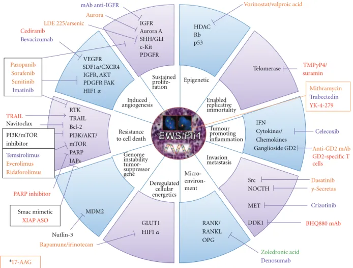
Sustained prolife-ration Epigenetic Enabled replicative immortality Tumour promoting inflammation Invasion metastasis Deregulated cellular energetics Genome instability tumor-suppressor gene Resistance to cell death Induced angiogenesis GLUT1 RTK TRAIL Bcl-2 PI3K/AKT/ mTOR PARP IAPs HDAC Rb p53 Telomerase MDM2 Src NOCTH MET DDK1 VEGFR IGFR, AKT PDGFR FAK Dasatinib Vorinostat/valproic acid suramin RANK/ RANKL OPG IFN Cytokines/ Chemokines Ganglioside GD2 Celecoxib IGFR Aurora A SHH/GLI c-Kit PDGFR mAb anti-IGFR Nutlin-3 Zoledronic acid Denosumab Rapamune/irinotecan LDE 225/arsenic Aurora Cediranib Bevacizumab Smac mimetic XIAP ASO Mithramycin Trabectedin YK-4-279 Navitoclax Pazopanib Sorafenib Sunitinib Imatinib TRAIL PI3K/mTOR inhibitor Crizotinib Anti-GD2mAb GD2-specific T cells PARP inhibitor Temsirolimus Everolimus Ridaforolimus NCT 01614795 Micro- environ-ment SDF1α/CXCR4 HIF1α HIF1α TMPγP4/ γ-Secretas BHQ880 mAb 17-AAG ∗
Figure 1: Targets and therapies in preclinical and clinical development in children and adolescent bone sarcomas. (A) Ewing sarcomas. (B) Osteosarcomas. The different colors described the current clinical development of the drugs. (Red) Preclinical: EW and OS; (Orange) Phase I: all paediatric studies; (Blue) Phase II: specific EW, OS, bone tumours; (Green) Phase III: specific EW and/or OS; (Black) Phase I or II in adults: all solid tumours. ∗17-AAG is an HSP90 inhibitor which targets client proteins involved in all tumour characteristics.
2. Biology of Bone Tumours
2.1. Biology of Ewing Sarcoma: A Cell of Mesenchymal Origin Driven by an Aberrant Fusion Protein, EWS-Ets. EW is
characterised by a group of translocations which oppose a gene from the EWS family with a gene from the ETS family arising in cells of mesenchymal origin [6]. The most frequent translocation is t(11;22). It leads to an aberrant fusion protein which is responsible for the malignant phenotype [7]. EWS-Ets is a transcription factor with a DNA binding domain (Ets; FLI1 in 85%) and a transcription enhancer domain (EWS) [8]. The altered intrinsic EWS-FLI1 region facilitates the formation of protein-protein interactions that regulate the transcription of numerous genes and mRNA alternative splicing [8]. Numerous biological pathways are modulated by EWS-FLI1 activity: IGFR, PDGFR, VEGFR, SHH pathway activation; Wnt, TGFβRII pathway inhibition, and lead to the EW malignant phenotype: proliferation, angiogenesis, immune system escape, metastatic potential, and treatment resistance [8].
2.2. Biology of Osteosarcoma: Osteoblast or Cells of Mesenchy-mal Origin with a Complex Biology Producing Osteoid Matrix.
OS is a malignant tumour that produces osteoid tissue. Different mesenchymal components found in different OS subtypes suggest that OS arise from a more pluripotent cell than the osteoblast.
OS belong to the spectrum of genetic predisposition to cancer syndromes (Li Fraumeni, hereditary retinoblas-toma, Rothmund-Thomson, Werner syndromes). Molec-ular abnormalities influence various tumour characteris-tics and may be implicated in several biological path-ways: sustaining proliferative signalling (IGFR, SHH/GLI, PDGFR, c-KIT), evading cell growth suppressors (p53, RB, CDK), resisting cell death (ERK activation, proapop-totic molecule inhibition, antiapopproapop-totic molecule activation Bcl2, syndecan-2, autophagy inhibition), enabling repli-cative immortality (telomerase), increasing angiogenesis (VEGFR, IGFR, PDGFR, HIF1α), and activating invasion and metastasis, genome instability (p53, Rad51, GADD45), evading immune destruction (IFN), reprogramming energy
GLUT1 RTK TRAIL Syndecan-2 PI3K/AKT/ mTOR IAPs p53, Rb WRN, RecQ Rad51, GADD45 MDM2 HMGB1 VEGFR IGFR, AKT IGFR SHH/GLI PDGFR Nutlin-3 Temsirolimus Everolimus Ridaforolimus Aurora Cediranib Bevacizumab Smac mimetic XIAP ASO Navitoclax Pazopanib Sorafenib Sunitinib Imatinib PI3K/mTOR inhibitor Telomerase Src NOCTH MMP2 MET CXCR4 Integrin Dasatinib Rexin-G RANK/ RANKL OPG Denosumab Samarium Crizotinib HDAC CyclinG1 CDK4/6 PLK1 Rb, p53 macrophages NK GM-CSF GD2 Sargramostim ch14.18mAb Cilengitide Dinaciclib Trabectedin TRAIL BI2536 2-O, 3-O-Disulfate heparin GIN Plerixafor Plerixafor L-MTP-PE Il15 Zoledronic acid Vorinostat/valproic acid 17-AAG ∗ Rapamune/irinotecan mAb anti-IGFR LDE 225/arsenic SDF1α/CXCR4 HIF1α PDGFR FAK c-Kit TMPγP4 γ-Secretase Bcl-2 HIF1α Sustanied prolife-ration Epigenetic Invasion metastasis Deregulated cellular energetics Genome instability tumor-suppressor gene Resistance to cell death Induced angiogenesis Micro- environ-ment Tumour promoting inflammation Enabled replicative immortality GSK3β
IL1β, IL6, IL12, TNFα
INnα
INFα Osteosarcoma
Aurora A
Figure 2
metabolism and hypoxic driven therapeutic resistance (HIF1α, GLUT1), and interacting with the bone microenvi-ronment (RANK/RANKL/OPG).
3. Therapeutic Options for Bone Sarcomas
3.1. EWS-FLI1 Inhibition in Ewing Sarcomas. The fusionprotein EWS-FLI1, exclusively expressed in EW tumour cells, is an ideal target for specifically treating EW without affecting normal cells.
Decreased EWS-FLI1 expression by antisense oligonu-cleotides [9] or RNA [10], small interference RNA (siRNA) through nanoparticles [11], inhibits cell proliferation and tumour growth of EW xenografts. The pharmacological delivery of these large molecules in patients is not yet solved. Mithramycin has been identified by high-throughput screening as another inhibitor of the EWS-FLI1 oncogenic transcription factor and has shown in vitro and in vivo activ-ity against EW [12]. Mithramycin is currently being tested at the NCI against EW in children and adults (NCT01601570). An alternative strategy is to target the interaction between EWS-FLI1 and its partner proteins in the transcriptional complexes in order to inhibit EWS-FLI1 function. YK-4-279
inhibits EWS-FLI1/RNA helicase A (RHA) interaction and induces apoptosis and tumour regression in EW models [13]. Trabectedin is an alkylating agent with increased efficacy in EW compared to other paediatric sarcomas (e.g., OS; rhabdomyosarcoma) through EWS-FLI1 inhibition [14,15]. However, in children/adolescents, compassionate use of trabectedin and phases I/II trials yielded only one complete response (CR) lasting 6 months and stable diseases (SD) in 5 EW [14,16–18]. In OS, only 2 partial responses (PR) out of 27 treated patients were observed. Tolerance in paediatric phases I/II trials [14,16] was acceptable (thrombocytopenia, reversible hepatic toxicity).
Combined inhibition of EWS-FLI1 (oligonucleotide) and EWS-FLI1-modulated pathways (e.g., mTOR) increased the antitumour effect (apoptosis, in vivo tumour regression) [19].
3.2. Inhibition of Growth Factor Signalling Pathways. Most
of the signalling pathways are involved in cell proliferation and resistance to apoptosis. They are mediated by proteins with kinase activity (tyrosine TK or serine SK kinases), located on the tumour cell surface, in the cytoplasm, or the nucleus. These proteins could be inhibited by two dif-ferent approaches: monoclonal antibodies directed against
extramembrane receptor and small molecule inhibitors of the intracellular kinase domain.
3.2.1. The IGF-1R/PI3K/AKT/mTOR Pathway. The IGF-1R
pathway plays an important role in paediatric cancers, including OS/EW [20]. Both tumours have a peak incidence at puberty, and OS occur in an area of a high bone growth rate at long bone metaphyses, suggesting a role of growth hormone and IGF-1. Like others, the IGF-1R pathway acti-vates downstream pathways PI3K/Akt/mTOR and stimulates OS/EW cell survival and angiogenesis through HIF-1α and VEGF secretion.
With different anti-IGF-1R monoclonal antibodies, chil-dren/adolescents suffering from relapsed/refractory EW achieved SD in phase I trials [21] and an objective response rate of 10–15% in paediatric/adult phase II trials [22–24]. SD was observed in relapsed/refractory OS patients (SCH 717454, P04720, unpublished data, NCT00617890) [25].
Predictive factors of response remain insufficiently known. Reduced activity in an IGF system might be asso-ciated with tumour progression and poor response to treat-ment [26], high expression levels of IGF-IR, IR, and IGF-I mRNAs with increased survival, and high circulating IGF-1 levels with a low risk of progression [27].
Unfortunately, the median duration of EW response was only 5–7 months [22, 23], probably because tumour cells escape IGF-1R inhibition, through AKT or through activation of other signalling pathways (e.g., other TK receptors, mTOR) [28]. These observations have prompted clinical researchers to consider using either a combination of monotargeted inhibitors or multitargeted inhibitors.
Rapamycin, the mTOR inhibitor, was first used in children to prevent graft rejection. mTOR is an intracy-toplasmic SK regulated by AKT. In OS cells, rapamycin inhibits proliferation through ezrin [29], a protein involved in intracellular signal transduction and migration [30]. In paediatric EW, phospho-mTOR overexpression is correlated with survival [31]. Paediatric phase I trials of everolimus [32] and temsirolimus [33] have demonstrated a good tolerance profile. One OS patient treated with everolimus achieved prolonged SD out of 5 patients treated with mTOR inhibitors [32]. The phase II trial of ridaforolimus in advanced bone and soft tissue sarcomas obtained a low response rate<2% (2/4 responders had OS), but 28% obtained a clinical benefit [34]. A double blind phase III maintenance trial comparing ridaforolimus and placebo (SUCCEED trial) in advanced bone and soft tissue sarcoma after stabilisation or response with CT has included 50 bone sarcoma patients showing an increased progression free survival (PFS) in patients treated with ridaforolimus [35]. A paediatric phase II is ongoing in refractory/relapsed OS, in Brazil (NCT01216826). All these mTOR inhibitors inhibit TORC1. However, two mTOR complexes participate in two functionally disparate protein complexes, TORC1 and TORC2, both being associated with oncogenesis. TORC2 and subsequent AKT activation is suggested to induce resistance to TORC1 inhibition, and the dual TORC1/TORC2 small molecule inhibitor is being developed in adults (OSI-027, NCT00698243).
Strategies targeting the IGF-1R/PI3K/AKT/mTOR path-way simultaneously at several levels are being evaluated. An adult phase I combination of the anti-IGF1-R antibody cixutumumab and temsirolimus showed good tolerance and tumour regression of more than 20% in 5/17 (29%) EW patients who remained on study for 8 to 27 months, with a CR in 1/6 of EW patients who previously developed resistance to a different IGF-1R inhibitor antibody [36]. The phase II in younger patient with refractory (1–30 tears) or relapsed sarcomas is ongoing (NCT01614795) in USA. A phase I-II trial of ridaforolimus combined with the anti-IGF1R antibody Dalotuzumab is ongoing (NCT01431547) in children in Europe and USA. Dual PI3K/mTOR inhibitors are being tested in an adult phase I trial and a dual mTOR/DNA-PK inhibitor (CC-115) in an adolescent/adult phase I trial (NCT01353625).
3.2.2. Multitarget Inhibitors. Imatinib mesylate inhibits
PDGFR, c-KIT, and BCR-ABL. High expression of c-KIT and PDGFR is observed in EW/OS [37] and associated with low EFS but not with poor response to CT [37]. Imatinib appeared to exhibit anti-EW activity in vitro and in xenografts [38]. Expression of imatinib targets is not sufficient to confer drug sensitivity [39]. Several phase II trials have shown some stabilisation of bone sarcomas (3/20 EW, 7/26 OS) with a median PFS<2 months [40,41]. In a COG paediatric phase II trial, only 1/24 EW achieved a PR [42]. Preclinical data showed increased antitumour activity of imatinib when combined with doxorubicin and vincristine [43] in EW or ifosfamide in OS.
Dasatinib which inhibits Src and BCR-ABL shows in
vitro cytostatic and antimigration effects and no apoptosis
in EW [44]. Src plays a role in OS cell adhesion/migration through a decrease in FAK, but its inhibition does not prevent metastasis [45], suggesting that Src plays a secondary role in this process. A phase I paediatric trial showed similar dasatinib pharmacokinetics in children and adults [46].
Sorafenib inhibits BRaf, c-KIT, PDGFR, VEGFR, and RET. In OS, sorafenib inhibits proliferation of tumour growth, angiogenesis (VEGF), invasion (MMP2), and the emergence of pulmonary metastases (Erzin/β4-int´egrin/ PI3K) and induces apoptosis [47]. A phase II trial of 35 patients ≥14 years with OS under 2nd/3rd-line therapy achieved 14% of objective responses (3PR, 2MR) and 29% of tumour control (12 additional SD). Tumour control lasted
≥6 months for 8 patients. The median PFS and survival were 4 and 7 months, respectively [48].
Sunitinib inhibits Flt3, c-KIT, PDGFR, and VEGF. Effi-cacy was observed with in vivo models of most paediatric tumours, including 4/5 EW xenografts [49]. In a paediatric phase I trial, the main toxicities were haematological and cardiac for children previously treated with anthracyclines [50,51].
Pazopanib inhibits VEGFR1–3, PDGFRα/β, and c-KIT. Pazopanib showed activity in paediatric in vivo tumour mod-els when used as a single agent (EW, EFS [52]) or combined with metronomic topotecan (OS, tumour regression [53]). A phase II study of pazopanib in relapsed bone sarcomas
is ready to begin in Europe. The phase I in children with solid tumours showed good tolerance [54]. The combination pazopanib/everolimus is currently being tested in an adult phase I (NCT01430572). Furthermore, there is increasing information that mTOR inhibition can reverse resistance to growth receptor inhibition in other solid tumours including breast cancer [55,56].
3.2.3. Cell Growth Inhibition Dependent on Cell Cycle Regula-tors. The CDK (cyclin-dependent kinase) inhibitor
dinaci-clib induces in vitro OS apoptosis [57]. The phase I/II trial of Rexin-G, a pathotrophic nanoparticle bearing a cytocidal cyclin G1 construct, in relapsed OS showed low toxicity, 2/3 SD, and survival lasting 7 months- [58]. Aurora A plays a crucial role during mitosis. The Aurora A inhibitor, MLN8237, led to prolonged CR in in vivo EW/OS models [59]. Two Aurora A inhibitors, MLN8237 (NCT01154816/NCT00739427) and AT9283 (NCT00985-868/NCT01431664), are under development in paediatric phase I/II studies. The Polo-like kinase 1 (PLK1) selective inhibitor, BI 2536, exerted antiproliferative effects and induced mitotic death in OS cell lines [60].
MDM2 is an oncoprotein that negatively regulates p53 and is overexpressed in p53 wild-type cancers. The MDM2 inhibitor, nutlin-3, activates the p53 signalisation pathway leading to major tumour regressions in OS xenografts through apoptosis [61,62]. This effect is also seen in p53
wild-type EW and can be increased by either NF-κB inhibi-tion [63] through TNF-alpha [64] or HDAC inhibitors [65]. An adult phase I of an oral MDM2 inhibitor (RO5503781) is ongoing in solid cancers (NCT01462175) and a study in sarcoma in preparation.
3.3. Resistance to Cell Death. Resistance to apoptosis is a
key element in tumour progression and chemoresistance [66]. Its mechanisms are increased survival signals (growth factors/TK receptors, downstream pathways), overexpression of antiapoptotic molecules (Bcl-2, Bcl-XL, FAK in OS), underexpression of proapoptotic molecules (Bim in OS), or resistance to cell death receptors Fas/FasL (Fas ligand) or TRAIL. The BCL2 inhibitor, navitoclax, is developed in adult refractory tumours in combination with docetaxel. Toxicity is acceptable, and a few responses (2 PR, 5 SD) have been achieved [67]. TRAIL-induced apoptosis in murine models inhibits EW/OS tumour growth, decreases osteolysis, prolongs survival, and decreases lung metastases from OS [68]. Combining them with imatinib further increased TRAIL effect on tumour growth and metastases in in vivo EW models [69]. The fully human monoclonal antibody directed against DR5 (human death receptor 5), conatumumab, activates caspases, and induces apoptosis [70]. Phases I/II of conatumumab combined with the anti-IGF1R antibody AMG479 in advanced sarcomas showed only SD (1OS/1EW) [71] and combined with doxorubicin did not show advantages compared to doxorubicin alone in advanced soft-tissue sarcomas [72]. IAPs (inhibitor of apop-tosis proteins) inhibit caspase-dependent apopapop-tosis. Smac, a mitochondrial protein, binds to IAPs, impedes the formation
of the protective complex IAP/caspase, and facilitates caspase degradation by the proteasome. The Smac mimetic, LCL161, increases survival of paediatric in vivo models, including 5/6 OS and glioblastomas [73]. The adult phase I trial of LCL161 in solid tumours (NCT01098838) has just been completed, and a combination trial with paclitaxel is ongo-ing (NCT01240655). The X-linked IAP antisense oligonu-cleotide (XIAP ASO-AEG35156) in paediatric tumour cell lines decreases XIAP in OS, RMS, and EW and sensitizes OS to doxorubicin, etoposide, and vincristine [74]. Poly(ADP-ribose) polymerase (PARP) inhibitors induce apoptosis and tumour CR in EW models, and EWS-FLI1 fusion genes maintain the expression of PARP1, a DNA damage response protein and transcriptional coregulator, thereby enforcing oncogene-dependent sensitivity to PARP-1 inhibition [75]. Inhibition of survivin induces apoptosis [76] and reverts CT resistance (etoposide, cisplatin, and doxorubicin) in OS cell lines [77].
Autophagy, a cell survival process implicated in tumouri-genesis and chemoresistance [78], participates, through HMGB1, in OS resistance to doxorubicin, cisplatin, and methotrexate. HMGB1 inhibition by siRNA restores chemosensitivity [79]. HMGB1 binds to Beclin1, which regulates the formation of the Beclin1-PI3KC3 complex and promotes autophagy. The 2-O,3-O-disulfate heparin (ODSH) is a low molecular weight anticoagulant with anti-inflammatory activity but low anticoagulant activity [80]. It might exhibit an antitumour action through inhibition of heparinase (invasion), selectins (pulmonary metastatic spread), and RAGE II which is no longer able to bind to HMGB1 (proinflammatory and proautophagy roles).
Replicative immortality through the restoration of telomerase activity in cancer cells induces resistance to cell death. Telomerase activity is present in 85% of metastases (100% EW, 75% OS), but in only 12% of primary OS/EW tumours and associated with shortened telomeres and decreased patient survival [81]. The telomerase inhibitor, TMPyP4, inhibits telomerase enzyme activity, but inhibi-tion of cell growth depends on the cellular context [82]. Telomerase activity is induced by EWS-FLI [83]. Telomerase is inhibited by suramin in OS [84] and imatinib [85], doxorubicin [86], or irradiation [87] in EW.
3.3.1. Inhibition of Angiogenesis and Hypoxia-Driven Resis-tance via mTOR Inhibition. Angiogenesis forms new
cap-illaries from preexisting vessels, and vasculogenesis is the formation of new vessels from bone-marrow-derived pro-genitor cells [88]. PDGFR, VEGF, VEGFR and their down-stream pathways (PI3K/AKT) are implicated in angiogenesis, VEGFR, and Notch (DLL4) in vasculogenesis, explaining the antiangiogenic effect of the multitargeted therapies described above. These receptors are overexpressed in OS/EW and associated with a poor prognosis [89, 90]. After cyto-toxic CT, the number of bone marrow progenitor cells increases, promoting expansion of residual tumour cells or micrometastases [88]. Hypoxia increases these phenomena, especially through induction of HIF1α expression [91], a factor associated with increased OS/EW aggressiveness
[92,93] and metastatic potential. HIF1α expression is also induced by PI3K/AKT/mTOR, RAS/MAPK pathways, and calcium signalling. HIF1α plays an additional role in bone sarcoma cell proliferation and apoptosis [94] and modulates EWS-FLI expression in EW [92].
Bevacizumab is an VEGF IgG1 monoclonal anti-body which inhibits VEGF/VEGFR-1 and VEGFR-2 inter-actions and VEGF-dependent angiogenesis. Tolerance in children/adolescents is good with a few side effects (pro-teinuria, thrombotic risk). A randomised phase II trial of bevacizumab combined with vincristine/topotecan/cyclo-phosphamide in first recurrent EW showed good tolerance (COG-AEWS0521, NCT00516295). A phase II trial com-bining bevacizumab with CT (MAP/MAPIE: methotrex-ate/adriamycin/platinum/ifosfamide/etoposide) as 1st-line therapy in OS is ongoing (NCT00667342).
Cediranib which inhibits VEGFR delayed tumour growth in 3/3 EW and 4/5 OS (1 CR) in in vivo models [95]. This delay in tumour growth was further increased when cediranib was combined with rapamycin, an mTOR inhibitor but not when combined with CT (vincristine, cyclophos-phamide, cisplatin) [96]. DLL4 inhibitors are being tested in phase I in adults (neutralising antibody REGN421, NCT00871559). SDF-1α/CXCR4 inhibition might also make it possible to target vasculogenesis, especially in tumours resistant to anti-VEGF therapies [88].
mTOR and topoisomerase I inhibitors decrease HIF-1α accumulation leading to a major antitumour effect mainly when combined [97]. An SFCE (Soci´et´e Franc¸aise des Cancers
de l’Enfant) paediatric phase I trial (RAPIRI, NCT01282697)
combining rapamycin/irinotecan is ongoing.
3.4. Inhibition of the Metastatic Phenotype. Each step of the
metastatic process could be targeted by different therapeutic classes [98]. OS invasion of the host extracellular matrix depends on the Notch/Hes1 pathway [99]. Its inhibition by gamma secretase inhibitors prevents the formation of metastases and induces tumour regression [9]. In EW, Notch is involved in neural differentiation, proliferation, and apoptosis, but its inhibition in established tumour models yielded a poor antitumour effect [100]. Paediatric phase I trials with the gamma secretase inhibitors MK-0752 in leukemia and CNS tumours showed good tolerance [101,
102].
Migration and the passage in the systemic circulation depend on the Met/HGF pathway [103,104]. The ALK/MET inhibitor, crizotinib (PF-2341066), decreased proliferation, survival, invasion, and clonogenicity in vitro, tumour growth, and osteolysis in in vivo OS models [103, 105,
106]. A phase II for patients≥15 years is about to start in patients with MET or ALK-driven sarcoma and lymphomas (CREATE, NCT01524926).
Resistance to anoikis and the capacity to escape the immune system allow tumour cells to survive in the blood-stream. Anoikis is an apoptotic death induced by the loss of intercellular and cell/extracellular matrix contacts and depends on Src/PI3K/AKT and Wnt/β-catenin/NF-κB pathways. In OS, GIN, the GSK3beta inhibitor stimulates
the Wnt/β-catenin pathway and induces intranuclear pas-sage of β-catenin [107]. A phase I of the LY2090314 (GSK3 inhibitor)/pemetrexed/carboplatin combination is ongoing in adults with progressive solid tumours, with good tolerance and restoration of β-catenin expression [108]. DDK1 inhibitors interfere with the Wnt pathway and bone metabolism. Adult phase I studies with monoclonal anti-DDK1 antibodies (LY2812176, NCT01457417; BHQ880, NCT00741377) are ongoing.
The arrival of circulating metastatic tumour cells in the lungs depends on chemokines and adhesion, then extrava-sation into target tissues depends on proteinases (MMP2, MMP9). CXCR4 is the main chemokine involved in OS [98]. CXCR4 inhibitors are used in humans to treat HIV infection and to mobilise hematopoietic stem cells (AMD3100, plerix-afor). A paediatric phase I trial of plerixafor as chemosensi-tiser is ongoing in children with relapsed acute leukemia and myelodysplastic syndrome (NCT01319864). Adhesion and survival in the novel microenvironment depend on Erzin/ β4-integrin/PI3K pathway and Fas/FasL-mediated resistance to apoptosis [109].
Dormancy is the prolonged survival in a quiescent state of isolated cells or micrometastases that might be responsible for late metastatic recurrences or resistance to cytotoxics. Dormancy depends onαvβ1 integrin activation of NF-κB, antiapoptotic molecule Bcl-XL, and the ERK/p38-MAPK ratio [110]. β4 and β3 integrins are expressed in OS and implicated in resistance to TNFα-dependent apoptosis [111,
112]. Their inactivation is sufficient to revert the metastatic phenotype, but not inactivation ofβ1 integrin. Cilengitide is the unique integrin inhibitor (high affinity selective antago-nist ofαvβ3/αvβ5) currently under development in children. It induces the detachment of endothelial and tumour cells, disorganises the cytoskeleton and the tight junctions, induces apoptosis, and inhibits angiogenesis [98]. A paediatric phase I trial in brain tumours showed similar pharmacokinetics akin to that observed in adults and no dose limiting toxicity [113]. A paediatric phase I trial in combination with irradiation is ongoing for children/adolescents with diffuse brainstem high grade gliomas (CILENT-0902, trial NCT01165333).
3.5. Modulation of the Antitumour Immune Response. The
immune system may play a major role in EW and OS cancer control. Interestingly, more rapid recovery of absolute lymphocyte count after the very first cycle of chemotherapy is associated with significantly improved survival for both EW and OS [114,115].
In EW, the proinflammatory microenvironment (inter-feron, IFN) is more often seen in metastasis than in pri-mary tumours and participates in neoangiogenesis (VEGFR secretion) and the metastatic potential (MMP9 secretion) [116,117]. The IFN/ifosfamide combination decreases these factors and inhibits tumour growth [116, 117] but at doses that cannot be reached in humans. The intratumour increase in proinflammatory type I cytokines/chemokines correlates with intratumour infiltration by cytotoxic T CD8+ lymphocytes which correlates with tumour progression
[118]. In vivo, elevated C-reactive protein, a white blood cell count, and profuse vascularisation are associated with tumour macrophage infiltration which correlates with decreased survival [119]. In EW patients, fever is a prognostic factor whatever the metastatic status is [120]. Celecoxib, a COX2 inhibitor, exerts an antiproliferative effect in vitro and increases the cisplatin proapoptotic effect [121]. In vivo, it prevents pulmonary metastases without any effect on the primary tumour and its vascularisation [122].
The ganglioside, GD2, is expressed on the surface of EW/OS cells [123, 124]. This neuroectodermic marker is targeted by an anti-GD2 monoclonal antibody, which, combined with IL2 and GM-CSF, has significantly increased the survival of metastatic neuroblastoma [125]. One OS patient treated in a phase I trial of ch14.18 had PD [126]. T cells were specifically modified to express the GD2-specific chimeric receptor 14. G2a-28zeta efficiently interacted with EW cells, resulting in antigen-specific secretion of cytokines. Moreover, chimeric receptor gene-modified T cells from healthy donors and from a patient exerted potent, GD2-specific cytolytic responses to allogeneic and autologous EW, including tumour cells grown as multicellular, anchorage-independent spheres. GD2-specific T cells further had activ-ity against EW xenografts [127]. Sargramostim (rhGM-CSF) induces myeloid dendritic cell differentiation facilitating the immune response mediated by T helper lymphocytes. However, the few objective responses were transient [128]. Inhaled sargramostim showed no detectable immunostim-ulatory effect in pulmonary metastases or improved out-come postrelapse (phase II NCT00066365) [129]. Recently, the identification of the first EFT-specific immunogenic T-cell epitope might lead to a better understanding of EFT immunology and may improve dendritic cell-based immunotherapy [130].
In OS, INFα/β expression correlates with a better out-come [131], and the presence of infiltrative macrophages is associated with a decreased incidence of metastasis and prolonged survival [132]. IFNα induces HLA class I molecule expression and exerts an antiproliferative effect [133]. The results of the randomised combination of IFNα with first-line CT in OS (EURAMOS I) are pending. IFNγ increases tumour cell surface expression of FAS and lymphocyte Tγδ cytotoxicity [134]. L-MTP-PE (muramyl tripeptide phosphatidyl ethanolamine liposomal) stimulates the antitumour effect of monocytes/macrophages, facilitates the secretion of proinflammatory cytokines with direct cytotoxic anti-tumour effects (IL1β, IL6, TNFα) [135], and induces IL12 which destroys circulating OS cells [109]. The US randomised phase III study of L-MTP-PE com-bined with 1st-line MAP/MAPIE CT in OS (INT-00133) appeared to be in favour of the combination, with a possible positive interaction between L-MTP-PE/ifosfamide [135]. However, the US Food and Drug Administration (FDA) did not approve MTP-PE use in OS, while the European Medicines Agency (EMA) allowed it. OS cells, including chemoresistant variants (doxorubicin, methotrex-ate, cisplatin), are highly susceptible to lysis by IL-15-induced NK cells of both allogeneic and autologous origin [132].
3.6. Modulation of the Bone Microenvironment. Bone
tumours are characterized by a vicious cycle between tumour growth and osteolysis, marked by the activity of RANK and its ligand (RANKL), key mediators of osteoclast differentiation, function, and survival [136]. RANKL facilitates osteoclastogenesis, bone resorption, growth factor secretion which participates in bone destruction, tumour growth, and intraosseous migration of RANK+ cells [137]. For OS patients, RANKL tumour expression is associated with a poor response to preoperative CT, high expression with decreased survival, and high TRACP5b plasma levels (osteoclastic activity marker) with the occurrence of metastases [138,139].
Zoledronic acid, a potent inhibitor of bone resorption by inducing osteoclast apoptosis, also inhibits RANK expression and osteoclast progenitor migration during osteoclastogene-sis and increases osteoprotegerin (OPG) expression [140]. In preclinical OS models, it exerts direct antiproliferative [141], proapoptotic/anoikis [142–144], and antiangiogenic effects
[145], decreases bone resorption, and exhibits antitumour activity [140, 146–148]. Contradictory data on metastases suggest preventive [143, 148, 149], inexistent [147], or prometastatic effects [150]. It overcomes OS resistance to cisplatin [151], irradiation [152], and mTOR inhibitors [145], in vitro, and to paclitaxel [140] and ifosfamide [146], in vivo. Zoledronic acid combined with 1st-line methotrexate or adriamycin/platinum/ifosfamide-based CT in OS is currently being tested in the French randomised phase III trial (OS2006, NCT00470223). In in vivo EW models, zoledronic acid alone is only active against the bone tumour. An effect on extraosseous tumour components is obtained when zoledronic acid is combined with ifosfamide [153]. The use of zoledronic acid in combination with 1st-line chemotherapy is being addressed for localised EW in Europe, in randomised phase III trials (the current Ewing 2008 and future Euro-EWING2012). In juvenile models, zoledronic acid decreases enchondral bone growth in a reversible manner [154].
In preclinical OS models, inhibition of RANKL signalling by a decoy receptor OPG or with a soluble form of its membranous receptor RANK (RANK-Fc) inhibits tumour-associated osteolysis and reduces tumour incidence, local growth, invasion, migration, and lung metastases, leading to increased survival in animals [155–157]. However, RANKL inhibition has no effect in OS cells in vitro [155–157]. An additive effect of RANKL inhibition with CT was observed in OS models [158]. Fewer data are available in EW, but indirect RANKL inhibition leads to inhibition of osteoclastic activity [159,160].
Denosumab is a humanised monoclonal antibody (IgG2) with high affinity and specificity against RANKL and is interesting in several cancers with bone metastases [161]. A phase II safety study of denosumab in subjects≥12 years with a recurrent/unresectable bone giant cell tumour is ongoing (NCT00680992) [162].
In addition to the antiangiogenic effects, DDK1 inhibi-tion (Wnt pathway) by the monoclonal antibody BHQ880 might restore bone formation but without a direct anti-tumour effect. BHQ880 is currently being investigated in
adult phase I/II trials for multiple myeloma, alone (NCT01-302886;NCT01337752) or associated with zoledronic acid (NCT00741377).
Bone-seeking radiopharmaceuticals provide another bone-specific means to target OS cells, which make bone. The standard 99mTc-MDP bone scan is the screening test of this characteristic needed for targeting. The beta emitting 153Sm-EDTMP (Samarium) is FDA approved for osteoblastic bone metastases and is useful for palliation of pain. A newer alpha-emitting radiopharmaceutical, 223Ra (Alpharadin), may be not only more safe (less marrow toxicity) but also more effective because the dense energy deposited by alpha particles produce double strand breaks [163–166].
3.7. Other Exploitable Therapeutic Pathways
3.7.1. Hedgehog Pathway Inhibitors (SHH/PATCH/Smo/GLI).
The Hedgehog signalling pathway plays an key role in growing organisms (embryogenesis, morphogenesis) and is activated in OS/EW (GLI is an EWS-FLI1 target) [167,
168]. Its inhibition by cyclopamine in OS [169] and arsenic trioxide, a GLI inhibitor, in EW [168], stunts tumour growth. Arsenic trioxide reverts multi-CT resistance in OS cell lines [170]. A paediatric phase I study is ongoing testing LDE225, a smoothened inhibitor (NCT01125800). Its effects on bone growth might be of concern. Another inhibitor of this pathway is Itraconazole at an antifungal dose [171].
3.7.2. Histone Deacetylase (HDAC) Inhibitors. HDAC and
histone acetyltransferase (HAT) are enzymes which catalyse histone deacetylation and acetylation, respectively, and mod-ify chromatin access to transcription factors and gene tran-scription. Two paediatric phase I trials have been completed with two HDAC inhibitors (vorinostat and valproic acid) [172,173].
In OS models, HDAC inhibitors decrease DNA repair capacity [174], sensitising cells to irradiation [175] and doxorubicin [176,177], facilitate Fas-dependent cell death by increasing Fas expression on tumour cells which die through apoptosis in the presence of FasL (lung) [178], and decrease FLIP expression, a negative regulator of caspase 8 [179]. SNDX-275 nasal administration exerts a preventive action against pulmonary metastases in murine OS models [178]. Valproic acid increases membrane HLA class I molecule expression, sensitizing OS cells to NK cytotoxicity [180]. HDAC inhibitors are suspected of negative effects in OS, through the induced expression of Notch genes and invasion, which might facilitate the OS metastatic potential [99].
In EW cells, EWS-FLI1 represses HAT and activates HDAC [181]. HDAC inhibition restores HAT activity, inhibits cell growth, and induces apoptosis [182]. FK228 decreases EWS-FLI1 expression and EW proliferation [181] and induces TRAIL-dependent apoptosis [183].
Acquired resistance to the cyclic tetrapeptide family HDAC inhibitor (FK228) is mediated by P glycoprotein (PgP), a drug efflux pump and the MAPK pathway, and
might be reverted with verapamil (EW) [184] and MEK inhibitors (OS) [185].
3.7.3. Heat Shock Protein 90 (HSP90) Inhibitors. HSP90 is
a chaperone protein implicated in numerous cancers. It is overexpressed in 21/54 EW patient samples [186]. Anti-HSP90 antibodies in sera are associated with a poor response to CT in OS [84].
HSP90 inhibitors induce proteasome-mediated degrada-tion of many oncogenic proteins involved in all hallmark characteristics of cancer. 17-AAG induces in vitro apoptosis [29] and in vivo tumour growth retardation in OS as a single agent and in combination with cisplatin [187] and restores the efficacy of the IGF1R inhibitor and imatinib in EW models [186]. No objective response was observed in two paediatric phase I trials (SD 1/3 EW, 0/7 OS). However, acquired resistance to 17-AAG is rapid [188], and new generations of HSP90 inhibitors might be more promising (adult phase I/II trials ongoing).
4. Conclusion
The multiplicity of targets in primary malignant bone tumours in children/adolescents, the increasing number of new molecular therapies becoming available, and the rarity of these tumours will not allow testing all of the strategies which are discussed in this paper. Consequently, prioritisa-tion in drug development as well as new methodologies for the development of therapeutic trials will be required.
In EW, the development of therapies targeting the EWS-FLI founder genetic abnormalities is crucial, but currently at an extremely early stage. The experience with anti-IGF1R antibodies suggests that the inhibition of EWS-FLI targets might be useful to control the disease in some patients but not in a prolonged manner if used as monotherapy. Combi-nation with CT should be tested, and a better understanding of the predictive factors of response is compulsory. In addition, due to the multiplicity of EWS-FLI targets and the pathways redundancies, simultaneous inhibition of growth factor receptor and downstream pathways might be useful to overcome some resistance, as well as, targeting different characteristics of the tumour and the environment such as bone microenvironment (Zometa phase III), angiogenesis (bevacizumab phase II), and antitumoural immunity (anti-GD2 humoral or cellular immunity).
In OS, no founder mutation is known and more efforts are necessary to understand the biological processes impli-cated in OS oncogenesis. Strategies targeting antitumoural immunity (MTP-PE, phase III first-line trial), angiogenesis (sorafenib, phase II trial), and bone microenvironment (zoledronic acid, preclinical data) appear promising, includ-ing in association with cytotoxic CT. Combininclud-ing these strate-gies together and with first-line CT as well as developing therapies directed against the metastatic process (e.g., MET inhibitors) might further improve OS outcome. In conclu-sion, future therapeutic strategies against bone tumours will reside in the way we combine therapies targeting different characteristics of the malignant cells and their environment.
Abbreviations
CR: Complete response CT: Chemotherapy EFS: Event-free survival
EMA: European Medicines Agency EW: Ewing sarcoma
FDA: The US Food and Drug Administration HAT: Histone acetyltransferase
HDAC: Histone deacetylase
IAP: Inhibitor of apoptosis proteins IGF-1R: Insulin-like-growth-factor 1 receptor OPG: Osteoprotegerin
OS: Osteosarcoma PD: Progressive disease PFS: Progression free survival
PPTP: Pediatric Preclinical Testing Program PR: Partial response
RANK: Receptor activator of nuclear factor-κB RANKL: RANK ligand
SD: Stable disease
SFCE: Soci´et´e Franc¸aise des Cancers de l’Enfant siRNA: Small interference RNA.
Acknowledgments
The authors thank Lorna Saint-Ange for the editing and Karim Fizazi, Clarisse Baumann, Christophe Glorion and Michel Ducreux for the review of this paper.
References
[1] R. Ladenstein, U. P¨otschger, M. C. Le Deley et al., “Primary disseminated multifocal Ewing sarcoma: results of the Euro-EWING 99 trial,” Journal of Clinical Oncology, vol. 28, no. 20, pp. 3284–3291, 2010.
[2] O. Oberlin, M. C. L. Deley, B. N. G. Bui et al., “Prognostic factors in localized Ewing’s tumours and peripheral neuroec-todermal tumours: the third study of the french society of paediatric oncology (EW88 study),” British Journal of Cancer, vol. 85, no. 11, pp. 1646–1654, 2001.
[3] M. C. Le Deley, J. M. Guinebreti`ere, J. C. Gentet et al., “SFOP OS94: a randomised trial comparing preoperative high-dose methotrexate plus doxorubicin to high-dose methotrexate plus etoposide and ifosfamide in osteosarcoma patients,” European Journal of Cancer, vol. 43, no. 4, pp. 752–761, 2007. [4] E. E. Pakos, A. D. Nearchou, R. J. Grimer et al., “Prognostic factors and outcomes for osteosarcoma: an international collaboration,” European Journal of Cancer, vol. 45, no. 13, pp. 2367–2375, 2009.
[5] M. Stahl, A. Ranft, M. Paulussen et al., “Risk of recurrence and survival after relapse in patients with Ewing sarcoma,” Pediatric Blood and Cancer, vol. 57, no. 4, pp. 549–553, 2011. [6] F. Tirode, K. Laud-Duval, A. Prieur, B. Delorme, P. Charbord, and O. Delattre, “Mesenchymal stem cell features of Ewing tumors,” Cancer Cell, vol. 11, no. 5, pp. 421–429, 2007. [7] S. Benini, M. C. Manara, V. Cerisano et al., “Contribution of
MEK/MAPK and PI3-K signaling pathway to the malignant behavior of Ewing’s sarcoma cells: therapeutic prospects,” International Journal of Cancer, vol. 108, no. 3, pp. 358–366, 2004.
[8] H. V. Erkizan, V. N. Uversky, and J. A. Toretsky, “Oncogenic partnerships: EWS-FLI1 protein interactions initiate key pathways of Ewing’s sarcoma,” Clinical Cancer Research, vol. 16, no. 16, pp. 4077–4083, 2010.
[9] K. Tanaka, T. Iwakuma, K. Harimaya, H. Sato, and Y. Iwamoto, “EWS-Fli1 antisense oligodeoxynucleotide inhibits proliferation of human Ewing’s sarcoma and primitive neu-roectodermal tumor cells,” Journal of Clinical Investigation, vol. 99, no. 2, pp. 239–247, 1997.
[10] A. Maksimenko, G. Lambert, J. R. Bertrand, E. Fattal, P. Couvreur, and C. Malvy, “Therapeutic potentialities of EWS-Fli-1 mRNA-targeted vectorized antisense oligonucleotides,” Annals of the New York Academy of Sciences, vol. 1002, pp. 72– 77, 2003.
[11] S. Hu-Lieskovan, J. D. Heidel, D. W. Bartlett, M. E. Davis, and T. J. Triche, “Sequence-specific knockdown of EWS-FLI1 by targeted, nonviral delivery of small interfering RNA inhibits tumor growth in a murine model of metastatic Ewing’s sarcoma,” Cancer Research, vol. 65, no. 19, pp. 8984–8992, 2005.
[12] P. J. Grohar, G. M. Woldemichael, L. B. Griffin et al., “Iden-tification of an inhibitor of the EWS-FLI1 oncogenic tran-scription factor by high-throughput screening,” Journal of the National Cancer Institute, vol. 103, no. 12, pp. 962–978, 2011. [13] H. V. Erkizan, Y. Kong, M. Merchant et al., “A small molecule blocking oncogenic protein EWS-FLI1 interaction with RNA helicase A inhibits growth of Ewing’s sarcoma,” Nature Medicine, vol. 15, no. 7, pp. 750–756, 2009.
[14] S. Baruchel, A. Pappo, M. Krailo et al., “A phase 2 trial of trabectedin in children with recurrent rhabdomyosarcoma, Ewing sarcoma and non-rhabdomyosarcoma soft tissue sarcomas: a report from the Children’s Oncology Group,” European Journal of Cancer, vol. 48, no. 4, pp. 579–585, 2012. [15] P. J. Grohar, L. B. Griffin, C. Yeung et al., “Ecteinascidin 743 interferes with the activity of EWS-FLI1 in ewing sarcoma cells,” Neoplasia, vol. 13, no. 2, pp. 145–153, 2011.
[16] L. Lau, J. G. Supko, S. Blaney et al., “A phase I and pharma-cokinetic study of ecteinascidin-743 (Yondelis) in children with refractory solid tumors. A Children’s Oncology Group study,” Clinical Cancer Research, vol. 11, no. 2 I, pp. 672–677, 2005.
[17] S. Delaloge, A. Yovine, A. Taamma et al., “Ecteinascidin-743: a marine-derived compound in advanced, pretreated sar-coma patients—preliminary evidence of activity,” Journal of Clinical Oncology, vol. 19, no. 5, pp. 1248–1255, 2001. [18] A. Le Cesne, A. Yovine, J. Y. Blay et al., “A retrospective pooled
analysis of trabectedin safety in 1,132 patients with solid tumors treated in phase II clinical trials,” Investigational New Drugs, vol. 30, pp. 1193–1202, 2012.
[19] S. Mateo-Lozano, P. C. Gokhale, V. A. Soldatenkov, A. Dritschilo, O. M. Tirado, and V. Notario, “Combined transcriptional and translational targeting of EWS/FLI-1 in Ewing’s sarcoma,” Clinical Cancer Research, vol. 12, no. 22, pp. 6781–6790, 2006.
[20] W. D. Tap, “AMG 479 in relapsed or refractory Ewing’s family tumors (EFT) or desmoplastic small round cell tumors (DSRCT): phase II results,” Journal of Clinical Oncology, vol. 28, supplement, abstract 10001, p. 15s, 2010.
[21] D. Olmos, S. Postel-Vinay, L. R. Molife et al., “Safety, phar-macokinetics, and preliminary activity of the anti-IGF-1R antibody figitumumab (CP-751,871) in patients with sarcoma and Ewing’s sarcoma: a phase 1 expansion cohort study,” The Lancet Oncology, vol. 11, no. 2, pp. 129–135, 2010.
[22] A. S. Pappo, S. R. Patel, J. Crowley et al., “R1507, a mono-clonal antibody to the insulin-like growth factor 1 receptor, in patients with recurrent or refractory Ewing sarcoma family of tumors: results of a phase II Sarcoma Alliance for Research through Collaboration study,” Journal of Clinical Oncology, vol. 29, no. 34, pp. 4541–4547, 2010.
[23] H. Juergens, N. C. Daw, B. Geoerger et al., “Preliminary efficacy of the anti-insulin-like growth factor type 1 receptor antibody figitumumab in patients with refractory Ewing sarcoma,” Journal of Clinical Oncology, vol. 29, no. 34, pp. 4534–4540, 2011.
[24] S. Malempati, B. Weigel, A. M. Ingle et al., “Phase I/II trial and pharmacokinetic study of cixutumumab in pediatric patients with refractory solid tumors and Ewing sarcoma: a report from the Children’s Oncology Group,” Journal of Clinical Oncology, vol. 30, no. 3, pp. 256–262, 2012.
[25] P. Anderson, K. Skubitz, R. Miller et al., “Activity of SCH 717454 in subjects with relapsed osteosarcoma or Ewing’s sarcoma (study P04720),” in Proceedings of the 14th Annual Meeting of the Connective Tissue Oncology Society, Abstract #35094, London, UK, 2007.
[26] A. L. Ho and G. K. Schwartz, “Targeting of insulin-like growth factor type 1 receptor in Ewing sarcoma: unfulfilled promise or a promising beginning?” Journal of Clinical Oncology, vol. 29, no. 34, pp. 4581–4583, 2011.
[27] K. Scotlandi, M. C. Manara, M. Serra et al., “Expression of insulin-like growth factor system components in Ewing’s sar-coma and their association with survival,” European Journal of Cancer, vol. 47, no. 8, pp. 1258–1266, 2011.
[28] C. Garofalo, M. C. Manara, G. Nicoletti et al., “Efficacy of and resistance to anti-IGF-1R therapies in Ewing’s sarcoma is dependent on insulin receptor signaling,” Oncogene, vol. 30, no. 24, pp. 2730–2740, 2011.
[29] Y. Gazitt, V. Kolaparthi, K. Moncada, C. Thomas, and J. Freeman, “Targeted therapy of human osteosarcoma with 17AAG or rapamycin: characterization of induced apoptosis and inhibition of mTOR and Akt/MAPK/Wnt pathways,” International Journal of Oncology, vol. 34, no. 2, pp. 551–561, 2009.
[30] X. Wan, A. Mendoza, C. Khanna, and L. J. Helman, “Rapamycin inhibits ezrin-mediated metastatic behavior in a murine model of osteosarcoma,” Cancer Research, vol. 65, no. 6, pp. 2406–2411, 2005.
[31] J. Mora, E. Rodr´ıguez, C. de Torres et al., “Activated growth signaling pathway expression in Ewing sarcoma and clinical outcome,” Pediatric Blood & Cancer, vol. 58, no. 4, pp. 532– 538, 2012.
[32] M. Fouladi, F. Laningham, J. Wu et al., “Phase I study of everolimus in pediatric patients with refractory solid tumors,” Journal of Clinical Oncology, vol. 25, no. 30, pp. 4806–4812, 2007.
[33] S. L. Spunt, S. A. Grupp, T. A. Vik et al., “Phase I study of tem-sirolimus in pediatric patients with recurrent/refractory solid tumors,” Journal of Clinical Oncology, vol. 29, no. 21, pp. 2933–2940, 2011.
[34] S. P. Chawla, A. P. Staddon, L. H. Baker et al., “Phase II study of the mammalian target of rapamycin inhibitor ridaforolimus in patients with advanced bone and soft tissue sarcomas,” Journal of Clinical Oncology, vol. 30, no. 1, pp. 78– 84, 2012.
[35] S. P. Chawla, “Results of the phase III, placebo-controlled trial (SUCCEED) evaluating the mTOR inhibitor ridaforo-limus (R) as maintenance therapy in advanced sarcoma patients (pts) following clinical benefit from prior standard
cytotoxic chemotherapy (CT),” Journal of Clinical Oncology, vol. 29, supplement, abstract 10005, 2011.
[36] A. Naing, P. LoRusso, S. Fu et al., “Insulin Growth Factor-Receptor (IGF-1R) antibody cixutumumab combined with the mTOR inhibitor temsirolimus in patients with refractory Ewing’s sarcoma family tumors,” Clinical Cancer Research, vol. 18, no. 9, pp. 2625–2631, 2012.
[37] T. Kubo, S. Piperdi, J. Rosenblum et al., “Platelet-derived growth factor receptor as a prognostic marker and a thera-peutic target for imatinib mesylate therapy in osteosarcoma,” Cancer, vol. 112, no. 10, pp. 2119–2129, 2008.
[38] M. S. Merchant, C. W. Woo, C. L. Mackall, and C. J. Thiele, “Potential use of imatinib in Ewing’s sarcoma: evidence for in vitro and in vivo activity,” Journal of the National Cancer Institute, vol. 94, no. 22, pp. 1673–1679, 2002.
[39] M. Hotfilder, C. Lanvers, H. J¨urgens, J. Boos, and J. Vormoor, “C-KIT-expressing Ewing tumour cells are insensitive to imatinib mesylate (STI571),” Cancer Chemotherapy and Pharmacology, vol. 50, no. 2, pp. 167–169, 2002.
[40] R. Chugh, J. K. Wathen, R. G. Maki et al., “Phase II multi-center trial of imatinib in 10 histologic subtypes of sarcoma using a bayesian hierarchical statistical model,” Journal of Clinical Oncology, vol. 27, no. 19, pp. 3148–3153, 2009. [41] J. Chao, G. T. Budd, P. Chu et al., “Phase II clinical trial
of imatinib mesylate in therapy of KIT and/or PDGFRα-expressing ewing sarcoma family of tumors and desmoplastic small round cell tumors,” AntiCancer Research, vol. 30, no. 2, pp. 547–552, 2010.
[42] M. Bond, M. L. Bernstein, A. Pappo et al., “A phase II study of imatinib mesylate in children with refractory or relapsed solid tumors: a children’s oncology group study,” Pediatric Blood and Cancer, vol. 50, no. 2, pp. 254–258, 2008. [43] I. Gonz´alez, E. J. Andreu, A. Panizo et al., “Imatinib
inhibits proliferation of Ewing tumor cells mediated by the stem cell factor/KIT receptor pathway, and sensitizes cells to vincristine and doxorubicin-induced apoptosis,” Clinical Cancer Research, vol. 10, no. 2, pp. 751–761, 2004.
[44] F. Timeus, N. Crescenzio, A. Fandi, A. Doria, L. Foglia, and L. C. di Montezemolo, “In vitro antiproliferative and antimi-gratory activity of dasatinib in neuroblastoma and Ewing sarcoma cell lines,” Oncology Reports, vol. 19, no. 2, pp. 353– 359, 2008.
[45] P. Hingorani, W. Zhang, R. Gorlick, and E. A. Kolb, “Inhi-bition of Src phosphorylation alters metastatic potential of osteosarcoma in vitro but not in vivo,” Clinical Cancer Research, vol. 15, no. 10, pp. 3416–3422, 2009.
[46] R. Aplenc, S. M. Blaney, L. C. Strauss et al., “Pediatric phase I trial and pharmacokinetic study of dasatinib: a report from the children’s oncology group phase I consortium,” Journal of Clinical Oncology, vol. 29, no. 7, pp. 839–844, 2011. [47] Y. Pignochino, G. Grignani, G. Cavalloni et al., “Sorafenib
blocks tumour growth, angiogenesis and metastatic potential in preclinical models of osteosarcoma through a mechanism potentially involving the inhibition of ERK1/2, MCL-1 and ezrin pathways,” Molecular Cancer, vol. 8, article 118, 2009. [48] G. Grignani, E. Palmerini, and P. Dileo, “A phase II trial
of sorafenib in relapsed and unresectable high-grade 10 osteosarcoma after failure of standard multimodal therapy: an Italian Sarcoma Group study,” Annals of Oncology, vol. 23, no. 2, pp. 508–516, 2012.
[49] J. M. Maris, J. Courtright, P. J. Houghton et al., “Initial testing (stage 1) of sunitinib by the pediatric preclinical testing program,” Pediatric Blood and Cancer, vol. 51, no. 1, pp. 42– 48, 2008.
[50] S. G. DuBois, S. Shusterman, J. M. Reid et al., “Tolerability and pharmacokinetic profile of a sunitinib powder formula-tion in pediatric patients with refractory solid tumors: a Chil-dren’s Oncology Group study,” Cancer Chemother Pharmacol, vol. 69, no. 4, pp. 1021–1027, 2012.
[51] S. G. DuBois, S. Shusterman, A. M. Ingle et al., “Phase I and pharmacokinetic study of sunitinib in pediatric patients with refractory solid tumors: a children’s oncology group study,” Clinical Cancer Research, vol. 17, no. 15, pp. 5113–5122, 2011. [52] S. T. Keir, J. M. Maris, R. Lock et al., “Initial testing (stage 1) of the multi-targeted kinase inhibitor sorafenib by the pediatric preclinical testing program,” Pediatric Blood and Cancer, vol. 55, no. 6, pp. 1126–1133, 2010.
[53] S. Kumar, R. B. Mokhtari, R. Sheikh et al., “Metronomic oral topotecan with pazopanib is an active antiangiogenic regi-men in mouse models of aggressive pediatric solid tumor,” Clinical Cancer Research, vol. 17, no. 17, pp. 5656–5667, 2011. [54] J. L. Glade Bender, A. Lee, P. C. Adamson et al., “Phase I study of pazopanib in children with relapsed or refractory solid tumors (ADVL0815): a Children’s Oncology Group Phase I Consortium Trial,” Journal of Clinical Oncology, vol. 29, supplement, abstract 9501, 2011.
[55] T. Bachelot, C. Bourgier, C. Cropet et al., “Randomized phase II trial of everolimus in combination with tamoxifen in patients with hormone receptor-positive, human epidermal growth factor receptor 2-negative metastatic breast cancer with prior exposure to aromatase inhibitors: a GINECO study,” Journal of Clinical Oncology, vol. 30, no. 22, pp. 2718– 2724, 2012.
[56] H. S. Rugo and S. Keck, “Reversing hormone resistance: have we found the golden key?” Journal of Clinical Oncology, vol. 30, no. 22, pp. 2707–2709, 2012.
[57] W. Fu, M. Le, B. Chu et al., “The cyclin-dependent kinase inhibitor SCH 727965 (dinacliclib) induces the apoptosis of osteosarcoma cells,” Molecular Cancer Therapeutics, vol. 10, no. 6, pp. 1018–1027, 2011.
[58] S. P. Chawla, V. S. Chua, L. Fernandez et al., “Phase I/II and phase II studies of targeted gene delivery In vivo: intravenous rexin-g for chemotherapy-resistant sarcoma and osteosar-coma,” Molecular Therapy, vol. 17, no. 9, pp. 1651–1657, 2009.
[59] J. M. Maris, C. L. Morton, R. Gorlick et al., “Initial testing of the Aurora kinase a inhibitor MLN8237 by the Pediatric Preclinical Testing Program (PPTP),” Pediatric Blood and Cancer, vol. 55, no. 1, pp. 26–34, 2010.
[60] A. G. Morales, M. S. Brassesco, J. A. Pezuk et al., “BI 2536-mediated PLK1 inhibition suppresses HOS and MG-63 osteosarcoma cell line growth and clonogenicity,” Anti-Cancer Drugs, vol. 22, pp. 995–1001, 2011.
[61] L. T. Vassilev, B. T. Vu, B. Graves et al., “In Vivo Activation of the p53 Pathway by Small-Molecule Antagonists of MDM2,” Science, vol. 303, no. 5659, pp. 844–848, 2004.
[62] C. Tovar, J. Rosinski, Z. Filipovic et al., “Small-molecule MDM2 antagonists reveal aberrant p53 signaling in can-cer: implications for therapy,” Proceedings of the National Academy of Sciences of the United States of America, vol. 103, no. 6, pp. 1888–1893, 2006.
[63] J. Sonnemann, C. D. Palani, S. Wittig et al., “Anticancer effects of the p53 activator nutlin-3 in Ewing’s sarcoma cells,” European Journal of Cancer, vol. 47, no. 9, pp. 1432–1441, 2011.
[64] D. Javelaud and F. Besanc¸on, “NF-κB activation results in rapid inactivation of JNK in TNFα-treated Ewing sarcoma cells: a mechanism for the anti-apoptotic effect of NF-κB,” Oncogene, vol. 20, no. 32, pp. 4365–4372, 2001.
[65] C. D. Palani, J. F. Beck, and J. Sonnemann, “Histone deacety-lase inhibitors enhance the anticancer activity of nutlin-3 and induce p53 hyperacetylation and downregulation of MDM2 and MDM4 gene expression,” Investigational New Drugs, pp. 1–12, 2010.
[66] S. Bruheim, Y. Xi, J. Ju, and O. Fodstad, “Gene expression profiles classify human osteosarcoma xenografts according to sensitivity to doxorubicin, cisplatin, and ifosfamide,” Clinical Cancer Research, vol. 15, no. 23, pp. 7161–7169, 2009. [67] M. Puglisi, L. van Doorn, M. Blanco-Codesido et al., “A phase
I safety and pharmacokinetic (PK) study of navitoclax (N) in combination with docetaxel (D) in patients (pts) with solid tumors,” Journal of Clinical Oncology, vol. 29, supplement, abstract 2518, 2011.
[68] G. Picarda, F. Lamoureux, L. Geffroy et al., “Preclinical evi-dence that use of TRAIL in Ewing’s sarcoma and osteosar-coma therapy inhibits tumor growth, prevents osteolysis, and increases animal survival,” Clinical Cancer Research, vol. 16, no. 8, pp. 2363–2374, 2010.
[69] Y. Wang, D. Mandal, S. Wang et al., “Platelet-derived growth factor receptorβ inhibition increases tumor necrosis factor-related apoptosis-inducing ligand (TRAIL) sensitivity: ima-tinib and TRAIL dual therapy,” Cancer, vol. 116, no. 16, pp. 3892–3902, 2010.
[70] P. J. Kaplan-Lefko, J. D. Graves, S. J. Zoog et al., “Conatu-mumab, a fully human agonist antibody to death receptor 5, induces apoptosis via caspase activation in multiple tumor types,” Cancer Biology and Therapy, vol. 9, no. 8, pp. 618–631, 2010.
[71] S. P. Chawla, A. C. Lockhart, N. Azad et al., “Efficacy and safety of conatumumab plus AMG 479 in patients with advanced sarcoma,” in Proceedings of the CTOS Meeting, Chicago, Ill, USA, 2010.
[72] G. D. Demetri, A. Le Cesne, S. P. Chawla et al., “First-line treatment of metastatic or locally advanced unresectable soft tissue sarcomas with conatumumab in combination with doxorubicin or doxorubicin alone: a phase I/II open-label and double-blind study,” European Journal of Cancer, vol. 48, no. 4, pp. 547–563, 2012.
[73] P. J. Houghton, M. H. Kang, C. P. Reynolds et al., “Initial test-ing (stage 1) of LCL161, a SMAC mimetic, by the pediatric preclinical testing program,” Pediatr Blood Cancer, vol. 58, no. 4, pp. 636–639, 2012.
[74] S. V. Holt, K. E. Brookes, C. Dive, and G. W. J. Makin, “Down-regulation of XIAP by AEG35156 in paediatric tumour cells induces apoptosis and sensitises cells to cyto-toxic agents,” Oncology Reports, vol. 25, no. 4, pp. 1177–1181, 2011.
[75] J. C. Brenner, F. Y. Feng, S. Han et al., “PARP-1 inhibition as a targeted strategy to treat Ewing’s sarcoma,” Cancer Research, vol. 72, no. 7, pp. 1608–1613, 2012.
[76] Y. F. Wu, X. J. Liang, Y. Y. Liu et al., “+Antisense oligonu-cleotide targeting survivin inhibits growth by inducing apoptosis in human osteosarcoma cells MG-63,” Neoplasma, vol. 57, no. 6, pp. 501–506, 2010.
[77] J. Zou, M. Gan, N. Mao, X. Zhu, Q. Shi, and H. Yang, “Sensi-tization of osteosarcoma cell line SaOS-2 to chemotherapy by downregulating survivin,” Archives of Medical Research, vol. 41, no. 3, pp. 162–169, 2010.
[78] J. Huang, K. Liu, Y. Yu et al., “Targeting HMGB1-mediated autophagy as a novel therapeutic strategy for osteosarcoma,” Autophagy, vol. 8, no. 2, pp. 275–277, 2012.
[79] J. Huang, J. Ni, K. Liu et al., “HMGB1 promotes drug resistance in osteosarcoma,” Cancer Research, vol. 72, no. 1, pp. 230–238, 2012.