HAL Id: hal-02615658
https://hal.archives-ouvertes.fr/hal-02615658
Submitted on 16 Nov 2020HAL is a multi-disciplinary open access archive for the deposit and dissemination of sci-entific research documents, whether they are pub-lished or not. The documents may come from teaching and research institutions in France or abroad, or from public or private research centers.
L’archive ouverte pluridisciplinaire HAL, est destinée au dépôt et à la diffusion de documents scientifiques de niveau recherche, publiés ou non, émanant des établissements d’enseignement et de recherche français ou étrangers, des laboratoires publics ou privés.
Spatially Resolved Localization of Lanthanum and
Cerium in the Rare Earth Element Hyperaccumulator
Fern Dicranopteris linearis from China
Wen-Shen Liu, Antony van der Ent, Peter Erskine, Jean Louis Morel,
Guillaume Echevarria, Emmanuelle Montargès-Pelletier, Kathryn Spiers,
Rong-Liang Qiu, Ye-Tao Tang
To cite this version:
Wen-Shen Liu, Antony van der Ent, Peter Erskine, Jean Louis Morel, Guillaume Echevarria, et al.. Spatially Resolved Localization of Lanthanum and Cerium in the Rare Earth Element Hyperaccu-mulator Fern Dicranopteris linearis from China. Environmental Science and Technology, American Chemical Society, 2020, 54 (4), pp.2287-2294. �10.1021/acs.est.9b05728�. �hal-02615658�
1
Spatially-resolved localization of Rare Earth Elements (REEs)
1in the fern Dicranopteris linearis from China
23
Wen-Shen Liu1,2,3, Antony van der Ent4,5, Peter D. Erskine4, Jean Louis Morel5, 4
Guillaume Echevarria4,5, Emmanuelle Montargès-Pelletier6, Kathryn M. Spiers7, 5
Rong-Liang Qiu1,2,3, Ye-Tao Tang1,2,3,* 6
7
1School of Environmental Science and Engineering, Sun Yat-sen University, Guangzhou 510275, China.
8 9
2Guangdong Provincial Key Laboratory of Environmental Pollution Control and Remediation Technology
10
(Sun Yat-sen University), Guangzhou 510275, China. 11
12
3Guangdong Provincial Engineering Research Center for Heavy Metal Contaminated Soil Remediation, Sun
13
Yat-sen University, Guangzhou 510275, China. 14
15
4Centre for Mined Land Rehabilitation, Sustainable Minerals Institute, The University of Queensland, St
16
Lucia, Queensland 4072, Australia. 17
18
5Université de Lorraine, INRA, Laboratoire Sols et Environnement, Nancy 54000, France.
19 20
6Laboratoire Interdisciplinaire des Environnements Continentaux, CNRS - Université́ de Lorraine,
21
Vandoeuvre-lès-Nancy F-54501, France. 22
23
7Photon Science, Deutsches Elektronen-Synchrotron DESY, Hamburg 22607, Germany.
24 25
*Corresponding author: Ye-Tao Tang 26
2
Address: School of Environmental Science and Engineering, Sun Yat-sen University, Guangzhou 28 510275, China 29 Telephone: +86 020-84113454 30 Fax: +86 020-84110508 31
E-mail: [email protected] (Y.T. Tang) 32
3 Summary 33 34
• The fern Dicranopteris linearis (Gleicheniaceae) from China is a hyperaccumulator of Rare 35
Earth Elements (REEs), but little is known about its ecophysiology in relation to REEs. This 36
study aimed to clarify tissue-tissue and organ-level distribution of REEs via synchrotron-37
based X-ray fluorescence microscopy (XFM). 38
39
• The results show that La + Ce are mainly co-localized with Mn in the pinnae and pinnules, 40
with the highest concentrations in necrotic tissues. 41
42
• In the cross-sections of the pinnules, midveins, petioles and stolons La + Ce and Mn are 43
enriched in the epidermis, vascular bundles, and pericycle (midvein). In these tissues, Mn is 44
localised mainly in the cortex and mesophyll compared to La + Ce in these cross-sections. 45
46
• We hypothesize that REEs transpiration flow in the veins is initially restricted by the 47
pericycle between vascular bundle and cortex, whilst excess REEs are transported by 48
evaporation and co-compartmentalized with Mn in the necrotic tissues and epidermis in an 49
immobile form, possibly an Si-coprecipitate. 50
51
Key words: X-ray fluorescence microscopy; compartmentalization; necrosis; vein; manganese; 52
silicon. 53
4 Introduction 55 56
Rare earth elements (REEs), which include 14 lanthanides and yttrium (Y), have a range of 57
applications in modern technologies, such as high-strength magnets, electric vehicles and medical 58
devices, and increasing demand for these technologies has resulted in a growing need for REEs 59
(Binnemans et al., 2013). Consequently, mining activities and a subsequent release of wastes into the 60
environment, may pose a threat to agricultural crops and human health (Migaszewski & Galuszka, 61
2015). 62
63
Some REEs can be beneficial to plants at low concentrations (Redling, 2006), yet can be toxic to 64
plants at higher concentrations (Thomas et al., 2014; Wang et al., 2014). However, 65
hyperaccumulator plants are able to accumulate and tolerate high concentrations of potentially toxic 66
elements in their living shoots (Baker & Brooks, 1989; Reeves, 2003; van der Ent et al., 2013). Thus 67
far ~700 plant species have been reported globally to hyperaccumulate a large variety of metals and 68
metalloids (Reeves et al., 2017 & 2018), but only 22 plant species are currently recognized as REEs 69
(hyper)accumulators (Liu et al., 2018). The threshold concentration for REEs hyperaccumulation is 70
set at 1000 mg kg-1 in the dry biomass of the aerial parts (Wei et al., 2006). This criterion is the same 71
as used for other trace metals (such as Ni) and metalloids (As), which are typically two or three 72
orders of magnitude higher than concentrations present in ‘normal’ plants (van der Ent et al., 2013). 73
Moreover, the bioaccumulation factor (BF), which is the quotient of REE concentration in shoots to 74
that in soil, is typically >1 in hyperaccumulator plants; which is indicative of a high ability of soil-to-75
plant metal(loid) transfer (van der Ent et al., 2013). 76
77
Hyperaccumulator plants potentially offer an environmentally-friendly and cost-effective option for 78
phytoremediation of REE polluted soils and recovery of REEs from low-grade ores and mining 79
wastes (van der Ent et al., 2015; Liu et al., 2018). Dicranopteris linearis collected from ion-80
adsorption REE mine tailings in Ganzhou, Jiangxi Province, can yield 10–15 t of dry biomass per ha 81
per year containing 0.2 wt% REEs, yielding 20 to 30 kg REEs ha-1 (unpublished data). Knowledge 82
5
on the ecophysiology of REE hyperaccumulator plants is important to better understand the 83
mechanisms for uptake, translocation and sequestration of REEs into living shoots. To date, most 84
studies have focused on the uptake of Ni, zinc (Zn), cadmium (Cd) and arsenic (As) in various 85
hyperaccumulator plant models (Hokura et al., 2006; Tappero et al., 2007; Vogel-Mikuš et al. 86
2008; Tian et al., 2009; Hu et al., 2015), while much less is known about the ecophysiology of REE 87
hyperaccumulator plants (Li et al., 2018). 88
89
Elucidating the spatial distribution of metal(loid)s and their associations with other elements is key to 90
understanding the mechanisms of tolerance in hyperaccumulator plants (Zhao et al., 2014; van der 91
Ent et al., 2018). Over the past decades, elemental distribution in tissues, cells and organelles of a 92
selection of hyperaccumulator plants have been studied extensively (e.g. van der Ent et al., 2018). 93
Excess metal(loid)s are typically concentrated in bio-inactive tissues of the leaves to minimize the 94
damage to biological activities (e.g. Zhao et al., 2014). This includes Ni localised in foliar epidermal 95
layers, vascular tissues and basal parts of trichomes of Alyssum murale (Tappero et al., 2007), As in 96
the venules and the edges of the pinnae of Pteris vittata (Hokura et al., 2006), Zn in the vascular and 97
epidermal tissues of Sedum alfredii and Sedum plumbizincicola (Tian et al., 2009; Hu et al., 2015), 98
and selenium (Se) in the tips and leaf edge of Astragalus bisulcatus and Stanleya pinnata (Freeman 99
et al., 2006). Meanwhile, vacuole compartmentalization is thought to be an indispensable component
100
of metal(loid)s detoxification in leaves (Sharma et al., 2016). The vascular system clearly has a 101
critical role in transporting metal(loid)s through xylem and phloem loading and unloading (Tappero 102
et al., 2007; Kitajima et al., 2008; Tian et al., 2009). In the hyperaccumulator Iberis intermedia,
103
thallium (Tl) is distributed in the vascular bundles of the leaves (Scheckel et al., 2007), whereas in 104
Phyllanthus balgooyi the phloem sap is extremely enriched in Ni (van der Ent and Mulligan, 2015)
105
and in Pycnandra acuminata the latex is the main storage for Ni (Jaffré et al., 1976). 106
107
Except for cerium (Ce) (+3 and +4 valences) and europium (Eu) (+2 and +3valences), the REEs are 108
a group of trivalent (+3) elements which show biochemical behavior that differs from other metals 109
e.g. higher affinity to O-containing ligands but lower affinity to S-/N-containing ligands than Zn, Ni,
6
Cd, etc. (Nieboer & Richardson, 1980). Thus, REEs in (hyper)accumulators may have distinct uptake 111
and transport strategies yet to be fully understood. 112
113
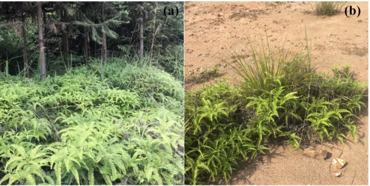
Dicranopteris linearis (Gleicheniaceae) is a native pioneer fern common throughout the Old World
114
(sub)tropics and Oceania (USDA, 2018). This species can grow in acidic (pH 4–5) and poor soils, 115
and exhibits high phosphorus use efficiency (Russell et al., 1998; Chen et al., 2016a). Moreover, D. 116
linearis often forms patches under an open canopy and dominates the understory of many plant
117
communities in Southern China (e.g. Pinus massoniana, Cunninghamia lanceolata, Eucalyptus 118
robusta) (Fig. 1). This plant has a crucial role in the ecosystem, retaining nutrients and organic
119
matter (Cohen et al., 1995); sustaining soil microclimates (Zhao et al., 2012); facilitating growth of 120
Eucalyptus trees (Wan et al., 2014); and controlling REE migration and gully erosion (Chen et al.,
121
2016b). To date, only the D. linearis accessions from Southern China have been recognized as a 122
REEs hyperaccumulator (Wei et al., 2001 & 2005; Shan et al., 2003). Field surveys confirm that D. 123
linearis can accumulate up to 1240–1760 mg kg-1 REEs in the aboveground parts when growing on 124
(ion-adsorption) REE mine tailings (Table S1). Dicranopteris linearis thus may be used for 125
phytoremediation and phytomining at ion-adsorption REE mine tailings in Southern China, which 126
can contain 409–1035 mg kg-1 REEs (Chao et al., 2016), and occupy an area of more than 100 km2 127
(Liu et al., 2015). Most of the REEs are accumulated in the cell walls in the pinnae of D. linearis, as 128
shown by differential extractions; although some of the REEs are also accumulated within the 129
protoplast, such as the organelles and cytosol, vacuoles and cell membranes (Wei et al., 2005). Light 130
REEs (LREEs, i.e. La, Ce, Pr and Nd) deposits have been reported from the cell wall, intercellular 131
space, plasmalemma, vesicles, and vacuoles of the root endodermis and stele cells of the adventitious 132
root by scanning electron microscopy with energy-dispersive spectroscopy (SEM-EDS) (Shan et al., 133
2003). Other studies reported that chlorophyll La and chlorophyll Ce (i.e. La and Ce bound to 134
chlorophyll) formed in the pinnae of D. linearis (Zhao et al., 1999; Wei et al., 2005), and the 135
chlorophyll REE can partly replace chlorophyll Mg (Wei et al., 2004). Moreover, binding to proteins 136
is likely one of the mechanisms against physiological toxicity of REEs in D. linearis pinnae (Wang 137
et al., 2003). Overall, the different techniques and samples preparations used to unravel those
7
different observations may be responsible for some artefacts. The high concentration of histidine 139
reported in the pinnae of this plant (Shan et al., 2003) may result from various physiological 140
activities and its presence is not sufficient to prove or demonstrate the chemical status of REEs, i.e. 141
histidine complexes of REEs. The investigation of REE distribution and speciation at a 142
submicrometric scale might provide key information about the mechanisms driving the uptake and 143
storage of those elements by identifying the analogies and co-distributions with other elements, 144
including nutrients. 145
146
Synchrotron-based X-ray fluorescence microscopy (XFM) is a non-destructive method that has been 147
successfully used to study the in-situ distribution of trace elements in hyperaccumulator plants (for 148
recent reviews see Lombi & Susini, 2009; van der Ent et al., 2018; Kopittke et al., 2018). The aim of 149
this study was to better understand the ecophysiological mechanisms that enable tolerance to REEs 150
in this fern. To that end, XFM elemental images of the sum of two light REEs (referred to as La + Ce 151
and being the sum of La and Ce), of K, Ca and Mn in the tissues and cells of D. linearis frond were 152
acquired. 153
154
Materials and methods 155
156
Collection of plant tissues 157
Live samples of D. linearis grown on the ion-adsorption REE mine tailings of Ganzhou, Jiangxi 158
Province, China (24 57 N, 115 05 E), and the corresponding soil material were collected. The 159
plants were transplanted in pots containing soil material and placed in a greenhouse, Sun Yat-sen 160
University, Guangzhou, China. Three mature plants with rhizosphere soil were brought alive to the 161
P06 beamline (PETRA III Synchrotron, DESY, Hamburg, Germany) for the experiments described 162
below. In parallel, four mature live pinna and three standing litter pinna (the dead pinna was still 163
connected to the stolon) samples from a D. linearis population on an ion-adsorption REE mine 164
tailing were sampled for bulk chemical analysis (Fig. S1). 165
8 167
9
Chemical analysis of bulk tissue samples 168
Plant samples were washed with pure water (18Ω, 25°C), then dried in an oven at 105°C for 2 h and 169
60°C for 72 h. The analysis methods of total concentrations of Al, REEs, Si, and other trace elements 170
(Mn, K, Ca, P) were adapted from Liu et al. (2019). 171
172
X-ray fluorescence microscopy (XFM) 173
The X-ray fluorescence microscopy (XFM) experiment was undertaken at Beamline P06 at the 174
PETRA III (Deutsches Elektronen-Synchrotron; DESY, Hamburg, Germany), a 6 GeV synchrotron 175
(Boesenberg et al., 2016). The undulator beam was monochromatized with a cryogenically cooled 176
Si(111) channel-cut monochromator to an energy of 12 keV with a flux of 1010 photon/s. A 177
Kirkpatrick-Baez mirror pair was used to focus the incident beam to 700 × 530 nm (hor × ver). The 178
samples were scanned in fly-scan mode, with the resultant sample X-ray signal detected using the 179
Maia 384C detector system, operated in backscatter geometry (Kirkham et al., 2010; Ryan et al., 180
2010 & 2014; Siddons et al., 2014). Typically, a quick ‘survey scan’ was first conducted to allow 181
for the selection of the appropriate portion of the sample. For the survey scan, the resolution was 50– 182
100 µm with a dwell of 1–2 ms and generally took ca. 5 min to complete. After that a ‘detailed scan’ 183
was conducted, with a resolution of 2–10 µm and a dwell time of 8–20 ms. For the whole 184
experiment, the incident energy of 12 keV was used in order that the fluorescence lines of the 185
elements of interest are well below the inelastic and elastic scatter peaks. 186
187
Live/fresh pinnule samples were analysed whole, or as cross-sections which hand cut with a 188
stainless-steel razor blade (‘dry knife’ method); whole or sectioned samples were then mounted 189
between two sheets of 4 µm Ultralene thin film in a tight sandwich to limit evaporation, and analysed 190
within 10 minutes after excision. The fresh samples mounted between two sheets of Ultralene thin 191
film (4 µm) were stretched over a 3D-printed frame magnetically attached to the x-y-z sample stage 192
at atmospheric temperature (~23°C). X-ray micro-fluorescence was performed using fast scanning to 193
keep the scan time, and hence sample degradation, to a minimum. 194
10
Data processing and statistics 195
The XRF event stream was analysed using the Dynamic Analysis method (Ryan & Jamieson, 1993; 196
Ryan, 2000) as implemented in GeoPIXE (Ryan et al., 1990 & 2005). It was necessary to use a 197
precise matrix file for the spectra fitting to account for X-ray fluorescence absorption of the 198
relatively low energy of the REEs L-lines, and the matrix was based on the stoichiometry of the 199
mean concentrations of major elements (>0.1 wt%) in dried D. linearis pinna samples (Table S2). 200
The matrix composition assumed hydration of the fresh samples, and was formulated as 201
C7O31H59N0.7S0.2Al0.3Si1.5Ca0.2Mn0.1K0.3 (on the basis of average concentrations of these elements in
202
bulk samples) with a density of 0.90 g cm-3, considering two layers of Ultralene foil (4 µm) and with 203
varying sample thicknesses. Pinnule samples were considered to have uniform thickness. The 204
rhodium coating of the KB mirror focusing optics available for this experiment results in a 205
high-energy cutoff of 23 keV, well below that required to excite the K-lines of the REEs (33.44 keV 206
for La), so therefore the experiment had to rely on exciting the L-lines (ranging from 4.65 keV for La 207
to 7.41 keV for Yb). The L-lines of Al and Si were around 1.5–3 keV, thus could not be detected. 208
Although the K-line of Y could be reached at 14.96 keV, its concentration was too low (mean of 106 209
µg g-1 Y in pinna) to be reliably detected. The reported resolution of the Maia 384C detector is 220 210
eV and it is hence unable to distinguish between the numerous L-lines of various the REEs. 211
Moreover, the L-lines of Gd, Eu and Sm are in the range of the K-lines of Mn and thus these 212
elements, at these relative concentrations, are not possible to distinguish reliably using the Maia 213
detector. Consequently, only La and Ce were used to represent the REEs. 214
215
Results 216
217
Bulk elemental concentrations in Dicranopteris linearis 218
The bulk pinna concentrations of REEs, Al, Si, Mn, K, Ca, and P are given in Table 1. The 219
concentrations of Al, Si and REEs in live pinnae are around half that of the standing litter pinnae – 220
Al 2850 vs 4850 mg kg-1, Si 14700 vs 33900 mg kg-1 and REEs 1900 vs 3500 mg kg-1 respectively.
11
Alternatively, the concentrations of Mn, P and K in live pinnae are always higher than the standing 222
litter pinnae – Mn 1480 vs 310 mg kg-1, P 211 vs 129 mg kg-1 and K 3000 vs 268 mg kg-1 223
respectively. The concentration of Ca has no significant difference between the two types of pinnae 224
(Table 1). Among the 15 REEs, the sum of La and Ce accounts for ~50% of the total REEs in the 225
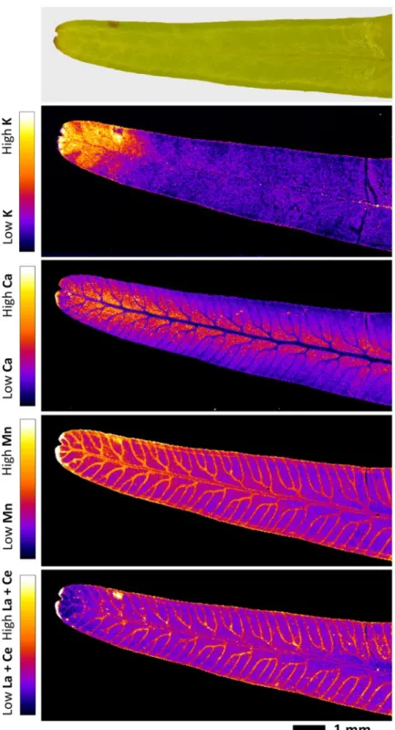
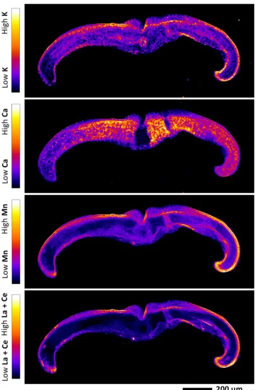
pinnae, without significant differences between live and standing litter pinnae (Table S2). 226
227
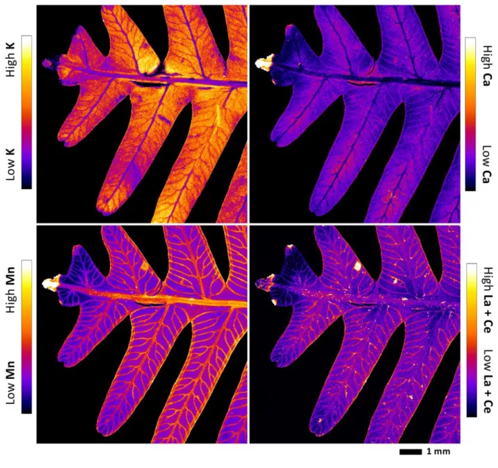
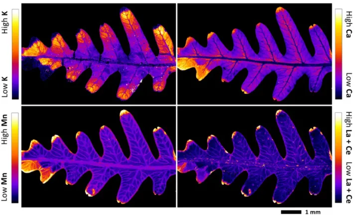
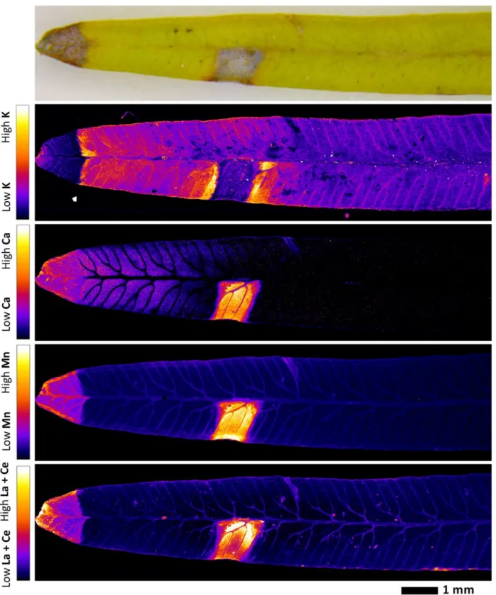
Localization of REEs in pinna and pinnule 228
The X-ray fluorescence element maps evidenced a distinct mapping for K, Ca, Mn and the summed 229
REEs La + Ce in the tissues of the pinna (Fig. 2; Fig. 3) and the pinnule (Fig. 4; Fig. 5; Fig. S2) of D. 230
linearis. The most significant enrichment of La + Ce and Mn is in the necrotic tissues, which are by
231
definition the bio-inactive regions of the pinna. The preferential accumulation of REEs in these 232
necrotic spots appear to in all of the tissue areas (pinna tips, margins and blades) of the necrosis. 233
Significant La + Ce and Mn enrichments are also in the midvein, secondary veins, tertiary veins and 234
in the margins of the pinna and its pinnule. The La + Ce and Mn were not strictly restricted within 235
the veins and necrotic tissues; their signals were also visibly and uniformly localized outside the 236
veins and necrotic tissues. Calcium is mainly distributed in the necrotic tissues and blade, but low in 237
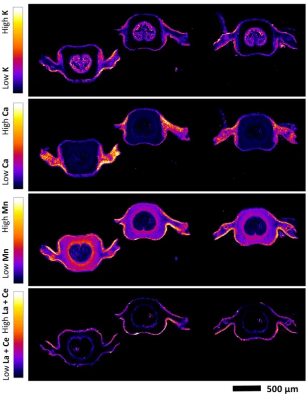
the veins. Potassium has a distinctly different distribution in the pinna and pinnule, and is mostly 238
localized in the blade, and absent in the necrotic tissues. 239
240
In order to compare the elements spatial co-occurrences and correlations, tri-color (Ce, Mn and Ca) 241
composites maps and Ce-Mn frequency plots of a pinna and a pinnule are provided in Fig. S3. The 242
results further confirm the co-localization of Ce and Mn both inside and outside the necrotic tissues, 243
with higher concentrations inside the necrotic tissues and lower outside. However, tri-color elemental 244
maps and element association frequency plots also show that Ce and Mn are not totally co-localized, 245
e.g. there are some high Ce areas at the right of the pinnule (Fig. S2), where Mn is relatively low.
246 247 248
12
Localization of REEs in pinnule and midvein cross-section 249
In order to establish the localization of REEs at the cellular level, XFM mapping was performed on 250
cross-sections of midvein and pinnule. The elemental maps revealed that La + Ce and Mn are co-251
localized in the epidermis (Fig. 6; Fig. 7). The La + Ce and Mn in the upper epidermis are much 252
more concentrated than in the low epidermis. In the midvein cross-section, there is a “ring” shaped 253
peak of La + Ce and Mn between the vascular bundle and the cortex, likely the pericycle. In the 254
pinnule cross-section, the vascular bundle and cortex are difficult to differentiate. However, 255
compared to La + Ce, Mn signals are more prominent in the cortex and mesophyll, while less marked 256
in the vascular bundle. Potassium is low in the cortex, but concentrated in the mesophyll, epidermis 257
and vascular bundle. Calcium is predominantly localized in the mesophyll and epidermis, but low in 258
the vascular bundle and cortex. The subcellular elemental distribution could not be differentiated for 259
any element. 260
261
Localization of REEs in petiole and stolon cross-section 262
The elemental XFM maps of the petiole and stolon cross-sections show La + Ce enrichment in the 263
epidermis and vascular bundle, while in the cortex and the pericycle of the petiole cross-section 264
prevailing concentrations are very low (Fig. 8 and Fig. S4). In contrast, in both the petiole and stolon 265
cross-sections, the K, Ca and Mn are similar and mostly concentrated in the epidermis, pericycle and 266
vascular bundle, with distinct localizations in the cortex. Within the vascular bundle of petiole cross-267
sections, La + Ce, Mn, K and Ca are mainly localized in the protoxylem and metaxylem. 268
269
Discussion 270
271
Dicranopteris linearis accumulates the highest concentrations of REEs in necrotic tissues, which
272
differs significantly from the behavior of most other metal hyperaccumulators. Potassium is an 273
essential element that plays a critical role in cellular osmotic regulation in the young and fresh leaves, 274
and the absence of K in necrotic tissues suggests that these areas are physiologically inactive. The 275
13
markedly higher concentrations of REEs, Al and Si in the standing litter pinnae, as compared to the 276
live pinnae further affirms that these elements are rather immobile as opposed to K which is strongly 277
depleted in litter tissues (Fig. S1; Table 1). The concentrations of Mn in the pinna of D. linearis 278
(Table 1) are much greater than what is typically toxic in most of the plants (e.g. < 200 mg kg-1 in
279
maize) (Shao et al., 2017). Therefore, the necrosis in these tissues is possibly, among other reasons, 280
the result of Mn accumulation, oxidation and localised toxicity within the pinnules, which then 281
induces cell death, necrotic spots, substantially larger necrotic tissue and then finally acts as a “dump 282
site” for REEs. A similar phenomenon has been reported in the leaf of soybean and cowpea in 283
response to Mn toxicity; the toxicity started with Mn pumped under the cuticle via the apoplast, or 284
expelled via hydathodes towards the leaf tip, and then increasing concentrations of Mn leading to Mn 285
oxidation (+2 to +3 and +4 valences) and necrotic lesions, which in turn stimulate more Mn 286
translocation as a result of higher evaporation (Blamey et al., 2018a & 2018b). However, Ce and Mn 287
were not totally co-localized, it is therefore also possible that the necrotic tissues were induced by Ce 288
accumulation and oxidation (+3 to +4 valences). It could be also interpreted as a tolerance 289
mechanism in which some cells are sacrificed and used as a dump, while in the others, 290
photosynthesis and normal activity can continue (Küpper et al., 2007). The underlying mechanisms 291
as to why REEs prefer to compartmentalize into the upper epidermis, margins and veins are not fully 292
understood. Previous studies on hyperaccumulator plants suggest that it may reflect a physiological 293
and/or defense-related response – protection of photosynthetically active tissues such as mesophyll, 294
and defense from predation by herbivores and pathogens (Martens & Boyd, 1994; Cappa & Pilon-295
Smits, 2014). 296
297
The distinct localization in vascular bundles, while prevailing concentrations are low in the cortex of 298
petiole and midvein, suggests an efficient REE transport system in this plant. The transport of REEs 299
to the pinna probably occurs as mass flow through the vascular tissue, driven by transpiration. In 300
non-accumulating species of beech and oak (Fagaceae), REEs transport within xylem was suggested 301
to be associated with general nutrient flux (Brioschi et al., 2013). However, in the vascular bundles 302
of D. linearis, we found both a small area of La + Ce peaks within vascular bundle and a very bright 303
14
“ring” shaped peak between vascular bundle and cortex (Fig. 7). The significant La + Ce enrichment, 304
with no obvious Ca and Mn enrichment within the vascular bundle of veins, suggests that REEs are 305
translocated to the blade to a lesser extent than Ca and Mn. This might be due to the features of the 306
pericycle tissue where cells are dense, preventing REEs translocation from the xylem into the blade 307
via the intercellular space. It is possible that once the REEs enter into the vascular system of veins 308
(xylem), they are partly fixed by the pericycle, and then transported to the D. linearis blades 309
(especially to the epidermis and necrotic tissues) via transpiration flow. In this case, the transport and 310
accumulation of REEs should mainly be passive convection processes. Thus, the distribution of 311
REEs in D. linearis pinna could be influenced by various parameters that are associated with the 312
evaporation (e.g. biological variety, seasonal and climatic changes) and Mn toxicity (e.g. light, 313
pinnule age and height at sampling) (Küpper et al., 2004; Fernando & Lynch, 2015; Bartoli et al., 314
2018). The highest concentrations recorded within the necrotic tissues of pinna may be ascribed to a 315
lack of wax coats at the surface of pinna, and/or the damage of the pericycle tissue or cells between 316
vascular bundle and cortex in the veins, thus leading to a higher evaporation rate. The higher 317
evaporation rate may also lead to higher concentrations of REEs in the upper epidermis, compared to 318
the lower epidermis (Fig. 6; Fig. 7). 319
320
As vacuoles in necrotic tissues of the pinnae disintegrate, compartmentalization and accumulation in 321
these areas indicate that the REEs are likely to be sequestrated by cell walls or exist under immobile 322
forms. Many studies found that REEs tend to complex with phosphate and form deposits in the cell 323
walls and more generally in the intercellular space of plant tissues (Ding et al., 2006; Ruíz-Herrera et 324
al., 2012). However, the molar ratio of P in D. linearis pinnae was much lower than that of REEs
325
(Table 1). Moreover, we found that D. linearis also accumulates high concentration of Al and Si 326
(Table 1; Chour et al., 2018). Detoxification of Al and other heavy metals by Si has been observed in 327
many plants (Liang et al., 2007). For example, Al localization coincided with Si distribution in cell 328
walls of the Al hyperaccumulator Rudgea viburnoides (Malta et al., 2016). The co-deposition of Si 329
and Cd in the cell walls as a [Si-wall matrix]Cd co-complex inhibited Cd ion uptake by rice cell (Liu 330
et al., 2013). In the Al and Si hyperaccumulator Faramea marginata, Al and Si are thought to be
15
deposited as phytolith Al (Britez et al., 2002). Also, silicon may co-deposit with Mn at the apoplast 332
and decrease the oxidation of Mn in cowpea, soybean and sunflower (Blamey et al., 2018a). Rare 333
earth elements are a group of trivalent elements, which exhibit many chemical similarities to Al 334
(Pletnev & Zernov, 2002). Considering the high concentration of REEs, Mn, Al and Si, a co-335
deposition of REEs, Mn and Al with Si may be involved in the homeostasis of high concentrations of 336
REEs, Mn and Al in D. linearis. Therefore, it would be crucial to study the localization of Al and Si 337
in D. linearis tissues with other methods to confirm these hypotheses. 338
339
Author contributions 340
341
WSL conducted the fieldwork and collected the samples. AvdE, GE, PDE and KMS conducted the 342
synchrotron XFM experiment. KMS, AvdE and EMP performed the XFM data processing and 343
analysis. WSL conducted and bulk elemental analysis. All authors contributed to writing the 344 manuscript. 345 346 Acknowledgements 347 348
This work was financially supported by the National Natural Science Foundation of China 349
(41771343), and the 111 Project (B18060). This work is a contribution of the joint lab ECOLAND, 350
established between Sun Yat-sen University, University of Lorraine and INRA. We acknowledge 351
DESY (Hamburg, Germany), a member of the Helmholtz Association HGF, for the provision of 352
experimental facilities. Parts of this research were carried out at PETRA III and we would like to 353
thank Dr. Jan Garrevoet and Dr. Gerald Falkenberg for assistance in using beamline P06. The 354
research leading to this result has been supported by the project CALIPSOplus under the Grant 355
Agreement 730872 from the EU Framework Programme for Research and Innovation HORIZON 356
2020. The French National Research Agency through the national “Investissements d’avenir” 357
program (ANR-10-LABX-21, LABEX RESSOURCES21) and through the ANR-14-CE04-0005 358
Project “Agromine” is acknowledged for funding support. 359
16 360 361
17 References 362 363
Baker AJM, Brooks RR. 1989. Terrestrial higher plants which hyper accumulate metallic elements. 364
Biorecovery 1: 81–126.
365
Bartoli F, Royer M, Coinchelin D, Thiec DL, Rose C, Robin C, Echevarria G. 2018. Multiscale 366
and age-dependent leaf nickel in the Ni-hyperaccumulator Leptoplax emarginata. Ecological 367
Research 33: 1–14.
368
Binnemans K, Jones PT, Blanpain B, Gerven TV, Yang Y, Walton A, Buchert M. 2013. 369
Recycling of rare earths: a critical review. Journal of Cleaner Production 51: 1–22. 370
(1) BlameyPlant Soil. 2019, 437, 427–437. 371
(2) Boesenberg, U.; Ryan, C. G.; Kirkham, R.; Siddons, D. P.; Alfeld, M.; Garrevoet, J.; Núñez, T.; 372
Claussen, T.; Kracht, T.; Falkenberg, G. Fast X-ray microfluorescence imaging with 373
submicrometer-resolution integrating a Maia detector at beamline P06 at PETRA III. J. 374
Synchrotron Radiat. 2016, 23, 1550−1560.
375
Kirkham, R.; Dunn, P. A.; Kuczewski, A. J.; Siddons, D. P.; Dodanwela, R.; Moorhead, G. F.; Ryan 376
C. G.; de Geronimo, G.; Beuttenmuller, R.; Pinelli, D.; Pfeffer, M.; Davey, P.; Jensen, M.; 377
Paterson, D. J.; de Jonge, M. D.; Howard, D. L.; Kusel, M.; McKinlay, J FPC, Mckenna BA, 378
Li C, Cheng MM, Tang CX, Jiang HB, Howard DL, Paterson DJ, Kappen P, Wang P, 379
Menzies NW, Kopittke PM. 2018a. Manganese distribution and speciation help to explain the 380
effects of silicate and phosphate on manganese toxicity in four crop species. New Phytologist 381
217: 1146–1160. 382
Blamey FPC, Paterson DJ, Walsh A, Afshar N, McKenna BA, Cheng MM, Tang CX, Horst 383
WJ, Menzies NW, Kopittke PM. 2018b. Time-resolved X-ray fluorescence analysis of 384
element distribution and concentration in living plants: An example using manganese toxicity 385
in cowpea leaves. Environmental and Experimental Botany 156: 151–160. 386
18
Boesenberg U, Ryan CG, Kirkham R, Siddons DP, Alfeld M, Garrevoet J, Núñez T, Claussen 387
T, Kracht T, Falkenberg G. 2016. Fast X-ray microfluorescence imaging with 388
submicrometer-resolution integrating a Maia detector at beamline P06 at PETRA III. Journal of 389
Synchrotron Radiation 23: 1550−1560.
390
Brioschi L, Steinmann M, Lucot E, Pierret MC, Stille P, Prunier J, Badot PM. 2013. Transfer 391
of rare earth elements (REE) from natural soil to plant systems: implications for the 392
environmental availability of anthropogenic REE. Plant and Soil 366: 143–163. 393
Britez RM, Watanabe T, Jansen S, Reissmann CB, Osaki M. 2002. The relationship between 394
aluminium and silicon accumulation in leaves of Faramea marginata (Rubiaceae). New 395
Phytologist 156: 437–444.
396
Cappa JJ, Pilon-Smits EA. 2014. Evolutionary aspects of elemental hyperaccumulation. Planta 239: 397
267–275. 398
Chao YQ, Liu WS, Chen YM, Chen WH, Zhao LH, Ding QB, Wang SZ, Tang YT, Zhang T, 399
Qiu RL. 2016. Structure, variation, and co-occurrence of soil microbial communities in 400
abandoned sites of a rare earth elements mine. Environmental Science & Technology 50: 401
11481–11490. 402
Chen ZQ, Chen ZB, Yan XY, Bai LY. 2016a. Stoichiometric mechanisms of Dicranopteris 403
dichotoma, growth and resistance to nutrient limitation in the Zhuxi watershed in the red soil
404
hilly region of China. Plant and Soil 398: 367–379. 405
Chen ZQ, Chen ZB, Bai LY. 2016b. Rare earth element migration in gullies with different 406
Dicranopteris dichotoma covers in the Huangnikeng gully group, Changting county, Southeast
407
China. Chemosphere 164: 443–450. 408
Chour Z, Laubie B, Morel JL, Tang YT, Qiu RL, Simonnot MO, Muhr L. 2018. Recovery of 409
rare earth elements from Dicranopteris dichotoma by an enhanced ion exchange leaching 410
process. Chemical Engineering and Processing 130: 208–213. 411
19
Cohen AL, Singhakumara BMP, Ashton PMS. 1995. Releasing rain forest succession: a case 412
study in the Dicranopteris linearis fernlands of Sri Lanka. Restoration Ecology 3: 261–270. 413
Ding SM, Liang T, Zhang CS, Huang ZC, Xie YN, Chen TB. 2006. Fractionation mechanisms of 414
rare earth elements (REEs) in hydroponic wheat: an application for metal accumulation by 415
plants. Environmental Science & Technology 40: 2686–2691. 416
Fernando DR, Lynch JP. 2015. Manganese phytotoxicity: new light on an old problem. Annals of 417
Botany 116: 313–319.
418
Freeman JL, Zhang LH, Marcus MA, Fakra S, Mcgrath SP, Pilon-Smits EAH. 2006. Spatial 419
imaging, speciation, and quantification of selenium in the hyperaccumulator plants Astragalus 420
bisulcatus and Stanleya pinnata. Plant Physiology 142: 124–134.
421
Hokura A, Omuma R, Terada Y, Kitajima N, Abe T, Saito H, Yoshida S, Nakai I. 2006. 422
Arsenic distribution and speciation in an arsenic hyperaccumulator fern by x-ray spectrometry 423
utilizing a synchrotron radiation source. Journal of Analytical Atomic Spectrometry 21: 321– 424
328. 425
Hu PJ, Wang YD, Przybyłowicz WJ, Li Z, Barnabas A, Wu LH, Mesjasz-Przybyłowicz J. 426
2015. Elemental distribution by cryo-micro-PIXE in the zinc and cadmium hyperaccumulator 427
Sedum plumbizincicola grown naturally. Plant and Soil 388: 267–282.
428
Jaffré T, Brooks RR, Lee J, Reeves RD. 1976. Sebertia acuminata: a hyperaccumulator of nickel 429
from New Caledonia. Science 193: 579–580. 430
Kirkham R, Dunn PA, Kuczewski AJ, Siddons DP, Dodanwela R, Moorhead GF, Ryan CG, de 431
Geronimo G, Beuttenmuller R, Pinelli D, Pfeffer M, Davey P, Jensen M, Paterson DJ, de 432
Jonge MD, Howard DL, Kusel M, McKinlay J. 2010. The Maia spectroscopy detector 433
system: engineering for integrated pulse capture, low latency scanning and real time 434
processing. AIP Conference Proceedings 1234: 240–243. 435
20
Kitajima N, Kashiwabara T, Fukuda N, Endo S, Hokura A, Terada Y, Nakai I. 2008. 436
Observation of arsenic transfer in leaf tissue of hyperaccumulator fern by utilizing synchrotron 437
radiation micro-XRF imaging. Chemistry Letters 37: 32–33. 438
Kopittke PM, Punshon T, de Jonge MD, Paterson DJ, Tappero RV, Ryan CG, Wang P, 439
Blamey FPC, McKenna BA, van der Ent A, Lombi E. 2018. Synchrotron-based X-ray 440
fluorescence microscopy as a technique for imaging of elements in plants. Plant Physiology 441
178: 507–523. 442
Küpper H, Mijovilovich A, Meyerklaucke W, Kroneck PMH. 2004. Tissue- and age-dependent 443
differences in the complexation of cadmium and zinc in the cadmium/zinc hyperaccumulator 444
Thlaspi caerulescens (Ganges ecotype) revealed by x-ray absorption spectroscopy. New
445
Phytologist 134: 748–757.
446
Küpper H, Parameswaran A, Leitenmaier B, Trtílek M, Šetlík I. 2007. Cadmium induced 447
inhibition of photosynthesis and long term acclimation to cadmium stress in the 448
hyperaccumulator Thlaspi caerulescens. New Phytologist 175: 655–674. 449
Li JT, Gurajala HK, Wu LH, van der Ent A, Qiu RL, Baker AJ, Tang YT, Yang XE, Shu WS. 450
2018. Hyperaccumulator plants from China: A synthesis of the current state of knowledge. 451
Environmental Science & Technology 52: 11980–11994.
452
Liang Y, Sun W, Zhu YG, Christie P. 2007. Mechanisms of silicon-mediated alleviation of abiotic 453
stresses in higher plants: a review. Environmental Pollution 147: 422–428. 454
Liu C, Yuan M, Liu WS, Guo MN, Huot H, Tang YT, Laubie B, Simonnot MO, Morel JL, Qiu 455
RL. 2018. Agromining: farming for metals. In Element case studies: rare earth elements; van 456
der Ent A, Echevarria G, Baker AJM, Morel JL, eds, Mineral Resource Reviews series, Cham: 457
Springer International Publishing pp. 297–308.
458
Liu J, Ma J, He CW, Li XL, Zhang WJ, Xu FS, Lin YJ, Wang LJ. 2013. Inhibition of cadmium 459
ion uptake in rice (Oryza sativa) cells by a wall-bound form of silicon. New Phytologist 200: 460
691–699. 461
21
Liu WS, Liu C, Wang ZW, Teng WK, Tang YT, Qiu RL. 2015. Limiting factors for restoration of 462
dumping sites of ion-adsorption rare earth tailing. Acta Ecologica Sinica 52: 879–887. 463
Liu WS, Zheng HX, Guo MN, Liu C, Huot H, Morel JL, van der Ent A, Tang YT, Qiu RL. 464
2019. Co-deposition of silicon with rare earth elements (REEs) and aluminium in the fern 465
Dicranopteris linearis from China. Plant and Soil 437: 427–437.
466
Lombi E, Susini J. 2009. Synchrotron-based techniques for plant and soil science: opportunities, 467
challenges and future perspectives. Plant and Soil 320: 1–35. 468
Malta PG, Arcanjo-Silva S, Ribeiro C, Campos NV, Azevedo AA. 2016. Rudgea viburnoides 469
(Rubiaceae) overcomes the low soil fertility of the Brazilian Cerrado and hyperaccumulates 470
aluminum in cell walls and chloroplasts. Plant and Soil 408: 369–384. 471
Martens SN, Boyd RS. 1994. The ecological significance of nickel hyperaccumulation: a plant 472
chemical defense. Oecologia 98: 379–384. 473
Migaszewski ZM, Gałuszka A. 2015. The characteristics, occurrence, and geochemical behavior of 474
rare earth elements in the environment: a review. Critical Reviews in Environmental Science 475
and Technology 45: 429–471.
476
Nieboer E, Richardson HS. 1980. The replacement of the nondescript term ‘heavy metals’ by a 477
biologically and chemically significant classification of metal ions. Environmental Pollution 478
series B 1: 3–26.
479
Pletnev IV, Zernov VV. 2002. Classification of metal ions according to their complexing properties: 480
a data-driven approach. Analytica Chimica Acta 455: 131–142. 481
Redling K. 2006. Rare earth elements in agriculture with emphasis on animal husbandry. Ludwig-482
Maximilians-Universität München.
483
Reeves RD, Baker AJM, Jaffré T, Erskine PD, Echevarria G, van der Ent A. 2017. A global 484
database for hyperaccumulator plants of metal and metalloid trace elements. New Phytologist 485
218: 407–411. 486
22
Reeves RD, van der Ent A, Baker AJM. 2018. Agromining: farming for metals. In Global 487
distribution and ecology of hyperaccumulator plants; van der Ent A, Echevarria G, Baker AJM, 488
Morel JL, eds, Mineral Resource Reviews series, Cham: Springer International Publishing pp. 489
75–92. 490
Reeves RD. 2003. Tropical hyperaccumulators of metals and their potential for phytoextraction. 491
Plant and Soil 249: 57–65.
492
Ruíz-Herrera LF, Sánchez-Calderón L, Herrera-Estrella L, López-Bucio J. 2012. Rare earth 493
elements lanthanum and gadolinium induce phosphate-deficiency responses in Arabidopsis 494
thaliana, seedlings. Plant and Soil 353: 231–247.
495
Russell AE, Raich JW, Vitousek PM. 1998. The ecology of the climbing fern Dicranopteris 496
linearis, on windward Mauna Loa, Hawaii. Journal of Ecology 86: 765–779.
497
Ryan CG, Achterbergh EV, Jamieson DN. 2005. Advances in dynamic analysis, PIXE imaging: 498
correction for spatial variation of pile-up components. Nuclear Instruments and Methods in 499
Physics Research Section B 231: 162–169.
500
Ryan CG, Cousens DR, Sie SH, Griffin WL, Suter GF, Clayton E. 1990. Quantitative PIXE 501
microanalysis of geological material using CSIRO proton microprobe. Nuclear Instruments and 502
Methods in Physics Research Section B 47: 55–71.
503
Ryan CG, Jamieson DN. 1993. Dynamic analysis: on-line quantitative PIXE microanalysis and its 504
use in overlap-resolved elemental mapping. Nuclear Instruments and Methods in Physics 505
Research Section B 77: 203–214.
506
Ryan CG, Kirkham R, Hough RM, Moorhead G, Siddons DP, de Jonge MD, Paterson DJ, de 507
Geronimo G, Howard DL, Cleverley JS. 2010. Elemental X-ray imaging using the Maia 508
detector array: the benefits and challenges of large solid-angle. Nuclear Instruments and 509
Methods in Physics Research Section A 619: 37–43.
23
Ryan CG, Siddons DP, Kirkham R, Li ZY, de Jonge MD, Paterson DJ, Kuczewski A, Howard 511
DL, Dunn PA, Falkenberg G, Boesenberg U, de Geronimo G, Fisher LA, Halfpenny A, 512
Lintern MJ, Lombi E, Dyl KA, Jensen M, Moorhead GF, Cleverley JS, Hough RM, Godel 513
B, Barnes SJ, James SA, Spiers KM, Alfeld M, Wellenreuther G, Vukmanovic Z, Borg S. 514
2014. Maia X-ray fluorescence imaging: Capturing detail in complex natural samples. Journal 515
of Physics: Conference Series 499: 012002.
516
Ryan CG. 2000. Quantitative trace element imaging using PIXE and the nuclear microprobe. 517
International Journal of Imaging Systems and Technology 11: 219–230.
518
Scheckel KG, Hamon R, Jassongne L, Rivers M, Lombi E. 2007. Synchroton x-ray absorption-519
edge computed microtograpy imaging of thallium compartmentalization in Iberis intermedia. 520
Plant and Soil 294: 305–306.
521
Shan XQ, Wang HO, Zhang SZ, Zhou HF, Zheng Y, Yu H, Wen B. 2003. Accumulation and 522
uptake of light rare earth elements in a hyperaccumulator Dicropteris dichotoma. Plant Science 523
165: 1343–1353. 524
Shao JF, Yamaji N, Shen RF, Ma JF. 2017. The key to Mn homeostasis in plants: regulation of 525
Mn transporters. Trends in Plant Science 22: 215–224. 526
Sharma SS, Dietz K, Mimura T. 2016. Vacuolar compartmentalization as indispensable component 527
of heavy metal detoxification in plants. Plant, Cell & Environment 39: 1112–1126. 528
Siddons DP, Kirkham R, Ryan CG, de Geronimo G, Dragone A, Kuczewski AJ, Li ZY, Carini 529
GA, Pinelli D, Beuttenmuller R, Elliott D, Pfeffer M, Tyson TA, Moorhead GF, Dunn PA. 530
2014. Maia X-ray microprobe detector array system. Journal of Physics: Conference Series 499: 531
012001. 532
Tappero R, Peltier E, Gräfe M, Heidel K, Ginder-Vogel M, Livi KJT, Rivers ML, Marcus MA, 533
Chaney RL, Sparks DL. 2007. Hyperaccumulator Alyssum murale relies on a different metal 534
storage mechanism for cobalt than for nickel. New Phytologist 175: 641–654. 535
24
Thomas PJ, Carpenter D, Boutin C, Allison JE. 2014. Rare earth elements (REEs): effects on 536
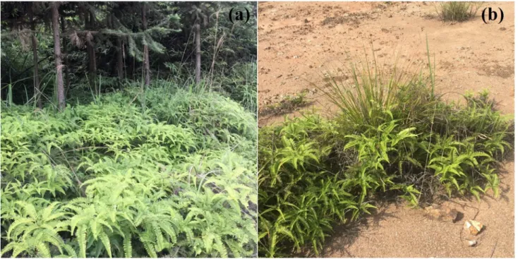
germination and growth of selected crop and native plant species. Chemosphere 96: 57–66. 537
Tian SK, Lu LL, Yang XE, Labavitch JM, Huang YY, Brown P. 2009. Stem and leaf 538
sequestration of zinc at the cellular level in the hyperaccumulator Sedum alfredii. New 539
Phytologist 182: 116–126.
540
USDA. 2018. Agricultural Research Service, National Plant Germplasm System Germplasm 541
Resources Information Network (GRIN-Taxonomy). 542
van der Ent A, Baker, AJM, Reeves RD, Pollard AJ, Schat H. 2013. Hyperaccumulators of metal 543
and metalloid trace elements: facts and fiction. Plant and Soil 362: 319–334. 544
van der Ent A, Baker AJM, Reeves RD, Chaney RL, Anderson CWN, Meech JA, Erskine PD, 545
Simonnot MO, Vaughan J, Morel JL, Echevarria G, Fogliani B, Qiu QL, Mulligan DR. 546
2015. Agromining: farming for metals in the future? Environmental Science & Technology 49: 547
4773–4780. 548
van der Ent A, Mulligan D. 2015. Multi-element concentrations in plant parts and fluids of 549
Malaysian nickel hyperaccumulator plants and some economic and ecological considerations. 550
Journal of Chemical Ecology 41: 396–408.
551
van der Ent A, Przybyłowicz WJ, de Jonge MD, Harris HH, Ryan CG, Tylko G, Paterson DJ, 552
Barnabas AD, Kopittke PM, Mesjasz-Przybyłowicz J. 2018. X ray elemental mapping 553
techniques for elucidating the ecophysiology of hyperaccumulator plants. New Phytologist 218: 554
432–452. 555
Vogel-Mikuš K, Regvar M, Mesjasz-Przybyłowicz J, Przybyłowicz WJ, Simčič J, Pelicon P, 556
Budnar M. 2008. Spatial distribution of cadmium in leaves of metal hyperaccumulating 557
Thlaspi praecox using micro-PIXE. New Phytologist 179: 712–721.
25
Wan SZ, Zhang CL, Chen YQ, Zhao J, Wang XL, Wu JP, Zhou LX, Lin YB, Liu ZF, Fu SL. 559
2014. The understory fern Dicranopteris dichotoma facilitates the overstory eucalyptus trees in 560
subtropical plantations. Ecosphere 5: 1–12. 561
Wang H, Shan XQ, Zhang S, Wen B. 2003. Preliminary characterization of a light-rare-earth-562
element-binding peptide of a natural perennial fern Dicranopteris dichotoma. Analytical and 563
Bioanalytical Chemistry 376: 49–52.
564
Wang LH, Li JG, Zhou Q, Yang GM, Ding XL, Li XD, Cai CX, Zhang Z, Wei HY, Lu TH, 565
Deng XW, Huang XH. 2014. Rare earth elements activate endocytosis in plant cells. 566
Proceedings of the National Academy of Sciences of the United States of America 111: 12936–
567
12941. 568
Wei ZG, Hong FS, Yin M, Li HX, Hu F, Zhao GW, Wong JWC. 2004. Off-line separation and 569
determination of rare earth elements associated with chloroplast pigments of hyperaccumulator 570
Dicranopteris dichotoma by normal-phase liquid chromatography and ICP-MS. Analytical and
571
Bioanalytical Chemistry 380: 677–682.
572
Wei ZG, Hong FS, Yin M, Li HX, Hu F, Zhao GW, Wong WJ. 2005. Subcellular and molecular 573
localization of rare earth elements and structural characterization of yttrium bound chlorophyll 574
a in naturally grown fern Dicranopteris dichotoma. Microchemical Journal 80: 1–8. 575
Wei ZG, Yin M, Zhang X, Hong FS, Li B, Zhao GW, Yan CH. 2001. Rare earth elements in 576
naturally grown fern Dicranopteris linearis in relation to their variation in soils in South-577
Jiangxi region (Southern China). Environmental Pollution 114: 345–355. 578
Wei ZG, Zhang HJ, Li HX, Hu F. 2006. Research trends on rare earth element hyperaccumulator. 579
Journal of the Chinese Rare Earth Society 24: 1–11. (in Chinese) 580
Zhao FJ, Moore KL, Lombi E, Zhu YG. 2014. Imaging element distribution and speciation in 581
plant cells. Trends in Plant Science 19: 183–192. 582
26
Zhao GW, Hong FS, Wei ZG, Gu YH, Hu TD, Xie YN, Liu T, Tao Y. 1999. Light rare earth 583
element speciation of chlorophyll a in naturally grown fern Dicranopteris linearis by EXAFS. 584
Progress in Natural Science 9: 1133–1135.
585
Zhao J, Wan SZ, Li ZA, Shao YH, Xu GL, Liu ZF, Zhou LX, Fu SL. 2012. Dicranopteris-586
dominated understory as major driver of intensive forest ecosystem in humid subtropical and 587
tropical region. Soil Biology and Biochemistry 49: 78–87. 588
27 GRAPHICAL ABSTRACT 589 590 591 592
28 593 594 595
Fig. 1. Live Dicranopteris linearis grown at (a) the understory of Cunninghamia lanceolata; (b) an 596
ion-adsorption REE mine tailing abandoned for about 10 years at Ganzhou, Jiangxi Province, 597
Southern China. 598
29 600 601
Fig. 2. Elemental µ-XRF maps of fresh Dicranopteris linearis pinna. The maps measure 8.86 × 8.56 602
mm. The elemental image was acquired in step size 15 µm with dwell 15 ms per pixel, 12.0 keV, 603
incident beam. 604
30 606 607
Fig. 3. Elemental µ-XRF maps of fresh Dicranopteris linearis pinna. The maps measure 13.1 × 8.58 608
mm. The elemental image was acquired in step size 25 µm with dwell 15 ms per pixel, 12.0 keV, 609
incident beam. 610
31 612 613
Fig. 4. Elemental µ-XRF maps of fresh Dicranopteris linearis pinnule. The maps measure 10.2 × 614
4.26 mm. The elemental image was acquired in step size 20 µm with dwell 20 ms per pixel, 12.0 615
keV, incident beam. 616
32 618
Fig. 5. Elemental µ-XRF maps of fresh Dicranopteris linearis pinnule. The maps measure 12.95 × 619
2.88 mm. The elemental image was acquired in step size 8 µm with dwell 15 ms per pixel, 12.0 keV, 620
incident beam. 621
33 622 623
Fig. 6. Elemental µ-XRF maps of fresh Dicranopteris linearis pinnule cross-section. The maps 624
measure 2.25 × 0.86 mm. The elemental image was acquired in step size 5 µm with dwell 15 ms per 625
pixel, 12.0 keV, incident beam. 626
34 628 629
Fig. 7. Elemental µ-XRF maps of fresh Dicranopteris linearis midvein cross-section of pinna. The 630
maps measure 4.70 × 1.57 mm. The elemental image was acquired in step size 8 µm with dwell 15 631
ms per pixel, 12.0 keV, incident beam. The concave side represents the adaxial side in the figure. 632
35 633
Fig. 8. Elemental µ-XRF maps of fresh Dicranopteris linearis petiole cross-section of pinna. The 634
maps measure 5.62 x 3.41 mm. The elemental image was acquired in step size 12 µm with dwell 15 635
ms per pixel, 12.0 keV, incident beam.
36 TABLES 637 638
Table 1. Bulk elemental concentrations in pinna of Dicranopteris linearis (mg kg-1 dry weight). 639 640 Type n Al Si REEs* Ca Mn P K Live pinna 1 2860 13400 1540 1330 1350 232 3330 2 3180 19500 2450 2160 1340 179 2600 3 1490 11800 1400 1160 1020 213 3390 4 3860 14100 2190 2580 2230 220 2700
Mean ± sd 2850±997a 14700±3310a 1900±505a 1810±676a 1480±523b 211±23.0b 3000±413b
Standing litter pinna
1 4910 36600 3620 2300 541 130 240
2 5280 36800 3750 988 241 156 432
3 4370 28200 3130 908 149 100 131
Mean ± sd 4850±455b 33900±4910b 3500±327b 1400±779a 310±205a 129±28.0a 268±152a
* The summed concentration of La, Ce, Pr, Nd, Sm, Eu, Gd, Tb, Dy, Ho, Er, Tm, Yb, Lu and Y, see the concentration of each rare earth element
641
in Table S2.
1
Supporting Information
1 2
Spatially-resolved localization of lanthanum and cerium in the rare earth element
3
hyperaccumulator fern Dicranopteris linearis from China
4 5
Wen-Shen Liu1,2,3, Antony van der Ent4,5, Peter D. Erskine4, Jean Louis Morel5, Guillaume
6
Echevarria4,5, Kathryn M. Spiers6, Emmanuelle Montargès-Pelletier7, Rong-Liang Qiu1,2,3, Ye-Tao
7
Tang1,2,3,*
8 9
1School of Environmental Science and Engineering, Sun Yat-sen University, Guangzhou 510275,
10
China.
11
2Guangdong Provincial Key Laboratory of Environmental Pollution Control and Remediation
12
Technology, Sun Yat-sen University, Guangzhou 510275, China.
13
3Guangdong Provincial Engineering Research Center for Heavy Metal Contaminated Soil
14
Remediation, Sun Yat-sen University, Guangzhou 510275, China.
15
4Centre for Mined Land Rehabilitation, Sustainable Minerals Institute, The University of
16
Queensland, St Lucia, Queensland 4072, Australia.
17
5Université de Lorraine, INRA, Laboratoire Sols et Environnement, Nancy 54000, France.
18
6Photon Science, Deutsches Elektronen-Synchrotron DESY, Hamburg 22607, Germany.
19
7CNRS – Université́ de Lorraine Laboratoire Interdisciplinaire des Environnements Continentaux,
20
Vandoeuvre-lès-Nancy F-54500, France.
2
* Corresponding author
22
Address: School of Environmental Science and Engineering, Sun Yat-sen University, Guangzhou
23 510275, China 24 E-mail: [email protected] 25 Tel./Fax. +86-020-39332743 26 27
3 28 29
Fig. S1. Live Dicranopteris linearis grown at (a) the understory of Cunninghamia lanceolata; (b) an
30
ion-adsorption REE mine tailing abandoned for about 10 years at Ganzhou, Jiangxi Province,
31
Southern China.
32 33
4 34 35
Fig. S2. Dicranopteris linearis growing at an ion-adsorption REE mine tailings. (a) D. linearis
36
thicket with both live pinnae and dead standing litter pinnae; (b) dead standing litter pinnae; (c)
37
morphological characteristics of D. linearis.
5 39 40
Fig. S3. Elemental µ-XFM maps of hydrated Dicranopteris linearis pinna. The maps measure 13.1 ×
41
8.58 mm. The elemental image was acquired in step size 25 µm with dwell 15 ms per pixel, 12.0
42
keV, incident beam.
6 44 45
Fig. S4. Elemental µ-XFM maps of hydrated Dicranopteris linearis pinnule. The maps measure 10.2
46
× 4.26 mm. The elemental image was acquired in step size 20 µm with dwell 20 ms per pixel, 12.0
47
keV, incident beam.
48 49
7 50 51
Fig. S5. Elemental µ-XRF maps of hydrated Dicranopteris linearis pinnule. The maps measure
52
11.25 × 2.79 mm. The elemental image was acquired in step size 8 µm with dwell 15 ms per pixel,
53
12.0 keV, incident beam. The REE content was below detection limits.
54 55
8 56 57
Fig. S6. Tri-color elemental maps (red = Ce, green = Mn, blue = Ca) in a pinna (top left) and element
58
association (Ce and Mn) frequency plots (top right). Two areas were marked and plotted on the pinna
59
elemental map (bottom two panels) showing occurrence of Mn-Ce associations in green.
60 61
9 62 63
Fig. S7. Elemental µ-XFM maps of hydrated Dicranopteris linearis petiole cross-section of pinna.
64
The maps measure 5.62 x 3.41 mm. The elemental image was acquired in step size 12 µm with dwell
65
15 ms per pixel, 12.0 keV, incident beam.
66 67
10 68
69
Fig. S8. Elemental µ-XRF maps of hydrated Dicranopteris linearis stolon cross-section. The maps
70
measure 2.84 x 2.81 mm. The images were acquired in step size 12 µm with dwell 15 ms per pixel,
71
12.0 keV, incident beam. The REE content was below detection limits.
11
Table S1. Rare earth elements concentrations in Dicranopteris linearis and rhizosphere soil
73
collected from different sites.
74 75 Type Aboveground part / mg kg-1 Underground part / mg kg-1 Rhizosphere soil / mg kg-1 Aboveground part / Rhizosphere soil Aboveground part / Underground part MT 1470±199 a 727±426 a 518±208 a 3.27±1.55 a 2.77±1.89 a TE 875±449 ab 198±66.0 bc 304±159 ab 4.09±3.57 a 5.12±3.49 a NM 1430±733 a 393±71.8 b 573±307 a 2.92±1.45 a 3.52±1.18 a HB 1160±806 ab 189±93.3 bc 363±81.9 ab 3.21±1.88 a 8.01±6.94 a LB 297±296 b 24.8±18.2 d 117±52.6 b 2.68±2.32 a 18.4±22.1 a 76
Dicranopteris linearis samples and rhizosphere soils were collected in non-mining area near ion-adsorption REE
77
mine tailings (NM), ion-adsorption REE mine tailings (MT), edge of the ion-adsorption REE mine tailings (TE),
78
high levels of REEs background (HB) from Ganzhou, Jiangxi province, and low levels of REEs background (LB)
79
from Guangzhou, Guangdong province of China. Different letters in the figure represent significant difference
80
among each group (ANOVA, Duncan, p<0.05). Plant samples were divided into aboveground part and
81
underground part, thoroughly washed with deionized water, dried in an oven at 105 for 1 hour and then 60 for
82
48 hours, and then carefully crushed. Soil samples were air-dried and sieved to 0.15 mm. 50 ± 0.5 mg of dry plant
83
samples and soil samples were introduced in a Teflon crucible equipped with cylinder sleeve, heated to 195 in an
84
oven for 48 hours after adding 3 mL HNO3 (UP, 65%) and 1 mL HF, and then evaporated on a hot plate to dryness.
85
One mL 20% HNO3 was added to the Teflon crucible and heated to 195 for 2 hours (1). The digestion solution
86
was transferred to 15 mL tubes with 2% HNO3. The REEs were determined using an X Series 2 ICP-MS (Thermo,
87
USA). The internal standard used was 115In. The external standard used during analysis was shrub branches and
88
leaves (GBW07602) and yellow-red soil (GBW07405). The recovery of In during the testing was 95–105%. The
89
measured values of the external standards were in the range of standard values of references.
12
Table S2. Rare earth elements concentrations of live pinnae and dead standing litter pinnae collected from an ion-adsorption REE mine tailing in
91
Southern China (mg kg-1 dry matter).
92 93 Type La Ce Pr Nd Sm Eu Gd Tb Dy Ho Er Tm Yb Lu Y Living pinna (n=4) 686±2 84 a 355±2 38 a 121±30 .2 a 366±63 .2 a 82.9±6. 68 a 12.4±0. 74 a 80.0±6. 51 a 9.00±1. 16 a 37.6±8. 15 a 5.92±1. 71 a 15.2±3. 51 a 1.58±0. 55 a 8.11±3. 07 a 1.07±0. 44 a 114±29 .2 a Dead standing pinna (n=3) 1710±420 b 277±110 a 325±63.0 b 948±161 b 229±63.9 b 35.1±8.86 b 188±39.1 b 21.7±5.42 b 85.2±22.0 b 12.7±3.39 b 33.3±8.81 b 3.25±0.97 b 17.2±5.56 b 2.28±0.76 b 236±60.0 b
Different letters in the figure represent significant difference among each group (ANOVA, Duncan, p<0.05).
13
Table S3 Rare earth elements concentrations in the shoot of accumulator plants
95 96
Family(a) Species REE / mg kg-1 Sampling
sites Reference
Gleicheniaceae Dicranopteris linearis Up to 7000 China (2)
Dicranopteris strigose 12; BF>1(b) Japan (3)
Juglandaceae Carya cathayensis 2300(c)
USA (4)
Carya tomentosa 136 (5)
Thelypteridaceae Pronephrium simplex 1230
China
(1)
Pronephrium triphyllum 1030 (6)
Phytolaccaceae Phytolacca americana 1040 (7)
Blechnaceae Blechnum orientale 1020
(8)
Woodwardia japonica 367
Lindsaeaceae Stenoloma chusana 725
Athyriaceae Athyrium yokoscence 202
Japan
(9)
Dryopteridaceae Dryopteris erythrosora 32; BF>1
(b) (3) Dryopteris fuscipes 10; BF>1(b) Aspleniaceae Asplenium filipes 25; BF>1(b) Asplenium hondoense 14; BF>1(b) Asplenium ruprechtii 40; BF>1(b) Asplenium ritoense 12; BF>1(b) Asplenium subnomale 14; BF>1(b) Asplenium trichomanes 21; BF>1(b)
Blechnaceae Blechnum niponicum 9.7; BF>1(b)
Adiantaceae Adiantum monochlamys 11; BF>1(b)
97
The table is adapted from Liu et al. (10) with minor modifications. (a) The lanthanum (La)
98
concentration in Glochidion triandrum (Phyllanthaceae) leaf < 1 mg kg-1, thus not a REEs
14
accumulator and removed from the figure; (b) means the La concentration in the shoot, BF means
100
bioaccumulation factor (i.e. La in the shoot / La in the soil); (c) the REE concentrations in the ash.
101 102