HAL Id: inserm-00663547
https://www.hal.inserm.fr/inserm-00663547
Submitted on 27 Jan 2012
HAL is a multi-disciplinary open access
archive for the deposit and dissemination of
sci-entific research documents, whether they are
pub-lished or not. The documents may come from
teaching and research institutions in France or
abroad, or from public or private research centers.
L’archive ouverte pluridisciplinaire HAL, est
destinée au dépôt et à la diffusion de documents
scientifiques de niveau recherche, publiés ou non,
émanant des établissements d’enseignement et de
recherche français ou étrangers, des laboratoires
publics ou privés.
Upstream ORF affects MYCN translation depending on
exon 1b alternative splicing.
Roger Besançon, Sandrine Valsesia-Wittmann, Clara Locher, Céline
Delloye-Bourgeois, Lydie Furhman, Giovani Tutrone, Christophe Bertrand,
Anne-Catherine Jallas, Elisabeth Garin, Alain Puisieux
To cite this version:
Roger Besançon, Sandrine Valsesia-Wittmann, Clara Locher, Céline Delloye-Bourgeois, Lydie
Furhman, et al.. Upstream ORF affects MYCN translation depending on exon 1b alternative splicing..
BMC Cancer, BioMed Central, 2009, 9 (1), pp.445. �10.1186/1471-2407-9-445�. �inserm-00663547�
Open Access
Research article
Upstream ORF affects MYCN translation depending on exon 1b
alternative splicing
Roger Besançon*
1,2, Sandrine Valsesia-Wittmann
3, Clara Locher
1,
Céline Delloye-Bourgeois
1, Lydie Furhman
1, Giovani Tutrone
1,
Christophe Bertrand
1, Anne-Catherine Jallas
3, Elisabeth Garin
3and
Alain Puisieux
1,2,3Address: 1INSERM UMR590, 28 rue Laënnec, Lyon, France, 2Université de Lyon, université Lyon 1, Institut des Sciences Pharmaceutiques et Biologiques, 8 avenue Rockefeller, Lyon, 69008, France and 3Centre Léon Bérard, Laboratoire de Recherche Translationnelle, 28 rue Laënnec, Lyon, France
Email: Roger Besançon* - [email protected]; Sandrine Valsesia-Wittmann - [email protected];
Clara Locher - [email protected]; Céline Delloye-Bourgeois - [email protected]; Lydie Furhman - [email protected]; Giovani Tutrone - [email protected]; Christophe Bertrand - [email protected];
Anne-Catherine Jallas - [email protected]; Elisabeth Garin - [email protected]; Alain Puisieux - [email protected] * Corresponding author
Abstract
Background: The MYCN gene is transcribed into two major mRNAs: one full-length (MYCN) and
one exon 1b-spliced (MYCNΔ1b) mRNA. But nothing is known about their respective ability to translate the MYCN protein.
Methods: Plasmids were prepared to enable translation from the upstream (uORF) and major
ORF of the two MYCN transcripts. Translation was studied after transfection in neuroblastoma SH-EP cell line. Impact of the upstream AUG on translation was evaluated after directed mutagenesis. Functional study with the two MYCN mRNAs was conducted by a cell viability assay. Existence of a new protein encoded by the MYCNΔ1b uORF was explored by designing a rabbit polyclonal antibody against a specific epitope of this protein.
Results: Both are translated, but higher levels of protein were seen with MYCNΔ1b mRNA. An upstream ORF was shown to have positive cis-regulatory activity on translation from MYCN but not from MYCNΔ1b mRNA. In transfected SH-EP neuroblastoma cells, high MYCN dosage obtained with MYCNΔ1b mRNA translation induces an antiapoptotic effect after serum deprivation that was not observed with low MYCN expression obtained with MYCN mRNA. Here, we showed that MYCNOT: MYCN Overlap Transcript, a new protein of unknown function is translated from the upstream AUG of MYCNΔ1b mRNA.
Conclusions: Existence of upstream ORF in MYCN transcripts leads to a new level of MYCN
regulation. The resulting MYCN dosage has a weak but significant anti-apoptotic activity after intrinsic apoptosis induction.
Published: 17 December 2009
BMC Cancer 2009, 9:445 doi:10.1186/1471-2407-9-445
Received: 7 July 2009 Accepted: 17 December 2009 This article is available from: http://www.biomedcentral.com/1471-2407/9/445
© 2009 Besançon et al; licensee BioMed Central Ltd.
This is an Open Access article distributed under the terms of the Creative Commons Attribution License (http://creativecommons.org/licenses/by/2.0), which permits unrestricted use, distribution, and reproduction in any medium, provided the original work is properly cited.
BMC Cancer 2009, 9:445 http://www.biomedcentral.com/1471-2407/9/445
Background
The MYCN gene, located in 2p23-24 [1] has been demon-strated to be composed of three exons (Fig. 1): exon 1a/b, exon 2 and exon 3, and the coding sequence for MYCN has been mapped to exons 2 and 3 [2]. Several transcripts have been described; one full length: MYCN mRNA and two alternatively spliced mRNAs; one containing exons 1a, 2 and 3: MYCNΔ1b mRNA [2,3] and the other contain-ing exons 1a and 3: MYCNΔ1b,2 mRNA [4]. Only MYCN and MYCNΔ1b transcripts might translate the MYCN pro-tein. It was previously hypothesized but neither proven nor explored that an upstream AUG (uAUG) located in +1894 (according to GenBank: Y00664) of MYCNΔ1b mRNA might translate a protein (GenBank: AAG40001.1) [5]. Moreover, as proposed by van Bokhoven et al, MYCNΔ1b,2 mRNA could be able to initiate translation of a new protein (ΔMYCN) from the same uAUG [4].
The existence of upstream open reading frames (uORFs) has been described in detail for many genes, and these uORFs could represent as much as half of the whole tran-scriptome [6,7]. Most studies have shown a negative cis-regulatory function of the major ORF [6-8].
MYCN, a basic helix-loop-helix transcription factor that belongs to the MYC family, was initially identified as a gene amplified in neuroblastoma, the most frequent
pae-diatric extra-cranial solid tumour [9]. Numerous experi-ments have demonstrated its importance during ontogenesis. In the developing mouse embryo, MYCN is required for normal organogenesis of the heart, neural tube, spinal ganglia, visceral arches, liver, stomach, limb buds and eyes [10]. More precisely, MYCN is essential to maintain a population of undifferentiated and proliferat-ing progenitor cells in the brain [11] and the distal lung epithelium [12] of mouse embryos. MYCN is also able, in chick embryos, to initiate the ventral migration of cells from the neural crest to the sympathetic ganglia and to induce their differentiation into neurons [13].
The overexpression of MYCN in the presence of deregu-lated H-ras has been shown to contribute to the neoplastic transformation of rat embryo cells, suggesting an onco-genic role of MYCN [14]. This oncoonco-genic role was later confirmed using MYCN-transgenic mice: targeted expres-sion of MYCN to neural crest-derived cells under the con-trol of the Tyrosine Hydroxylase promoter leads to the development of neuroblastoma-like tumours in all homozygous mice [15].
The amplification and overexpression of MYCN primarily found in 25% of neuroblastomas are associated with advanced tumour stage, tumour progression and poor outcome [16]. MYCN can also be overexpressed in other
The MYCN oncogene and encoded proteins
Figure 1
The MYCN oncogene and encoded proteins. a) Annotated sequence of MYCN according to Genbank Y00664. b)
Alter-native splicing and putative proteins encoded by MYCN. c) MYCNOT sequence. Bold aminoacids: shared homology with the putative ΔMYCN [4]. Underlined aminoacids: selected epitope for polyclonal antibody production in rabbit.
tumours including medulloblastoma, retinoblastoma, small cell lung cancer, glioblastoma and other embryonal tumours [17] suggesting the implication of MYCN dosage in cancer development and aggressive expressivity. In sin-gle copy MYCN (SCN) neuroblastoma cell lines, MYCN has also been demonstrated to enhance proliferation after bFGF administration [18]. Contrariwise, the protein exhibits pro-apoptotic properties in particular conditions such as drug-triggered apoptosis [19,20] or induction of the death receptor machinery by TRAIL [21].
Thus, MYCN protein exhibits dual properties in prolifera-tion and apoptosis, both in physiological and in patho-logical conditions.
In the present work, we evidenced a link between MYCN transcription and the level of MYCN translation. In vitro translation of the two coding mRNAs led to different lev-els of MYCN expression, with MYCNΔ1b mRNA being the most efficient. Their translation was differently regulated by an uORF located in the long 5' untranslated region of exon 1a/b. Moreover, we observed that the uORF of MYCN mRNA was able to up-regulate MYCN translation, whereas the uORF of MYCNΔ1b mRNA was not. Our results demonstrated a MYCN dosage effect in SH-EP cells in which high amounts of MYCN were anti-apoptotic after serum deprivation, compared to low levels or absence of MYCN. Finally, the uORF of MYCNΔ1b mRNA directed the translation of a new protein of unknown function, MYCNOT.
Methods
Cell cultureSH-EP cell line (an epithelial substrate-adherent Schwann-like clone issued from SK-N-SH neuroblastoma cell line[22] that do not express MYCN protein was cul-tured in RMPI 1640 (Sigma) supplemented with 10% foe-tal calf serum (GIBCO), penicillin G (200 IU/mL; GIBCO), streptomycin sulphate (200 μG/mL; GIBCO) and L-glutamine (2 mM; GIBCO) at 37°C in an atmos-phere containing 5% CO2.
Plasmids preparation
Plasmid preparations were based on the Invitrogen Gate-way® technology. Inserts were prepared from human
foe-tal brain mRNAs (Clontech) with a forward primer specific to the very beginning of exon 1a/b at +1865 (according to Genbank Y00664) flanked with an attB1 sequence. Depending on the desired construct, three reverse primers extended with an attB2 sequence were designed. The first one, for p-uORF/MYCN was compli-mentary to the sequence of the penultimate nucleotides of the first stop codon of the MYCN mRNA in frame with the uAUG codon located at +1894. The second one, for p-uORF/MYCNΔ1b, was complimentary to the sequence of the penultimate nucleotides of the first stop codon of
MYCNΔ1b mRNA in frame with the same uAUG codon. The third reverse primer, for p-MYCN and p-MYCNΔ1b, was complementary to the sequence of the penultimate nucle-otides of the MYCN stop codon in exon 3. DNAs ampli-fied from MYCN and MYCNΔ1b mRNAs were inserted into pDEST40 according to Invitrogen Gateway® protocols with
a C-terminal V5 epitope tag for visualizing protein trans-lation.
The plasmids p-MYCNmut and p-MYCNΔ1b, mut were
obtained respectively from p-MYCN and p-MYCNΔ1b by mutation of the upstream AUG located in +1894 into an AUC using the Quickchange® Site-Directed Mutagenesis
kit (Stratagene).
Cell transfection
For translational studies, three independant experiments were conducted in duplicate: SH-EP cells were sowed at a density of 200000 cells in 6-wells plates and transfected the next day with 3.6 μg of purified MYCN-derived plas-mids or control pcDNA/GW-40/lacZ (Invitrogen), 0.4 μg p-EGFP (Clontech) and 8 μL Lipofectamine 2000 (Invitro-gen). Cells were harvested 24 hours after transfection. For apoptosis experiments, three independent experi-ments were conducted in quadruplate: SH-EP cells were sowed at a density of 200000 cells in 6-wells plates and cultured in RMPI 1640 supplemented with 0.5% foetal calf serum, penicillin G (200 IU/mL), streptomycin sul-phate (200 μG/mL) and L-glutamine (2 mM) for 48 hours at 37°C in an atmosphere containing 5% CO2, then trans-fected with 3.6 μg of p-MYCN, p-MYCNΔ1b or pcDNA/GW-40/lacZ, 0.4 μg pEGFP and 8 μL Lipofectamine 2000. After transfection, cells were cultured for an additional day in the same medium supplemented with 10% foetal calf serum.
Western blotting
Proteins (40 μg) from cell lysates or from a human foetal and adult brain (protein Medley™; BD Biosciences) were separated by SDS-PAGE and transferred to 0.2 μm-nitro-cellulose membranes (Biorad). Protein levels were deter-mined using:
- a mouse anti-V5 epitope monoclonal antibody (1:5000, Invitrogen),
- a mouse anti-human β-ACTIN monoclonal antibody (1:200000, Sigma),
- a rabbit anti-NGAERSPQSPAGRRA [anti-MYCNOT] peptide polyclonal antibody (1:100, Eurogentec), whose specificity was checked by manufacturer - a mouse anti-human MYCN monoclonal antibody, clone B8.4.B (1:1000, BD Pharmingen).
BMC Cancer 2009, 9:445 http://www.biomedcentral.com/1471-2407/9/445
Primary antibodies were detected using goat anti-mouse (1:10000, Sigma) or anti-rabbit (1:3000, Sigma) horse-radish peroxidase-conjugated secondary antibodies, and visualized using an enhanced chemiluminescence HRP substrate (Millipore).
Cell viability assay
Cell viability was measured with Uptiblue viable cell counting reagent (Uptima) before serum deprivation and one day after transfection: cells were incubated for two hours after addition of 10% Uptiblue, fluorescence was measured with 530 nm excitation wavelength and 590 nm emission wavelength.
A one-sided student-t test was performed on the serum deprivation-induced apoptosis calculated as the fluores-cence difference between the two measures.
Results
MYCN and MYCN1b mRNAs are differently translated Since alternative splicing of exon 1b affects a potential upstream open reading frame (uORF), we speculated that it could influence the translation of MYCN protein. Vec-tors p-MYCN and p-MYCNΔ1b were prepared to allow the translation of MYCN respectively from these two mRNAs. In SH-EP neuroblastoma cells, transfection assays fol-lowed by Western blot showed no MYCN expression in presence of the control plasmid (pcDNA/GW40/lacZ), weak but detectable translation from p-MYCN, whereas a very high amount of protein was obtained with p-MYCNΔ1b (Fig. 2a). The observed differences are not a sim-ple consequence of experimental variations in either transfection efficacy, because cotransfection of p-EGFP induced no difference in the number of GFP-expressing cells between experiments (data not shown), or RNA tran-scription or stability, because semi quantitative RT-PCR analysis showed no difference in the levels of MYCN and MYCNΔ1b transcripts brought by plasmids (data not shown). Upstream ORF is usually known for cis-acting inhibitory effect on translation of the major protein. To test this hypothesis, the upstream AUG was mutated to AUC to produce p-MYCNmut and p-MYCNΔ1b, mut vectors.
MYCN levels in SH-EP cells clearly diminished when it was translated from MYCN transcripts, but not from MYCNΔ1b transcripts (Fig. 2b).
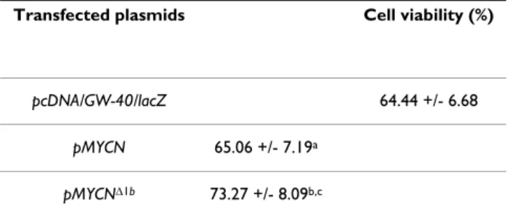
High MYCN dosage enhances cell viability
To explore the effect of MYCN dosage, SH-EP cells were subjected to apoptosis by 48-hour serum deprivation, then transfected with either control pcDNA/GW40/lacZ, p-MYCN, or p-MYCNΔ1b plasmids. As shown in Table 1, when SH-EP cells did not express (pcDNA/GW-40/lacZ transfected cells) or expressed low amount (p-MYCN transfected cells) of MYCN protein, serum deprivation induced similar cell loss (respectively 35.56% +/- 6.68% and 34.94 +/- 7.19%; p = 0.60). When SH-EP cells
expressed high levels (p-MYCNΔ1b transfected cells) of MYCN protein, apoptosis (26.72% +/- 8.09%) was signif-icantly (p = 0.01 and p = 0.02 respectively compared to previous conditions) reduced by one third.
MYCNOT is translated from a upstream AUG of MYCN1b
mRNA
We speculated that uAUG, located in +1894 (according to GenBank: Y00664, may initiate the translation and we assessed the importance of alternative splicing of exon 1b. In SH-EP cells, the transfection of p-uORF/MYCNΔ1b,
Exon 1b-splicing, uAUG and MYCN translation
Figure 2
Exon 1b-splicing, uAUG and MYCN translation. a)
Western blot analysis of 40 μg SH-EP neuroblastoma cells transfected with either control pcDNA/GW40/lacZ (lane 1), p-MYCN (lane 2) or p-p-MYCNΔ1b (lane 3). Results obtained with -upper panel: a monoclonal antibody against MYCN (1:1000) and -lower panel: a monoclonal antibody against β-Actin (1:10000). b) Western blot analysis of 40 μg SH-EP cells transfected with either p-MYCN (lane 1), p-MYCNmut (lane 2),
p-MYCNΔ1b (lane 3) or p-MYCNΔ1b, mut (lane 4). Results obtained with -upper panel: the anti-MYCN antibody (1:1000) and -lower panel: the anti β-Actin antibody (1:10000).
Table 1: MYCN dose-dependant anti-apoptotic effect after serum deprivation.
Transfected plasmids Cell viability (%)
pcDNA/GW-40/lacZ 64.44 +/- 6.68
pMYCN 65.06 +/- 7.19a pMYCNΔ1b 73.27 +/- 8.09b,c
MYCN dose-dependant enhanced SH-EP viability after serum deprivation measured with Uptiblue viable cell counting reagent (Uptima). a p= 0.60 compared with pcDNA/ GW-40/lacZ ; b p= 0.01 compared with pcDNA/GW-40/lacZ ; c p= 0.02 compared
which contains the uORF of MYCNΔ1b mRNA, allowed the production of a V5 epitope-tagged protein of approxi-mately 15 kDa, whereas the transfection of p-uORF/Myc, which contains the uORF of the full length MYCN tran-script, did not (Fig. 3a). This 11.8 kDa new protein was named MYCNOT: MYCN Overlap Transcript.
To validate the existence of endogenous MYCNOT, we performed a Western blot analysis using a specifically designed polyclonal antibody raised in rabbit against a N-terminal epitope (NGAERSPQSPAGRRA) [anti-MYC-NOT] of the hypothetical protein. The designed MYCNOT antibody was able to detect in transfected SH-EP cells with p-uORF/MYCNΔ1b the same protein detected with the V5 epitope antibody (Fig. 3b), The anti-MYCNOT antibody was able to detect a ~12 kDa protein in foetal but not in adult brain (Fig. 3c), thus confirming the existence of an endogenous MYCNOT protein. An analysis of MYCNOT
sequence with BLAST and PROSITE software did not show any sequence or motif homology.
Discussion
Because upstream ORF are known to influence protein translation [23], we investigated the MYCN translation from the full length MYCN mRNA and the exon 1b-spliced MYCNΔ1b mRNA. Only these two mRNAs are able to translate the MYCN protein. More recently, in 2005, van Bokhoven et al described a new exon 1a/2-spliced mRNA able to translate a new protein named ΔMYCN [4]. In SH-EP cells, we demonstrated that both MYCN and MYCNΔ1b were able to translate the MYCN protein, but MYCN translation from MYCNΔ1b mRNA was much more efficient. This difference may not be attributed to a differ-ential stability of the two transcripts because they have the same half-life of approximatively 15 minutes [2]. But, that could be due to differences in the regulating activities of uORFs in the two mRNAs. Because, the coding AUG is sev-eral hundreds nucleotides after the stop codon of the uORF in MYCN mRNA compared to the overlapping uORF in MYCNΔ1b transcript, we should have expected a higher translation of MYCN protein from full length mRNA due to reinitiation [6,23], but as already demon-strated, this mRNA contains an Internal Ribosome Entry Site (IRES) that could explain the higher initiation of translation at the major AUG in MYCNΔ1b mRNA [5,24,25].
Upstream ORFs are usually known for their cis-acting inhibitory activity on the translation of the major protein [7,26-30]. To test this hypothesis, uAUG was mutated into AUC. This mutation had no effect on MYCN translation from MYCNΔ1b mRNA but significantly impacted the translation from MYCN mRNA. Thus, MYCN uORFs have different cis-regulatory activities in MYCN translation depending on the alternative splicing of exon 1b. In our conditions, the uORF of the MYCNΔ1b mRNA did not affect MYCN translation. Alternatively, the uORF tran-scribed from full length MYCN mRNA has cis-enhancing activity on MYCN translation. To our knowledge, only one team has shown that an uORF is able to augment the translation of the HIV-1 Env protein from the HIV-1 mRNA [30]. Taken together, our results suggest the exist-ence of a new level of MYCN regulation. One hypothesis should be further explored: in cap-dependent conditions, low-level MYCN would be produced from the full length mRNA and this translation would be sustained by the activity of the uORF whereas in IRES-dependent condi-tions, high-level MYCN would be produced from exon 1b-spliced mRNA, independently of the uORF.
Numerous experiments have demonstrated important ontogenic [31] and oncogenic [14] roles. The amplifica-tion and overexpression of MYCN primarily found in 25% of neuroblastomas [16] and in other tumours
Translation of uORF and existence of MYCNOT
Figure 3
Translation of uORF and existence of MYCNOT. a)
SH-EP cells were transfected with either p-uORF/MYCN (lane 1) or p-uORF/MYCNΔ1b (lane 2). Western blot was conducted on 40 μg proteins, 48 hours after transfection with -upper panel: a monoclonal antibody against the V5 epitope (1:300) and -lower panel: the anti β-Actin antibody (1:10000). L: pro-tein ladder; b) SH-EP cells were transfected with p-uORF/ MYCNΔ1b. Western blot was conducted on 40 μg protein, 48 hours after transfection with -left panel: a monoclonal anti-body against the V5 epitope (1:300) and -right panel: a poly-clonal rabbit anti-NGAERSPQSPAGRRA peptide [anti-MYCNOT] polyclonal antibody (1:100) L: protein ladder; c) Western blot analysis of 40 μg protein extracts from adult (lane 1) and foetal (lane 2) brain. Results obtained with -upper panel: the anti-MYCNOT antibody (1:100) and -lower panel: the anti β-Actin antibody (1:10000).
BMC Cancer 2009, 9:445 http://www.biomedcentral.com/1471-2407/9/445
including medulloblastoma, retinoblastoma, small cell lung cancer, glioblastoma and other embryonal tumours [17] suggest the implication of MYCN dosage in cancer development and aggressive expressivity.
Our results showed that, in transfected SH-EP neuroblast-oma cells, high MYCN dosage obtained with MYCNΔ1b mRNA translation induces a weak but significant antiap-optotic effect after serum deprivation that was not observed with low MYCN expression obtained with MYCN mRNA. Our results do not fit the accepted pro-apoptotic properties of MYCN: using a MYCN-inducible SH-EP Tet21/N cell line, it has been demonstrated that MYCN overexpression enhances either doxorubicin- or platinum-induced apoptosis [19]. Using a similar approach, Cui et al have shown that the transfection of SH-EP cells with a pBabe-hygro/MYCN vector sensitises the cells to TRAIL-triggered apoptosis[21]. But, as we did, Jasty et al have reported a significant increase of cell viabil-ity with high MYCN expression after 6 days of serum dep-rivation [32]. These different results and ours suggest that MYCN overexpression in SH-EP cells might have different effects depending on the existence of an intrinsic (serum deprivation) or extrinsic (TRAIL or drug-induced apopto-sis) pathway-mediated apoptosis. Thus, MYCN dosage, depending on translation of the alternatively exon 1b splicing mRNA, enhances cell viability after intrinsic path-way-mediated apoptosis.
As previously proposed, the uAUG codon located in exon 1a may initiate the translation of ΔMYCN, a new isoform of the MYCN protein [4]. It has also been hypothesized, but not proven at this time, that this uAUG is able to translate a protein (GenBank: AAG40001.1) [5]. We now demonstrated the existence of this 11.8 kDa protein and named it MYCNOT: MYCN Overlap Transcript. Even if uORFs appear to apply to as much as 50% of the transcrip-tome [6,7,23], only few papers have reported their trans-lation in in-vitro experiments using chimeric tag-fusion proteins [23]. This is particularly true for the MYC gene, to which MYCN is related, which presents several uORFs: one uORF in exon 1 conducts to the translation of a pro-tein of unknown function called MYCHEX1 [33]. To our knowledge, only one team, using two-dimensional chro-matography and mass spectrometry analysis, has been able to prove the existence of eight proteins encoded by uORFs in human K562 and HEK293 cell lines [34]. More-over, these small proteins (< 20 kDa) were not subject to rapid proteasome degradation suggesting functional activ-ity [34]. The MYCNOT protein, which has no homology with MYCN, other known proteins or protein motifs, shares exon 1a-encoded amino acids with the putative ΔMYCN previously described [4]. Because MYCNOT is translated in foetal but not adult brain, we may hypothe-size that it plays a role during neural development. Our
results need further analysis to determine the functions of MYCNOT and might provide new insights into MYCN regulation.
Conclusion
The MYCN gene is transcribed into two major mRNAs which are differently translated. Higher levels of protein were seen with MYCNΔ1b mRNA. Existence of an upstream ORF was shown to have positive cis-regulatory activity on translation from MYCN but not from MYCNΔ1b mRNA. The high MYCN dosage obtained with MYCNΔ1b mRNA translation induces, in transfected SH-EP neuroblastoma cells, a weak but significant antiapoptotic effect after serum deprivation that was not observed with low MYCN expression.
Here, we show the translation of a new protein of unknown function from the upstream AUG of MYCNΔ1b mRNA.
In conclusion, further studies are now needed to explore the exact relationship between alternative splicing of exon 1b, MYCN dosage, and the mechanism, either extrinsic or intrinsic, underlying apoptosis.
Competing interests
The authors declare that they have no competing interests.
Authors' contributions
RB carried out the design of the study and manuscript writing and participated in plasmids preparation, cell cul-ture and Western blotting experiments. SWV participated to design of the study and manuscript writing. CL partici-pated to cell culture and Western blotting experiments. CDB participated to plasmids preparation, cell culture and Western blotting experiments. GT participated to cell culture and Western blotting experiments. ACJ partici-pated to cell culture and Western blotting experiments. EG participated to cell culture and Western blotting experi-ments. AP participated to design of the study and manu-script writing. All authors read and approved the final manuscript."
Acknowledgements
The authors are grateful to Dr. A. Denuzière for statistical assistance and to Mrs M.-D. Reynaud for editorial assistance. This work was supported by INCa Projet Libre 2006-2008 PL 06-76.
References
1. Schwab M, Varmus HE, Bishop JM, Grzeschik KH, Naylor SL, Sak-aguchi AY, et al.: Chromosome localization in normal human
cells and neuroblastomas of a gene related to c-myc. Nature
1984, 308:288-291.
2. Stanton LW, Bishop JM: Alternative processing of RNA
tran-scribed from NMYC. Mol Cell Biol 1987, 7:4266-4272.
3. Ibson JM, Rabbitts PH: Sequence of a germ-line N-myc gene and
amplification as a mechanism of activation. Oncogene 1988, 2:399-402.
Publish with BioMed Central and every scientist can read your work free of charge
"BioMed Central will be the most significant development for disseminating the results of biomedical researc h in our lifetime."
Sir Paul Nurse, Cancer Research UK Your research papers will be:
available free of charge to the entire biomedical community peer reviewed and published immediately upon acceptance cited in PubMed and archived on PubMed Central yours — you keep the copyright
Submit your manuscript here:
http://www.biomedcentral.com/info/publishing_adv.asp
BioMedcentral 4. van Bokhoven H, Celli J, van Reeuwijk J, Rinne T, Glaudemans B, van
Beusekom E, et al.: MYCN haploinsufficiency is associated with
reduced brain size and intestinal atresias in Feingold syn-drome. Nat Genet 2005, 37:465-467.
5. Jopling CL, Willis AE: N-myc translation is initiated via an
inter-nal ribosome entry segment that displays enhanced activity in neuronal cells. Oncogene 2001, 20:2664-2670.
6. Kochetov AV, Ahmad S, Ivanisenko V, Volkova OA, Kolchanov NA, Sarai A: uORFs, reinitiation and alternative translation start
sites in human mRNAs. FEBS Lett 2008, 582:1293-1297.
7. Calvo SE, Pagliarini DJ, Mootha VK: Upstream open reading
frames cause widespread reduction of protein expression and are polymorphic among humans. Proc Natl Acad Sci USA
2009, 106:7507-7512.
8. Landry JR, Mager DL, Wilhelm BT: Complex controls: the role of
alternative promoters in mammalian genomes. Trends Genet
2003, 19:640-648.
9. Schwab M, Alitalo K, Klempnauer KH, Varmus HE, Bishop JM, Gilbert F, et al.: Amplified DNA with limited homology to myc
cellu-lar oncogene is shared by human neuroblastoma cell lines and a neuroblastoma tumour. Nature 1983, 305:245-248.
10. Sawai S, Shimono A, Wakamatsu Y, Palmes C, Hanaoka K, Kondoh H:
Defects of embryonic organogenesis resulting from targeted disruption of the N-myc gene in the mouse. Development 1993, 117:1445-1455.
11. Knoepfler PS, Cheng PF, Eisenman RN: N-myc is essential during
neurogenesis for the rapid expansion of progenitor cell pop-ulations and the inhibition of neuronal differentiation. Genes
Dev 2002, 16:2699-2712.
12. Okubo T, Knoepfler PS, Eisenman RN, Hogan BL: Nmyc plays an
essential role during lung development as a dosage-sensitive regulator of progenitor cell proliferation and differentiation.
Development 2005, 132:1363-1374.
13. Wakamatsu Y, Watanabe Y, Nakamura H, Kondoh H: Regulation of
the neural crest cell fate by N-myc: promotion of ventral migration and neuronal differentiation. Development 1997, 124:1953-1962.
14. Schwab M, Varmus HE, Bishop JM: Human N-myc gene
contrib-utes to neoplastic transformation of mammalian cells in cul-ture. Nature 1985, 316:160-162.
15. Weiss WA, Aldape K, Mohapatra G, Feuerstein BG, Bishop JM:
Tar-geted expression of MYCN causes neuroblastoma in trans-genic mice. EMBO J 1997, 16:2985-2995.
16. Seeger RC, Brodeur GM, Sather H, Dalton A, Siegel SE, Wong KY, et al.: Association of multiple copies of the N-myc oncogene
with rapid progression of neuroblastomas. N Engl J Med 1985, 313:1111-1116.
17. Pession A, Tonelli R: The MYCN oncogene as a specific and
selective drug target for peripheral and central nervous sys-tem tumors. Curr Cancer Drug Targets 2005, 5:273-283.
18. Schweigerer L, Ledoux D, Fleischmann G, Barritault D: Enhanced
MYCN oncogene expression in human neuroblastoma cells is associated with altered FGF receptor expression and cel-lular growth response to basic FGF. Biochem Biophys Res
Com-mun 1991, 179:1449-1454.
19. Fulda S, Lutz W, Schwab M, Debatin KM: MycN sensitizes
neurob-lastoma cells for drug-triggered apoptosis. Med Pediatr Oncol
2000, 35:582-584.
20. Paffhausen T, Schwab M, Westermann F: Targeted MYCN
expres-sion affects cytotoxic potential of chemotherapeutic drugs in neuroblastoma cells. Cancer Lett 2007, 250:17-24.
21. Cui H, Li T, Ding HF: Linking of N-Myc to death receptor
machinery in neuroblastoma cells. J Biol Chem 2005, 280:9474-9481.
22. Ross RA, Spengler BA, Biedler JL: Coordinate morphological and
biochemical interconversion of human neuroblastoma cells.
J Natl Cancer Inst 1983, 71:741-747.
23. Iacono M, Mignone F, Pesole G: uAUG and uORFs in human and
rodent 5'untranslated mRNAs. Gene 2005, 349:97-105.
24. Cobbold LC, Spriggs KA, Haines SJ, Dobbyn HC, Hayes C, de Moor CH, et al.: Identification of internal ribosome entry segment
(IRES)-trans-acting factors for the Myc family of IRESs. Mol
Cell Biol 2008, 28:40-49.
25. Spriggs KA, Cobbold LC, Jopling CL, Cooper RE, Wilson LA, Stoneley M, et al.: Canonical initiation factor requirements of the Myc
family of internal ribosome entry segments. Mol Cell Biol 2009, 29:1565-1574.
26. Watatani Y, Ichikawa K, Nakanishi N, Fujimoto M, Takeda H, Kimura N, et al.: Stress-induced translation of ATF5 mRNA is
regu-lated by the 5'-untransregu-lated region. J Biol Chem 2008, 283:2543-2553.
27. Pendleton LC, Goodwin BL, Solomonson LP, Eichler DC:
Regula-tion of endothelial argininosuccinate synthase expression and NO production by an upstream open reading frame. J
Biol Chem 2005, 280:24252-24260.
28. Park EH, Lee JM, Pelletier J: The Tie2 5' untranslated region is
inhibitory to 5' end-mediated translation initiation. FEBS Lett
2006, 580:1309-1319.
29. Martin MM, Buckenberger JA, Knoell DL, Strauch AR, Elton TS:
TGF-beta(1) regulation of human AT(1) receptor mRNA splice variants harboring exon 2. Mol Cell Endocrinol 2006, 249:21-31.
30. Krummheuer J, Johnson AT, Hauber I, Kammler S, Anderson JL, Hau-ber J, et al.: A minimal uORF within the HIV-1 vpu leader
allows efficient translation initiation at the downstream env AUG. Virology 2007, 363:261-271.
31. Hurlin PJ: N-Myc functions in transcription and development. Birth Defects Res C Embryo Today 2005, 75:340-352.
32. Jasty R, van Golen C, Lin HJ, Solomon G, Heidelberger K, Polverini P, et al.: Bcl-2 and M-Myc coexpression increases IGF-IR and
fea-tures of malignant growth in neuroblastoma cell lines.
Neo-plasia 2001, 3:304-313.
33. Nanbru C, Prats AC, Droogmans L, Defrance P, Huez G, Kruys V:
Translation of the human c-myc P0 tricistronic mRNA involves two independent internal ribosome entry sites.
Oncogene 2001, 20:4270-4280.
34. Oyama M, Kozuka-Hata H, Suzuki Y, Semba K, Yamamoto T, Sugano S: Diversity of translation start sites may define increased
complexity of the human short ORFeome. Mol Cell Proteomics
2007, 6:1000-1006.
Pre-publication history
The pre-publication history for this paper can be accessed here:
http://www.biomedcentral.com/1471-2407/9/445/pre pub