HAL Id: hal-03065556
https://hal.archives-ouvertes.fr/hal-03065556
Submitted on 15 Dec 2020
HAL is a multi-disciplinary open access
archive for the deposit and dissemination of
sci-entific research documents, whether they are
pub-lished or not. The documents may come from
teaching and research institutions in France or
abroad, or from public or private research centers.
L’archive ouverte pluridisciplinaire HAL, est
destinée au dépôt et à la diffusion de documents
scientifiques de niveau recherche, publiés ou non,
émanant des établissements d’enseignement et de
recherche français ou étrangers, des laboratoires
publics ou privés.
Rapid identification and characterization of infected
cells in blood during chronic active Epstein-Barr virus
infection
Benjamin Fournier, David Boutboul, Julie Bruneau, Charline Miot, Cécile
Boulanger, Marion Malphettes, Isabelle Pellier, Bertrand Dunogué, Benjamin
Terrier, Felipe Suarez, et al.
To cite this version:
Benjamin Fournier, David Boutboul, Julie Bruneau, Charline Miot, Cécile Boulanger, et al.. Rapid
identification and characterization of infected cells in blood during chronic active Epstein-Barr
virus infection. Journal of Experimental Medicine, Rockefeller University Press, 2020, 217 (11),
�10.1084/jem.20192262�. �hal-03065556�
TECHNICAL ADVANCES AND RESOURCES
Rapid identification and characterization of infected
cells in blood during chronic active Epstein-Barr virus
infection
Benjamin Fournier1,2, David Boutboul3, Julie Bruneau2,4, Charline Miot5, C´ecile Boulanger6, Marion Malphettes3, Isabelle Pellier5, Bertrand Dunogu´e7, Benjamin Terrier7, Felipe Suarez8, St´ephane Blanche9, Martin Castelle9, Sarah Winter9, Henri-Jacques Delecluse10, Thierry Molina2,4, Capucine Picard1,2,11, Stephan Ehl12, Despina Moshous2,9, Lionel Galicier3, Vincent Barlogis13, Alain Fischer2,9,14,15, B´en´edicte Neven2,9, and Sylvain Latour1,2
Epstein-Barr virus (EBV) preferentially infects epithelial cells and B lymphocytes and sometimes T and NK lymphocytes.
Persistence of EBV-infected cells results in severe lymphoproliferative disorders (LPDs). Diagnosis of EBV-driven T or NK cell
LPD and chronic active EBV diseases (CAEBV) is difficult, often requiring biopsies. Herein, we report a flow-FISH cytometry
assay that detects cells expressing EBV-encoded small RNAs (EBERs), allowing rapid identification of EBV-infected cells
among PBMCs. EBV-infected B, T, and/or NK cells were detectable in various LPD conditions. Diagnosis of CAEBV in 22 patients
of Caucasian and African origins was established. All exhibited circulating EBV-infected T and/or NK cells, highlighting that
CAEBV is not restricted to native American and Asian populations. Proportions of EBV-infected cells correlated with blood EBV
loads. We showed that EBV-infected T cells had an effector memory activated phenotype, whereas EBV-infected B cells
expressed plasma cell differentiation markers. Thus, this method achieves accurate and unambiguous diagnoses of different
forms of EBV-driven LPD and represents a powerful tool to study their pathophysiological mechanisms.
Introduction
The EBV is an oncogenic virus of the Herpesviridae family re-stricted to humans with a selective tropism for epithelial cells and B lymphocytes. EBV is implicated in a range of cancers, in-cluding gastric and nasopharyngeal carcinomas, smooth muscle tumors, B cell neoplasms such as Hodgkin lymphoma, diffuse large B cell lymphoma, Burkitt lymphoma, and T and natural killer (NK) cell lymphomas. EBV infection is the first known risk of cancer-associated mortality in emerging countries (Young and Rickinson, 2004;Plummer et al., 2016).
EBV infects all human populations, with more than 90% being carriers by the age of 20 yr. In immunocompetent individuals,
EBV establishes long-term persistent asymptomatic infection of B cells. However, during the primary infection, some individuals develop infectious mononucleosis (IM), a clinical manifestation that reflects a strong but self-limiting immune response without persistent deleterious consequences. In immunocompromised subjects, EBV infection is associated with life-threatening pa-thologies such as hemophagocytic lymphohistiocytosizs (HLH) and B cell lymphoproliferative disorders (B-LPD), including lymphomas. These disorders are observed in HIV-infected pa-tients with AIDS and in papa-tients receiving immunosuppressive treatments following organ transplantation, usually denoted as
...
1Laboratory of Lymphocyte Activation and Susceptibility to EBV Infection, Institut National de la Sant´e et de la Recherche M´edicale UMR 1163, Imagine Institute, Paris,
France; 2Universit´e de Paris, Paris, France; 3Department of Clinical Immunology, Saint-Louis Hospital, Assistance Publique–Hˆopitaux de Paris, Paris, France; 4Department of Pathology, Necker-Enfants Malades Hospital, Assistance Publique–Hˆopitaux de Paris, Paris, France; 5Department of Pediatric Immunology Hematology and
Oncology, University Hospital, Angers, France; 6Institut Roi Albert II, Cancerology and Hematology Departments, University Clinics Saint-Luc Hospital, Brussels, Belgium; 7Department of Internal Medicine, Cochin Hospital, National Referral Centre for Systemic and Autoimmune Diseases, Cochin Hospital, Assistance Publique–Hˆopitaux de
Paris, Paris, France; 8Department of Adult Hematology, Necker-Enfants Malades Hospital, Assistance Publique–Hˆopitaux de Paris, Paris, France; 9Department of Pediatric
Immunology, Hematology and Rheumatology, Necker-Enfants Malades Hospital, Assistance Publique–Hˆopitaux de Paris, Paris, France; 10Unit F100, Institut National de la
Sant´e et de la Recherche M´edicale U1074, Deutsches Krebsforschungszentrum, German Cancer Research Center, Heidelberg, Germany; 11Study Center for Primary
Immunodeficiencies, Necker-Enfants Malades Hospital, Assistance Publique–Hˆopitaux de Paris, Paris, France; 12Institute for Immunodeficiency-Center for Chronic
Immunodeficiency, Department of Pediatrics and Adolescent Medicine, Medical Center - Faculty of Medicine, University of Freiburg, Germany; 13Department of Pediatric
Hematology-Oncology, La Timone Hospital, Marseille, France; 14Collège de France, Paris, France; 15Institut National de la Sant´e et de la Recherche M´edicale UMR 1163,
Paris, France.
Correspondence to Sylvain Latour:sylvain.latour@inserm.fr.
© 2020 Fournier et al. This article is distributed under the terms of an Attribution–Noncommercial–Share Alike–No Mirror Sites license for the first six months after the publication date (seehttp://www.rupress.org/terms/). After six months it is available under a Creative Commons License (Attribution–Noncommercial–Share Alike 4.0 International license, as described athttps://creativecommons.org/licenses/by-nc-sa/4.0/).
post-transplant lymphoproliferative disorders (PTLDs). Patients with inherited immunodeficiencies are also prone to develop EBV-related B-LPD. In particular, there is a subgroup of primary immunodeficiencies (PIDs) characterized by a high and specific predisposition to EBV infection and to development of EBV-related B-LPD. These PIDs are caused by autosomal recessive or X-linked mutations in genes required for the expansion and cytotoxic responses of EBV-specific T cells (Latour and Winter, 2018;Tangye et al., 2017;Latour and Fischer, 2019). Most of these conditions logically respond to depletion of the EBV reservoir in B cells by anti-CD20 antibodies such as rituximab.
More rarely, EBV infection is associated with T and NK lym-phoproliferative diseases (T/NK-LPDs), in which EBV infects T or NK cell subsets. T/NK-LPDs include malignant (i.e., extranodal NK/T lymphoma [ENKTL]) and the nonmalignant conditions (i.e., T and NK cell chronic active EBV infection diseases [CAEBV];
Swerdlow et al., 2016;Kim et al., 2019). CAEBV infections are further divided into systemic and cutaneous forms (i.e., severe mosquito bite allergy and hydroa vacciniforme–like LPD;Quintanilla-Martinez et al., 2013;Cohen et al., 2019).
T/NK-LPD and CAEBV have been mostly reported in native populations from Asia and the Americas ( Quintanilla-Martinez et al., 2013;Kimura et al., 2012). Their etiology is poorly understood, but polygenic factors and immune defects are thought to favor their occurrence. Along these lines, so-matic mutations in DDX3X have been shown to play a role in the progression of the disease (Okuno et al., 2019). Further-more, we recently identified a familial form of systemic T cell CAEBV caused by digenic homozygous loss-of-function mu-tations in TNFRSF9 (4-1BB/CD137) and PIK3CD resulting in defective immune control of EBV-infected T cells (Rodriguez et al., 2019).
One common feature of T/NK-LPD is the high EBV load in the blood of patients that is not relieved by anti-CD20 treatment and can persist for years. The persistence of EBV-infected T cells and/or NK cells may progress to life-threatening LPDs often associated with HLH. In contrast to B-LPD, these conditions have a poor prognosis (Kim et al., 2016;Kimura et al., 2012;Kawamoto et al., 2018). Indeed, they are often refractory to standard treatments, and although hematopoietic stem cell transplanta-tion (HSCT) represents the best curative therapy so far, man-agement remains difficult and differs from that of B-LPD (Sawada and Inoue, 2018;Bollard and Cohen, 2018;Sawada et al., 2017).
Most T/NK-LPDs, including CAEBV-related diseases, are not familiar to clinicians because of their relative rarity and have misleading and various clinical presentations that may impede the diagnosis. They range from chronic lymphoid organ enlargement and overt autoinflammatory diseases to unexplained recurrent and/or fulminant HLH. So far, the presence of EBV in T or NK cells has been mostly obtained by immunostaining of biopsies or by PCR of sorted cells. Both assays can be difficult to perform and give ambiguous re-sults. Even when CAEBV is suspected (especially in case of a poor clinical and biological response to B cell depletion), reaching an accurate diagnosis may be hampered by the absence of any lesions to analyze or by a poor clinical
condition that may prevent any invasive organ biopsy. However, providing an accurate and rapid diagnosis by showing EBV-infected T and/or NK cells is key to adjust the treatment. The lack of direct access to infected cells also restrains progress in the understanding of mechanisms un-derlying the pathophysiology of these diseases, which is so far largely not understood.
We report a minimally invasive, rapid method allowing re-liable detection and characterization of EBV-infected cells from peripheral blood mononuclear cells (PBMCs). We showed the suitability of this test to diagnose EBV-driven T/NK-LPD and to study EBV-infected cells.
Results
Detection of EBV-infected cell lines using a flow cytometry RNA assay
We developed a fluorescence in situ hybridization (FISH) flow cytometry (flow-FISH) method that detects EBER-1 and EBER-2 mRNAs (EBERs) expressed by EBV based on the PrimeFlow RNA assay (Henning et al., 2016). After cell fixation and per-meabilization, EBERs are recognized through hybridization with a specific oligonucleotide probe, which is then serially hy-bridized with two specific DNA probes to amplify the signal, the last one being fluorescent. Importantly, EBERs are expressed in all EBV-containing cells, irrespective of the lytic and latency programs (Lerner et al., 1981). To assess the specificity of the assay, several known EBV-positive (EBV+) and EBV-negative (EBV−) cell lines were tested, including two EBV+ and EBV− Burkitt lymphoma cell lines (Raji: EBV+; Ramos: EBV−), human B cells immortalized with EBV in vitro (termed“LCL” for lym-phoblastoid cell line), an EBV+ NK cell line (SNK-6), an EBV− T cell leukemia (Jurkat) cell line, and the human embryonic kidney HEK-293 cell line, stably infected or not with a GFP-tagged EBV (HEK and EBV-GFP+HEK; Delecluse et al., 1998). Cell lines were also tested with Bacillus and RLP13A probes as a control (Fig. 1 A). The Bacillus probe targets a bacterial RNA that is not expressed in human cells and is used to assess background staining, whereas the RPL13A probe recognizes a ubiquitous human ribosomal RNA present in all cell types and served as a positive control. Both EBER and Bacillus probes are coupled to the fluorochrome Alexa Fluor 647, whereas the RPL13A probe is coupled to the fluorochrome Alexa Fluor 488. All cells were in-cubated with a viability dye (eFluor 450 dye), and only eFluor-negative cells were analyzed. All cell lines known to contain EBV (Raji, SNK-6, LCL, and EBV-GFP+HEK) showed more than 90% of cells positively stained with the EBER probe. In contrast, cells known not to be infected by EBV (HEK, Ramos, and Jurkat) ex-hibited a very low percentage of EBER+cells. These latter can be viewed as false-positive background because their frequency was similar to or below that of cells stained with the Bacillus probe and because both stains had a low mean fluorescence in-tensity (MFI). Of note, in some experiments, we noticed that a proportion of LCL was negative for EBER. EBER expression was shown to be downregulated during the switch from viral latency to lytic cycle (Greifenegger et al., 1998) and to be negative in epithelial cells from hairy leukoplakia, in which EBV infection is
Figure 1. PrimeFlow assay for EBER allows specific detection of EBV-containing cell lines and circulating EBV-infected B cells during severe IM. (A) FACS dot plots of EBER (left panels), Bacillus (middle panels), and RPL13A (right panels) stains using the PrimeFlow RNA assay of EBV−cell lines (Jurkat, Ramos, and HEK) and EBV+cell lines (HEK EBV-GFP, SNK-6, Raji, and LCL). The Bacillus probe targets a bacterial RNA, whereas the RPL13A probe targets a
ubiquitously expressed ribosomal RNA, and they are used as negative and positive controls, respectively. The RPL13A probe is coupled to the fluorochrome Alexa Fluor 488, except for the analysis of HEK GFP-EBV, for which it is coupled to Alexa Fluor 647. (B) Upper panels show FACS dot plots of costaining with RPL13A and EBER probes (coupled to Alexa Fluor 488 and 647, respectively) of LCL cells treated for 72 h with TPA + ionomycin stimulation to induce the lytic cycle. LCL-1 and LCL-2 lines were obtained by infection with the BZLF1-KO BRLF1-KO B95-8 EBV strain and devoid of the essential BLZF1 and BRLF1 proteins required to induce the EBV lytic cycle. LCL cells were obtained by infection with the B95-8 EBV WT strain (WT B95-8 EBV). Lower panels show expression of BZLF1 (453 pb) and GAPDH (258 pb) fragment transcripts by RT-PCR in TPA + ionomycin–treated LCL-1, LCL-2, and LCL cell lines. GAPDH was used as an amplification control and DNA ladder (pb) on the left. (C) FACS dot plots of EBER expression by the PrimeFlow assay coupled with CD3, CD19, anti-CD16, or anti-CD56 staining of PBMCs from patients with IM. Dot plots are gated on eFluor−CD14−cells. The red gates in the dot plots highlight the
EBV-infected cell subsets. Patients (Pt) 1.1, 1.2, and 1.3 had mild IM symptoms and low plasma EBV loads. Pt 1.4 and Pt 1.5 had severe IM symptoms and high plasma EBV loads. (D) Tonsil biopsy section from Pt 1.4 that has been costained with an EBER probe and anti-CD3 or anti-CD79a antibodies, showing infiltration of EBV-infected B cells in tissues. Magnification, ×400. Scale bars, 40 µm.
considered to be purely lytic (Gilligan et al., 1990). To assess if the EBER-negative LCL cells could correspond to cells with EBV in the lytic program, the EBV lytic cycle was induced by 12-O-tetradecanoylphorbol-13-acetate (TPA) and ionomycin in WT LCL cells (infected with the EBV strain B95-8) or LCL cells that were obtained by infection with a defective B95-8 strain lacking BZLF1 and BRLF1, two viral factors required for EBV to undergo the lytic cycle (Feederle et al., 2000;Fig. 1 B). Although lytic cycle induction was detectable by BZLF1 expression by RT-PCR in WT LCL treated with TPA and ionomycin, no BZLF1 expression was found in BZLF1/BRLF1 double-deficient LCL, as expected. How-ever, in all conditions, EBER-negative cells were not stained with the RPL13A-positive control probe, indicating that they likely have not been permeabilized or hybridized with the probes rather than being true-negative cells. These data also suggest that when the EBV lytic cycle is activated in B cells, there is enough EBER expression to be detected with the PrimeFlow as-say. These findings are in line with recent evidence of increased EBER1/2 expression in LCL after EBV lytic cycle induction (Li et al., 2019).
PrimeFlow was then used in patients with proven EBV-driven B-LPD and T/NK-LPD by histology. Analysis was fur-ther extended to patients in whom standard histology was not performed (16 of 39), either because clinical and biological context was sufficient for a conclusive diagnosis (i.e., IM and hydroa vacciniforme–like LPD) or because no lesion was avail-able to biopsy. More details related to clinical manifestations and histology of patients are reported inTable 1. Analyses of infected subsets were performed after the exclusion of dead cells (eFluor+) and monocytes (CD14+), for which the absence of EBER+ was systematically verified.
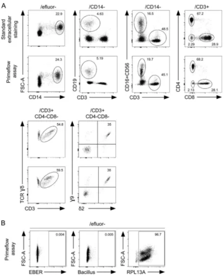
Detection of EBV-infected circulating cells from PBMCs of healthy individuals and patients with EBV-driven B-LPD We tested whether the PrimeFlow EBER assay could enable the detection and phenotyping of EBV-infected cells among PBMCs of EBV-infected healthy individuals and patients. In all following experiments, 1.5 million to 3 million PBMCs were tested. First, we verified that conditions for intracellular hybridization with the EBER probe did not alter extracellular costaining with an-tibodies to common specific markers of monocytes and B, T, and NK cell populations. The distributions of the different lympho-cyte subsets within PBMCs from healthy individuals (including B lymphocytes; CD3, CD4, CD8, andγδ T lymphocytes; NK cells; and monocytes) were comparable to those obtained under classic conditions, even though the MFI of each marker was slightly reduced when we used the PrimeFlow protocol (Fig. S1
and data not shown). Naive and memory T and B cell pheno-types were also found to be comparable (data not shown). No difference was noticed between fresh and frozen samples (data not shown). Tested antibodies suitable for the PrimeFlow EBER assay are listed in Table S1. We then analyzed PBMCs from 15 healthy control subjects, including both EBV-seronegative (3 individuals) and EBV-seropositive (12 individuals) subjects (Fig. S1,Fig. 7 A, and data not shown). When costained with the EBER probe and antibodies for membrane markers, only few cells were positive (<0.05%) in PBMCs of all individuals tested,
irrespective of their EBV status. Proportions of EBER+ cells were always below the threshold of Bacillus-positive cells, and the very few positive cells in some control subjects were dis-tributed across all lymphocyte subsets (in contrast to patients with B-LPD and T/NK-LPD shown inFigs. 2,3,4,5, and6; data not shown). These results strongly support that these few EBER+cells represent false-positive cells.
We next tested PBMCs of patients diagnosed with IM (Fig. 1 C). Compared with the Bacillus probe staining (negative control probe;Fig. S2), no significant EBER+cells were detected in any cell subset from PBMCs of three patients (patients [Pt] 1.1, 1.2, and 1.3) who had low detectable blood EBV loads by PCR (ranging from 2.6 to 2.9 log copies/ml). This may be explained by the fact that symptoms of IM result from a strong expansion of EBV-specific T cells and/or NK cells, a time at which most EBV-infected B cells may already have been eliminated (Balfour et al., 2005;Hadinoto et al., 2008). In contrast, significant amounts of EBER+cells re-stricted to CD19+cells were detected in two patients (Pt 1.4 and 1.5) diagnosed with more severe symptoms related to IM (Fig. 1 C) and high EBV loads (5 and 6.2 log copies/ml, respectively). No other positive cells were found among other lymphoid cell subsets. This specific B cell EBER staining was in accordance with EBER and CD79a costaining in standard histology of a lymph node biopsy from Pt 1.4 (Fig. 1 D).
Patients with EBV-associated B-LPD were further examined, including patients with solid B cell neoplasia (Hodgkin lym-phoma, n = 3; PTLD, n = 1;Fig. 2 A), inherited immune defi-ciencies predisposing to EBV-driven B-LPD, such as X-linked lymphoproliferative syndrome type 1 (XLP-1; n = 2), CD70 defi-ciency (Izawa et al., 2017), MAGT1 deficiency (Li et al., 2011), and CTPS1 deficiency (Martin et al., 2014,2020), or not particularly predisposing to EBV like the combined ZAP-70 immunodefi-ciency (Fig. 2 B). No significant EBV+ circulating cells were detected in PBMCs from two patients with EBV+Hodgkin lym-phoma, despite positive blood EBV load in one of them. The third patient with Hodgkin lymphoma had very rare EBER+cells in the B cell subset (0.29% of B cells). Patients with PTLD following liver transplant and those with CD70, CTPS1, and ZAP-70 defi-ciencies all had high chronic EBV load and exhibited between 0.4% and 3.5% EBV+ B cells (n = 4). Of note, only the CD70-deficient patient had signs of LPD. A MAGT-1–CD70-deficient patient was tested during primary EBV infection based on EBV serology and presented a low proportion of EBV+cells (0.1%). Both XLP-1 patients had B cell lymphocytosis during a fulminant IM-like episode with a high proportion of EBER+ cells restricted to B cells (between 33.9% and 20.5% among PBMCs). The specific B cell EBER staining was in agreement with EBER probe and anti-CD79a costaining on the formalin-fixed, paraffin-embedded liver biopsy from Pt 2.10 (XLP-1;Fig. 2 C). These data demonstrate the reliability of the PrimeFlow EBER assay for achieving specific detection of circulating EBV-infected B cells in a range of patho-logical conditions in which EBV infection is known to drive B-LPD. Detection of EBV-infected circulating cells among PBMCs of patients with post-transplant T-LPD and T/NK cell lymphoma We next tested whether this assay could also be useful to characterize EBV-associated lymphoproliferations that likely
Table 1. Characteristics of patients Patient Ethnicity/
origin
Age at onset (yr)
Clinical manifestations/genetic defects PrimeFlow: EBV+subset
Histology: EBV+subset
IM Pt. 1.1 Caucasian 26 IM Absent NP Pt 1.2 Caucasian 13 IM Absent NP Pt 1.3 Caucasian 32 IM Absent NP Pt 1.4 Sub-Saharan African
8 Severe IM (SMG and compressive lymph nodes requiring ventilation support)
CD19+ Tonsils and lymph node,
CD79a+
Pt 1.5 Caucasian 23 Severe IM (SMG, thrombopenia) CD19+ NP
B cell LPD
Pt 2.1 North African 7 Classical Hodgkin lymphoma Absent Lymph node CD30+CD15+
Pt 2.2 Caucasian 26 Classical Hodgkin lymphoma Absent Lymph node (EBER not performed)
Pt 2.3 Sub-Saharan African
17 Classical Hodgkin lymphoma, HLH CD19+ Lymph node CD30+CD15+
Pt 2.4 Caucasian 1 Liver transplantation, PTLD, high peripheral EBV load CD19+ NP
Pt S4 North African 5 Fulminant IM CD19−/low Lymph node, CD79a+
Genetically characterized PIDs
Pt 2.5 North African 8 B-cell LPD, HMG, SMG, ADP, CD70 deficiency(TNFSF7 hmz mutation: c.535C>T/p.R179X)
CD19+ Lymph node, spleen, CD79a+
Pt 2.6 Comorian 14 Chronic peripheral EBV load without clinical LPD, ZAP-70 deficiency (ZAP70 hmz mutation: c.252C>G/p.C84W), strongly decreased protein expression
CD19+ NP
Pt 2.7 Caucasian 3 Chronic peripheral EBV load without clinical LPD, CTPS1 deficiency (CTPS1 hmz mutation: c.1692-1G>C/ p.T566Dfs26X)
CD19+ NP
Pt 2.8 Caucasian 5 Primary EBV infection (ongoing), chronic
hypogammaglobulinemia, MAGT1 deficiency (MAGT1 hmz mutation: c.991C>T/p;R331X), strongly decreased protein expression
CD19+ NP
Pt 2.9 Caucasian 16 Fulminant IM, XLP-1 (SH2D1A hemiZ mutation: c.257-264delCATTTCAG p.A86EfsX15)
CD19+ Liver, CD79a+
Pt 2.10 North-African 9 Fulminant IM, XLP-1 (SH2D1A hemizygous exons 1-4 deletion)
CD19+ NP
T cell PTLDs
Pt 3.1 Caucasian 2 HSCT for AML, persistent peripheral EBV load after rituximab
CD3+ NP
Pt 3.2 Caucasian 7 Cardiac transplantation, persistent peripheral EBV load after rituximab, hepatosplenomegaly, enlarged lymph nodes
CD8+ NP
Pt 3.3 Caucasian 2 Liver transplantation, persistent peripheral EBV load after rituximab, IBD
CD8+ Liver, bone marrow, gut
CD8+
T/NK cell lymphomas
Pt 3.4 Caucasian 15 ENKTL, nasal type Absent Nasal cavity, CD3+CD5−CD56+
Pt 3.5 Caucasian 60 Angioimmunoblastic T cell lymphoma revealed by EBV+
LPD, eosinophilia with lung involvement
CD3−CD4+ Lymph node, CD3+CD4+
Pt 3.6 Caucasian 74 ENKTL, recurrent HLH CD3−CD56+ Skin, CD3+GrB+
Pt 3.7 Sub-Saharan African
33 ENKTL, HMG-SMG, bicytopenia, weight loss, fever CD3−CD16+ Spleen, CD3+CD5−GrB−
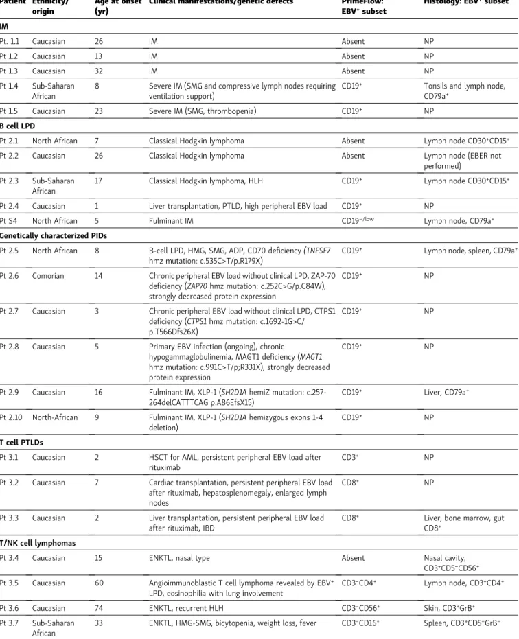
involve non–B cell subsets. Samples from three pediatric pa-tients referred to our laboratory with noncharacterized lym-phoproliferations associated with persistent elevated blood EBV load (>5 log copies/ml) following hematopoietic stem cell (Pt 3.1), heart (Pt 3.2), or liver transplant (Pt 3.3) were first evalu-ated (Fig. 3 A). In two of them (Pt 3.1 and Pt 3.2), EBV load remained high after anti-CD20 treatment, indicating the per-sistence of EBV-infected cells that may not be B cells. In all three patients, EBER staining was found to be highly positive in a subset of CD3+cells and to a lesser extent in the residual B cell compartment in two. In Pt 3.3, EBV infection of CD8+T cells was confirmed by standard histologic staining showing CD3+EBER+ lymphocytes in the gut (Fig. 3 B). Thus, these findings indicate that lymphoproliferations in these patients can be considered as T cell PTLD.
Five adult patients with a histology-based diagnosis of NK or T cell lymphoma were next assessed. The first patient (Pt 3.4) presented with a symptomatic nasal form, and no circulating EBER+cells were found in his PBMCs, in accordance with the negative result of blood EBV PCR. The four other patients had
more misleading and diverse clinical presentations with ele-vated EBV load that required lymph node biopsies (Pt 3.5), re-current skin biopsies (Pt 3.6 and Pt 3.8), a splenectomy (Pt 3.7), and a nasal biopsy in the absence of local symptoms (Pt 3.8) to complete the diagnosis (Table 1). In these latter patients, EBER staining revealed a positive subset among PBMCs likely corre-sponding to NK cells (CD3−CD2+CD56+ or CD3−CD2+CD16+), except for Pt 3.5, who was affected by CD3−CD4+ angioimmu-noblastic lymphoma (Fig. 3 C). Arguing for an NK cell ontogeny of these EBER+cells, CD16+cells from Pt 3.8, which were almost all infected by EBV, were all strongly positive for the NK cell– specific markers CD160, DNAM1, NKp46, and 2B4, whereas CD3+ cells were negative (Fig. S3). Some of the EBER+cells were also not stained by any extracellular marker (Pt 3.5, 3.7, and 3.8). They could represent malignant cells that have been shown to be positive for intracellular CD3 in ENKTL, which is not detected by our assay (Chan et al., 1996). Taken together, these results demonstrate that the PrimeFlow EBER assay enables a specific and sensitive detection and identification of EBV-infected cir-culating cells among PBMCs in various pathological conditions,
Table 1. Characteristics of patients (Continued) Patient Ethnicity/
origin
Age at onset (yr)
Clinical manifestations/genetic defects PrimeFlow: EBV+subset
Histology: EBV+subset
Pt 3.8 North-African 43 Disseminated ENTKL, nasal type, recurrent fever and skin rashes, asymptomatic nasal cavity involvement (on biopsy)
CD3−CD16+ Skin, nasal cavity,
CD16+CD56−
CAEBV, systemic EBV-positive T-cell lymphoma of childhood, EBV-positive CD8+HLH
Pt 4.1 Caucasian 4 Systemic EBV+T-cell lymphoma of childhood CD8+ Lymph node, CD8+
Pt 4.2 Caucasian 9 EBV+CD8+HLH during primary infection CD8+ Bone marrow, CD8+
Pt 5.1 Caucasian 12 Hydroa vacciniforme TCRγδ+ NP
Pt 5.2 Caucasian 5 Hydroa vacciniforme TCRγδ+ NP
Pt 5.3 North African 40 Hydroa vacciniforme TCRγδ+ NP
Pt 6.1 North African 3 Systemic T/NK cell CAEBV, mucocutaneous lymphoproliferation
CD8+, NK Skin, CD8+
Pt 6.2 Turkish 15 Systemic T/NK cell CAEBV, recurrent HMG and fever NK Liver, CD3+
Pt 6.3 Sub-Saharan African
14 Systemic T/NK cell CAEBV, IBD NK Gut, CD3+
Pt 6.4 North African 7 Systemic T/NK cell CAEBV, mucocutaneous lymphoproliferation
CD4+; TCRγδ+ Lymph node, CD4+
Pt 6.5 North African 22 Systemic T/NK cell CAEBV, large-vessel vasculitis TCRγδ+ NP
Pt 6.6 Sub-Saharan African
11 Systemic T/NK cell CAEBV, large-vessel vasculitis CD3lowCD4−CD8−
TCRγδ− NP
Pt 6.7 Pakistani 6 Persistent peripheral EBV load after rituximab, CD137/4-1BB deficiency(TNFRSF9 hmz mutation: c.170delG/ p.G57fsX91)
CD8+ NP
Pt 6.8 Caucasian 10 Systemic T/NK cell CAEBV, recurrent HMG and fever CD8+ Liver, CD3+
Pt 6.9 Caucasian 35 Systemic T/NK cell CAEBV, indolent floor of the mouth tumor
CD4+ Floor of the mouth, CD4+
Pt S5 North African 52 Systemic T/NK cell CAEBV, chronic liver disease, SMG CD4+; TCRγδ+ NP
Patients are numbered according to the figure number in which their PrimeFlow data are shown (first digit) and their place of appearance in the figure (second digit). GrB, granzyme B; hemiZ, hemizygous; HMG, hepatomegaly; hmz, homozygous; htz, heterozygous; IBD, inflammatory bowel disease; NP, not performed; PTCL-NOS, peripheral T cell lymphoma, not otherwise specified; SMG, splenomegaly.
irrespective of the infected cell subsets. The presence of EBV-infected lymphocytes correlates in most of the patients with the clinical lymphoproliferation manifestations, EBV load, and his-tological findings. Therefore, this method provides a direct, rapid, and valuable diagnosis of EBV-driven B, T, and NK cell LPD.
Characterization of EBV-driven T/NK cell LPDs
Persistent infection of T and/or NK cells by EBV is under-diagnosed in Western countries, but it should be considered in patients with recurrent elevated EBV load, unusual clinical presentations of lymphoproliferation and HLH episodes, and/or no response to treatment with anti-CD20 antibodies. Moreover, this diagnosis is difficult to be rapidly obtained because it
depends on the accessibility of tissues for histological staining to detect EBV+T and/or NK cells. Hence, the PrimeFlow EBER as-say could represent a useful and potent tool to reach a rapid and earlier diagnosis without invasive procedures. Over the last years, we collected cases with noncharacterized lym-phoproliferation and/or HLH manifestations associated with elevated and/or persistent blood/plasma EBV PCR over 6 mo and no biological or clinical response to B cell depletion af-ter treatment with anti-CD20 (Table 1). 22 patients were recruited over a period of 5 yr, whose PBMCs were tested using the PrimeFlow EBER assay. On the basis of their clinical symptoms, patients can be classified into three groups according toKim et al. (2019): (1) EBV+CD8+T cell HLH during primary infection (2 patients); (2) systemic EBV+T cell lymphoma
Figure 2. Detection by PrimeFlow EBER of peripheral EBV-infected B cells in PBMCs from patients with B-LPDs. (A and B) FACS dot plots of EBER expression by PrimeFlow assay coupled with anti-CD3, anti-CD19, anti-CD16, or anti-CD56 staining of PBMCs from patients affected with (A) Hodgkin lymphoma (Pt 2.1, Pt 2.2, and Pt 2.3) and post–liver transplantation B-LPD (Pt 2.4) or (B) B-LPD resulting from primary immune deficiencies, including CD70 (Pt 2.5), ZAP-70 (Pt 2.6), CTPS1 (Pt 2.7), and MAGT1 (Pt 2.8) deficiencies and XLP-1 (Pt 2.9, Pt 2.10). Dot plots are gated on eFluor−CD14−cells. The red gates in the
dot plots highlight the EBV-infected cell subsets. (C) Liver biopsy section from Pt 2.10 (XLP-1) that has been costained with an EBER probe and anti-CD79 antibody, showing infiltration of EBV-infected B cells. Magnification, ×400. Scale bar, 40 µm. Pt 2.10 analysis was repeated over time at different dates showing the same EBV-infected subset.
of childhood (1 patient); and (3) CAEBV of T and/or NK cell types (19 patients) that are further subdivided into cutaneous forms (hydroa vacciniforme–like LPD [3 patients] and muco-cutaneous lymphoproliferation [2 patients]) and systemic forms (14 patients), with symptoms ranging from chronic and/
or relapsing enlargement of lymphoid organs to various atyp-ical autoinflammatory syndromes (e.g., inflammatory bowel disease [IBD], systemic vasculitis). Table 1 summarizes data only for the 14 patients for whom PrimeFlow EBER assay dot plots are shown.
Figure 3. PrimeFlow EBER detection of peripheral EBV-infected T and NK cells in patients with post-transplant EBV+T/NK cell lymphoproliferation
and ENKTL. (A) FACS dot plots of EBER expression by PrimeFlow assay coupled with anti-CD3, anti-CD8, anti-CD16, anti-CD56, and anti-CD19 staining in PBMCs of patients with EBV+T/NK cell lymphoproliferation after hematopoietic stem cell (Pt 3.1), cardiac (Pt 3.2), and liver transplant (Pt 3.3). A red square
highlights the infected subset. Dot plots are gated on eFluor−CD14−cells. (B) Gut biopsy sections from Pt 3.3 stained with anti-CD3 or EBER probe showing
infiltration of EBV-infected T cells. Magnification, ×200. Scale bars, 40 µm. (C) FACS dot plots of EBER expression using the PrimeFlow assay coupled with anti-CD3, anti-CD19, anti-CD4, anti-CD5, anti-CD56, anti-CD16, and anti-CD2 staining of PBMCs from patients with ENKTL. PBMCs used in panels 1 and 2 are from different time points, explaining the slight differences in proportions of EBER+cells. Dot plots are gated on eFluor−CD14−cells. The red gates in the dot plots
highlight the EBV-infected cell subsets. Pt 3.3, Pt 3.5, Pt 3.6, and Pt 3.7 analyses were repeated over time at different dates, showing the same EBV-infected subsets.
Analysis of two patients meeting criteria of EBV+ T cell lymphoma of childhood (Pt 4.1) and severe EBV+CD8+ T cell lymphohistiocytosis (Pt 4.2) showed in both EBER+ cells re-stricted to CD8+ T cells in their PBMCs (1.84% and 0.54%,
respectively) that were also detected in the material from bone marrow aspiration performed at the same time for Pt 4.2 (2.5% of total cells;Fig. 4 A). EBV-infected CD8+T cells were observed by standard formalin-fixed, paraffin-embedded histology in
Figure 4. PrimeFlow EBER assay detects EBV-infected CD8+T cells among PBMCs from patients with systemic EBV+T cell lymphoma and
EBV-associated HLH. (A) FACS dot plots of EBER expression by PrimeFlow assay coupled with anti-CD3, anti-CD4, anti-CD8, anti-CD16, anti-CD56, and anti-CD19 of PBMCs from patients with systemic EBV+T cell lymphoma of childhood and EBV-associated HLH. Dot plots are gated on eFluor−CD14−cells. The red gates in
the dot plots highlight the EBV-infected cell subsets. (B) Lymph node biopsy section from Pt 4.1 costained with anti-CD3 and EBER probe (magnification, ×400) or stained with anti-CD8 (magnification, ×400) or anti-CD79a (magnification, ×50), showing infiltration by EBV-infected T cells. Liver biopsy section from Pt 4.2 costained with anti-CD8 and EBER probe showing infiltration of EBV-infected T cells (magnification, ×400). Scale bars, 40 µm, except for anti-CD79a, 200 µm.
Figure 5. Patients with hydroa vacciniforme–like LPD showed circulating EBV-infected γδ2 T cells by PrimeFlow EBER assay. FACS dot plots of EBER expression by PrimeFlow assay coupled with anti-CD19, anti-CD3, anti-CD4, anti-CD8, anti-TCRδ2, anti-CD16, and anti-CD56 of PBMCs from Pt 5.1, Pt 5.2, and Pt 5.3 with hydroa vacciniforme–like LPD. The red gates in the dot plots highlight the EBV-infected cell subsets. All dot plots were gated on eFluor−CD14−cells.
Fraction of positive cells in the fifth column (/CD3+) is calculated as the percentage of total CD3+T cells. Pt 5.1 analysis was repeated over time at different
dates showing the same EBV-infected subset.
tissues (with EBER probe and anti-CD8 costaining) from both patients (Fig. 4 B). Regarding hydroa vacciniforme–like LPD,
PBMC samples from three patients (Pt 5.1, 5.2, and 5.3) had an important proportion of EBV-infectedγδ T cells, as previously
described (Kimura et al., 2012). These T cells expressed the Vδ2
TCR chain (Fig. 5). Several patients with previously proven or suspected systemic forms of chronic active EBV infection of T/NK cell type with various clinical presentations were further
Figure 6. Characterization of peripheral EBV-infected cells by the PrimeFlow EBER assay in patients suspected of systemic T/NK cell CAEBV. FACS dot plots of EBER expression by PrimeFlow assay coupled with anti-CD19, anti-CD3, anti-CD4, anti-CD8, anti-CD16, and anti-CD56 of PBMCs from patients with systemic T/NK cell CAEBV. All dot plots are gated on eFluor−CD14−cells. The red gates in the dot plots highlight the EBV-infected cell subsets. PBMCs used for individual anti-CD16 and anti-CD56 staining (Pt 6.1, Pt 6.2, and Pt 6.3) are from different time points, explaining the slight differences in proportions of EBER+cells. Pt 6.1, Pt 6.2, Pt 6.3, Pt 6.5, and Pt 6.8 analyses were repeated over time at different dates, showing the same EBV-infected subsets.
examined (Fig. 6). We also included analysis of an asymptomatic female carrier (Pt 6.7) of a TNFRSF9 homozygous mutation previously reported inRodriguez et al. (2019), who had a per-sistent high blood EBV load not relieved by anti-CD20 therapy. In all of these cases, the PrimeFlow EBER assay revealed either a single EBV-infected cell type or two EBV-infected cell types consisting of T and/or NK cells. Pt 6.3 also had circulating EBER+ B cells that may be related to the chronic immunosuppressive treatment he received. Importantly, these data correlated well with the infected subset(s) found by standard histology when available for Pt 6.1, 6.4, 6.8, and 6.9 (Fig. 6andFig. S3). Of note, histology revealed that two patients (Pt 6.2 and 6.3) had EBER+ CD3ε+ cells, whereas EBV-infected circulating cells corre-sponded only to NK cells that were repeatedly detected (Fig. S3,
Table 1, and data not shown). This discrepancy is explained by intracellular expression of CD3ε of NK cells (Chan et al., 1996). Thus, the PrimeFlow EBER assay formally confirmed the diag-nosis of CAEBV in these patients. Standard histology was not available for two patients (Pt 6.5 and 6.6) with systemic vascu-litis who did not present any site to biopsy. In these cases, a systemic CAEBV diagnosis was only considered on the basis of results of the PrimeFlow EBER assay. Therefore, the PrimeFlow EBER assay appears to be an excellent and efficient tool to rap-idly diagnose various EBV+T/NK cell lymphoproliferations and to discriminate the infected cell subsets (as observed in these 22 cases). Hence, the assay can substitute for immunostaining of tissue biopsies, especially when no organ can be biopsied. Correlation analyses between EBV load, EBV-infected cells, and clinical phenotypes
A potential relationship between whole-blood EBV load and the percentage of circulating EBER+cells detected by the PrimeFlow EBER assay was examined. Individuals with B-LPD or T/NK-LPD tested with the PrimeFlow EBER assay were analyzed, including all patients listed inTable 1, in addition to seven other patients with NK/T-LPD and nine other patients with B-LPD. A signifi-cant positive correlation was found starting from EBV loads above 4 log copies/ml, whereas the number of EBER+cells was not significantly different from the false-positive background in most cases with EBV loads below 4 log copies/ml (Fig. 7 A). The percentage of infected cells among each cell subset was also evaluated according to the disease classification (Fig. 7 B). The proportion of circulating EBV-infected B cells was below 10% in patients with B cell lymphoproliferation, with the exception of the two patients with XLP-1 who had EBV+B cell lymphocytosis and one patient (Pt S4) shown in Fig. S4(>40% of infected B cells).
Both patients with systemic EBV+T cell lymphoma of child-hood and EBV+T cell HLH had a low proportion of circulating EBER+ CD8+ T cells (ranging from 1.1% to 10%; Fig. 7 B). In contrast, in patients who presented with systemic T/NK-LPD/ CAEBV, the proportion of infected cells was found to be much higher, as exemplified by patients with hydroa vacciniforme– like LPD, who all exhibited a high proportion of EBV-infected TCR Vδ2 T cells, ranging from 14.5% to 60%. In other patients with systemic CAEBV, distinct T cell subpopulations were found to be infected, including CD4, CD8, and NK cells. Three patients
had two infected subsets (CD4+and NK cells [n = 1] and CD4+and TCRγδ+[n = 2]). Thus, with the exception of patients with hy-droa vacciniforme–like LPD, HLH, and systemic lymphoma of childhood, no clear association could be made between the clinical phenotypes and the nature of the infected cell subsets. Advantage of PrimeFlow EBER assay over EBV PCR on sorted cells
Discrimination of EBV-infected cells among PBMCs can be per-formed by a specific PCR for EBV on sorted cells. However, the cell-sorted fraction may be falsely positive because of the purity of sorted cells, which is never 100%, and residual cell-free EBV DNA that may still be present. An example of such a false-positive result is depicted in Fig. S4. EBV loads only slightly decreased in a 5-yr-old patient with severe symptoms of IM suggestive of a primary infection (with anti-viral capsid antigen [VCA] IgM+, anti-VCA IgG−, and anti-EBNA IgG−) despite ef-fective B cell depletion after three injections of anti-CD20 (Pt S4 inTable 1). PCR for EBV on sorted cells was then performed and revealed 3.5 log copies/105cells and 4.9 log copies/105 cells of EBV in CD3+and CD3−cells, respectively. Because no B cell had been detected before sorting, no contamination of the CD3+ fraction was expected, and no purity check was therefore per-formed after sorting. The PrimeFlow EBER assay perper-formed in parallel showed EBER staining only in a subset expressing low amounts of CD19, whereas neither T nor NK cells were positive (Fig. S4 B). Additional courses of anti-CD20 treatment eventu-ally significantly reduced the EBV load (Fig. S4 A), and only EBV+ B cells without EBV+T or NK cells were detected in a lymph node biopsy detected by immunostaining (Fig. S4 C). Thus, the Pri-meFlow EBER assay appears to be more reliable at discrimi-nating EBV-infected cells than the detection of EBV by PCR on sorted cells.
Phenotypic characterization of EBV-infected B and T cells We evaluated whether the PrimeFlow EBER assay could be suitable for further characterization of EBV-infected cells. One strength of the PrimeFlow assay is the large panel of extracel-lular antibodies that can be used (Table S1). The precise phe-notypes of EBV-infected cells in EBV-related disorders are not well known. Thus, EBV-infected B cells and T cells were char-acterized in more detail in several patients. We first examined EBER+B cells of one XLP-1 patient (Pt 2.10; shown inFig. 2 B) during a fulminant IM course (Fig. 8 A). These cells divided into two subsets based on CD19, CD21, CD27, CD38, and IRF4 ex-pression. One was CD19highCD21highCD27lowCD38intIRF4low, and the second was CD19lowCD21lowCD27highCD38highIRF4int/high, evoking germinal center B cells and plasma cells, respectively (Kassambara et al., 2015). In contrast to the intracellular staining for IRF4, the PrimeFlow EBER assay did not work with intra-cellular BCL6 staining using our available antibody (data not shown). However, all CD19lowcells and a large proportion of CD19highcells were stained for BCL6, suggesting that the ma-jority of EBER+B cells expressed BCL6. Both subsets were IgM+, IgDlow/−, and CD80+. We next analyzed EBER+T cells from two patients with CAEBV (Pt 6.8 and Pt 6.9; shown in Fig. 6). A homogeneous effector memory CD27−CD45RA−phenotype with
an increased expression of the HLA-DR activation marker was observed in both (Fig. 8 B). In contrast, EBER− T cells were heterogeneous, comprising naive, effector, and, memory cell subsets. These data suggest that EBV triggers both T and B cells to differentiate into activated effector memory T cells and plasma cells, respectively.
To gain more insight into the type of latency that is associated with EBV-infected T cells, we used the PrimeFlow assay to detect concomitant expression of transcripts for LMP1 or EBNA2 using specific probes (coupled to a fluorochrome different from the one of the EBER probe). Although we were able to detect EBNA2
transcript expression and heterogeneous LMP1 transcript pression in LCL cells, we failed to detect EBNA2 or LMP1 ex-pression among EBER+cells of four CAEBV patients (Pt 3.3, 5.1, 6.4, and 6.8). However, we found a low proportion of cells ex-pressing EBNA2+transcripts among EBER+B cells from an XLP1 patient (Pt 2.9), but no expression of LMP1 transcripts (Fig. S5 A).
The mechanisms of infection of T and/or NK cells by EBV remain elusive. One hypothesis is that it is mediated by trogo-cytosis, a phenomenon whereby exchange of cellular material occurs at the immune synapse (Joly and Hudrisier, 2003). EBV
Figure 7. Relationships between EBV-infected cell fraction, EBV load, and clinical phenotypes. (A) Percentage of EBV-infected cells among PBMCs plotted against EBV loads detected from whole blood by PCR (log copies/ ml), including both healthy control and patient samples. Control corresponds to distinct data of 13 different healthy donors with EBV load below log 2.7, which is the limit of detection. 0.001% of EBER+cells is considered the limit of detection of
EBER+ cells. (B) Percentages of EBV-infected
cells among the infected cell subsets from PrimeFlow EBER analysis of B cells correspond to CD3−CD19+ cells, CD4 T cells correspond
to CD3+CD4+CD8−, CD8 T cells correspond to
CD3+CD4−CD8+, double-negative (DN) T cells
correspond to CD3+CD4−CD8−, CD3lowDN T cells
correspond to CD3lowCD4−CD8−, GD T cells
corre-spond to CD3+γδ TCR, and NK cells correspond to
CD3−CD16+ and/or CD56+. Line-connected dots
represent two infected subsets in a single patient. All data from dot plots in A and B are gated on eFluor−CD14− cells. Data from patients listed in
Table 1are depicted, supplemented with data from additional patients with B-LPD (n = 9) and T/NK CAEBV (n = 7).
may be transmitted to T or NK cells through a synapse between infected B cells and EBV-specific cytotoxic T or NK cells, either directly or through membrane exchange including CD21, the receptor for EBV expressed on B cells (but not on T or NK cells;
Tabiasco et al., 2003;Lee et al., 2018). If this assumption was correct, EBV-infected T cells should be specific for EBV peptides presented by infected B cells. This was tested in a patient (Pt 6.8) presenting with a high proportion of blood EBV-infected CD8 T cells and expressing HLA-A2 molecules, for which tetramers specific for EBV-derived peptides are available (Fig. 6). 0.8% of CD8+ T cells were recognized by the HLA-A2-EBV pentamer
(0.21% of total cells). However, EBER+T cells were not stained with the HLA-A2-EBV pentamer (Fig. 8 C). These findings sug-gest that trogocytosis is not a major mechanism accounting for the entry of EBV into T cells, at least in the patient tested here. We next analyzed whether the presence of EBV in T cells could influence CD8+or CD4+T cell functions from PBMCs of Pt 6.8 and Pt 6.9, who harbored CD8+and CD4+EBV-infected T cell subsets, respectively (Fig. 9). CD3-induced T cell proliferation and degranulation assays were performed and compared be-tween infected and noninfected cells that were discriminated with the PrimeFlow EBER assay. Because these functional assays
Figure 8. Phenotypes of EBV-infected B and T cells. (A) FACS dot plots and histograms of EBV-infected CD19+cells using the PrimeFlow EBER assay
coupled with anti-IgM, anti-IgD, anti-CD27, anti-CD80, anti-CD21, anti-CD27, anti-CD38, and anti-IRF4 staining in addition to size (forward scatter [FCS]) and granularity (side scatter [SSC]) from PBMCs of a patient with XLP-1 during fulminant IM (Pt 2.10). Upper panels, cells gated on CD19low(/CD19low), CD19high
(/CD19high), or CD19 (/CD19). Middle and lower panels, cells gated on EBER+CD19low(/EBER+CD19low), EBER+CD19high(/EBER+CD19high), or EBER−CD19+
(/EBER−CD19+). (B) FACS dot plots using the PrimeFlow EBER assay coupled with anti-CD3, anti-CD4, anti-CD8, anti-CD27, anti-CD45RA, and anti-HLA-DR
staining from PBMCs of two patients, one with CAEBV associated with EBV-infected CD8+T cells (Pt 6.8) and one with EBV-infectedγδ T cells (Pt 6.5). Dot
plots from gating on EBER+CD8+(/EBER+CD8+) or EBER+CD3+CD4−CD8−(/EBER+CD3+DN). (C) FACS dot plots using the PrimeFlow EBER assay coupled with
anti-CD3, anti-CD8, and EBV-specific HLA-A2* pentamer staining showing EBV-specific T cells from PBMCs of Pt 6.8. All dot plots are gated on eFluor−CD14−cells.
require important numbers of cells to be analyzed, they were conducted on expanded T cell blasts from PBMCs. Phenotyping of T cell blasts in both patients revealed that most EBV+T cells had an effector memory phenotype (CD27−CD45RA–), were ac-tivated (HLA-DR+), and were not senescent (CD57−). By contrast, EBV−cells had rather an effector or central memory phenotype (CD27+CD45RA−), were not activated, and a small proportion was senescent (Fig. 9 A). Notably, all EBV+CD8+T cells (in Pt 6.8)
were found to express the transcription factor EOMES, well known to be associated with peripheral CD8+T cell differenti-ation. The proportion of EBV+T cells was stable in culture; thus, EBV+ cells did not seem to have a selective advantage in the conditions of culture used. Interestingly, EBV+CD8+ T cells al-ready expressed significant amounts of the degranulation markers CD107a/b without any stimulation, in contrast to noninfected CD8+ T cells (Fig. 9, B and E). Under stimulation
Figure 9. Functional characterization of EBV-infected T cells. (A) FACS dot plots using PrimeFlow EBER assay coupled with extracellular staining for CD3, CD4, CD8, CD27, CD45RA, and HLA-DR or with intranuclear staining for EOMES from T cell blasts of two patients affected with CAEBV associated with EBV-infected CD8+T cells (Pt 6.8) or EBV-infected CD4+T cells (Pt 6.9). Dot plots from gating on EBER+CD8+(/EBER+CD8+), EBER+CD3+CD4+(/EBER+CD4+), or
eFluor−cells (/eFluor−). (B) FACS dot plots (upper panels) and histograms (lower panels) of anti-CD3 (OKT3)–induced degranulation assay of T cell blasts from
Pt 6.8 assessed by staining for CD8 and CD107a/b surface, followed by the PrimeFlow EBER assay. FACS dot plots showing EBV-infected (EBER+CD8+) or
noninfected (EBER−CD8+) T cells and overlaid histograms showing CD107a/b expression in gated EBER+CD8+(blue) or EBER−CD8+(red) cells for each
stimulation condition. Concentrations inμg/ml of OKT3 are indicated along with nonstimulated cells (NS). (C and D) FACS dot plots (upper panels) and histograms (lower panels) of anti-CD3 (OKT3)–induced proliferation of T cell blasts from Pt 6.8 (C) and Pt 6.9 (D) assessed with staining with CellTrace Violet (CTV) dye, followed by PrimeFlow EBER coupled with staining for CD4 (D), CD8 (C), and HLA-DR. FACS dot plots showing EBV-infected (EBER+) or noninfected
(EBER−) T cells (upper panels), CTV, and HLA-DR expression in gated CD8+EBER+or CD8+EBER−cells (C, middle panels) and CD4+EBER+or CD4+EBER−cells (D,
middle panels) and overlaid histograms showing CTV expression in gated EBER+(blue) or EBER−(red) cells for each stimulation condition. Concentrations inμg/ml
of OKT3 are indicated along with nonstimulated (NS) cells. All dot plots are gated on eFluor−CD14−cells in A–D. (E) Graphed data of the percentage of degranulation (CD107+cells among CD8+) from the amounts shown in B. (F) Index of proliferation calculated from the experiments showed in C and D.
with a low dose of anti-CD3, CD107a/b expression was strongly increased on the vast majority of EBV+CD8+T cells. This may point to a particular role of EBV in activation of infected T cells. Proliferation of EBV+CD8+and CD4+T cells was also increased at low doses of anti-CD3, whereas it was decreased at higher doses and in response to CD3/CD28 stimulation (Fig. 9, C, D, and F). This decrease correlated with a strong reduction of the proportion of EBV+T cells, which may at first sight indicate a loss by activation-induced cell death (AICD). However, we did not find any evidence of increased AICD of these cells (data not shown). Because AICD was apparently not increased in infected cells, it may suggest a potential downregulation or decay of EBERs. Additional experiments are warranted to test this hy-pothesis. Thus, these findings suggest that EBV modulates terminal differentiation and threshold of TCR activation in CD4+and CD8+infected cells. Finally, we investigated cytokine production by intracellular staining for IFN-γ, TNF-α, and IL-2. In PBMCs from four EBV+T/NK-LPD patients (Pt 3.3, 5.1, 6.4, and 6.8), no cytokine production was detected (Fig. S5 B). However, a slight increase in the proportion of stimulated T cell blasts from Pt S5, but with a lower MFI (indicating probably a lower amount per cell), was detected (Fig. S5, C and D). Taken together, these observations show that functional studies on either infected or noninfected cells can be performed at the same time using the PrimeFlow EBER assay.
Discussion
In this study, we describe the PrimeFlow RNA assay, which al-lows both rapid and reliable detection and characterization of EBV-infected cells from fluid samples, including blood and bone marrow. The assay uses conventional flow cytometry with antibody-based staining of membrane extracellular markers in combination with a specific DNA probe for EBERs, thus enabling the discrimination of the EBV-infected cell subset(s) (Henning et al., 2016). This method has been previously used in a hu-manized mouse model of EBV infection (McHugh et al., 2017). Because EBERs are abundantly expressed in all infected cells, regardless of the type of latency involved (Lerner et al., 1981), they represent the best markers for detection of EBV presence within cells. Some laboratories have used EBV-specific PCR on sorted cells to detect and characterize circulating EBV-infected cells as an alternative to conventional histology (Zhang et al., 2019), but this approach is cumbersome and may lead to false-positive findings through contamination of the T or NK cell or non–B cell fraction by either infected B cells or plasma EBV DNA, as shown herein inFig. S4.
The PrimeFlow EBER assay revealed good reproducibility, showing the same EBV-infected cell populations when per-formed on several occasions in the same patients (Pt 2.10, 3.3, 3.5, 3.6, 3.7, 5.1, 6.1, 6.2, 6.3, 6.5, and 6.8; see figure legends), and was found to be concordant with immunohistochemistry and coimmunostaining data when available. In most of the patients, circulating EBV-infected lymphocyte subsets (identified by PrimeFlow) corresponded to the same infected cell populations detected in tissue biopsies by immunostaining. Flow-FISH as-says using probes for EBERs have been developed previously
(Kimura et al., 2009;Kawabe et al., 2012); however, they worked only with a restricted panel of extracellular antibodies for which buffers and reagents were not specifically developed to sustain proper extracellular staining (Kimura et al., 2009). Moreover, signal amplification of the EBER probe in these assays was low. These technical limitations may impair diagnosis, especially for patients with a very low number of circulating infected cells. None of these tests are available for routine laboratory diag-nostics. By contrast, the PrimeFlow FISH assay reported here was able to preserve cell surface and intracellular staining with a wide range of antibodies and to sustain a high EBER signal, al-lowing us to easily discriminate infected cells. Moreover, in our assay, we use a viability dye, which further improves the gating on living cells and decreases the risk of positive or false-negative staining. The previous studies used forward and side scatter profiles, which are strongly altered after fixation and permeabilization.
We failed to detect infected cells in PBMCs of healthy donors seropositive for EBV. Circulating EBV-infected memory B cells that represent the EBV reservoir are considered to be very scarce, below 1 in 100,000 total circulating B lymphocytes (Laichalk et al., 2002). This is very likely under the threshold of detection of our method. No or rare circulating infected cells were noticed in patients with EBV-related Hodgkin lymphoma, although EBV was detected in the serum or whole blood by PCR. This is not surprising, because Hodgkin lymphoma cells are restricted to lymphoid tissues with no or few circulating cells, and detection of EBV in blood by PCR relies much more on EBV plasma content than on mononuclear cells in this context (Hohaus et al., 2011). The positive blood EBV load in patients with Hodgkin lymphoma is likely explained by EBV released in the plasma from apoptotic neoplastic cells from tissues (Ryan et al., 2004; Kimura and Kwong, 2019). Hence, because our method cannot detect tissue-resident EBV-infected cells, it is not suitable for pathologies in which EBV-infected cells are not circulating in the blood.
In individuals with IM, circulating EBV-infected cells were detectable only in the most severe forms with elevated EBV loads. Thus, we inferred that the PrimeFlow EBER assay is powerful enough to detect infected cells during ongoing infec-tion but not for convalescent or asymptomatic carriers. The low proportion of infected B cells detected in the periphery is within the same range as in previous studies that used PCR for EBV with limited cell dilution conditions in individuals with IM and patients with solid organ transplantation (Babcock et al., 1999;
Hochberg et al., 2004). Overall, our data show a positive cor-relation between the proportion of circulating EBV-infected cells and EBV loads, but they reveal limitations regarding sensitivity: In patients with low EBV loads (below 4 log copies/ml of EBV), detection of EBV-infected cells is ineffective and thus noninformative.
In patients in whom very few circulating infected cells are detected, results need to be interpreted with caution, and careful interpretation of positive cells is required to confirm the diag-nosis. Acquisition of a higher number of cells (>3 million) can help to highlight one or two small clusters of cells with high MFI corresponding to EBV-infected cells. Inclusion of control probes
is necessary and contributes to an accurate diagnosis in this situation. Importantly, staining with the Bacillus probe helps to exclude false-positive EBER+cells related to autofluorescence or rare events of nonspecific hybridization (seeFig. S2). Indeed, in contrast to truly EBER+cells, the Bacillus-staining MFI is lower and heterogeneous, generating a smear shape rather than a homogeneous cluster. When cellular material is scarce with poor viability (possibly due to the poor clinical condition of the pa-tient), it is advisable to include both a viability dye and the positive control probe (targeting the ubiquitous RPL13A ribo-somal RNA) to secure both procedure and diagnosis.
Based on the most recent diagnosis criteria from the World Health Organization classification of tumors of hematopoietic and lymphoid tissues (Swerdlow et al., 2016) and the Committee on Measures against Intractable Diseases of the Ministry of Health, Labour and Welfare of Japan (Arai, 2019), demonstration of EBV-infected T or NK cells is required to establish the diag-nosis of T/NK CAEBV or EBV+ T/NK LPD. This diagnosis is usually achieved by immunostaining of tissue biopsies. How-ever, clinical conditions for organ biopsies are not always ful-filled, especially when facing organ failure associated with severe HLH. Importantly, our study shows that all patients with histologically proven CAEBV had detectable blood EBV-infected T or NK cells, which are easier to access. In addition, the Pri-meFlow EBER assay enables correct diagnosis of patients with-out an identified lesion to biopsy, as illustrated by two patients with atypical systemic vasculitis (Pt 6.5 and Pt 6.6). Similarly, circulating EBV+NK-like cells were detected by the PrimeFlow EBER assay in two patients (Pt 3.5 and Pt 3.7) before diagnosis of ENKTL by histology. This was rather an unexpected finding because positive EBV load in these patients is thought to be due to release of EBV DNA from in situ tumoral cells (Kimura and Kwong, 2019). Importantly, this may indicate a transition from a latent form of CAEBV to overt lymphoma. Hence, the PrimeFlow EBER assay has three important advantages: (1) It avoids harmful invasive procedures in patients with a poor clinical condition; (2) it permits a diagnosis in the absence of access to pathological tissue; and (3) it leads to an early and rapid diag-nosis and thus to adapted therapies and can be used as a bio-marker of treatment efficacy. Furthermore, the mere detection of infected cells in some diseases could give insight into their progression and their pathophysiology.
EBV+T/NK-LPD, including T/NK CAEBV diseases, is mostly prevalent in native Asian and American populations. In Western countries, EBV+T/NK-LPD diagnosis is often not considered or considered last. Nevertheless, we recruited more than 20 pa-tients of Caucasian or African origin with atypical (e.g., vascu-litis, autoimmunity) or typical (HLH, hydroa vacciniforme) clinical presentations of T/NK-LPD associated with a high blood EBV load, poor response to anti-CD20 treatment, and/or per-sistent EBV load. Thus, our observations stress that patients with T/NK-LPD are not as geographically and ethnically restricted as reported previously. On the whole, our patients showed infected-cell subsets quite similar to those of Asian/Japanese patients previously analyzed (Kimura et al., 2012).
We did not observe any correlation between clinical pre-sentation and the type of EBV-infected subsets, with the
exception of patients with hydroa vacciniforme–like LPD and HLH, who exhibited EBV-infectedγδ T cells and CD8+T cells, respectively. The presence of EBV-infectedγδ T cells has pre-viously been associated with hydroa vacciniforme–like LPD (Kimura et al., 2012). Here, the three patients with hydroa vacciniforme exhibited circulating EBV-infected TCR Vδ2 T cells. The reason for this selective expansion of EBV+TCR Vδ2 T cells remains undetermined. As previously reported (Kimura et al., 2012), circulating EBV-infected CD8+ T cells were de-tected only at a low proportion in two patients, one with EBV+ T/NK-LPD–related fulminant HLH (Pt 4.1) and one with EBV+ CD8 T cell lymphoma of childhood (Pt 4.2), although they had a significant EBV load (>4 log copies/ml). Likely, the low pro-portion of circulating EBV+ cells does not reflect the level of infected cells in tissues. In fact, a higher proportion of infected CD8+T cells was found in the bone marrow sample of Pt 4.2 by the PrimeFlow assay, and an important EBV+CD8 T cell infil-tration was shown by histology in Pt 4.1.
The conserved extracellular, intracytoplasmic, or intranu-clear staining may be especially relevant regarding the study of the pathophysiology of EBV-related disorders. Notably, we show that this assay can be used to directly assess properties and characteristics of infected cells in a more physiological setting without the need of cell sorting. In particular, it allows detailed phenotyping of EBV-infected B, T, or NK cells and functional studies. This is first illustrated by the XLP-1 case with a fulmi-nant IM course (Pt 2.10), in whom we found that EBV-infected B cells harbored germinal center and plasmablast markers. In-terestingly, these findings may support the germinal center hypothesis stating that EBV-infected naive B cells are driven to germinal center cell differentiation (Thorley-Lawson and Bab-cock, 1999;Thorley-Lawson et al., 2013). However, XLP-1 pa-tients have an impaired capacity to form germinal centers and to undergo immunoglobulin class switching (Ma et al., 2006), indicating that EBV could also directly provide signals that mimic germinal center differentiation without requiring T cell help and/or germinal center transit, although those would not be sufficient for class switching, because EBER+ B cells are only IgM+. These data are concordant with the previous study ofChaganti et al. (2008) showing that EBV+ infected B cells which persist in XLP-1 patients after IM are CD27+IgM+.
Regarding T/NK-LPD, we showed that EBV-infected CD8 and γδ T cells (in Pt 6.8 and 6.5, respectively) had an effector memory cell phenotype in contrast to noninfected cells. EBV-infected T cell blasts from patients also had an effector memory cell phenotype with strong activation markers, including in-creased degranulation capacity and no expression of the senes-cent CD57 marker despite a strong expression EOMES (known to be associated with T cell exhaustion and senescence). Conse-quently, EBV may be directly involved in activating or lowering the threshold of activation in CD4+and CD8+infected cells. Al-though we failed to detect increased IFN-γ content or production in EBV+T cells in the five tested patients, it will be of interest to determine if increased IFN-γ production by EBV+T cells could correlate with the appearance of HLH symptoms, which are known to be driven by excessive IFN-γ production by T cells.