HAL Id: inserm-00187003
https://www.hal.inserm.fr/inserm-00187003
Submitted on 28 Oct 2008
HAL is a multi-disciplinary open access
archive for the deposit and dissemination of sci-entific research documents, whether they are pub-lished or not. The documents may come from teaching and research institutions in France or
L’archive ouverte pluridisciplinaire HAL, est destinée au dépôt et à la diffusion de documents scientifiques de niveau recherche, publiés ou non, émanant des établissements d’enseignement et de recherche français ou étrangers, des laboratoires
Shaping of the Autoreactive Regulatory T Cell
Repertoire by Thymic Cortical Positive Selection.
Julie Ribot, Geneviève Enault, Sylvie Pilipenko, Anne Huchenq, Maryline
Calise, Denis Hudrisier, Paola Romagnoli, Joost van Meerwijk
To cite this version:
Julie Ribot, Geneviève Enault, Sylvie Pilipenko, Anne Huchenq, Maryline Calise, et al.. Shaping of the Autoreactive Regulatory T Cell Repertoire by Thymic Cortical Positive Selection.. Journal of Immunology, Publisher : Baltimore : Williams & Wilkins, c1950-. Latest Publisher : Bethesda, MD : American Association of Immunologists, 2007, 179 (10), pp.6741-6748. �inserm-00187003�
Cellular Immunology and Immune Regulation
Shaping of the auto-reactive regulatory T cell repertoire
by thymic cortical positive selection
1,2,3Julie Ribot*, Geneviève Enault*, Sylvie Pilipenko†, Anne Huchenq†, Maryline Calise†, Denis Hudrisier‡ §, Paola Romagnoli*, and Joost P.M. van Meerwijk* § ¶
*Centre de Physiopathologie de Toulouse Purpan, Institut National de la Santé et de la
Recherche Médicale (Inserm) U563, Toulouse, France; University Paul Sabatier, Toulouse, France; IFR 30, Institut Claude de Preval, Toulouse, France; †Transgenic mouse facility, IFR 30, Institut Claude de Preval, Toulouse, France; ‡Institut de Pharmacologie et de Biologie Structurale, CNRS, Toulouse, France; §Faculty of life-sciences (UFR-SVT), University Paul
Sabatier, Toulouse, France; and ¶Institut Universitaire de France, Paris, France
Running title: Selection of autospecific Treg in the thymic cortex
Keywords: Repertoire development, Tolerance/Suppression/Anergy, Thymus
This is an author-produced version of a manuscript accepted for publication in The Journal of Immunology (The JI). The American Association of Immunologists, Inc. (AAI), publisher of The JI, holds the copyright to this manuscript. This manuscript has not yet been
copyedited or subjected to editorial proofreading by The JI; hence it may differ from the final version published in The JI (online and in print). AAI (The JI) is not liable for errors or
HAL author manuscript inserm-00187003, version 1
HAL author manuscript
Footnotes
1Address correspondence to Joost P. M. van Meerwijk, INSERM U563, BP 3028, 31024
Toulouse Cedex 3, France, Phone: +33 562 748381, FAX: +33 562 744558, E-mail: Joost.van-Meerwijk@toulouse.inserm.fr
2Abbreviations used in this paper: Treg, regulatory T cell; Tconv, conventional T cell; cTEC,
cortical thymic epithelial cell; mTEC, medullary thymic epithelial cell; CD4SP, CD4-single positive (CD4+CD8-); LDA, limiting dilution analysis
3This work was supported by a grant from the European Community awarded to the
EuroThymaide consortium (contract # LSHB-CT-2003-503410).
This is an author-produced version of a manuscript accepted for publication in The Journal of Immunology (The JI). The American Association of Immunologists, Inc. (AAI), publisher
Abstract
The main function of regulatory T lymphocytes is to keep autoimmune responses at bay. Accordingly, it has been firmly established that the repertoire of CD4+CD25+Foxp3+ regulatory T cells is enriched in autospecific cells. Differences in thymic positive and/or negative selection may account for selection of the qualitatively distinct regulatory and conventional T cell repertoires. It has previously been shown that precursors for regulatory T cells are less sensitive to negative selection than conventional T cell-precursors. Studies with TCR/ligand doubly transgenic mice suggested that agonist ligand might induce positive selection of regulatory (but not conventional) T cells. However, massive deletion of conventional (but not regulatory) T cell precursors observed in these mice renders interpretation of such data problematic and a potential role for positive selection in generation of the autospecific regulatory T cell-repertoire has remained therefore incompletely understood. To study this important unresolved issue and circumvent use of TCR/ligand transgenic mice, we have developed transgenic mice expressing a single MHC class II/peptide ligand on positively selecting thymic cortical epithelial cells. We found that functional regulatory (but not conventional) T cells specific for the single ligand were preferentially selected from the naturally diverse repertoire of immature precursors. Our data therefore demonstrate that thymic cortical positive selection of regulatory and conventional T cell precursors is governed by distinct rules and that it plays an important role in shaping the autoreactive regulatory T cell repertoire.
This is an author-produced version of a manuscript accepted for publication in The Journal of Immunology (The JI). The American Association of Immunologists, Inc. (AAI), publisher of The JI, holds the copyright to this manuscript. This manuscript has not yet been
copyedited or subjected to editorial proofreading by The JI; hence it may differ from the final version published in The JI (online and in print). AAI (The JI) is not liable for errors or
Introduction
Regulatory T lymphocytes (Treg) play a critical role in control of immune responses. In experimental settings, these cells inhibit development of autoimmune responses and of inflammatory bowel disease, regulate immunity to the fetus and to tumors, and also control immunity to various infections (1-6). Moreover, when appropriately expanded in vitro, Treg can prevent allograft rejection as well as Graft-versus-Host Disease (7). Whereas the repertoire of Treg must therefore contain cells specific for non-self antigens, it has been firmly established that it is enriched in autospecific cells. Our studies using limiting dilution analysis showed that in the CD4+CD25+ Treg repertoire the frequency of autospecific cells is substantially higher than that of cells proliferating in response to allogeneic APC (8). We therefore concluded that the Treg repertoire is exquisitely shaped to recognize self-antigens. This conclusion was later elegantly confirmed by others (9-11).
It is thought that most, though not all (12-15), CD4+CD25+ Treg develop in the thymus (16). Moreover, data from our laboratory as well as from others, indicate that an autoreactive Treg repertoire is shaped in the thymus (11, 17). Given the stringent selection processes developing T cell precursors are submitted to in this organ, it is of utmost importance to evaluate how the autospecific Treg repertoire develops intrathymically. To allow for development of an autoreactive repertoire, Treg precursors must be less susceptible to negative selection than precursors of conventional T cells (Tconv). In mice in which negative selection is generally defective, e.g. in certain transgenic or bone marrow chimeric mice, a strongly autospecific Tconv repertoire develops (18-20). Development of the autoreactive Treg repertoire may therefore simply result from defective negative selection of Treg precursors. On the other hand, also positive selection of Treg vs. Tconv may be governed by different rules. Positive This is an author-produced version of a manuscript accepted for publication in The Journalof Immunology (The JI). The American Association of Immunologists, Inc. (AAI), publisher
selection of Tconv is very MHC/peptide specific and appears to be mediated by non-agonist self-ligands (21). By contrast, Treg precursors with high avidity for self-MHC/peptide ligands may be preferentially selected, thus contributing to shaping of the autoreactive Treg repertoire.
Initially, thymic selection of Treg was studied using TCR/ligand doubly transgenic mice (22-27). In these mice, strongly increased proportions of CD25+ cells with regulatory potential among mature CD4+ thymocytes were found, and authors concluded that interaction
with agonist ligand directs T cell precursors to the Treg lineage. However, it has since been argued that the increased proportion of Treg may largely be due to the massive deletion of conventional (but not regulatory) thymocytes commonly observed in TCR/ligand doubly transgenic mice (28). Understanding thymic selection of Treg therefore required analysis of Treg selection in mice in which T cell precursors have a normally diverse TCR-repertoire. We have recently shown that superantigens, when expressed exclusively by thymic epithelial cells, substantially and enhanced development of superantigen-specific Treg from precursors expressing a normally diverse TCR-repertoire (29). Collectively, these data clearly indicated that developing Treg must be relatively resistant to negative selection induced by agonist ligands.
A common and crucial feature of the above-cited reports was that transgenic or superantigen ligand was expressed by thymic epithelial cells (TEC) only. Using MHC-class II transfer from thymic stromal cells to developing thymocytes as a marker for the avidity of interaction between selecting and selected cells, we have recently shown that (in contrast to Tconv precursors) Treg precursors are relatively insensitive to induction of negative selection (i.e. deletion and/or anergy) upon recognition of agonist ligand at the surface of TEC (17). A similar conclusion was reached for TCR-transgenic or superantigen-specific Treg-precursors
This is an author-produced version of a manuscript accepted for publication in The Journal of Immunology (The JI). The American Association of Immunologists, Inc. (AAI), publisher of The JI, holds the copyright to this manuscript. This manuscript has not yet been
copyedited or subjected to editorial proofreading by The JI; hence it may differ from the final version published in The JI (online and in print). AAI (The JI) is not liable for errors or
(28, 29). By contrast, APC of bone-marrow origin efficiently induce deletion of self-reactive Treg precursors (8, 17, 29-31).
Therefore, relative resistance of autospecific Treg precursors to thymic negative selection by TEC (but not APC) may in part explain how the autospecific Treg repertoire is shaped in the thymus. On the other hand, positive selection of Treg precursors may or may not preferentially select autoreactive cells. Recently published data indicate that reduced negative selection results in a very cross-reactive T cell repertoire (32-34). It is therefore essential to study the potential role of thymic positive selection in shaping of the autoreactive Treg repertoire, but this issue has never been addressed directly. Development of conventional thymocytes requires interaction with MHC expressed by cortical TEC (cTEC) (18, 19, 35-37), and differentiation of Treg is not an exception to this rule (38). By contrast, whereas some contradictory data exist (refs. 39, 40, but also see ref. 41), it is now widely admitted that cTEC do not induce negative selection of developing autospecific T cells (18, 19, 42).
In the present report we directly address the issue of the role of cortical positive selection in shaping of the autoreactive Treg repertoire. We present data on the repertoire of Treg developing in non TCR-transgenic mice expressing a single MHC class II/peptide ligand on positively selecting cTEC. Our results indicate that functional, single ligand reactive Treg (but not Tconv) are preferentially selected from the precursor population expressing a normally diverse TCR repertoire in these mice. We conclude therefore that cortical positive selection selectively promotes survival of autoreactive Treg precursors and therefore plays an important role in shaping of the autoreactive Treg repertoire.
This is an author-produced version of a manuscript accepted for publication in The Journal of Immunology (The JI). The American Association of Immunologists, Inc. (AAI), publisher
Materials and Methods
Mice
C57BL/6 and DBA/2 mice were purchased from Janvier (Le Genest St Isle, France). B10.Q mice were obtained from Jackson laboratories (Bar Harbor, MN). IAβ° and Ii° C57BL/6 mice were obtained from the CDTA-CNRS (Orléans, France). All experiments involving animals were performed in compliance with the relevant laws and institutional guidelines (INSERM; approval no. 31-13) and were approved by the localethics committee (Midi-Pyrénees, France; ref MP/01/31/10/03).
Transgenic mice
Transgenic constructs were generated by RT-PCR amplification of IAαq and IAβq coding
sequences from DBA/1 splenic total RNA. Similarly to a previously published IAβb-peptide transgene (43), a flexible linker and the dominant IAq-associated bovine collagen type II (bcII) epitope 255-272 (44) were subsequently introduced (by PCR) in between amino-acids 31 and 32 of the native IAβq chain. The IAαq and IAβq-bcII sequences were verified by sequencing and cloned into the BamHI site of a K14 expression cassette (45). Linearized IAαq and IAβq-bcII transgenic constructs were separately injected or coinjected into fertilized C57BL/6 ovocytes and transgenic mouse lines established. Transgenic mice were bred to IAβ°Ii° and IAβ°Ii+ mice.
Antibodies and peptides
The following monoclonal antibodies (mAbs) were used for phenotypic analysis: anti HSA-FITC, anti CD5-HSA-FITC, anti CD69-HSA-FITC, anti H2Kb-FITC, anti H2Kq-FITC, anti B220-FITC, anti CD25-PE, anti MHC class II-PE (clone M5), anti MHC class II-biotin (M5), anti CD4-PE-Cy7, anti-Ly51 (clone 6C3)-FITC (BD Pharmingen, San José, CA), anti Foxp3-PE, anti CD8-APC, anti CD25-APC, anti CD8-Pacific blue, anti TCR-APC,
anti-CD45-APC-This is an author-produced version of a manuscript accepted for publication in The Journal of Immunology (The JI). The American Association of Immunologists, Inc. (AAI), publisher of The JI, holds the copyright to this manuscript. This manuscript has not yet been
copyedited or subjected to editorial proofreading by The JI; hence it may differ from the final version published in The JI (online and in print). AAI (The JI) is not liable for errors or
AlexaFluor750, and anti-MHC class II-APC (eBioscience, San Diego, CA). For analysis of thymic epithelial cells the following reagents were used: anti-Ly51 (clone 6C3)-FITC (BD Pharmingen, San José, CA); anti-Ep-CAM (clone G8.8) followed by anti-rat IgG2a (clone RG7/1.30)-biotin (BD Pharmingen, San José, CA) and streptavidin-PE; anti-MHC class II (clone M5/114)-APC; and anti-CD45 (clone 104)-APC/Alexa 750 (eBioscience, San Diego, CA).
Peptides used for in vitro experiments were purchased from NeoMPS (Strasbourg, France). Bovine collagen type II peptide 257-270 (bCII) sequence is NH2-ELGIAGFKGEQGPK-COOH. Mutants used as control peptides were: G266A (C1), A261I (C2), K264A (C3). The mutant peptides have a similar affinity for IAq as the wildtype peptide (46). We also used the C4 peptide in which all three mutations were combined.
Flow Cytometry
Cells were labeled with indicated antibodies and analyzed using a FACSCalibur or an LSR II cytometer (BD Biosciences, San Jose, CA). Intracellular Foxp3 staining was performed according to the instructions of the supplier (eBioscience). Data were analyzed using CellQuest (BD Biosciences) or FlowJo (Tree Star, Ashland, OR) software. For analysis of MHC class II expression by thymic stromal cells by flow-cytometry, thymic stroma was digested and epithelial cells enriched on a discontinuous Percoll density gradient, as previously described (47). After EDTA dissociation, non-specific staining was blocked by incubation of cells in 2.4G2 (anti-FcγR) culture supernatant for 20’ on ice. Cells were then incubated, on ice, with antibodies directed to indicated cell-surface molecules.
This is an author-produced version of a manuscript accepted for publication in The Journal of Immunology (The JI). The American Association of Immunologists, Inc. (AAI), publisher
Histological analysis
Thymi were snap frozen in an isopentane-bath cooled in liquid nitrogen. Cryostat sections (5µM) were air-dried on frosted microscope slides for 2 hours, placed in acetone at RT for 10 minutes and re-hydrated in PBS. Blocking was performed with peroxydase block (Dako, Glostrup, Denmark) (10 minutes) and with biotin blocking kit (Dako) (30 minutes). Sections were then incubated for 30 minutes with biotinylated M5 (2µg/ml). After washing with PBS / 0.3% BSA, slides were incubated for 30 minutes with streptavidin-HRP (Dako), then with diaminobenzidine (DAB) (Dako) for 5 minutes. Slides were finally counterstained with hematoxylin (Dako) for 3 minutes.
Purification of thymic T cell populations
Thymocytes were depleted of CD8+ cells by treatment with anti-CD8 mAb (31M) and
complement (HD Supplies, Aylesbury, UK). CD25- and CD25+ cells were magnetically sorted (Miltenyi Biotec, Paris, France). CD4+ cells were subsequently magnetically sorted. Cell purity was checked by flow cytometry. Positively sorted CD4+CD25- and CD4+CD25+ T cells were respectively always more than 98% and 87% pure.
Purification of splenic T cell populations
Splenocytes were incubated with a cocktail of the following rat mAbs : anti-FcRII/III (2.4G2), anti-CD8 (53.6.7) and anti-MHC class II (M5). Thus labeled cells were eliminated using Dynabeads (Dynal Biotech, Oslo, Norway). CD25+ and CD25- cells were magnetically sorted. CD4+ cells were magnetically sorted. Cell purity was checked by flow cytometry.
Sorted CD4+CD25- and CD4+CD25+ T cells were routinely more than 95% pure.
This is an author-produced version of a manuscript accepted for publication in The Journal of Immunology (The JI). The American Association of Immunologists, Inc. (AAI), publisher of The JI, holds the copyright to this manuscript. This manuscript has not yet been
copyedited or subjected to editorial proofreading by The JI; hence it may differ from the final version published in The JI (online and in print). AAI (The JI) is not liable for errors or
In vitro proliferation and inhibition-of-proliferation tests
105 responder T cells purified from thymus or spleen were cultured in presence of 5.105
irradiated APC (1750 Rad γ) from indicated strains of mice and indicated peptides, in RPMI 1640 supplemented with 10% FCS and 300 U IL-2/ml (EL-4 culture supernatant) for CD4+CD25+ T cells and no IL-2 for CD4+CD25- T cells. Cultures were performed in triplicate for 72 hours and 1 µCi 3H-thymidine (Amersham) was added for the last 16 hours. 3 H-thymidine incorporation was assessed using the Microbeta Trilux scintillation counter (Perkin Elmer).
Functional limiting dilution analysis
Titrated numbers (0-400) of purified CD4+CD25+ thymocytes were cultured in 96-well plates (48 wells/condition) containing 5.105 APC pulsed or not with bCII or control peptides (50 µΜ) in complete RPMI 1640 supplemented with 300 U/ml IL-2. After 14 days, 5.105 APC (pulsed or not with bCII or control peptides) and 104 allogenic (DBA/2) CD8+ cells were added. CTL activity was assessed 6 days later by redirected lysis using anti-CD3 mAb (2C11)-coated 51Cr labeled P815 cells. Cultures were scored as positive for inhibition of lysis if the percentage of lysis was inferior to the mean - 3SD of 48 control cultures performed in absence of added CD4+CD25+ T cells. Precursor frequencies were calculated as previously described (48).
This is an author-produced version of a manuscript accepted for publication in The Journal of Immunology (The JI). The American Association of Immunologists, Inc. (AAI), publisher
Results
Generation and characterization of transgenic mice expressing a single MHC class II/peptide ligand by cTEC
To study the potential role of agonist ligands in positive selection of T lymphocytes from precursors with a normally diverse TCR repertoire, we generated mice in which a single MHC class II/peptide ligand is expressed on positively selecting thymic stromal cells, i.e. cTEC. The chosen ligand was the bovine collagen type II (bCII) 255-272 peptide associated with the IAq complex, known to play a major role in an experimental model for rheumatoid arthritis, Collagen Induced Arthritis (44). A transgenic construct was generated coding for the IAβq chain covalently linked, via a flexible (Gly4Ser)3 linker, to the dominant IAq-associated bovine collagen type II epitope bCII255-272. Since the IAβq chain very inefficiently associates with the IAαb chain (R. Holmdahl, personal communication), we also generated an IAαq transgene. Both coding sequences were separately cloned into the human K14 expression cassette that has previously successfully been used to specifically target thymic transgene expression to cTEC (18, 19, 49). Mice carrying the IAαq and IAβq-bCII transgenes (Tg) were bred to IAβ-deficient (IAβ°) mice to assure exclusive expression of the transgenic IAβq chain. Since in invariant chain expressing (Ii+) cells peptides covalently linked to a transfected I-Aβ chain are proteolytically removed, we crossed these mice to Ii-deficient (Ii°) animals to assure exclusive presentation of the transgenic peptide (43). TgIAβ°Ii+ mice, that
express transgenic IAq presenting endogenous peptides, were used for specificity controls. Immunohistochemical analysis of thymus sections showed that TgIAβ° mice expressed the transgenic complex on cTEC but not on mTEC (Fig. 1A). No transgene expression was found on splenocytes (Fig. 1B). FACS analysis of thymic stromal cells confirmed expression of the single ligand by CD45This is an author-produced version of a manuscript accepted for publication in The Journalof Immunology (The JI). The American Association of Immunologists, Inc. (AAI), publisher-EpCAM+Ly51+ cTEC (Fig. 1C). It also showed that less than 2% of
of The JI, holds the copyright to this manuscript. This manuscript has not yet been
copyedited or subjected to editorial proofreading by The JI; hence it may differ from the final version published in The JI (online and in print). AAI (The JI) is not liable for errors or
CD45-EpCAM+Ly51- mTEC express very low levels of the transgene (approximately 100-fold lower MHC class II levels than wt mTEC, Fig. 1D). IAβ°Ii° mice carrying the IAβq-bCII but not the IAαq transgene did not express detectable levels of the transgenic β-chain, demonstrating that the endogenous IAαb chain does not associate with the transgenic IAβq -bCII chain (data not shown).
Treg development in pK14-IAαβqbCII transgenic mice
We analyzed the development of Treg in our transgenic mice in which a single MHC class II/peptide ligand is expressed on cTEC. The best marker for Treg is the forkhead/winged helix transcription factor Foxp3 (50-54) and, as shown in figures 2A and 2B, substantial percentages of Foxp3+ CD4+CD8- (CD4SP) thymocytes were found in TgIAβ°Ii° and TgIAβ°Ii+, but not in non-transgenic (nTg) IAβ°Ii° mice. The percentage of Treg among CD4SP cells in TgIAβ°Ii+ was lower than that in TgIAβ°Ii° mice (Fig. 2B). However, the percentage of CD4SP cells was higher in Ii+ than in Ii° mice and the total number of thymocytes was similar in these mice, and the absolute numbers of Treg developing in Ii+ and Ii° transgenic mice were similar (Fig. 2B).
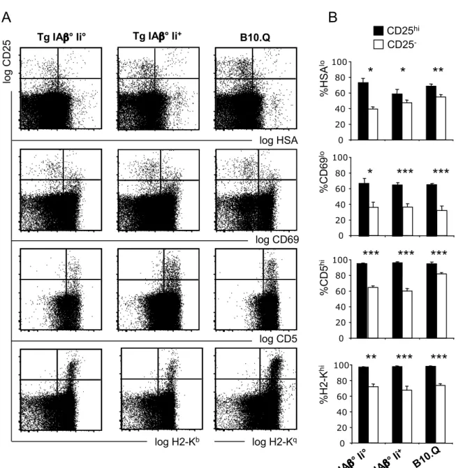
The phenotype of Treg from transgenic mice was similar to that of Treg from control B10.Q animals. Most of the Foxp3+ cells expressed high levels of CD25 (Fig. 2A). They expressed relatively low levels of the maturation marker HSA (CD24) and the activation marker CD69, and expressed high levels of CD5 and MHC class I (Figs. 3A, B). Therefore, transgenic regulatory CD4SP thymocytes had a more mature phenotype than conventional CD4SP cells, similar to that found on wt CD4SP CD25+ thymocytes.
In transgenic mice, mature CD4SP Treg migrated to the periphery (Figs. 2C, D). Peripheral transgenic Foxp3+ Treg mostly expressed high levels of CD25. Similar to what we observed in the thymus, IiThis is an author-produced version of a manuscript accepted for publication in The Journal+ mice had lower percentages of Foxp3+ cells among CD4+ splenocytes.
of Immunology (The JI). The American Association of Immunologists, Inc. (AAI), publisher
However, the percentage of CD4+ splenocytes was higher in Ii+ mice and the absolute numbers of Foxp3+ Treg was similar in transgenic Ii° and Ii+ mice (Fig. 2D).
The repertoire of Treg positively selected by a single MHC class II/peptide ligand expressed by cTEC
We next evaluated the specificity of Treg developing in transgenic mice. Since only minute numbers of Treg (i.e. CD4SP CD25high cells) can be obtained from thymi, we developed a very sensitive functional limiting dilution analysis (LDA). Magnetic bead-sorted CD25high
CD4SP thymocytes were seeded at limiting numbers in microtiter plates containing B10.Q APC with bCII or control peptides and high concentrations of IL-2. Two weeks later, the time it took for specific Treg to proliferate, new APC/peptide as well as allogeneic CD8+ responder T cells were added. Alloreactive CTL were allowed to proliferate during one week, after which 51Cr -labeled, anti-CD3ε mAb-coated targets were added. In cultures in which no Treg had proliferated, alloreactive CTL would proliferate and kill targets in the redirected lysis assay. By contrast, in cultures in which Treg had proliferated, these cells would inhibit proliferation of allospecific CTL (55) and thus reduced lysis of targets would be seen in the redirected lysis assay. Using this functional LDA to evaluate the repertoire of regulatory T lymphocytes from TgIAβ°Ii+ mice (in which IAq molecules are expressed by cTEC but in which the transgenic peptide is cleaved off and replaced by endogenous peptides, ref. 43), we observed similar frequencies of suppressed cultures in presence or in absence of bCII peptide (Figs. 4A, C, Table I). Presence of autospecific Treg in TgIAβ°Ii+ mice is at least in part due to absence of negative selection in our transgenics. By contrast, when analyzing Treg from TgIAβ°Ii° mice, in which exclusively the transgenic MHC/peptide ligand is expressed by cTEC, we reproducibly observed a significantly, approximately two-fold, higher frequency of suppressed cultures in presence of bCII peptide than in presence of control or no peptide (Figs. 4B, C, Table I). These results showed that an agonist ligand expressed by cTEC
This is an author-produced version of a manuscript accepted for publication in The Journal of Immunology (The JI). The American Association of Immunologists, Inc. (AAI), publisher of The JI, holds the copyright to this manuscript. This manuscript has not yet been
copyedited or subjected to editorial proofreading by The JI; hence it may differ from the final version published in The JI (online and in print). AAI (The JI) is not liable for errors or
positively selects regulatory T cells from a precursor population with a normally diverse TCR repertoire.
To consolidate these results, we also performed more conventional in vitro inhibition-of-proliferation assays using splenic Treg from transgenic mice. Titrated numbers of TgIAβ°Ii+ Treg were cultured in presence of B10.Q APC, the bCII or a control peptide, and allogeneic CD4+CD25- responder T cells. Similar inhibition curves were reproducibly obtained in absence and in presence of the bCII peptide (Fig. 4D). Again, inhibition was at least in part due to defective negative selection in our transgenic mice. By contrast, TgIAβ°Ii° Treg inhibited T cell proliferation substantially more efficiently in presence than in absence of specific bCII peptide (Fig. 4E).
We also assessed splenic Treg activation by measuring their proliferation in response to IAq -expressing APC presenting bCII or control peptide in presence of high concentrations of IL-2. As shown in figures 4F and H, Treg from TgIAβ°Ii+ mice proliferated in response to APC expressing IAq (because of defective negative selection), but addition of bCII or control peptide did not modify this proliferation. By contrast, addition of bCII peptide led to increased proliferation of Treg from TgIAβ°Ii° mice, whereas addition of control peptide had no effect (Figs. 4G and H).
In contrast to the observed preferential specificity of Treg for the positively selecting I-Aq/bCII ligand, in proliferation assays we failed to observe any evidence that the repertoire of
(thymic or splenic) conventional T cells is enriched in cells specific for this ligand (Figs. 4 I, J, and K). Proliferation in these cultures, in which Tconv had been stimulated with syngeneic APC, is due to absence of negative selection in mice expressing MHC molecules on cTEC only (18, 19, 38).
Based on these results, obtained with three distinct experimental setups, we conclude that autoreactive Treg can be positively selected from a precursor-population with a naturally This is an author-produced version of a manuscript accepted for publication in The Journalof Immunology (The JI). The American Association of Immunologists, Inc. (AAI), publisher
diverse TCR-repertoire upon interaction with high avidity (agonist) ligand expressed by cTEC.
This is an author-produced version of a manuscript accepted for publication in The Journal of Immunology (The JI). The American Association of Immunologists, Inc. (AAI), publisher of The JI, holds the copyright to this manuscript. This manuscript has not yet been
copyedited or subjected to editorial proofreading by The JI; hence it may differ from the final version published in The JI (online and in print). AAI (The JI) is not liable for errors or
Discussion
In this report we show that an agonist ligand, when expressed by cTEC only, positively selects autospecific regulatory CD4+ T cells from a precursor population with a naturally diverse TCR repertoire. A significant fraction of Treg positively selected on a single MHC class II/peptide ligand proliferated in response to the same ligand and had suppressive activity in a functional LDA for regulatory T cells we developed, as well as in more classical inhibition assays. By contrast, whereas conventional T cells developing in mice expressing a single ligand by cTEC are reactive to APC expressing syngeneic MHC class II (because of defective negative selection), no increased reactivity towards the transgenic MHC class II/peptide ligand could be detected in in vitro proliferation assays. Cortical positive selection of Treg vs. Tconv therefore appears to be governed by fundamentally different rules: low avidity interactions allow for positive selection of Tconv while high(er) avidity interactions are involved in positive selection of Treg. Cortical positive selection of Treg precursors therefore preferentially selects autoreactive cells and thus substantially contributes to the generation of the autospecific Treg repertoire.
The conclusion that agonist ligand can induce selection of Treg has earlier been reached based on TCR/antigen doubly transgenic mice (22-26, 29). However, in a study in which other TCR/antigen doubly transgenic mice were used it was shown that the increase in the proportion of Treg was accompanied by rather limited increase in their absolute numbers. Moreover, this moderate increase was not limited to transgenic antigen specific Treg. The relative increase in Treg in TCR/ligand doubly transgenic mice was explained by massive negative selection of conventional T cells combined with resistance to deletion of Treg precursors (28). This explanation cannot apply to the original experimental system reported here since immature precursors have a naturally diverse TCR-repertoire. Rather, positive selection of Treg appears to depend on high avidity interactions. Given that in the reported This is an author-produced version of a manuscript accepted for publication in The Journalof Immunology (The JI). The American Association of Immunologists, Inc. (AAI), publisher
experimental model immature precursors expressed a naturally diverse (as opposed to a monoclonal transgenic) TCR-repertoire, we assessed specificity of selected cells using three independent in vitro assays (functional LDA of thymic Treg, in vitro inhibition assays and proliferation assays using splenic Tregs). Moreover, the specificity-control was carefully performed using Ii+ vs. Ii°, MHC class II/peptide transgenic mice. Whereas in Ii° mice only the transgenic peptide was presented, in Ii+ mice transgenic peptide is cleaved off the transgenic IAβ-chain, and MHC class II molecules therefore presented endogenous peptides (43). In contrast to Treg from Ii° mice, cells from Ii+ mice did not show any biased specificity toward the IAq/bCII ligand. Therefore, our data on selection of Treg from naturally diverse precursors indicate that agonist self-MHC class II/peptide ligands, when expressed on cTEC, induce positive selection of autoreactive Treg.
Due to defective negative selection, in vitro reactivity of transgenic Treg (and Tconv) to APC expressing syngeneic MHC (not presenting transgenic peptide) was quite high, as previously observed (38). When we added transgenic peptide to the functional LDA, a two-fold increase in the frequency of activated Treg was observed. Given the high level of autoreactivity, this as compared to TCR/ligand doubly transgenic systems at first sight moderate increase, actually corresponds to a very substantial increase in ligand-specific Treg-reactivity. Moreover, in in vitro inhibition-of-proliferation assays, addition of transgenic peptide induced an almost 10-fold shift in the inhibition curve. Combined the data showed that cTEC-expression of agonist ligand substantially increased the generation of a transgenic MHC/peptide complex-specific Treg-repertoire.
Using mice in which expression of transgenic MHC class II was targeted to cTEC, Bensinger and colleagues earlier showed that cTEC induced development of functional Treg (38). Treg from these mice were autoreactive, but so were Tconv. These results were therefore explained by defective negative selection and did not show that positive selection of This is an author-produced version of a manuscript accepted for publication in The Journalof Immunology (The JI). The American Association of Immunologists, Inc. (AAI), publisher
of The JI, holds the copyright to this manuscript. This manuscript has not yet been
copyedited or subjected to editorial proofreading by The JI; hence it may differ from the final version published in The JI (online and in print). AAI (The JI) is not liable for errors or
Treg precursors is preferentially induced by agonist ligand. However, these data indicated that interaction of Treg precursors with MHC/peptide ligand in the medulla is not required for their full differentiation and are therefore consistent with the results presented here.
The hypothesis that ligands expressed by mTEC might also be involved in selection of Treg is particularly attractive since mTEC, in contrast to all other thymic stromal cellular compartments, ectopically express tissue specific antigens (56). The autoimmune syndrome developing in mice deficient in AIRE, a transcription factor involved in ectopic expression of antigens by mTEC, however, does not appear to be due to defective selection of Treg (57). On the other hand, using an elegant transgenic mouse model, it was recently shown that interactions with transgenic agonist ligand expressed by mTEC (under control of the AIRE promoter) promoted thymic accumulation of TCR transgenic Treg in an as yet unidentified manner (27). It was argued that the type of medullary cells expressing self-ligands determines if an autospecific T cell precursor is deleted or redirected to the Treg lineage. Treg lineage commitment would therefore take place late during T cell development, but (since RAG-sufficient TCR transgenic animals were used) the nature of the TCR signal involved in positive selection in the cortex remained unclear. Given these two contradictory reports, the precise role of mTEC in selection of a polyclonal repertoire of autospecific Treg remains incompletely understood.
In our transgenic mice, a very small subpopulation of mTEC appears to express very low levels of the transgenic MHC class II/peptide ligand. However, conventional T cells developing in our transgenic mice react vigorously to stimulation by syngeneic MHC class II-expressing APC, even in absence of added IL-2. This result demonstrates that negative selection by mTEC (via induction of anergy, ref. 58) is defective, and therefore strongly suggests that the very low MHC class II-expression level is functionally not significant. Whereas we formally cannot exclude a minor role of mTEC, we therefore favor the This is an author-produced version of a manuscript accepted for publication in The Journalof Immunology (The JI). The American Association of Immunologists, Inc. (AAI), publisher
conclusion that cTEC are entirely responsible for selection of the Treg repertoire in our transgenic mice.
The data currently available in the literature support the notion that agonist ligands favor development of Treg. However, in mice expressing a single MHC/peptide ligand by all normally MHC class II-expressing stromal cells, a diverse Treg repertoire develops. In these mice, also negatively selecting dendritic cells express the single ligand, and Treg from such mice do not respond to the selecting ligand (30, 59, 60). Therefore, even if agonist self-ligands expressed by TEC clearly favor development of Treg, they probably are not the only ones to do so. We therefore privilege a model in which positive selection of Treg is mediated by higher avidity interactions than that of Tconv, including but not limited to agonist TCR-ligand interactions.
In conclusion, our data indicate that thymic positive selection in the cortex substantially contributes to shaping of the autoreactive Treg repertoire. However, they do not exclude a potential role for ligands expressed in the thymic medulla. It will therefore now be important to evaluate how cortical and medullary selection mechanisms collaborate in shaping the autoreactive Treg repertoire.
This is an author-produced version of a manuscript accepted for publication in The Journal of Immunology (The JI). The American Association of Immunologists, Inc. (AAI), publisher of The JI, holds the copyright to this manuscript. This manuscript has not yet been
copyedited or subjected to editorial proofreading by The JI; hence it may differ from the final version published in The JI (online and in print). AAI (The JI) is not liable for errors or
Acknowledgements
The authors would like to thank the staff of the IFR30 animal facility for excellent animal husbandry, Dr. Fatima-Ezzahra L’Faqihi-Olive for flow cytometry, Dr. E. Fuchs for the K14 promoter construct, Dr. Talal Al Saati and Florence Capilla for histological analysis, and Drs. Antonio Bandeira, Benoit Salomon, Philippe Naquet, and Roland Liblau for critical reading of the manuscript.
This is an author-produced version of a manuscript accepted for publication in The Journal of Immunology (The JI). The American Association of Immunologists, Inc. (AAI), publisher
References
1. Sakaguchi, S., M. Ono, R. Setoguchi, H. Yagi, S. Hori, Z. Fehervari, J. Shimizu, T. Takahashi, and T. Nomura. 2006. Foxp3+ CD25+ CD4+ natural regulatory T cells in dominant self-tolerance and autoimmune disease. Immunol. Rev. 212:8-27.
2. Shevach, E. M., R. A. DiPaolo, J. Andersson, D. M. Zhao, G. L. Stephens, and A. M. Thornton. 2006. The lifestyle of naturally occurring CD4+ CD25+ Foxp3+ regulatory T cells. Immunol. Rev. 212:60-73.
3. Izcue, A., J. L. Coombes, and F. Powrie. 2006. Regulatory T cells suppress systemic and mucosal immune activation to control intestinal inflammation. Immunol. Rev. 212:256-271.
4. Belkaid, Y., R. B. Blank, and I. Suffia. 2006. Natural regulatory T cells and parasites: a common quest for host homeostasis. Immunol. Rev. 212:287-300.
5. Rouse, B. T., P. P. Sarangi, and S. Suvas. 2006. Regulatory T cells in virus infections.
Immunol. Rev. 212:272-286.
6. Beyer, M., and J. L. Schultze. 2006. Regulatory T cells in cancer. Blood 108:804-811. 7. Joffre, O., and J. P. M. van Meerwijk. 2006. CD4+CD25+ regulatory T lymphocytes
in bone marrow transplantation. Sem. Immunol. 18:128-135.
8. Romagnoli, P., D. Hudrisier, and J. P. M. van Meerwijk. 2002. Preferential recognition of self-antigens despite normal thymic deletion of CD4+CD25+ regulatory T cells. J. Immunol. 168:1644-1648.
9. Hsieh, C. S., Y. Liang, A. J. Tyznik, S. G. Self, D. Liggitt, and A. Y. Rudensky. 2004. Recognition of the peripheral self by naturally arising CD25+ CD4+ T cell receptors.
Immunity 21:267-277.
This is an author-produced version of a manuscript accepted for publication in The Journal of Immunology (The JI). The American Association of Immunologists, Inc. (AAI), publisher of The JI, holds the copyright to this manuscript. This manuscript has not yet been
copyedited or subjected to editorial proofreading by The JI; hence it may differ from the final version published in The JI (online and in print). AAI (The JI) is not liable for errors or
10. Fisson, S., G. Darrasse-Jeze, E. Litvinova, F. Septier, D. Klatzmann, R. Liblau, and B. L. Salomon. 2003. Continuous Activation of Autoreactive CD4+ CD25+ Regulatory T Cells in the Steady State. J. Exp. Med. 198:737-746.
11. Hsieh, C. S., Y. Zheng, Y. Liang, J. D. Fontenot, and A. Y. Rudensky. 2006. An intersection between the self-reactive regulatory and nonregulatory T cell receptor repertoires. Nat. Immunol. 7:401-410.
12. Chen, W., W. Jin, N. Hardegen, K.-j. Lei, L. Li, N. Marinos, G. McGrady, and S. M. Wahl. 2003. Conversion of Peripheral CD4+CD25- Naive T Cells to CD4+CD25+ Regulatory T Cells by TGF-{beta} Induction of Transcription Factor Foxp3. J. Exp.
Med. 198:1875-1886.
13. Walker, M. R., D. J. Kasprowicz, V. H. Gersuk, A. Benard, M. Van Landeghen, J. H. Buckner, and S. F. Ziegler. 2003. Induction of FoxP3 and acquisition of T regulatory activity by stimulated human CD4+CD25- T cells. J. Clin. Invest. 112:1437-1443. 14. Liang, S., P. Alard, Y. Zhao, S. Parnell, S. L. Clark, and M. M. Kosiewicz. 2005.
Conversion of CD4+ CD25- cells into CD4+ CD25+ regulatory T cells in vivo requires B7 costimulation, but not the thymus. J. Exp. Med. 201:127-137.
15. Apostolou, I., and H. von Boehmer. 2004. In vivo instruction of suppressor commitment in naive T cells. J. Exp. Med. 199:1401-1408.
16. Itoh, M., T. Takahashi, N. Sakaguchi, Y. Kuniyasu, J. Shimizu, F. Otsuka, and S. Sakaguchi. 1999. Thymus and autoimmunity: production of CD25+CD4+ naturally anergic and suppressive T cells as a key function of the thymus in maintaining immunologic self-tolerance. J. Immunol. 162:5317-5326.
17. Romagnoli, P., D. Hudrisier, and J. P. M. van Meerwijk. 2005. Molecular signature of recent thymic selection events on effector and regulatory CD4+ T lymphocytes. J.
Immunol. 175:5751-5758.
This is an author-produced version of a manuscript accepted for publication in The Journal of Immunology (The JI). The American Association of Immunologists, Inc. (AAI), publisher
18. Laufer, T. M., J. DeKoning, J. S. Markowitz, D. Lo, and L. H. Glimcher. 1996. Unopposed positive selection and autoreactivity in mice expressing class II MHC only on thymic cortex. Nature 383:81-85.
19. Capone, M., P. Romagnoli, F. Beermann, H. R. MacDonald, and J. P. M. van Meerwijk. 2001. Dissociation of thymic positive and negative selection in transgenic mice expressing major histocompatibility complex class I molecules exclusively on thymic cortical epithelial cells. Blood 97:1336-1342.
20. Hudrisier, D., S. Feau, V. Bonnet, P. Romagnoli, and J. P. M. van Meerwijk. 2003. In vivo unresponsiveness of T lymphocyte induced by thymic medullary epithelium requires antigen presentation by radioresistant cells. Immunol. 108:24-31.
21. Starr, T., J. Jameson, and K. A. Hogquist. 2003. Positive and negative selection of T cells. Annu. Rev. Immunol. 32:139-176.
22. Jordan, M. S., A. Boesteanu, A. J. Reed, A. L. Petrone, A. E. Holenbeck, M. A. Lerman, A. Naji, and A. J. Caton. 2001. Thymic selection of CD4+CD25+ regulatory T cells induced by an agonist self-peptide. Nat. Immunol. 2:301-306.
23. Kawahata, K., Y. Misaki, M. Yamauchi, S. Tsunekawa, K. Setoguchi, J.-i. Miyazaki, and K. Yamamoto. 2002. Generation of CD4+CD25+ Regulatory T Cells from Autoreactive T Cells Simultaneously with Their Negative Selection in the Thymus and from Nonautoreactive T Cells by Endogenous TCR Expression. J. Immunol. 168:4399-4405.
24. Apostolou, I., A. Sarukhan, L. Klein, and H. von Boehmer. 2002. Origin of regulatory T cells with known specificity for antigen. Nat. Immunol. 3:756-763.
25. D'Cruz, L. M., and L. Klein. 2005. Development and function of agonist-induced CD25+Foxp3+ regulatory T cells in the absence of interleukin 2 signaling. Nat.
Immunol. 6:1152-1159.
This is an author-produced version of a manuscript accepted for publication in The Journal of Immunology (The JI). The American Association of Immunologists, Inc. (AAI), publisher of The JI, holds the copyright to this manuscript. This manuscript has not yet been
copyedited or subjected to editorial proofreading by The JI; hence it may differ from the final version published in The JI (online and in print). AAI (The JI) is not liable for errors or
26. Cabarrocas, J., C. Cassan, F. Magnusson, E. Piaggio, L. Mars, J. Derbinski, B. Kyewski, D.-A. Gross, B. L. Salomon, K. Khazaie, A. Saoudi, and R. S. Liblau. 2006. Foxp3+ CD25+ regulatory T cells specific for a neo-self-antigen develop at the double-positive thymic stage. Proc. Natl. Acad. Sci. U.S.A. 103:8453-8458.
27. Aschenbrenner, K., L. M. D'Cruz, E. H. Vollmann, M. Hinterberger, J. Emmerich, L. K. Swee, A. Rolink, and L. Klein. 2007. Selection of Foxp3(+) regulatory T cells specific for self antigen expressed and presented by Aire(+) medullary thymic epithelial cells. Nat. Immunol. 8:351-358.
28. van Santen, H.-M., C. Benoist, and D. Mathis. 2004. Number of T Reg Cells That Differentiate Does Not Increase upon Encounter of Agonist Ligand on Thymic Epithelial Cells. J. Exp. Med. 200:1221-1230.
29. Ribot, J., P. Romagnoli, and J. P. M. van Meerwijk. 2006. Agonist Ligands Expressed by Thymic Epithelium Enhance Positive Selection of Regulatory T Lymphocytes from Precursors with a Normally Diverse TCR Repertoire. J. Immunol. 177:1101-1107. 30. Pacholczyk, R., P. Kraj, and L. Ignatowicz. 2002. Peptide specificity of thymic
selection of CD4+CD25+ T cells. J. Immunol. 168:613-620.
31. Shih, F. F., L. Mandik-Nayak, B. T. Wipke, and P. M. Allen. 2004. Massive Thymic Deletion Results in Systemic Autoimmunity through Elimination of CD4+ CD25+ T Regulatory Cells. J. Exp. Med. 199:323-335.
32. Huseby, E. S., J. White, F. Crawford, T. Vass, D. Becker, C. Pinilla, P. Marrack, and J. W. Kappler. 2005. How the T cell repertoire becomes peptide and MHC specific.
Cell 122:247-260.
33. Huseby, E. S., F. Crawford, J. White, P. Marrack, and J. W. Kappler. 2006. Interface-disrupting amino acids establish specificity between T cell receptors and complexes of major histocompatibility complex and peptide. Nat. Immunol. 7:1191-1199.
This is an author-produced version of a manuscript accepted for publication in The Journal of Immunology (The JI). The American Association of Immunologists, Inc. (AAI), publisher
34. Huseby, E. S., F. Crawford, J. White, J. Kappler, and P. Marrack. 2003. Negative selection imparts peptide specificity to the mature T cell repertoire. Proc. Natl. Acad.
Sci. U.S.A. 100:11565-11570.
35. Benoist, C., and D. Mathis. 1989. Positive selection of the T cell repertoire: where and when does it occur? Cell 58:1027-1033.
36. Cosgrove, D., S. H. Chan, C. Waltzinger, C. Benoist, and D. Mathis. 1992. The thymic compartment responsible for positive selection of CD4+ T cells. Int. Immunol. 4:707-710.
37. DeKoning, J., L. DiMolfetto, C. Reilly, Q. Wei, W. L. Havran, and D. Lo. 1997. Thymic cortical epithelium is sufficient for the development of mature T cells in relB-deficient mice. J. Immunol. 158:2558-2566.
38. Bensinger, S. J., A. Bandeira, M. S. Jordan, A. J. Caton, and T. M. Laufer. 2001. Major Histocompatibility Complex Class II-positive Cortical Epithelium Mediates the Selection of CD4+25+ Immunoregulatory T Cells. J. Exp. Med. 194:427-438.
39. Goldman, K. P., C. S. Park, M. Kim, P. Matzinger, and C. C. Anderson. 2005. Thymic cortical epithelium induces self tolerance. Eur. J. Immunol. 35:709-717.
40. Mayerova, D., and K. A. Hogquist. 2004. Central Tolerance to Self-Antigen Expressed by Cortical Epithelial Cells. J. Immunol. 172:851-856.
41. Baldwin, T. A., M. M. Sandau, S. C. Jameson, and K. A. Hogquist. 2005. The timing of TCR alpha expression critically influences T cell development and selection. J.
Exp. Med. 202:111-121.
42. Laufer, T. M., L. Fan, and L. H. Glimcher. 1999. Self-reactive T cells selected on thymic cortical epithelium are polyclonal and are pathogenic in vivo. J. Immunol. 162:5078-5084.
This is an author-produced version of a manuscript accepted for publication in The Journal of Immunology (The JI). The American Association of Immunologists, Inc. (AAI), publisher of The JI, holds the copyright to this manuscript. This manuscript has not yet been
copyedited or subjected to editorial proofreading by The JI; hence it may differ from the final version published in The JI (online and in print). AAI (The JI) is not liable for errors or
43. Ignatowicz, L., G. Winslow, J. Bill, J. Kappler, and P. Marrack. 1995. Cell surface expression of class II MHC proteins bound by a single peptide. J. Immunol. 154:3852-3862.
44. Brand, D. D., L. K. Myers, K. Terato, K. B. Whittington, J. M. Stuart, A. H. Kang, and E. F. Rosloniec. 1994. Characterization of the T cell determinants in the induction of autoimmune arthritis by bovine alpha 1(II)-CB11 in H-2q mice. J. Immunol. 152:3088-3097.
45. Vassar, R., M. Rosenberg, S. Ross, A. Tyner, and E. Fuchs. 1989. Tissue-specific and differentiation-specific expression of a human K14 keratin gene in transgenic mice.
Proc. Natl. Acad. Sci. U.S.A. 86:1563-1567.
46. Rosloniec, E. F., K. B. Whittington, D. D. Brand, L. K. Myers, and J. M. Stuart. 1996. Identification of MHC Class II and TCR Binding Residues in the Type II Collagen Immunodominant Determinant Mediating Collagen-Induced Arthritis. Cell. Immunol. 172:21-28.
47. Derbinski, J., A. Schulte, B. Kyewski, and L. Klein. 2001. Promiscuous gene expression in medullary thymic epithelial cells mirrors the peripheral self. Nat.
Immunol. 2:1032-1039.
48. Taswell, C. 1981. Limiting dilution assays for the determination of immunocompetent cell frequencies. I. Data analysis. J. Immunol. 126:1614-1619.
49. Stefanski, H. E., D. Mayerova, S. C. Jameson, and K. A. Hogquist. 2001. A Low Affinity TCR Ligand Restores Positive Selection of CD8+ T Cells In Vivo. J.
Immunol. 166:6602-6607.
50. Khattri, R., T. Cox, S. A. Yasayko, and F. Ramsdell. 2003. An essential role for Scurfin in CD4(+)CD25(+) T regulatory cells. Nat. Immunol. 3:3.
This is an author-produced version of a manuscript accepted for publication in The Journal of Immunology (The JI). The American Association of Immunologists, Inc. (AAI), publisher
51. Hori, S., T. Nomura, and S. Sakaguchi. 2003. Control of regulatory T cell development by the transcription factor Foxp3. Science 299:1057-1061.
52. Fontenot, J. D., M. A. Gavin, and A. Y. Rudensky. 2003. Foxp3 programs the development and function of CD4(+)CD25(+) regulatory T cells. Nat. Immunol. 3:3. 53. Yagi, H., T. Nomura, K. Nakamura, S. Yamazaki, T. Kitawaki, S. Hori, M. Maeda, M.
Onodera, T. Uchiyama, S. Fujii, and S. Sakaguchi. 2004. Crucial role of FOXP3 in the development and function of human CD25+CD4+ regulatory T cells. Int. Immunol. 16:1643-1656.
54. Fontenot, J. D., J. P. Rasmussen, L. M. Williams, J. L. Dooley, A. G. Farr, and A. Y. Rudensky. 2005. Regulatory T cell lineage specification by the forkhead transcription factor foxp3. Immunity 22:329-341.
55. Piccirillo, C. A., and E. M. Shevach. 2001. Cutting edge: control of CD8+ T cell activation by CD4+CD25+ immunoregulatory cells. J. Immunol. 167:1137-1140. 56. Kyewski, B., and J. Derbinski. 2004. Self-representation in the thymus: an extended
view. Nat. Rev. Immunol. 4:688-698.
57. Anderson, M. S., E. S. Venanzi, Z. Chen, S. P. Berzins, C. Benoist, and D. Mathis. 2005. The cellular mechanism of AIRE control of T cell tolerance. Immunity 23:227-239.
58. Ramsdell, F., T. Lantz, and B. J. Fowlkes. 1989. A nondeletional mechanism of thymic self tolerance. Science 246:1038-1041.
59. Pacholczyk, R., H. Ignatowicz, P. Kraj, and L. Ignatowicz. 2006. Origin and T cell receptor diversity of Foxp3+CD4+CD25+ T cells. Immunity 25:249-259.
60. Ignatowicz, L., J. Kappler, and P. Marrack. 1996. The repertoire of T cells shaped by a single MHC/peptide ligand. Cell 84:521-529.
This is an author-produced version of a manuscript accepted for publication in The Journal of Immunology (The JI). The American Association of Immunologists, Inc. (AAI), publisher of The JI, holds the copyright to this manuscript. This manuscript has not yet been
copyedited or subjected to editorial proofreading by The JI; hence it may differ from the final version published in The JI (online and in print). AAI (The JI) is not liable for errors or
Legends to the Figures
Figure 1
The transgenic IAαβqbCII complex is expressed by cTEC but not by mTEC or splenic APC
A) Histological analysis of thymi from transgenic and control mice. Thymus sections were
stained for MHC class II. The cortical (c) and medullary (m) regions are indicated. B) Flow cytometry analysis of splenocytes from transgenic and control mice detecting indicated surface molecules. Depicted data are representative of 3 independent experiments. C) Flow cytometry analysis of MHC class II expression by CD45- EpCAM+Ly51+ cTEC of indicated mice in red. D) Flow cytometry analysis of MHC class II expression by CD45-EpCAM+Ly51 -mTEC of indicated mice in red. Blue histograms: staining with isotype-matched control antibody.
Figure 2
Conventional and regulatory T cells develop in pK14-IAαβqbCII transgenic mice
Flow cytometry analysis of CD4 vs. CD8 expression by thymocytes (A) and splenocytes (C) from transgenic and control mice (upper panels). TCR and CD25 vs. Foxp3 expression on electronically gated CD4+CD8- cells (middle and lower panels). Percentages of CD4SP among total cells and Foxp3+ cells among CD4SP cells using gates as indicated in A in thymus (B, upper and middle panels) and in spleen (D, upper and middle panels). Absolute numbers of Foxp3+ cells in thymi (B, lower panel) and spleens (D, lower panel). ***, p < 0.001; NS, not significant; Student’s t test. Error bars indicate SD (n=3).
This is an author-produced version of a manuscript accepted for publication in The Journal of Immunology (The JI). The American Association of Immunologists, Inc. (AAI), publisher
Figure 3
Conventional and regulatory T cells developing in pK14-IAαβqbCII transgenic mice display normal phenotypes
A) Expression of CD25 vs. HSA, CD69, CD5 and H2-K by electronically gated CD4SP
thymocytes. B) Percentages of (most mature) HSAlow, CD69low, CD5high and H2-Khigh cells among CD25- and CD25high CD4SP populations. ***, p < 0.001; **, p < 0.01; *, p < 0.05; Student’s t test. Error bars indicate SD (n=3).
Figure 4
Treg, but not Tconv, from pK14-IAαβqbCII mice display increased reactivity toward the IAq/bCII complex
A, B, C) Functional Treg LDA. Titrated numbers (as indicated) of purified CD4SP CD25+
thymocytes from TgIAβ°Ii+ (A) and TgIAβ°Ii° (B) mice were cultured with irradiated B10.Q splenocytes pulsed or not with peptides, as indicated. After 14 days, same APC/peptide and allogeneic DBA/2 CD8+ cells were added. Seven days later redirected lysis was assessed. Data shown in A and B are representative of 4 independent experiments. C) Precursor frequency-ratios of Treg specific for bCII vs. C1 control peptide observed in the four independently performed experiments. Individual experiments are shown and vertical bars indicate mean values. Raw-data from individual experiments are shown in Table I. D, E) In
vitro inhibition of proliferation assays. CD4+CD25+ splenic Treg from TgIAβ°Ii+ (D) and TgIAβ°Ii° mice (E) were cocultured, at indicated ratios, with 105 CD4SP CD25- conventional T cells from B6 mice in the presence of irradiated B10.Q splenocytes pulsed with bCII () or C1 control peptides () (50 µM). 3H-thymidine incorporation was assessed after 5 days. Data are representative of 3 independent experiments. F, G, H) In vitro Treg-proliferation assays.
CD4This is an author-produced version of a manuscript accepted for publication in The Journalof Immunology (The JI). The American Association of Immunologists, Inc. (AAI), publisher+CD25+ splenic Treg from TgIAβ°Ii+ (F) and TgIAβ°Ii° mice (G) were cultured with
of The JI, holds the copyright to this manuscript. This manuscript has not yet been
copyedited or subjected to editorial proofreading by The JI; hence it may differ from the final version published in The JI (online and in print). AAI (The JI) is not liable for errors or
B10.Q APC pulsed with peptides, in presence of IL-2. 3H-thymidine incorporation was assessed after 4 days. H) Ratio of proliferation of Treg from indicated mice in response to B10.Q APC pulsed with bCII vs. control C1 peptides (50 µM). I, J, K) In vitro Tconv proliferation assays. CD4+CD25- Tconv from TgIAβ°Ii° thymi (I) or spleen (J) were cultured with irradiated B10.Q splenocytes pulsed with titrated doses of peptides ( bCII, C1, C2, C3 and C4), in absence of IL-2. 3H-thymidine incorporation was assayed after 4 days. K) Ratio of proliferation of thymic () or splenic () Tconv in response to B10.Q APC pulsed with bCII vs. control C1 peptides (50 µM). In F, G, I, and J, background proliferation, as determined by stimulating T cells with MHC-deficient APC, was inferior to 1000 cpm. **, p < 0.01; NS, not significant; Student’s t test.
This is an author-produced version of a manuscript accepted for publication in The Journal of Immunology (The JI). The American Association of Immunologists, Inc. (AAI), publisher
Figure 1
co un tsA
B
lo g B 22 0 log MHC class II cTEC mTECC
D
log MHC class II nTg IA β ° Ii ° Tg I A β ° Ii ° c m 0 102 103 104 105 0 20 40 60 80 100 0 102 103 104 105 0 20 40 60 80 100 0 102 103 104 105 0 20 40 60 80 100 0 10 2 10 3 10 4 10 5 0 20 40 60 80 100 0 102 103 104 105 0 20 40 60 80 100 Tg IA β ° Ii + c m 0 102 103 104 105 0 20 40 60 80 100 0 10 2 10 3 10 4 10 5 0 20 40 60 80 100 0 102 103 104 105 <APC 660/20-A> 0 20 40 60 80 100 % o f M ax 0 102 103 104 105 <APC 660/20-A> 0 20 40 60 80 100 % o f M ax 0 102 103 104 105 0 20 40 60 80 100 c m 0 102 103 104 105 0 20 40 60 80 100 0 10 2 10 3 10 4 10 5 0 20 40 60 80 100 0 102 103 104 105 <APC 660/20-A> 0 20 40 60 80 100 % o f M ax 0 102 103 104 105 <APC 660/20-A> 0 20 40 60 80 100 % o f M ax B10.Q 0 102 103 104 105 0 20 40 60 80 100 0 102 103 104 105 0 20 40 60 80 100 0 102 103 104 105 0 20 40 60 80 100 0 10 2 10 3 10 4 10 5 0 20 40 60 80 100 c mThis is an author-produced version of a manuscript accepted for publication in The Journal of Immunology (The JI). The American Association of Immunologists, Inc. (AAI), publisher of The JI, holds the copyright to this manuscript. This manuscript has not yet been
copyedited or subjected to editorial proofreading by The JI; hence it may differ from the final version published in The JI (online and in print). AAI (The JI) is not liable for errors or
Figure 2
B
A
log TCR co un ts log CD8 lo g C D 4 % F o xp 3 + a m o n g C D 4 S P t h ym o cy te s log CD25 lo g F ox p3 Tg IA β° Ii + B10.Q Tg IA β° Ii° nTg IAβ° I i°Tg IAβ° Ii° Tg IAβ° Ii+ nTg IAβ° Ii° B10.Q
0 5 10 15 20 % C D 4 S P a m o n g to ta l t h ym o cy te s
***
1 2 3 4 5 6***
0 0.1 0.2 0.3 0.4 # F o xp 3 + / t h ym u s ( x 1 0 6) NS IAβ° Ii+ IAβ° Ii° IAβ° I i° % F o xp 3 + a m o n g C D 4 + s p le n o cy te s 0 5 10 15 20C
D
log CD8 log TCR log CD25 lo g C D 4 co un ts lo g F ox p3Tg IAβ° Ii° Tg IAβ° Ii+ nTg IAβ° Ii° B10.Q
B10.Q 0 5 10 15 20 % C D 4 +a m o n g to ta l s p le n o cy te s
***
***
0 1 1.5 2 0.5 # F o xp 3 + / s p le e n ( x 1 0 6) NSThis is an author-produced version of a manuscript accepted for publication in The Journal of Immunology (The JI). The American Association of Immunologists, Inc. (AAI), publisher
Figure 3
B
0 20 40 60 80 100 % H S A lo 0 20 40 60 80 100 % C D 5 h i 0 20 40 60 80 100 Tg IA β° Ii + B10.Q Tg IA β° Ii° % H 2-K h i CD25hi CD25-*
*
**
0 20 40 60 80 100 % C D 69 lo*
***
***
***
*** ***
**
***
***
log HSA lo g C D 25A
log CD69 log CD5Tg IAβ° Ii° Tg IAβ° Ii+ B10.Q
log H2-Kb log H2-Kq
This is an author-produced version of a manuscript accepted for publication in The Journal of Immunology (The JI). The American Association of Immunologists, Inc. (AAI), publisher of The JI, holds the copyright to this manuscript. This manuscript has not yet been
copyedited or subjected to editorial proofreading by The JI; hence it may differ from the final version published in The JI (online and in print). AAI (The JI) is not liable for errors or
Figure 4
This is an author-produced version of a manuscript accepted for publication in The Journal of Immunology (The JI). The American Association of Immunologists, Inc. (AAI), publisher
A
B
Table I:
Treg from pK14-IAαβ
qbCII mice have increased reactivity toward IA
q/bCII
complex.
Tg Ii° CD4SP CD25+ thymocytes
Tg Ii+ CD4SP CD25+ thymocytes
Summary of functional limiting dilution analysis performed as in fig. 4
# exp peptide f R2 f(bCII)/f(C1)
none 1/185 0.66 C1 1/163 0.56 bCII 1/90 0.94 none 1/526 0.86 C1 1/526 0.88 bCII 1/333 0.96 C1 1/1666 0.96 bCII 1/714 0.97 none 1/555 0.96 C1 1/370 0.83 bCII 1/133 0.81 4 2.78 2 1.57 3 2.33 1 1.81
# exp peptide f R2 f(bCII)/f(C1)
none 1/123 0.89 C1 1/103 0.89 bCII 1/93 0.96 none 1/128 0.63 C1 1/172 0.77 bCII 1/172 0.81 C1 1/666 0.78 bCII 1/833 0.83 none 1/555 0.75 C1 1/769 0.87 bCII 1/666 0.72 5 1.11 6 1.00 7 0.80 8 1.15
This is an author-produced version of a manuscript accepted for publication in The Journal of Immunology (The JI). The American Association of Immunologists, Inc. (AAI), publisher of The JI, holds the copyright to this manuscript. This manuscript has not yet been
copyedited or subjected to editorial proofreading by The JI; hence it may differ from the final version published in The JI (online and in print). AAI (The JI) is not liable for errors or