HAL Id: inserm-02481105
https://www.hal.inserm.fr/inserm-02481105
Submitted on 17 Feb 2020
HAL is a multi-disciplinary open access
archive for the deposit and dissemination of
sci-entific research documents, whether they are
pub-lished or not. The documents may come from
teaching and research institutions in France or
abroad, or from public or private research centers.
L’archive ouverte pluridisciplinaire HAL, est
destinée au dépôt et à la diffusion de documents
scientifiques de niveau recherche, publiés ou non,
émanant des établissements d’enseignement et de
recherche français ou étrangers, des laboratoires
publics ou privés.
Soluble HLA-I/Peptide Monomers Mediate
Antigen-Specific CD8 T Cell Activation through Passive
Peptide Exchange with Cell-Bound HLA-I Molecules
Mathilde Allard, Romain Oger, Houssem Benlalam, Laetitia Florenceau,
Klara Echasserieau, Karine Bernardeau, Nathalie Labarrière, François Lang,
Nadine Gervois
To cite this version:
Mathilde Allard, Romain Oger, Houssem Benlalam, Laetitia Florenceau, Klara Echasserieau, et al..
Soluble HLA-I/Peptide Monomers Mediate Antigen-Specific CD8 T Cell Activation through Passive
Peptide Exchange with Cell-Bound HLA-I Molecules. Journal of Immunology, Publisher : Baltimore :
Williams & Wilkins, c1950-. Latest Publisher : Bethesda, MD : American Association of
Immunolo-gists, 2014, 192 (11), pp.5090-5097. �10.4049/jimmunol.1303226�. �inserm-02481105�
of September 30, 2014.
This information is current as
Cell-Bound HLA-I Molecules
through Passive Peptide Exchange with
Antigen-Specific CD8 T Cell Activation
Soluble HLA-I/Peptide Monomers Mediate
Nathalie Labarrière, François Lang and Nadine Gervois
Florenceau, Klara Echasserieau, Karine Bernardeau,
Mathilde Allard, Romain Oger, Houssem Benlalam, Laetitia
http://www.jimmunol.org/content/192/11/5090
doi: 10.4049/jimmunol.1303226
April 2014;
2014; 192:5090-5097; Prepublished online 21
J Immunol
Material
Supplementary
DCSupplemental.html
http://www.jimmunol.org/content/suppl/2014/04/21/content.1303226.
References
http://www.jimmunol.org/content/192/11/5090.full#ref-list-1
, 14 of which you can access for free at:
cites 34 articles
This article
Subscriptions
http://jimmunol.org/subscriptions
is online at:
The Journal of Immunology
Information about subscribing to
Permissions
http://www.aai.org/ji/copyright.html
Submit copyright permission requests at:
Email Alerts
http://jimmunol.org/cgi/alerts/etoc
Receive free email-alerts when new articles cite this article. Sign up at:
Print ISSN: 0022-1767 Online ISSN: 1550-6606.
Immunologists, Inc. All rights reserved.
Copyright © 2014 by The American Association of
9650 Rockville Pike, Bethesda, MD 20814-3994.
The American Association of Immunologists, Inc.,
is published twice each month by
The Journal of Immunology
at Universite Nantes BU Sante on September 30, 2014
http://www.jimmunol.org/
Downloaded from
at Universite Nantes BU Sante on September 30, 2014
http://www.jimmunol.org/
The Journal of Immunology
Soluble HLA-I/Peptide Monomers Mediate Antigen-Specific
CD8 T Cell Activation through Passive Peptide Exchange
with Cell-Bound HLA-I Molecules
Mathilde Allard,*
,†,‡Romain Oger,*
,†,‡Houssem Benlalam,*
,†,‡Laetitia Florenceau,*
,†,‡Klara Echasserieau,*
,†,‡,xKarine Bernardeau,*
,xNathalie Labarrie`re,*
,†,‡Franc¸ois Lang,*
,†,‡,xand Nadine Gervois*
,†,‡Accumulating evidence that serum levels of soluble class I HLA molecules (sHLA-I) can, under various pathological conditions, correlate with disease stage and/or patient survival, has stimulated interest in defining whether sHLA-I can exert immunological functions. However, despite a mounting number of publications suggesting the ability of sHLA-I to affect immune effectors in vitro, the precise underlying mechanism still remains controversial. In this article, we address potential functions of both classical and nonclassical sHLA-I, using soluble recombinant HLA-I/peptide monomers, and clearly demonstrate their ability to trigger Ag-specific activation of CD8 T cells in vitro. Furthermore, we provide strong evidence that this behavior results from the passive transfer of peptides from monomers to T cell–bound HLA-I molecules, allowing for fratricide representation and activation. Hence, we proposed a unifying model of T cell activation by HLA-I/peptide monomers, reappraising the potential involvement of sHLA-I molecules in the immune response. The Journal of Immunology, 2014, 192: 5090–5097.
T
he human MHC encodes two sets of class I molecules: class Ia or classical molecules, (HLA-A, -Bm and -C) characterized by a high degree of polymorphism and a broad tissue expression (1), and class Ib or nonclassical molecules (HLA-E, -F, and -G) presenting a more restricted polymorphism and expression profile (2). Both classical and nonclassical HLA-I molecules need to accommodate endogenously processed peptides and to associate with the invariantb2-microglobulin (b2m) chainto be stably expressed at the cytoplasmic membrane (3). Cell-surface HLA-I/peptide/b2m complexes can then be scanned by
CD8 T lymphocytes and NK cell receptors, allowing immune effectors to distinguish healthy cells from altered ones (i.e., stressed, infected, or transformed ones) (4, 5).
Besides being expressed at the membrane of most nucleated cells, HLA-I molecules are also present in body fluids in soluble forms [soluble class I HLA molecules (sHLA-I)], mostly generated by proteolytic cleavage of membrane-bound molecules, that can be either empty or associated with peptide and/orb2m (6, 7). Of
interest, although serum sHLA-I levels are rather stable in physio-logical conditions, they are modified in various pathophysio-logical conditions, including inflammation, autoimmunity, infections,
transplant rejection, and cancer, and can even correlate with disease stage, patient survival, and/or prognosis (7–9). Moreover, several studies have reported, in vitro, the ability of sHLA-I molecules to modulate the activity of NK cells and T lymphocytes expressing cognate NK or TCRs, suggesting that sHLA-I molecules may play significant immunoregulatory roles in vivo (10–16).
Although accumulated evidence points out the immunological properties of sHLA-I molecules, the precise underlying mecha-nisms remain poorly understood and debated. Most studies argue that sHLA-I effects result from the triggering of their cognate NK or TCRs (or even with the CD8 coreceptors) (10–16). However, most sHLA-I molecules are found in monomeric forms, and should therefore not be able to mediate multivalent (i.e., productive) engagement of their receptors. An alternative mechanism relying on the transfer of class I MHC–derived peptides toward cell-bound MHC molecules has been suggested and could reconcile these observations with the well-accepted model of multivalent en-gagement (17, 18).
We previously reported the ability of both endothelial (19) and tumor cells (20, 21) to produce sHLA-E molecules, especially in proinflammatory conditions, and documented a significant increase of serum sHLA-E levels in melanoma patients, compared with healthy donors (20). Moreover, we and others (19, 22) observed that naturally produced and recombinant sHLA-E monomers decrease the allogeneic and antitumor activity of NK and CD8 T cells expressing the related inhibitory NK receptor CD94/ NKG2-A. In this study, taking advantage of an HLA-E–restricted CD8 T cell clone (23), we investigated whether sHLA-E functions rely on engagement of their cognate receptors or, alternatively, on peptide transfer from sHLA-E onto cell-bound HLA-E molecules. To check whether these mechanisms are shared by classical HLA-I molecules, the same experiments were conducted in an HLA-A2 model.
In agreement with previous reports, results clearly showed that both classical and nonclassical sHLA-I monomers could efficiently activate Ag-specific CD8 T cells. In addition, we demonstrated that
*INSERM, U892, Nantes F-44007, France;†Centre National de la Recherche
Sci-entifique, U6299, Nantes F-44007, France;‡Universite´ de Nantes, Nantes F-44007
France; andxRecombinant Protein Production Facility, Federative Research Structure Franc¸ois Bonamy, Nantes, F-44007, France
Received for publication December 3, 2013. Accepted for publication March 21, 2014.
This work was supported by grants awarded by LabEx IGO and Ligue Re´gionale contre le Cancer.
Address correspondence and reprint requests to Dr. Nadine Gervois, INSERM/Centre National de la Recherche Scientifique/Universite´ Nantes, 8 Quai Moncousu, Nantes, 44007, France. E-mail address: nadine.gervois@inserm.fr
The online version of this article contains supplemental material.
Abbreviations used in this article: 7-AAD, 7-aminoactinomycin D;b2m,b2
-micro-globulin; RFI, relative fluorescence intensity; SCT, single-chain trimer; sHLA-I, soluble class I HLA molecule.
CopyrightÓ 2014 by The American Association of Immunologists, Inc. 0022-1767/14/$16.00
www.jimmunol.org/cgi/doi/10.4049/jimmunol.1303226
at Universite Nantes BU Sante on September 30, 2014
http://www.jimmunol.org/
the underlying mechanism relied on a passive peptide transfer from sHLA-I complexes to membrane-bound HLA-I molecules, hence prompting reappraisal of the ability of sHLA-I molecules to modulate immune responses in vivo.
Materials and Methods
T cell clones and cell lines
The human CMV-reactive CD8ab T cell clone specific for UL4015–23
epitope (MART.22) was derived from the PBMCs of a kidney transplant patient (23). The human melanoma-reactive CD8ab T cell clone specific for NA17-A1–9epitope (H2) was derived from peptide-stimulated PBMCs
(21). T cell clones were expanded by stimulation with PHA-L (Sigma-Aldrich, Saint Quentin Fallavier, France) in the presence of irradiated feeder cells (24). The human mutant CEMx721 T2 (T2), the B-EBV 721.221 (0.221), and the adenocarcinoma HeLa cell lines, purchased from the American Type Culture Collection, were maintained in RPMI 1640 10% FCS.
Peptides and recombinant peptide/HLA-E monomers
The peptides (Millegen, Labege, France; purity . 80%) used were: HLA-A*0201–binding peptides, NA17-A1–9(VLPDVFIRC) (25), and
Melan-A26–35(ELAGIGILTV) (26) derived from two human
melanoma-associated Ags, HLA-E*0101/03–binding peptides, UL4015–23(VMAPRTLLL
and VMAPRTLV) derived from human CMV strains (Towne and Toledo) (27, 28), and HLA-B*3501–binding self-peptide 37F (LPFDFTPGY) (29). HLA-A*0201/VLPDVFIRC, HLA-A*0201/ELAGIGILTV, HLA-E*0101/ VMAPRTLLL, and HLA-E*0101/VMAPRTLVL monomers were gener-ated by the local recombinant protein facility (SFR Sante´, Nantes, France), as previously described (30). Briefly, recombinant proteins were pro-duced as inclusion bodies in Escherichia coli XA90’Lacq1, dissolved in 8 M urea, and refolded 5 d at 4˚C with the peptide of interest. The solution was then concentrated and the buffer changed on Amicon mem-brane 10 kDa (Millipore, Bedford, MA). Folded MHC/peptide complexes were biotinylated with the BirA enzyme (Avidity, Denver, CO) for 2 h at 30˚C and desalted on Hiprep 26/10 Desalting Column (GE Healthcare). MHC/peptide complexes were then purified by anion exchange Q-Sepharose chromatography to remove unfolded proteins, any free heavy L chain, or free peptide. Biotinylation was tested by tetramerization with streptavidin (Sigma-Aldrich) at a molar ratio of 4:1. Stability of HLA-E*0101/ VMAPRTLLL and HLA-A*0201/VLPDVFIRC monomers under assay conditions was analyzed by gel filtration (Superdex 200 column) after#6 h of incubation at 37˚C in 5% CO2atmosphere (Supplemental Fig. 1).
Single-cell Ca2+video imaging
Fura-2/AM loaded T cells (1mM) (Invitrogen, Saint Aubin, France) for 1 h at room temperature in HBSS (Invitrogen) were resuspended in HBSS 1% FCS and seeded on Lab-Tek glass chamber slides (Nunc, Naperville, IL) coated with poly-L-lysine (Sigma-Aldrich). After addition of monomers
(10mg/ml equivalent to 0.2 mM) or anti-CD3 Ab (10 mg/ml) (UCHT1; Beckman Coulter, Marseille, France), measurements of intracellular Ca2+
were performed at 37˚C with a DMI 6000 B microscope (Leica Micro-systems, Nanterre, France). Cells were illuminated every 15 s with a 300-W xenon lamp, using 340/10 nm and 380/10 nm excitation filters. Emission at 510 nm was captured with a Cool Snap HQ2 camera (Roper, Tucson, AZ) and analyzed at single-cell level with Metafluor 7.1 imaging software (Universal Imaging, Downington, PA).
CD69 and CD107a upregulation
A total of 23 105T cell clones were stimulated for 6 h by antigenic or irrelevant monomers (10mg/ml equivalent to 0.2 mM). Anti-CD3 stimu-lation (1mg/ml, coated) (OKT3; Beckman Coulter) was used as positive control. CD69 and CD107a markers were analyzed by flow cytometry using specific mAb (Becton Dickinson, Le Pont de Claix, France). Relative fluorescence intensity (RFI) was calculated as sample mean fluorescence divided by isotype control mean fluorescence.
51
Cr release assay
Cytotoxicity was assessed in a standard chromium release assay. Target cells were labeled for 1 h with 100mCi Na51CrO4(NEN Life Science, France),
washed, and incubated with monomers for 1 h. The 103target cells were then mixed with T cells at different effector/target ratio (10:1 to 2:1). After 4 h, supernatant radioactive content was measured on ab plate counter (EG&G Wallac, Evry, France). The percentage of lysis was calculated as follows: (sample release2 spontaneous release)/(maximum release 2 spontaneous release)3 100.
TNF-a intracellular staining
A total of 105T cells were stimulated for 6 h in the presence of brefeldin A (10 mg/ml) (Sigma-Aldrich) with peptides or monomers (0.2 mM) in autopresentation assays or with 23 105peptide- or monomer-loaded target cells. For loading, target cells were incubated with peptides or monomers for 1 h and washed before incubation with T cells. In some experiments, target cells were fixed for 30 s with glutaraldehyde (0.05%; Sigma-Aldrich) prior to incubation with peptides or monomers. When T cells were used as target cells, they were stained for 1 h with calcein acetoxymethyl ester (1mM) (Invitrogen). Cells were then fixed with 4% paraformaldehyde (Sigma-Aldrich), labeled with anti–TNF-a mAb (Becton Dickinson) and analyzed by flow cytometry. Results are expressed as per-centages of TNF-a–producing T cells.
Annexin V and 7-aminoactinomycin D double staining
A total of 105T cells were stimulated for 6 h with monomers (0.2mM) or
anti-CD3 mAb (OKT3, 1mg/ml), then stained with Annexin VFITCand 7-aminoactinomycin D (7-AAD) (Becton Dickinson) and analyzed by flow cytometry. Results are expressed as the percentages of death cells, in-cluding apoptotic (annexin V+7-AAD2) and secondary necrotic (annexin V+7-AAD+) cells.
Competition binding experiments
T cell clones were stimulated in the presence of brefeldin A (10mg/ml) with a constant concentration (0.2mM) of specific monomers and increasing concentrations of competitor or irrelevant peptides. After 6 h, cells were analyzed for TNF-a production.
Dose and kinetic responses
T cell clones were stimulated for 6 h with various concentrations (from 1029to 1026M) of monomers or peptides. For kinetic studies, T2 cells were loaded for 15–120 min with monomers or peptides and washed before in-cubation with T cells. T cell activation was then assessed by TNF-a staining.
Results
Activation of CD8 T cell clones by classical and nonclassical sHLA class I monomers
We assessed the ability of soluble recombinant HLA-I/peptide monomers (HLA-E*0101/VMAPRTLLL and HLA-A*0201/ VLPDFIRC) to activate CD8 T cells expressing cognate TCRs. To this end, experiments were carried out in parallel on two hu-man CD8 T cell clones: a CMV-reactive clone, MART.22, specific for the UL4015–23HLA-E–restricted VMAPRTLLL epitope (23),
and a melanoma-reactive clone, H2, specific for the NA17-A1–9
HLA-A*0201–restricted VLPDFIRC epitope (21). To prevent pro-teolytic degradation of HLA–peptide complexes, all experiments were carried out in serum-free AIM V medium. Moreover, to ex-clude that HLA-I/peptide monomer activity could rely on their adventitious immobilization on plastic, we preincubated culture wells with a saturating dose of BSA (1 mg/ml).
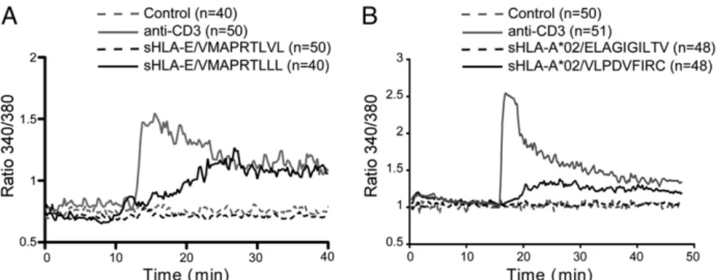
As shown in Fig. 1, we observed that incubation with antigenic sHLA-I/peptide monomers (10 mg/ml) triggered an increase in intracellular free calcium (Ca2+) concentration in both MART.22 (Fig. 1A) and H2 (Fig. 1B) clones. In contrast, no significant Ca2+ increase was detected with monomers refolded with irrelevant peptides (HLA-E*0101/VMAPRTLVL for MART.22 clone and HLA-A*0201/ELAGIGILTV for H2 clone). We next analyzed several activation and function markers after 6 h of incubation with anti-CD3 mAb or sHLA-I/peptide monomers (Fig. 2, Supplemental Fig. 2 for MART.22 T and H2 clones, respectively). T cells stim-ulated by antigenic soluble monomers displayed an activated phe-notype characterized by an increased expression of the early activation marker CD69 (Fig. 2A, Supplemental Fig. 2A). In ad-dition, we observed the induction of TNF-a production (48% and 87% of TNF-a–producing T cells for MART.22 and H2 clones, respectively) (Fig. 2B, Supplemental Fig. 2B) and CD107a surface mobilization reflecting T cell degranulation (Fig. 2C, Supplemental Fig. 2C). Finally, we showed that incubation of CD8 T cells with
The Journal of Immunology 5091
at Universite Nantes BU Sante on September 30, 2014
http://www.jimmunol.org/
soluble monomers induced significant T cell apoptosis (36% and 46% Annexin V+7AAD+for MART.22 and H2 clones, respectively) (Fig. 2D, Supplemental Fig. 2D). In agreement with Ca2+flux ex-periments, T cell activation was not observed with irrelevant monomers. Thus, these data indicated that sHLA-I monomers were able to induce Ag-specific activation of CD8 T cell clones.
Activation by soluble monomers relied on autopresentation of antigenic peptide by T cells
The data presented above could be interpreted as the result of productive engagement of TCR by sHLA-I/peptide monomers, but this hypothesis would disagree with the well-accepted multivalent engagement of receptor model. Thorough analysis of Ca2+ flux FIGURE 1. Induction of calcium mobilization upon activation of CD8 T cells with sHLA-I monomers. HLA-E– (A) and HLA-A2–restricted (B) T cells were stimulated by antigenic (HLA-E*0101/VMAPRTLLL and HLA-A*0201/VLPDFIRC, respectively) or irrelevant (HLA-E*0101/VMAPRTLVL and HLA-A*0201/ELAGIGILTV, respectively) monomers (0.2mM). Anti-CD3 stimulation (1 mg/ml) was used as a positive control. Graphs represent the kinetics of intracellular Ca2+levels (340/380 nm ratio). Values correspond to the mean of emission measured among all T cells present in the field. Results
are representative of two independent experiments.
FIGURE 2. CD8 T cell activation after stimulation by sHLA-E monomers. HLA-E–restricted T cells were stimulated by antigenic HLA-E*0101/VMAPRTLLL or irrelevant HLA-E*0101/VMAPRTLVL monomers (0.2mM). Anti-CD3 stimulation (1 mg/ml) was used as positive control. (A) Surface expression of CD69 activation marker (RFI). (B) TNF-a production. Data are expressed as mean percentage of T cells producing cytokine. (C) Surface expression of CD107a de-granulation marker (RFI). In histograms (A–C) specific mAbs are presented by solid lines and isotype controls by dotted lines. (D) Detection of apoptosis by Annexin VFITCand 7-AAD staining. Data are expressed as percentages of dead cells (Annexin V+). Results are representative of three independent experiments.
5092 CD8 T CELL ACTIVATION BY HLA-I/PEPTIDE MONOMERS
at Universite Nantes BU Sante on September 30, 2014
http://www.jimmunol.org/
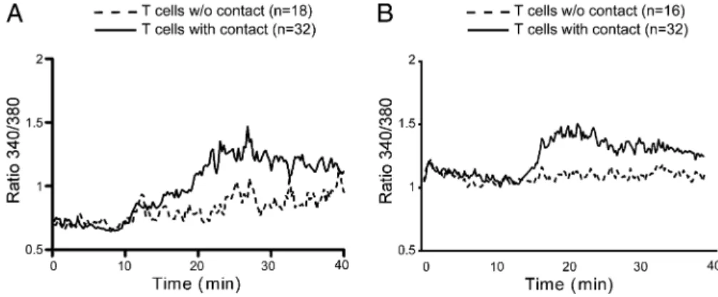
experiments suggested that T cell activation by soluble monomers required cellular contact because T cells in direct physical contact displayed higher calcium influx than did isolated T cells (Fig. 3). Thus, these results suggested that, rather than arising directly from monomeric engagement of the TCR, the underlying activa-tion mechanism is related to inducactiva-tion of Ag autopresentaactiva-tion by T cells, as previously suggested in rodent models (17, 31).
To document this proposed representation mechanism, we tested whether T cell clones preincubated with sHLA-I/peptide mono-mers (10mg/ml equivalent to 0.2 mM, for 1 h) can present the antigenic peptides on their surface and activate T cell clones that have not been directly exposed to the Ags. To differentiate “tar-get” (directly exposed to Ag) and “effector” cells (indirectly ex-posed to Ag), we previously stained the effector population with calcein acetoxymethyl ester (Fig. 4A). As illustrated in Fig. 4B and 4C, T cells preincubated with their antigenic peptide, both in a free or in HLA-complexed form, were able to elicit the pro-duction of TNF-a by effector T lymphocytes, thus suggesting that soluble monomer–induced activation depends on peptide repre-sentation and fratricide responses.
To further substantiate these data, we addressed the requirement of cognate HLA-I expression to mediate T cell activation. To this end, we assessed whether soluble monomers could sensitize target cells, expressing, or not, the restricting HLA-I molecule, for rec-ognition by CD8 T cells. We show that preincubation of T2 and
0.221 cells (HLA-E+), but not HeLa cells (HLA-E2), with HLA-E*0101/VMAPRTLLL monomers induced their recognition by MART.22 clone, monitored by lytic activity and TNF-a pro-duction (Fig. 5A, 5B). Similarly, T2 cells (HLA-A2+), but not 0.221 and HeLa cells (HLA-A22), preincubated with HLA-A*0201/VLPDFIRC monomers induced activation of H2 clone (Fig. 5C, 5D).
Altogether, these experiments support the hypothesis that CD8 T cell activation induced by sHLA-I/peptide monomers depends on the ability of monomer-derived peptides to load onto membrane-bound HLA-I molecules, thus allowing Ag autopresentation. Activation mechanism: a passive peptide transfer from monomers to T cell HLA-I molecules
The next step was to determine whether activation by sHLA-I/ peptide monomers depends on a passive peptide transfer, that is, direct loading of exogenous short peptides onto surface-bound HLA-I molecules, or on an active mechanism, which could in-volve internalization of HLA-I/peptide complexes, their deg-radation in intracellular compartments, followed by peptide processing and presentation by endogenous HLA molecules. Thus we reproduced the indirect T cell stimulation experiments de-scribed above (Fig. 4), with or without glutaraldehyde fixation of the target T cell population prior to incubation with monomers (Fig. 6). Control experiments were performed with short peptides to check FIGURE 3. CD8 T cell activation by
sHLA-I monomers relies on cellular contact. HLA-E– (A) and HLA-A2–restricted (B) T cells were stimulated by antigenic mono-mers (10mg/ml). Kinetic profiles of intra-cellular Ca2+were performed on T cells with or without contact. Values correspond to the mean of emission measured among all T cells present in the field. Results are rep-resentative of two independent experiments.
FIGURE 4. CD8 T cell activation by sHLA-I monomers relies on autopresentation of monomer-derived peptide. (A) Experimental design and pro-tocol. Calcein staining was used to differentiate target and effector CD8 T cells. Calcein2T cells were pulsed 1 h with free or HLA-complexed peptide (0.2mM), washed, and cocultured with calcein+ T cells. Activation of calcein+and calcein2HLA-E– (B) and HLA-A2–restricted (C) T cells was assessed by TNF-a staining. Results are representative of three independent experiments.
The Journal of Immunology 5093
at Universite Nantes BU Sante on September 30, 2014
http://www.jimmunol.org/
that fixation itself did not alter Ag presentation by T cells. As shown in Fig. 6, fixation of target T cells did not significantly alter their capacity to activate effector T cells, demonstrating that internali-zation was not involved in monomer-mediated activation. In support of these results, the use of endocytosis inhibitors, such as cyto-chalasin D and dimethyl amiloride, abolishing phagocytic and pinocytotic activities, respectively, had no effect on the efficiency of monomer-mediated activation (Supplemental Fig. 3 and data not shown). These results invalidate the hypothesis of an active mech-anism, supporting the assumption of an exogenous passive transfer of monomer-derived peptide toward cell-bound HLA-I molecules of T cells.
On the basis of these results, we generated single-chain trimers [(SCT); a single polypeptide chain with a linear composition of antigenic peptide,b2m, and H chain connected by flexible linkers]
(32, 33) to address whether covalent binding of peptide within HLA-I monomers could affect their ability to activate T cells. First, one must demonstrate that immobilized SCTs are able to stimulate T cell clones. We observed that immobilized HLA-A*0201/VLPDFIRC SCTs were able to activate, even modestly, H2 T cell clones (8% of TNF-a–producing cells), but no activation at all could be observed with soluble forms of SCT (Supplemental Fig. 4). Thus, these data suggest that T cell–induced activation is strictly dependent on the ability of monomer-derived peptide to FIGURE 5. CD8 T cell activation by sHLA-I monomers relies on cognate membrane HLA expression. Target cells (T2 [HLA-A2+, HLA-E+], 0.221
[HLA-A22, HLA-E+, and HeLa [HLA-A22, HLA-E2]) were pulsed with either HLA-E*0101/VMAPRTLLL or HLA-A*0201/VLPDFIRC monomers (0.2mM), washed, and cocultured with HLA-E– (A and B) and HLA-A2–restricted (C and D) T cells. Cytotoxicity was assessed by a chromium release assay (A and C) and TNF-a (B and D). Results are representative of three independent experiments.
5094 CD8 T CELL ACTIVATION BY HLA-I/PEPTIDE MONOMERS
at Universite Nantes BU Sante on September 30, 2014
http://www.jimmunol.org/
dissociate from HLA-Ia complexes. Unfortunately, we could not confirm this result for nonclassical HLA-E because the preas-sembled nature of these SCTs makes them ineffective to activate CD8 T cells, even immobilized.
To formally demonstrate that monomer-induced activation relied on a passive peptide transfer toward surface HLA-I molecules, we tested whether this mechanism could be inhibited by competitor peptides (i.e., nonantigenic peptides binding the same HLA-I molecules). As shown in Fig. 7, addition of E– and HLA-A2–binding competitor peptides induced a dose-dependent inhi-bition of monomer-induced T cell activation, whereas irrelevant peptides (i.e., HLA-B35–binding peptides) had no impact. These data demonstrated that stimulatory properties of soluble mono-mers result from passive peptide transfer onto T cell endogenous HLA-I molecules and can be prevented in the presence of high concentrations of competing peptides.
Finally, we evaluated the efficiency and kinetic of this passive peptide transfer. To this end, we performed titration assays com-paring T cell–induced activation upon direct incubation with soluble monomers or with the corresponding peptides at equivalent molar concentration. As shown in Fig. 8A and 8C, TNF-a production by T cells could be detected from 0.05mM of both soluble monomers, and free peptides indicated that they were equally efficient in in-ducing CD8 T cell activation. Kinetic experiments were conducted by stimulating T cell clones with T2 cells that have been loaded for various times (from 0 to 2 h) with soluble monomers or peptides. Again, the two kinetic profiles were very similar, with 15 min of monomer/peptide loading being sufficient to induce 50% of max-imal T cell response (Fig. 8B, 8D). These data clearly showed that sHLA-I/peptide monomers and peptides exhibited a similar ability to activate CD8 T cells.
Discussion
To elucidate the minimal requirements for T cell activation, pre-vious studies reported that peptide class I MHC monomers, in contrast to peptide class II monomers, were sufficient to induce
activation of T cells (12, 34, 35). Stimulatory capacity of HLA-I monomers was initially attributed to a productive TCR–CD8 heterodimerization, implying that CD8 T cell activation could arise from monomeric engagement. However, subsequent inves-tigations have suggested an alternative mechanism, enabling rec-onciliation with previous observations of the consensual model of multivalent engagement of receptors. Using the 2C and OT-1 TCR mouse models, these studies provide evidence that stimulatory capacities of monomers were strictly dependent on the CD8 T cell ability to express the cognate MHC-I molecule. Therefore, an unexpected mechanism involving a peptide transfer from mono-mers to T cell endogenous MHC molecules has been proposed, thus leading to Ag autopresentation (17, 31). Similarly, using both a classical (HLA-A*0201) model and a nonclassical (HLA-E) model, we observed that monomer-induced CD8 T cell activation required cellular contacts and that CD8 T cells that had been previously exposed to monomers acquired the ability to activate CD8 T cells of the same antigenic specificity. Hence, rather than arising from monomeric engagement of the TCR, CD8 T cell activation ap-parently stems from the T cell ability to present monomer-derived peptide to each other, allowing fratricide responses. In support of this mechanism, we documented that HLA-I monomers could also sensitize target cells for recognition by CD8 T cells, but only if the target cells expressed the appropriate HLA-I molecules.
The aforementioned process raised the question of how such a peptide transfer could occur. Previous studies suggested that mon-omers could be internalized into T cell endocytic compartments (possibly after binding to TCR and/or CD8 molecules), where monomer-derived peptides could be reloaded onto endogenous HLA-I molecules (17, 31). However, we showed that cell fixation or use of endocytosis inhibitors did not alter monomer-derived peptide transfer, excluding this possibility and thereupon suggesting that peptide is transferred exogenously. In support of these data, we uncovered evidence that monomer-derived peptide transfer could be efficiently inhibited in the presence of exogenous com-petitor peptides. Moreover, we observed, in the HLA-A*0201 FIGURE 6. No impact of cell fixation on CD8 T cell activation by sHLA-I monomers. HLA-E– (A) and HLA-A2–restricted (B) calcein+T cells were
stimulated by calcein2T cells that had previously been loaded with VMAPRTLLL and VLPDFIRC peptides or E*0101/VMAPRTLLL and HLA-A*0201/VLPDFIRC monomers at 0.2mM before or after glutaraldehyde fixation (0.05%, 30 s). Calcein+T cell activation was assessed by TNF-a staining.
Results are representative of two independent experiments.
The Journal of Immunology 5095
at Universite Nantes BU Sante on September 30, 2014
http://www.jimmunol.org/
model, that the covalent linking of peptides onto HLA-I mon-omers (referred to as SCTs) preclude their ability to activate CD8 T cells. Altogether, these observations strongly support a mechanism of passive external loading of monomer-derived peptide onto surface HLA-I molecules.
Consistent with previous observations (17, 31), we also docu-mented that the transfer of monomer-derived peptides was as ef-ficient and fast as loading of free peptides. This finding could be explained by quantitative dissociation of monomers and subse-quent peptide release into the medium. However, this is incom-patible with estimated HLA-I/peptide complex lifetime, as we could detect only minor dissociation of soluble monomers when incubated in assay conditions (Supplemental Fig. 1). Thus, rather, we hypothesize that the release of peptide from monomers prob-ably occurs close to the cell membrane, allowing an immediate reloading on surface HLA molecules.
Several lines of evidence suggest that sHLA-I molecules are immunologically functional and may play a significant role in vivo (9). In this regard, others and we (19, 22) previously documented in vitro that sHLA-E molecules can modulate the effector func-tions of NK and CD8 T cells expressing the cognate inhibitory NK receptor CD94/NKG2-A (19, 22). We performed this study to find out whether this behavior relies on the ability of sHLA-E to di-rectly interact with their receptors, as mostly suggested (15, 16), or on an indirect mechanism of peptide transfer, as described for classical MHC-I molecules in murine models (17, 31). In this article, we clearly demonstrated that sHLA-E molecules have the potential to exert immunoregulatory functions through their ability to provide peptides that can be transferred to cell-bound HLA-E molecules, hence raising the question of their potential involve-ment in the global immune response. Thus, we propose that prior observations made with CD94/NKG2-A+cells are more likely to stem from a similar mechanism of peptide transfer toward surface HLA-E molecules of targets or immune cells than from the direct triggering of NK receptors.
In conclusion, we reported that both classical and nonclassical HLA-I/peptide monomers can efficiently activate Ag-specific CD8 T cells in vitro, via a mechanism that involves the passive trans-fer of monomer-derived peptide to cell-bound HLA-I molecules. These findings support the peptide-representation theory previ-ously investigated in rodent models and provide a molecular basis for the capacity of sHLA-I molecules to modulate immune ef-fectors expressing cognate receptors and thus open up the possi-bility that this mechanism could occur in vivo.
FIGURE 7. Inhibition of CD8 T cell activation by sHLA-I monomers with competitor peptides. HLA-E– (A) and HLA-A2–restricted (B) T cells were stimulated in a 6-h assay with a fixed concentration of HLA-E*0101/ VMAPRTLLL or HLA-A*0201/VLPDFIRC monomers (0.2 mM) and various concentrations of HLA-E– (VMAPRTLVL) or HLA-A2–restricted (ELAGIGILTV) competitor peptides. Irrelevant HLA-B35–restricted pep-tide (LPFDFTPGY) has been used as negative control. T cell activation was assessed by TNF-a staining. Results are representative of two inde-pendent experiments.
FIGURE 8. Comparable efficiency and kinetics of sHLA-I monomers versus free peptide transfer activation of CD8 T cells. Response of HLA-E– (A) and HLA-A2– restricted (C) T cells to incubation with various concentrations (from 1029to 1026M) of monomers or peptides. Kinetics of activa-tion of HLA-E– (B) and HLA-A2–restricted (D) T cells in response to incubation with monomer- or peptide-pulsed T2 cells. In all experiments, T cell activation was assessed by TNF-a staining. Results are representative of two independent experiments.
5096 CD8 T CELL ACTIVATION BY HLA-I/PEPTIDE MONOMERS
at Universite Nantes BU Sante on September 30, 2014
http://www.jimmunol.org/
Acknowledgments
We thank the Cellular and Tissular Imaging Core Facility “MicroPICell,” the Cytometry Facility “CytoCell,” and the P2R Facility of the Federative Research Structure Franc¸ois Bonamy for technical and scientific assistance.
Disclosures
The authors have no financial conflicts of interest.
References
1. Ploegh, H. L., H. T. Orr, and J. L. Strominger. 1981. Major histocompatibility antigens: the human (HLA-A, -B, -C) and murine (H-2K, H-2D) class I mole-cules. Cell 24: 287–299.
2. O’Callaghan, C. A., and J. I. Bell. 1998. Structure and function of the human MHC class Ib molecules HLA-E, HLA-F and HLA-G. Immunol. Rev. 163: 129– 138.
3. Yaneva, R., C. Schneeweiss, M. Zacharias, and S. Springer. 2010. Peptide binding to MHC class I and II proteins: new avenues from new methods. Mol. Immunol. 47: 649–657.
4. Germain, R. N., and I. Stefanova´. 1999. The dynamics of T cell receptor sig-naling: complex orchestration and the key roles of tempo and cooperation. Annu. Rev. Immunol. 17: 467–522.
5. Moretta, A., R. Biassoni, C. Bottino, M. C. Mingari, and L. Moretta. 2000. Natural cytotoxicity receptors that trigger human NK-cell-mediated cytolysis. Immunol. Today 21: 228–234.
6. Campoli, M., and S. Ferrone. 2008. Tumor escape mechanisms: potential role of soluble HLA antigens and NK cells activating ligands. Tissue Antigens 72: 321– 334.
7. McDonald, J. C., and I. Adamashvili. 1998. Soluble HLA: a review of the lit-erature. Hum. Immunol. 59: 387–403.
8. Adamashvili, I., R. E. Kelley, T. Pressly, and J. C. McDonald. 2005. Soluble HLA: patterns of expression in normal subjects, autoimmune diseases, and transplant recipients. Rheumatol. Int. 25: 491–500.
9. Tabayoyong, W. B., and N. Zavazava. 2007. Soluble HLA revisited. Leuk. Res. 31: 121–125.
10. Bahri, R., F. Hirsch, A. Josse, N. Rouas-Freiss, N. Bidere, A. Vasquez, E. D. Carosella, B. Charpentier, and A. Durrbach. 2006. Soluble HLA-G inhibits cell cycle progression in human alloreactive T lymphocytes. J. Immunol. 176: 1331–1339.
11. Contini, P., M. Ghio, A. Poggi, G. Filaci, F. Indiveri, S. Ferrone, and F. Puppo. 2003. Soluble HLA-A,-B,-C and -G molecules induce apoptosis in T and NK CD8+ cells and inhibit cytotoxic T cell activity through CD8 ligation. Eur. J. Immunol. 33: 125–134.
12. Delon, J., C. Gre´goire, B. Malissen, S. Darche, F. Lemaıˆtre, P. Kourilsky, J. P. Abastado, and A. Trautmann. 1998. CD8 expression allows T cell signaling by monomeric peptide-MHC complexes. Immunity 9: 467–473.
13. Fournel, S., M. Aguerre-Girr, X. Huc, F. Lenfant, A. Alam, A. Toubert, A. Bensussan, and P. Le Bouteiller. 2000. Cutting edge: soluble HLA-G1 triggers CD95/CD95 ligand-mediated apoptosis in activated CD8+ cells by interacting with CD8. J. Immunol. 164: 6100–6104.
14. Naji, A., A. Durrbach, E. D. Carosella, and N. Rouas-Freiss. 2007. Soluble HLA-G and HLA-G1 expressing antigen-presenting cells inhibit T-cell allo-proliferation through ILT-2/ILT-4/FasL-mediated pathways. Hum. Immunol. 68: 233–239.
15. Spaggiari, G. M., P. Contini, R. Carosio, M. Arvigo, M. Ghio, D. Oddone, A. Dondero, M. R. Zocchi, F. Puppo, F. Indiveri, and A. Poggi. 2002. Soluble HLA class I molecules induce natural killer cell apoptosis through the engage-ment of CD8: evidence for a negative regulation exerted by members of the inhibitory receptor superfamily. Blood 99: 1706–1714.
16. Spaggiari, G. M., P. Contini, A. Dondero, R. Carosio, F. Puppo, F. Indiveri, M. R. Zocchi, and A. Poggi. 2002. Soluble HLA class I induces NK cell apo-ptosis upon the engagement of killer-activating HLA class I receptors through FasL-Fas interaction. Blood 100: 4098–4107.
17. Ge, Q., J. D. Stone, M. T. Thompson, J. R. Cochran, M. Rushe, H. N. Eisen, J. Chen, and L. J. Stern. 2002. Soluble peptide-MHC monomers cause activation of CD8+ T cells through transfer of the peptide to T cell MHC molecules. Proc. Natl. Acad. Sci. USA 99: 13729–13734.
18. Schott, E., N. Bertho, Q. Ge, M. M. Maurice, and H. L. Ploegh. 2002. Class I negative CD8 T cells reveal the confounding role of peptide-transfer onto CD8 T cells stimulated with soluble H2-Kb molecules. Proc. Natl. Acad. Sci. USA 99: 13735–13740.
19. Coupel, S., A. Moreau, M. Hamidou, V. Horejsi, J. P. Soulillou, and B. Charreau. 2007. Expression and release of soluble HLA-E is an immunoregulatory feature of endothelial cell activation. Blood 109: 2806–2814.
20. Allard, M., R. Oger, V. Vignard, J. M. Percier, G. Fregni, A. Pe´rier, A. Caignard, B. Charreau, K. Bernardeau, A. Khammari, et al. 2011. Serum soluble HLA-E in melanoma: a new potential immune-related marker in cancer. PLoS ONE 6: e21118.
21. Derre´, L., M. Corvaisier, B. Charreau, A. Moreau, E. Godefroy, A. Moreau-Aubry, F. Jotereau, and N. Gervois. 2006. Expression and release of HLA-E by melanoma cells and melanocytes: potential impact on the response of cytotoxic effector cells. J. Immunol. 177: 3100–3107.
22. Morandi, F., C. Venturi, R. Rizzo, M. Castellazzi, E. Baldi, M. L. Caniatti, M. R. Tola, E. Granieri, E. Fainardi, A. Uccelli, and V. Pistoia. 2013. Intrathecal soluble HLA-E correlates with disease activity in patients with multiple sclerosis and may cooperate with soluble HLA-G in the resolution of neuroinflammation. J. Neuroimmune Pharmacol. 8: 944–955.
23. Allard, M., P. Tonnerre, S. Nedellec, R. Oger, A. Morice, Y. Guilloux, E. Houssaint, B. Charreau, and N. Gervois. 2012. HLA-E-restricted cross-recognition of allogeneic endothelial cells by CMV-associated CD8 T cells: a potential risk factor following transplantation. PLoS ONE 7: e50951. 24. Gervois, N., N. Labarriere, S. Le Guiner, M. C. Pandolfino, J. F. Fonteneau,
Y. Guilloux, E. Diez, B. Dreno, and F. Jotereau. 2000. High avidity melanoma-reactive cytotoxic T lymphocytes are efficiently induced from peripheral blood lymphocytes on stimulation by peptide-pulsed melanoma cells. Clin. Cancer Res. 6: 1459–1467.
25. Guilloux, Y., S. Lucas, V. G. Brichard, A. Van Pel, C. Viret, E. De Plaen, F. Brasseur, B. Lethe´, F. Jotereau, and T. Boon. 1996. A peptide recognized by human cytolytic T lymphocytes on HLA-A2 melanomas is encoded by an intron sequence of the N-acetylglucosaminyltransferase V gene. J. Exp. Med. 183: 1173–1183.
26. Valmori, D., J. F. Fonteneau, C. M. Lizana, N. Gervois, D. Lie´nard, D. Rimoldi, V. Jongeneel, F. Jotereau, J. C. Cerottini, and P. Romero. 1998. Enhanced gen-eration of specific tumor-reactive CTL in vitro by selected Melan-A/MART-1 immunodominant peptide analogues. J. Immunol. 160: 1750–1758.
27. Cerboni, C., M. Mousavi-Jazi, H. Wakiguchi, E. Carbone, K. Ka¨rre, and K. So¨derstro¨m. 2001. Synergistic effect of IFN-gamma and human cytomega-lovirus protein UL40 in the HLA-E-dependent protection from NK cell-mediated cytotoxicity. Eur. J. Immunol. 31: 2926–2935.
28. Tomasec, P., V. M. Braud, C. Rickards, M. B. Powell, B. P. McSharry, S. Gadola, V. Cerundolo, L. K. Borysiewicz, A. J. McMichael, and G. W. Wilkinson. 2000. Surface expression of HLA-E, an inhibitor of natural killer cells, enhanced by human cytomegalovirus gpUL40. Science 287: 1031.
29. Takamiya, Y., C. Scho¨nbach, K. Nokihara, M. Yamaguchi, S. Ferrone, K. Kano, K. Egawa, and M. Takiguchi. 1994. HLA-B*3501-peptide interactions: role of anchor residues of peptides in their binding to HLA-B*3501 molecules. Int. Immunol. 6: 255–261.
30. Bodinier, M., M. A. Peyrat, C. Tournay, F. Davodeau, F. Romagne, M. Bonneville, and F. Lang. 2000. Efficient detection and immunomagnetic sorting of specific T cells using multimers of MHC class I and peptide with reduced CD8 binding. Nat. Med. 6: 707–710.
31. Schott, E., and H. L. Ploegh. 2002. Mouse MHC class I tetramers that are unable to bind to CD8 reveal the need for CD8 engagement in order to activate naive CD8 T cells. Eur. J. Immunol. 32: 3425–3434.
32. Garboczi, D. N., D. T. Hung, and D. C. Wiley. 1992. HLA-A2-peptide com-plexes: refolding and crystallization of molecules expressed in Escherichia coli and complexed with single antigenic peptides. Proc. Natl. Acad. Sci. USA 89: 3429–3433.
33. Hansen, T., Y. Y. Yu, and D. H. Fremont. 2009. Preparation of stable single-chain trimers engineered with peptide, beta2 microglobulin, and MHC heavy chain. Curr. Protoc. Immunol. 17: Unit 17.15.
34. Goldstein, J., H. Mostowsky, J. Tung, H. Hon, M. Brunswick, and S. Kozlowski. 1997. Naive alloreactive CD8 T cells are activated by purified major histocom-patibility complex class I and antigenic peptide. Eur. J. Immunol. 27: 871–878. 35. Sykulev, Y., M. Joo, I. Vturina, T. J. Tsomides, and H. N. Eisen. 1996. Evidence
that a single peptide-MHC complex on a target cell can elicit a cytolytic T cell response. Immunity 4: 565–571.
The Journal of Immunology 5097
at Universite Nantes BU Sante on September 30, 2014
http://www.jimmunol.org/