HAL Id: hal-02976870
https://hal.archives-ouvertes.fr/hal-02976870
Submitted on 23 Oct 2020
HAL is a multi-disciplinary open access
archive for the deposit and dissemination of
sci-entific research documents, whether they are
pub-lished or not. The documents may come from
teaching and research institutions in France or
abroad, or from public or private research centers.
L’archive ouverte pluridisciplinaire HAL, est
destinée au dépôt et à la diffusion de documents
scientifiques de niveau recherche, publiés ou non,
émanant des établissements d’enseignement et de
recherche français ou étrangers, des laboratoires
publics ou privés.
Structure, microstructure, optical and magnetic
properties of cobalt aluminate nanopowders obtained by
sol-gel process
Youssef El Jabbar, Hind Lakhlifi, Rachida El Ouatib, Lahcen Er-Rakho,
Sophie Guillemet, Bernard Durand
To cite this version:
Youssef El Jabbar, Hind Lakhlifi, Rachida El Ouatib, Lahcen Er-Rakho, Sophie Guillemet, et
al.. Structure, microstructure, optical and magnetic properties of cobalt aluminate nanopowders
obtained by sol-gel process.
Journal of Non-Crystalline Solids, Elsevier, 2020, 542, pp.120115.
Any correspondence concerning this service should be sent
to the repository administrator:
tech-oatao@listes-diff.inp-toulouse.fr
This is an author’s version published in:
http://oatao.univ-toulouse.fr/26799
To cite this version: El Jabbar, Youssef and Lakhlifi, Hind and
El Ouatib, Rachida and Er-Rakho, Lahcen and Guillemet,
Sophie
and Durand, Bernard
Structure, microstructure,
optical and magnetic properties of cobalt aluminate
nanopowders obtained by sol-gel process. (2020) Journal of
Non-Crystalline Solids, 542. 120115. ISSN 0022-3093
Official URL
DOI :
https://doi.org/10.1016/j.jnoncrysol.2020.120115
Open Archive Toulouse Archive Ouverte
OATAO is an open access repository that collects the work of Toulouse
researchers and makes it freely available over the web where possible
Structure, microstructure, optical and magnetic properties of cobalt
aluminate nanopowders obtained by sol-gel process
Y. El Jabbar
"
'
*
, H. Lakhlifi
"
, R. El Ouatib
"
, L. Er-Rakho
"
, S. Guillemet-Fritsch\ B. Durand
ba Laboratoire de Physico-chimie des Matériaux Inorganiques, Faculté des sciences Ain chock, Université Hassan II, Bb. 5366 Mâarif, Casablanca, Morocco b Institut Canwt CIRIMAT, CNRS Université de Toulouse, 118 route de Narbonne, 31062 Toul.ouse Cedex 9, France
ABSTRACT Keywords: Nanoparticles Morphology Spinel Ammphous Blue Complexing agent
The aim of this study was to detennine the effect of different complexing agents and of the annealing tem perature on the structural, morphological and optical properties of the synthesized precursors. Thus, cobalt aluminate nanoparticles were prepared by the sol-gel method using polyacrylic acid, glycine or citric acid as complexing agents. The synthesis performed at 500°C for 5 hours led to dark green powders composed of solid solutions denoted Co2+ [Agci-x)Co�;i]O4 and of amorphous alumina (39.8wt%). Calcination at temperatures above 900°C caused the powder colour to change from dark green to blue. A direct spinel structure Co2+ [Al�+] 04 is all the more achieved as the annealing temperature is high. The powder obtained using polyacrylic acid as the complexing agent at 900°C for 5 hours revealed the best morphology; it consisted of agglomerates of primary particles with a quasi- spherical shape and a size in the range 20-40 nm.
1. Introduction
Transition-metal aluminates witb general formula MAl2O4 (M: Ni,
Co, Fe) have attracted significant attention in recent decades because of tbeir fundamental and applied interest. The excellent physical proper ties of tbese materials offer many possibilities for use in different fields, such as coatings, pigmentation and photocatalysis [1--6].
Cobalt aluminate spinel(CoAl2O4) is known for its blue colour and it
is widely used as pigment.s for ceramics, paint.s, fibres and so on. Among its advantages, this compound presents high thermal and excellent chemical stability [7-10]. The spinel oxides powders are generally prepared by solid state reactions[ll,12]. This metbod has tbe ad vantage of being simple and inexpensive, but it requires long tbermal treatments at high temperatures (T~l300°C) [13,14]. Therefore, tbe
resulting powders exhibit a low specific surface area, witb morpholo gies tbat are most often heterogeneous. Recently, several research studies have focussed on tbe preparation of ultrafine CoAl,O4 powder
for its use in the decoration of ceramics by ink-jet printers, which re quires nanometric sizes to avoid clogging of tbe nozzles [15].
For tbe reasons described above, several soft-chemistry metbods have been studied to prepare tbese materials, namely, tbe hydrotbermal metbod [16], tbe co-precipitation route, the molten sait syntbesis [17],
tbe sol-gel process [18] and recently, tbe polymerisable complex metbod [19,20].This last metbod provides nano-sized oxide powders
• Corresponding author.
E-mail aMress: ysf.eljabbar@grnail.com (Y.E. Jabbar).
with high chemical and morphological homogeneities. This process uses a carboxylic acid as a chelating agent towards metal cations, which allows a mixed precursor to be obtained. The calcination of tbis latter leads to tbe formation of tbe mixed mdde at a relatively low tempera ture [21]. Severa! authors have shown that the morphological char acteristics of powders syntbesized by tbis process depend on tbe che mical nature of tbe chelating agent, tbe reaction pH, tbe calcination temperatureand so on [22-26].
The aim of this work is to prepare nanosized CoA12O4 powders using
sol-gel syntbesis witb different complexing agents (polyacrylic acid, citric acid and glycine) at pH 7 and with different calcination temperatures. Then, the effect of the synthesis parameters on the structural, morphological, optical and magnetic properties of CoAl2O4
nanoparticles is studied. The use of tbis powder as a blue pigment is discussed.
2. Materials and methods 2.1. Synthesis
Cobalt nitrate Co(NO3h•6H2O (99.0%, Aldrich) and aluminum ni
trate Al(NO3)s-9H2O (99.0%, Aldrich) were dissolved in water at a
molar ratio 1/2. Tuen an excess of one of tbe following complexing and gelling agents was added: polyacrylic acid (PM) at a concentration of
Y.E. Jabbar, et aL
100 g L -,, citric acid (CA) with molar ratio CA/cations=3 or glycine (Gly) with a molar ratio Gly /nitrate= 2/3. After adjusting the pH to 7 by adding an ammonium hydroxide solution under constant stirring, the solution was evaporated at 80"C until obtaining a viscous gel, that was dried at 120'C for 24 hours (precursor noted PRO). Then the or ganic malter was removed by a precalcination at 300'C for 12 hours under air and cobalt aluminate samples noted PRl, PR2 and PR3 were obtained by annealing for 5 hours at respectively 500, 900 and 11 00'C. 2.2. Characterization
The decomposition in air atmosphere of the pre-calcined powders (PRO) was investigated by differential thermal analysis and thermo gravimetric analysis using a simultaneous DTA/TGA analyzer (DTA-TG-60H Shimadzu); the heating rate was set at 2.5'C/min. The X-ray dif fraction patterns of the powders were recorded by means of a dif fractometer equipped with a detector Lynx Eye (Bruker D8 Advance, Â.cuKa = 1.5406 Â). The infrared spectra were taken using FfIR spec trometer (IR affinity-1S Shimadzu). The morphology and size of the powders were examined with a field emission scanning electronic mi croscope (JEOL JSM 6700F) and the specific surface area were de terrnined by the BET method (Micrometrics Flowsorb II 2300). The magnetic measurements were performed using a superconducting quantum interference magnetometer (SQUID, MPMS-XI). The absor bance spectra were determined by UV-Vis spectrophotometry (Perkin Elmer Lambda 35) and the colorimetric parameters (L*, a* and b*) were measured in the system (CIE) using a colorimeter (Chroma Meler CR-400/410, Konica Minolta).
3. Results and discussion 3.1. Thermal analysis
The thermal analysis curves of the PRO precursors prepared with different complexing and gelling agents and obtained after pyrolysis at 300'C for 12 hours are illustrated in Fig. 1. The TG curves of the PR0PAA and PR0my precursors (Figs.la and lb) showed a total weight Joss of about 15% and 12%, respectively, due to the elimination of the ad
sorbed water and the organic residues. The thermogram of the PR0CA
precursor presented a weight Joss of about 23%, and the OTA curve showed a strong exothermic peak around 415'C (Fig. le). This could be attributed to the combustion of organic matter [27]. An additional slight endothermic inflection at 920'C was observed for the three pre cursors decomposition; this phenomenon might be due to the reduction of trivalent cobalt.
3.2. Powder X-ray diffraction analysis
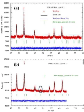
The XRD patterns of the samples prepared with PAA, CA and Gly as complexing agents and heat treated at different temperatures are given in Fig. 2. Those of PR0PAA and PR0my showed some small peaks, in dicating the beginning of crystallization, while the one of PR0CA showed an amorphous phase. The annealing at 500'C/5h of PRO pre cursors with different complexing and gelling agents led to dark green powders PRl. The XRD patterns of these powders showed several peaks at 26 values of 31.1 ', 36.8', 44.7', 55.T, 59.4', 65.3', 74.2'and 77.4'. These peaks could be indexed with space group Fd3m to (220), (311), (400), (422), (511), (440), (620) and (533) planes of a cubic unit cell. The Rietveld refinement pattern of the PRl sample obtained with PAA as complexing agent is given in Fig. 3. The corresponding data (Table 1) showed that cobalt ions were located both in the tetrahedral Ba and octaltedral 16d sites, whereas the aluminium ions predominantly oc cupied the octaltedral 16d sites. This could indicate a partial oxidation of Co2+ in Co3 +, giving the sample a greenish hue. Moreover, the structural properties (ce!! parameter, cell volume and density) deduced from the Rietveld refinement (Table 2) are interrnediate between those
0 ---
·,
50 ·-·- 15%o
_
1
,-..·-�
-._ <(r
10·-
,_
·-···-...
-50 O1
Exo -100 20(a)
1
Endo -150 100 200 300 400 500 600 700 800 900....
Temperature (°C)--'
12%�
r
·., 0�·-...
1023
···-�--,--,., <( ···-·-···----,_
F-- -50° 15 20 -100(b)
25 -150 100 200 300 400 500 600 700 800 900 Temperature (•C) 200 0··-
·-···--...
150 IO 23% 100_�
50 � <( 20 0 JO(c)
-50 '----c--�--,----�c--�--,--��-100 100 200 300 400 500 600 700 800 900 Temperalure ('C)Fig. 1. Thermal analysis (TGA-DTA) curves of dried gel precalcined at 300°C for 12h PROPAA (a), PROG!y (b) and PROAc (c).
of the direct spinel phases Co2+ [Co�+]O
4 and Co2+ [AJ�+]O4• It is then concluded that the PRl dark green powders are solid solutions between the two spinel phases, denoted Co2+ [Al�éi-,J Co:l:]O
4•
In these PRl powders, the unreacted aluminium is present, beside the crystallized spinel phase, under the forrn of amorphous alumina. In agreement with Yan et al [28], the internai standard method was in volved to quantify the amorphous phase in the samples. This method consists in using an exact quantity of well-crystallized a-Al2O3 powder to be mixed and milled with the studied sample. The calculation of the percentage of amorphous phase present in a powder was done using the Ful!Prof software by Rietveld refinement of diagram patterns. The percentages of the phases obtained by refinement were corrected with those of the prepared mixture. This allowed the rate of amorphous
phase to be deduced. The content in amorphous alumina of the PRPAA
sample is about 39.8 wt%.
Whatever the nature of the complexing agent, blue powders PR2 and PR3 were obtained by annealing the PRl powders at respectively 900'C/5h and 1100'C/5h. Their XRD patterns (Fig. 2) showed the same system of lines as the PRl samples but they were narrower and became
Y.E. Jabbar, et aL
(a)
s
1100° 500°C(b)
1100°c(c)
1100°c 00° 300°c 20 30e
;=-s
;;;
"'
é
::t.e
::t. 40 50 602 0(degree)
s
70 PRO PR3 Gly PRlcA PRO A 80Fig. 2. X-ray powder diffraction patterns of PRO, PRl, PR2 and PR3 samples. more intense, indicating higher crystallinity. Ali the diffraction peaks well matched with the standard JCPDS data for the direct spinel Co2+ [Al�+]O
4 (JCPDS file n° 70-0753). The appearance of the reflec
tion 331 at 49.3° (Figs. 2 and 3) is characteristic of the blue spinel
compound Co2+ [Al�+]O
4 [29]. The structural analysis by XRD re
vealed that no effect of the complexing agent was observed for the samples obtained at a annealing temperature equal or superior to 900°G. The structural refinement for PR2
PAA and PR3PAA samples
(Table 1) showed that the cobalt ions were located in tetrahedral sites and the aluminium ions occupied octahedral sites. This confirms a structure close to that of the direct spinel Co2+ [Al�+]O
4• The structural
properties (cell parameter, cell volume, density) also became doser to those of this spinel (Table 2). Simultaneously the amount of amorphous alumina decreased respectively to 8.3 and 2. 9 wto/o.
Consequently the transformation of the intermediate dark green solid solutions PR! obtained at 500°G in blue spinel phases close to
Co2+ [Al�+]O
4 can be explained, in agreement with Pacewska et al [30], by the incorporation of amorphous alumina according to Eq. (1):
Î 11800
�
'f 11000 ..:a.-"Î
10200 :: 9400 8600 7800 7000 6200 15000 -� 13000 -ÎIIOOOj
0000 7000"
3J 39 ?7 33 39 45 45 51 57 20{°) 63 51 57 63 20{".) 69 75 81 Il 69,.
81Fig. 3. Experimental, cakulated and difference signais for PR! (a) and PR2 (b) samples.
Table 1
Rietveld refinement for PRlPAA, PR2PAA and PR3PAA samples.
Sample Atom Wyckoff Occupancy Bisa Refinement Positions quality X=y=z parameters PRl (500°C/5h) Co 0.125 0.78 0.114(5) Rp-0.87% Al 0.125 0.08 0.114(5) Rwp=l.09% Co 0.5 0.51 0.207(3) :x:2=1.03 Al 0.5 0.43 0.207(3) 0 0.262(4) 1 0.502(6) PR2 (900'C/5h) Co 0.125 0.88 0.899(3) Rp=0.87% Al 0.125 0.02 0.899(3) Rwp=l.08% Al 0.5 0.84 0.642(7) X 2=1.05% Co 0.5 0.10 0.642(7) 0 0.263(3) 1 0.744(5) PR3 (ll00°C/5h) Co 0.125 0.90 0.468(4) Rp-0.95% Al 0.125 0.10 0.468(4) Rwp=l.20% Al 0.5 0.92 0.314(3) X 2=1.18% Co 0.5 0.06 0.314(3) 0 0.264(6) 0.268(5) Co2+[Al'+2c1-xJC03+
2xJO4
+
3xAl20, ➔ (1+
2x)Co2+[A/3+2JO4+
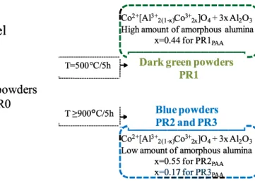
1/202(1) The transformation is ail the more complete as the annealing tem perature is high. The mechanism of amorphous alumina insertion during the calcination of the gels is illustrated in Fig. 4.
3. 3. FI'IR spectroscopy analysis
The inlrared spectra of the prepared samples are given in Fig. 5. The PR0PAA and PR0Gly spectra show two weak bands in the spectral range
Y.E. Jabbar, et aL
Table 2
Structural properties and amorphous phase ratio for PRl, PR2 and PR3 sample obtained by Rietveld method refinements.
Structural properties
Cell parameters (Â) 8.072 Volume (Â)3 525.95
Density (g/cm3) 6.08
Amorphous phase ratio (Internai standard method)
Samples PRl,AA (500"C/5h) 8.076(4) 526.68 5.74 39.8
The PRl spectrum shows two bands in the spectral range 500-700 cm-1 whatever the nature of the complexing agent. These bands could be attributed to the Co2+ [ AIM.-xl Co�:]0
4 solid solution. The width of the
peaks indicates the presence of amorphous phases in the sample [13]. When the thermal treatment raised-up 900"C (PR2 and PR3), a sup plementary band appeared at 501 cm -l and the two other bands be came more visible at 563 and 669 cm -l. These three bands are attri butable to the vibration modes of the CoAl204 phase [31]. These results
are in good agreement with XRD analysis. 3.4. Magnetic measurement
The mixed and direct spinel structures were characterized using magnetic measurements. The PRlpAA and PR2PAA powders were ana
lysed using a SQUID magnetometer in the temperature range 50-300K. lsothermal magnetization measurements ( o) as a function of the a pp lied field (Hl were performed at 50 and 300K. The evolutions of o= f (Hl
curves illustrated in Fig. 6a and b showed that the magnetization o is a linear fonction of the applied field, according to the following equation:
r;=x·H
(2)x: the magnetic susceptibility of the compound. H: the applied field (Oe).
Therefore, the PRlpAA and PR2PAA compounds exhibited a para
magnetic behaviour at 50 and 300K. A small deviation of the slope of o=f(H) was observed between PRlpAA and PR2PAA; this indicated a
significant influence of the calcination temperature on the magnetic properties of the samples studied.
Magnetic susceptibility measurements
x
were made under a con stant field of 10,000 Oe in the temperature range 50-300K, as presented in Fig. 7. The obtained curves evolutions were in good agreement with the Curie-Weiss law and could therefore be given by the following equation:Gel
X PR2,AA (900"C/5h) 8.092(4) 529.71 4.72 8.3 C T + 0p C: Curie constant. T: Temperature (K). PR3,AA (1100"C/5h) 8.097(1) 530.85 4.69 2.90p: Paramagnetic Curie temperature (K).
CoAbO4 JCPDS 70-0753
8.095 530.46 4.43
(3)
Based on the linear evolution of the inverse molar magnetic sus ceptibility, the effective magnetic moment could be extracted for dif ferent samples. The value obtained for PR2PAA was close to 4.65µ8• This
value characterizes Co2 + in a tetrahedral site with a spin-orbit inter
action (Lande g-factor [g] = 2.4 and a spin [S] = 3/2) [12]. This con
firmed the Rietveld refinements, which presented a direct spinel structure for the PR2PAA sample. However, the observed value for PRl
was 3.74 µ8 far from that of isolated Co2+ (µ.," = 4.65µ8). This sug
gested that a mixture of Co2 + and Co3 + existed in this compound
[32,33]. Table 3 shows the comparison of the experimental values of the effective magnetic moments with those theoretically obtained by applying the following equation [34]:
(4) n: percentage of the crystalline phase obtained by the internai standard method.
In Eq. (4), x and y are the fraction of cobalt Co2+ in tetrahedral sites
(Td) and the fraction of Co3+ in octahedral sites (Oh), respectively. The
theoretical values of µ,ff(Co};t) = 4.65µ8 and µ,ff(Cobtl = 0µ8 (Co3+
was considered in low spin). The effective magnetic moments calcu lated by Eq. ( 4) were close to those obtained experimentally; and confirmed the results obtained by Rietveld refinement analysis. 3.5. Colour characterization
The heat treatment (500 ,:; T,:; 11 OO"C) under air of the precursors
,---,
fco
2+[Al3
+2
(1-xJC0
3+ 2xJO
4+ 3xA1
2O
3:
:
High amount of amorphous alumina
:
:
x
=0.44 for PRlp
AA1
Pyrolysis
300
°C/12h
l
________
\
..,
.---�_,
Black powders
PRO
T=500°C/5h �
Dark green powders
'---!
PRl
T:::>:900
°C/5h �
Bluepowders
.__ ____ ,
t ,._ __________ � '
,
PR2 and PR3
_
1Co
2+[Al3
+2
(1-x)Co
3+2x]Ü
4+ 3xA1
2O
3:
:
Low amount of amorphous alumina
:
:
x
=0.55 for PR2
pAA1
'.., _ -- _
x
.,:c
Q
-
1
7
_f
,2
iJ'.!l1
�
---
_)
Fig. 4. Mechanism of alumina insertion.Y.E. Jabbar, et aL
(b)
1200 1100 1000 900 800 700
Wavenumber (cm-')
600 500
Fig. 5. Ff!R spectra of PRO, PRl, PR2 and PR3 samples prepapred by different complexing agents.
pre-calcined at 300°C for 12h (PRO black) led to different shades of
coloured powders. Indeed, the green colour was obtained at 500°C/5h
(PRl) while the blue colour was observed from 900°C/5h (PR2 and
PR3) (Fig. 8). The colorimetric and UV-Visible analyses were performed in order to correlate the optical properties with the chemical compo sition of the obtained powders. The PRl powder obtained at 500°C had
weak parameters a* and b* where the observed colour is of green shade. At temperatures above 900°C (PR2 and PR3), the parameter
(-b*) indicating the degree of blueing increased strongly so the blue shade was then observed. The colour shift from green to blue could be explained by the reduction of Co3 + to Co2 + with a change of co
ordination [35]. This blue color which represents CoAl,04 compound
was confirmed by three absorption bands in UV-Visible observed at 551, 590 and 628 nm (Fig. 9) which are due to the permissible spin
4 �2 E 1
�
§
0�
�-1 C: gi-2�
-3 4 ::ê,2 ::, E1 Q) 001-1
Q) C: �2�
-3 -30000 -20000 -10000 10000 20000 30000Applied magnetic field (Oe)
(b)
300K
-4
+-�-�-��-��-�-��-����-��
-30000 -20000 -10000 10000 20000 30000
Applied magnetic field (Oe)
Fig. 6. Isothermal magnetization versus applied magnetic field for PRl and PR2 samples. 0,016
i
0,012 E!�
0,008 0.004 50 100 150 200 250 T(K)Fig. 7. Magnetic susceptibility for PRl and PR2 samples. 50 300
Y.E. Jabbar, et aL Table 3
Effective magnetic moments of samples PRlpAA and PR2PAA. Samples 3.59 4.52 14,ffi.experimental)(µB) 3.74 4.58
transitions of 3d electrons of Co2+ ions in tetrahedral coordination
[4A2 (F) - 4Tl (P)] which are responsible for the blue colouring [36]. 3. 6. Microstructure characterizations
The FE-SEM micrographs of the Co2+ [Al�ci-xiCO�,i]Ü
4 blue powders
(PR2 and PR3) prepared with different complexing agents are presented in Fig. 10. The images of PR2PAA (Fig. 10a) revealed ultrafine powders
with quasi-spherical shapes and a size of 20-40 nm. In PR3PAA, the
particle sizes grew with increasing calcination temperatures, as shown in Fig. 10b. The powders, obtained at 900"C for 5 hours and synthesized using citric acid as the complexing agent (PR2c,J, showed agglomerates of variable shapes and the particle diameter was in the range 15-40 nm (Fig. 10c). The increase of the annealing temperature (ll00"C for 5 hours) favoured the densification of the powder (PR3c,J, which in dicated a pre-sintering. Grain growth was also observed (Fig. 10d). In contras!, in the powders obtained using glycine as the complexing agent (PR2Giy), the powders were formed by porous agglomerates consisting
of very fine particles that were difficult to individualize (Fig. lOe). The increasing of the annealing temperature created compact agglomerates, indicating an early pre-sintering (Fig. lOf). These powders had the particularity of being porous, which is due to the sudden departure of gases produced during the combustion of the precursors.
Surface area measurements (BET) were carried out for the different samples. As shown in Table 4, the specific surface area decreased with the annealing temperature and reached a high value (34 m2g-1) for the
PR2PAA powders.
L*=30.37
a*= -1.57
b*= 1.18
L*= 38.15
a*= -2.04
b*= -22.09
628nm 5 0 5 0 6 0 6 0 7 0 7 0 Wavelength (nm) Fig. 9. PR2PAA and PR3PAA UV-Visible spectra. 4. ConclusionCobalt aluminates nano powders were prepared through sol-gel method. Further heat treatment performed at 500"C for 5 hours led to green powders. The composition was a mixed valence spinel structure denoted Co2+ [AlM.-xJ Co�]O
4 with a 39.8% Wto/o amorphous phase.
Calcinations at temperatures above 900°C created blue powders with a
direct spinel structure (CoA'2O4). The colour shift from green to blue
was explained by the reduction of Co3+ to Co2+ with a change of co
ordination. The influence of the complexing agent on the texture of the elaborated powders was remarkable. Indeed, the powder prepared using polyacrylic acid as the complexing agent and obtained at 900"C for 5 hours (PR2PAAl shows homogenous morphology; it consists in
agglomerates of primary particles of quasi-spherical shape with a size in the range 20-40 nm.
1100
°C/5h
L*=40.84
a*= 12.2
b*= -37.43
Y.E. Jabbar, et aL
PR2 (900
°C)
PR3 (1100
°C)
PAA
CA
Gly
Fig. 10. SEM micrographs of samples obtained at 900°C (a,c,e) and 1100°C (b,d,D with different complexing agents. Table 4
BET surface area for PR2 and PR3 as a function of the complexing agent.
Sample/Complexing agent PR2
PR3
Surface specific area (m2/g)
PAA Citric acid 34.26
14.63 31.63 15.55
CRediT authorship contribution statement
Glycine 26.39 10.42
Y. El Jabbar: Conceptualization, Writing - original draft. H. Lakhlifi: Software, Formai analysis. R. El Ouatib: Supervision, Project administration, Validation. L. Er-Rakho: Methodology. S. Guillemet Fritsch: Resources. B. Durand: Investigation, Visualization, Supervision, Data curation.
Declaration of Competing Interest Ail authors declare no conflict of interests. References
[1] R.M. Bouclt, A.M. Anderson, C. Prasad, M.E. Hagerman, M.K. Carroll, Cobalt-alu mina sol gels: effects of heat treatment on structure and catalytic ability, J. Non Cryst. Solids 453 (2016) 94-102, https://doi.org/10.1016/j.jnoncrysol.2016.09. 013.
[2] P.M.T. Cavalcante, M. Dondi, G. Guarini, M. Raimondo, G. Baldi, Colour perfor mance of ceramic nana-pigments, Dyes Pigments 80 (2009) 226-232, https://doi. org/10.1016/j.dyepig.2008.07.004.
[3] L.S. Loba, A.R. Kumar, Structural and electrical properties of ZnCo2O4 spinel syn
thesized by sol-gel combustion method, J. Non-Cryst. Solids 505 (2019) 301-309,
https:/ / doi.org/10.1016/j .jnoncrysol. 2018.11.004.
[4] M. Mq.czka, M. Ptak, M. Kurnatowska, J. Hanuza, Synthesis, phonon and optical properties ofnanosized CoCr2O4, Mater. Chem. Phys. 138 (2013) 682-688, https:// doi.org/10.1016/j.matchemphys.2012.12.039.
[5] Y. Tong, H. Zhang, S. Wang, z. Chen, B. Bian, Highly Disperse<i re-doped CoA1O4 nanopigments: synthesis and chromatic properties, J. Nanomater (2016) (2016) 1-7, https:/ /doi.org/10.1155/2016/4169673.
Y.E. Jabbar, et aL
[6] Y. El Jabbar, M. ElHafdi, M. Benchikhi, R. El Ouatib, L. Er-Rakho, A. Essadki, Photocatalytic degradation of navy blue textile dye by nanoscale cobalt aluminate prepared by polymeric precursor method, Environ. Nanotechnol. Monit. Manag. 12
(2019) 100259, , https://doi.org/10.1016/j.enmm.2019.100259.
[7] N.M. Deraz, M.M. Foud.a, structural Synthesis, morphological properties of cobalt aluminum nano-composite, Int. J. Electrochem. Sei. 8 (2013) 2756--2767. [8] J. Gilabert, M.P. Gômez-Tena, V. Sanz, S. Mestre, Effect of secondary thermal
treatment on crystallinity of spinel-type Co(Cr,Alh 04 pigments synthesized by solution combustion route, J. Non-Cryst. Solids (2018), https://doi.org/10.1016/j. jnoncrysol.2018.02.026.
[9] Y. Song, Y.L. Zheng, Y.F. Tang, H.B. Yang, Fabrication and stability of CoA12O4
ceramic pigment for 3D printing, Mater. Sei. Forum 898 (2017) 1935-1939,
https:/ /doi.org/l0.4028/www.scientific.neVMSF.898.1935.
[10] Q. Wang, Q. Chang, Y. Wang, X. Wang, J. Zhou, Ultrafine CoA12O4 ceramic pigment
prepared by Pechini-sacrificial agent method, Mater. Lett. 173 (2016) 64-67,
https://doi.org/10.1016/j.matleL2016.03.014.
[11] N. Srisawad, W. Chaitree, O. Mekasuwandumrong, P. Praserthdam, J. Panpranot, Formation of CoA12O4 nanoparticles via low-temperature solid-state reaction of fine
gibbsite and cobalt precursor, J. Nanomater 2012 (2012) 1--8, https://doi.org/10. l 155/2012/108369.
[12] B. Roy, A. Pandey, Q. Zhang, T.W. Heinnann, D. Vaknin, D.C. Johnston,
Y. Furukawa, Experimental evidence of a collinear antiferromagnetic ordering in the frustrated CoA12O4 spinel, Phys. Rev. B 88 (2013) 174415, , https://doi.org/10.
l 103/PhysRevB.88.174415.
[13] N. Ouahdi, S. Guillemet, J.J. Demai, B. Durand, L. Er Rakho, R. Moussa, A. Samdi, Investigation of the reactivity of A1Cl3 and CoCl2 toward molten alkali-metal ni
trates in order to synthesize CoA12O4, Mater. Lett. 59 (2005) 334--340, https://doi.
org/10.1016/j.matlet.2004.10.013.
[14] F. Yu, J. Yang, J. Ma, J. Du, Y. Zhou, Preparation of nanosized CoAl2O4 powders by
sol-gel and sol-gel-hydrothermal methods, J. Alloys Compd. 468 (2009) 443--446,
https://doi.org/10.1016/j.jallcom.2008.01.018.
[15] Z. Pan, Y. Wang, H. Huang, Z. Ling, Y. Dai, S. Ke, Recent development on pre paration of ceramic inks in ink-jet printing, Ceram. Int. 41 (2015) 12515-12528,
https://doi.org/10.1016/j.ceramint.2015.06.124.
[16] J.-H. Kim, B.-R. Son, D.-H. Yoon, K.-T. Hwang, H.-G. Noh, W.-S. Cho, U.-S. Kim, Characteriza.tion of blue CoA12O4 nana-pigment synthesized by ultrasonic hydro
thennal method, Ceram. Int. 38 (2012) 5707-5712, https://doi.org/10.1016/j. ceramint.2012.04.015.
[17] N. Ouahdi, S. Guillemet, B. Durand, R. El Ouatib, L. Er-Rakho, R. Moussa, A. Samdi,
Synthesis of CoA12O4 by double decomposition reaction between LlA1O2 and molten
KCoC13, J. Eur. Ceram. Soc. 28 (2008) 1987-1994, https://doi.org/10.1016/j.
jeurceramsoc.2007.12.035.
[18] N. Bayal, P. Jeevanandam, Synthesis of metal aluminate nanoparticles by sol-gel method and studies on their reactivity, J. Alloys Compd. 516 (2012) 27-32, https:// doi.org/10.1016/j.jallcom.2011.11.080.
[19] M. Jafari, S.A. Hassanzadeh-Tabrizi, M. Ghashang, R. Poum.ajaf, Characterization of Ba2+ -added alumina/cobalt nanoceramic pigment prepared by polyacrylamide gel
method, Ceram. Int. 40 (2014) 11877-11881, https://doi.org/10.1016/j.ceramint. 2014.04.022.
[20] M. Jafari, S.A. Hassanzadeh-Tabrizi, Preparation of CoA12O4 nanoblue pigment via
polyacrylamide gel method, Powder Technol. 266 (2014) 236--239, https://doi. org/10.1016/j.powtec.2014.06.018.
[21] L. Gama, M.A. Ribeiro, B.S. Barras, R.H.A. Kiminami, I.T. Weber, A.C.F.M. Costa, Synthesis and characterization of the NiAl2O4, CoA12O4 and ZnA12O4 spinels by the
Journal of Non-Ctysta.Uine Solids 542 (2020) 120115 polymeric precursors method, J_ Alloys Compd. 483 (2009) 453--455, https:/ /doi.
org/10.1016/j.jallcom.2008.08.111.
[22] Y. El Jabbar, R. El Ouatib, L. Er-Rakho, B. Durand, Influence of temperature and pH on the morphology and the color of the CoAI2O4 prepared by sol gel method, J. Mater. Env. Sei. 6 (2015) 3452-3456.
[23] Y.F. Gomes, P.N. Medeiros, M.R.D. Bomio, I.M.G. Santos, C.A. Paskocimas, R.M. Nascimento, F.V. Motta, Optimizing the synthesis of cobalt aluminate pigment using fractional factorial design, Ceram. Int. 41 (2015) 699-706, https:/ /doi.org/ 10.1016/j.ceramint.2014.08.125.
[24] M.C.G. Merino, A.L. Estrella, M.E. Rodriguez, L. Acufia, M.S. Lassa, G.E. La.scalea, P. Vâzquez, Combustion syntheses of CoA12O4 powders using different fuels,
Procedia Mater. Sei. 8 (2015) 519-525, https://doi.org/10.1016/j.mspro.2015.04. 104.
[25] S. Salem, Effect of calcination temperature on colorant behavior of cobalt-aluminate nano-particles synthesized by combustion technique, J. Ind. Eng. Chem. 20 (2014) 818-823, https://doi.org/10.1 0l 6/j.jiec.2013.06.011.
[26] L. Torkian, M. Daghighi, Effects of �-alanine on morphology and optical properties of CoAl2O4 nanopowders as a blue pigment, Adv. Powder Technol. 25 (2014)
739-744, https://doi.org/10.1016/j.apt.2013.11.003 ..
[27] S.R. Prim, A. Garcia, R. Galindo, S. Cerro, M. Llusar, M.V. Folgueras, G. Monr6s, Pink ceramic pigments based on chromium doped M(Al2_I Crx)O4, M=Mg, Zn,
normal spinel, Ceram. Int 39 (2013) 6981--6989, https://doi.org/10.1016/j. ceramint.2013.02.035.
[28] K. Yan, Y. Guo, Z. Ma, Z. Zhao, F. Cheng, Quantitative analysis of crystalline and amorphous phases in pulverized coal fly ash based on the Rietveld method, J. Non Cryst. Solids. 483 (2018) 37-42.
[29] AM. Wahba, N.G. Imam, M.B. Mohamed, Flower-like morphology of blue and greenish-gray ZnCoIA12_x 04 nanopigments, J. Mol. Struct. 1105 (2016) 61-69,
https://doi.org/10.1016/j.molstruc.2015.10.052.
[30] B. Pacewska, M. Keshr, Thermal transformations of aluminium nitrate hydrate, Thennochim. Acta 385 (2002) 73--80, https://doi.org/10.1016/S0040-6031(01) 00703-1.
[31] X. Duan, M. Pan, F. Yu, D. Yuan, Synthesis, structure and optical properties of CoA12O4 spinel nanocrystals, J. Alloys Compd. 509 (2011) 1079-1083, https://doi.
org/10.1016/j.jallcom.2010.09.199.
[32] M. Gabrovska, n. Stanica, d. Crisan, d. Nikolova, l. Bilyarska, M. Crisan, R. Edreva kardjieva, Co-Al layered double hydroxides as precursors of ceramic pigment CoA12O4. Part II: magnetic and tint properties, Rev. Roum. Chim. 59 (2014) 451-455.
[33] L. Markov, K. Petrov, V. Petrov, On the thermal decomposition of some cobalt
hydroxide nitrates, Thennochim. Acta 106 (1986) 283--292, https://doi.org/10. 1016/0040-6031(86)85140-1.
[34] M. Taguchi, T. Nakane, K. Hashi, S. Ohki, T. Shimizu, Y. Sakka, A. Matsushita,
H. Abe, T. Funazukuri, T. Naka, Reaction temperature variations on the crystal lographic state of spinel cobalt aluminate, Dalton Trans. 42 (2013) 7167, https:// doi.org/10.1039/c3dt32828g.
[35] S. Kurajica, J. Popovié, E. Tkalèec, B. Gl'Zeta, V. Mandié, The effect of annealing
temperature on the structure and optical properties of sol-gel derived nanocrys talline cobalt aluminate spinel, Mater. Chem. Phys. 135 (2012) 587-593, https:// doi.org/10.1016/j.matchemphys.2012.05.030.
[36] I.S. Ahmed, A simple route to synthesis and characterization of CoA12O4 nano
crystalline via combustion method using egg white (ovalbumine) as a new fuel, Mater. Res. Bull. 46 (2011) 2548-2553, https:/ /doi.org/10.1016/j.materresbull. 2011.08.005.