HAL Id: hal-02314403
https://hal.archives-ouvertes.fr/hal-02314403
Submitted on 12 Oct 2019HAL is a multi-disciplinary open access archive for the deposit and dissemination of sci-entific research documents, whether they are pub-lished or not. The documents may come from teaching and research institutions in France or abroad, or from public or private research centers.
L’archive ouverte pluridisciplinaire HAL, est destinée au dépôt et à la diffusion de documents scientifiques de niveau recherche, publiés ou non, émanant des établissements d’enseignement et de recherche français ou étrangers, des laboratoires publics ou privés.
Giant hybrid polymer/lipid vesicles (Chapter 27)
Thi Phuong Tuyen Dao, K. Ferji, Fabio Fernandes, Manuel Prieto, Sébastien
Lecommandoux, Emmanuel Ibarboure, Olivier Sandre, Jean-François Le
Meins
To cite this version:
Thi Phuong Tuyen Dao, K. Ferji, Fabio Fernandes, Manuel Prieto, Sébastien Lecommandoux, et al.. Giant hybrid polymer/lipid vesicles (Chapter 27). Rumiana Dimova; Carlos Marques. The Giant Vesicle Book, CRC Press - Taylor and Francis group, pp.543-561, 2019, 9781498752176. �10.1201/9781315152516.ch27�. �hal-02314403�
Author manuscript version of Chapter 27 published In book: The Giant Vesicle Book,
Rumiana Dimova & Carlos Marques Eds. (CRC PRESS Taylor and Francis group, Oct. 7th 2019)
1
GIANT HYBRID POLYMER/LIPID VESICLES
Thi Phuong Tuyen Dao1,2,3, Khalid Ferji1,2, Fabio Fernandes3, Manuel Prieto3, Sébastien Lecommandoux1,2, Emmanuel Ibarboure1,2, Olivier Sandre1,2, Jean-François Le Meins1,2
1. Univ. Bordeaux, Laboratoire de Chimie des Polymères Organiques, UMR5629 ENSCBP 16 avenue Pey Berland 33607 Pessac cedex, France
2. CNRS, LCPO UMR5629, 33607 Pessac cedex, France
3. Centro de Quimica-Fisica Molecular, Complexo Interdisciplinar, IST Universidade de Lisboa, Av. Rovisco Pais 1049-001 Lisbon, Portugal
Contact Author Email: [email protected]
Motto:
A compromise is the art of dividing a cake in such a way that everyone believes he has the biggest piece.
Ludwig Erhard
Index words:
Amphiphilic copolymer Cell membrane model Domain formation Drug delivery Hybrid membrane Hybrid vesicle Hydrophobic mismatch Line tension Lipid segregation Liposome Membrane properties Membrane structures Membrane tension Microdomain Multicomponent membrane Nanodomain Phase coexistence Phase separation Phospholipid Polymersome
Author manuscript version of Chapter 27 published In book: The Giant Vesicle Book,
Rumiana Dimova & Carlos Marques Eds. (CRC PRESS Taylor and Francis group, Oct. 7th 2019)
2
List of abbreviations:
AFM Atomic-force microscopy
Biotinyl DSPE 1,2-distearoyl-sn-glycero-3-phosphoethanolamine- N-[biotinyl(poly(ethylene glycol))-2000
Chol Cholesterol
Ch-PEG-b-hbPG Cholesteryl-poly(ethylene oxide)-b- poly(glycerol) DC13PC 2-Ditridecanoyl-sn-glycerophosphocholine
DLPC 1,2-dilauroyl-sn-glycero-3-phosphocholine DMPC 1,2-dimyristoyl-sn-glycero-3-phosphocholine DOPC 1,2-dioleoyl-sn-glycero-3-phosphocholine DPPC
DDS 1,2-di-palmitoyl-sn-glycero-3-phosphocholine Drug delivery system
Egg Liss Rhod PE L-α- Egg-phosphatidylethanolamine-N-(Lissamine Rhodamine B-Sulfonyl) hLUVs
FITC Hybrid large unilamellar vesicles Fluorescein isothiocyanate
FRAP Fluorescence recovery after photobleaching FRET Fluorescence resonance energy transfer hGUVs Hybrid giant unilamellar vesicles
HSPC L-α-phosphatidylcholine, hydrogenated (Soy)
Liss-Rhod PE 1,2-dioleoyl-sn-glycero-3-phosphoethanolamine-N-(lissamine rhodamine B sulfonyl)
NBD nitrobenzoxadiazole
PBd Poly(butadiene)
PBd-b-PEO Polybutadiene-b-poly(ethylene oxide)
PC Egg-phosphatidylcholine
PDMS Poly(dimethyl siloxane)
PDMS-b-PMOXA Poly(dimethylsiloxane)-block-poly(methyloxazoline) PDMS-g-PEO Poly(dimethylsiloxane)-graft-poly(ethylene oxide)
PE Egg-phosphatidylethanolamine
PEO Poly(ethylene oxide)
PIB Poly(isobutylene)
PIB-b-PEO Poly(isobutylene)-b-poly(ethylene oxide)
PMOXA Poly(2-methyloxazoline)
PMOXA-b-PDMS-b-PMOXA Poly(methyloxazoline)-block-poly(dimethylsiloxane)-block- poly(methyloxazoline) POPC 1-palmitoyl-2-oleoyl-sn-glycero-3-phosphocholine
ROI Region of interest
RT Room temperature
Rh-DHPE 1,2-dihexadecanoyl-sn-glycero-3-phosphothanolamine-N-(lissamine rhodamine B sulfonyl)
SLS Static light scattering
Author manuscript version of Chapter 27 published In book: The Giant Vesicle Book,
Rumiana Dimova & Carlos Marques Eds. (CRC PRESS Taylor and Francis group, Oct. 7th 2019)
3
Glossary of symbols:
δ Hildebrand solubility parameter
KA Area compressibility modulus
κ Bending rigidity of the membrane
c0 spontaneous curvature
lme Hydrophobicmembrane thickness
Tg Glass transition temperature
Tm Melting temperature
Author manuscript version of Chapter 27 published In book: The Giant Vesicle Book,
Rumiana Dimova & Carlos Marques Eds. (CRC PRESS Taylor and Francis group, Oct. 7th 2019)
4
Outline:
1. INTRODUCTORY WORDS ... 5
2. CRITERIA TO BE FULFILLED TO OBTAIN HYBRID GIANT VESICLES ... 7
2.1. A BRIEF OVERVIEW OF MULTICOMPONENT LIPID VESICLES ... 7
2.2. THE EFFECT OF KEY MOLECULAR PARAMETERS: LIPID FLUIDITY, CHEMICAL INCOMPATIBILITY AND HYDROPHOBIC MISMATCH ... 10
2.3. THE EFFECT OF THE CHEMICAL MODIFICATIONS AND ADDITIVES ... 21
2.4. THE EFFECT OF MACROSCOPIC PARAMETERS: TEMPERATURE, MEMBRANE TENSION ... 23
3. SPECIFIC ASPECTS OF THE FORMATION OF GIANT HYBRID UNILAMELLAR VESICLES ... 26
4. UNDERSTANDING MEMBRANE PROPERTIES FROM MEMBRANE STRUCTURE ... 32
4.1. MECHANICAL PROPERTIES, FLUIDITY ... 33
4.2. PERMEABILITY ... 44
4.3. SPECIFIC PROPERTIES, BIOFUNCTIONALITY ... 47
5. CONCLUSION AND PERSPECTIVES ... 48
Author manuscript version of Chapter 27 published In book: The Giant Vesicle Book,
Rumiana Dimova & Carlos Marques Eds. (CRC PRESS Taylor and Francis group, Oct. 7th 2019)
5
1. INTRODUCTION
About 15 years ago, vesicles resulting from the self-assembly of amphiphilic copolymers referred to as “polymersomes” emerged as a potential alternative to liposomes for fundamental research and applications, such as cell membrane models, nano/micro
-reactors or drug delivery systems (DDS). (Discher, Won et al. 1999; Discher, Hammer et al. 2000; Discher and Ahmed 2006; LoPresti, Lomas et al. 2009; Le Meins, Sandre et al. 2011; Liao, Wang et al. 2012; Thevenot, Oliveira et al. 2013). Their major characteristics are described and discussed in Chapter 26, to be consulted by readers who are non-specialist in the field before going through the present chapter. This one deals with hybrid, i.e. intimately mixed polymer/lipid, vesicles that can be viewed as advanced vesicular structures as compared to their liposome and polymersome forerunners, as they potentially marry in a single membrane the best characteristics of the two separate components. Ideally, these structures could present biocompatibility and bio-functionality of liposomes, as well as robustness, low permeability and functional variability conferred by the copolymer chains. This should be of great interest in pharmaceutical applications for which only a few formulations based on liposomes are commercially available despite decades of research, (e.g. DaunoXome®,Doxil®/Caelyx®, Visudyne®), but also in personal care. In particular such moderate use of liposomes in clinics could be due to their lack of mechanical stability in the high shear rate of blood circulation through tiny vessels. Liposomal DDS can also often exhibit un-controlled leakage phenomena (seen as a “burst release” effect on their pharmacokinetic profiles). As a consequence, the controlled release of encapsulated molecules at the pre-determined biological target (e.g. a tumor site) remains a difficult
Author manuscript version of Chapter 27 published In book: The Giant Vesicle Book,
Rumiana Dimova & Carlos Marques Eds. (CRC PRESS Taylor and Francis group, Oct. 7th 2019)
6 challenge. Besides the obvious interest of the association of lipids and amphiphilic copolymers into a single membrane of LUVs for biomedical applications, hybrid giant unilamellar vesicles (hGUVs) can also be an excellent tool to get more insight into molecular and macroscopic parameters that govern the membrane domain formation, fusion or fission. Literature on the subject is still relatively limited (Le Meins, Schatz et al. 2013; Schulz and Binder 2015) although the scientific output is increasing with growing interest from different scientific communities (biophysicists, biologists, physico-chemists). Promising results have been obtained regarding their drug targeting ability and biomolecular recognition properties (Cheng, Elias et al. 2011; Schulz, Werner et al. 2013).
To date, the physical and molecular factors governing the phase separation in these hybrid copolymer/lipid membranes are only partially understood. In addition to the expected chemical incompatibility between copolymer block chains and phospholipids, one also has to consider the respective dimensions of the molecules as well as those of the corresponding bilayers. In order to perfectly benefit from the potential of such systems, the membrane structure must be tuned either towards homogeneous mixing of the molecular components, or on the contrary towards lateral phase separation leading to the presence of nano/micrometric domains. The relationship between membrane structure and physical and bio-functional properties must then be better understood in order to eventually optimize and validate the use of hybrid vesicles in future applications like drug delivery, tumor targeting, bio-recognition or bio-adhesion.
In the present chapter, an overview of hybrid copolymer/phospholipid vesicles will be given with a particular emphasis on hGUVs. The molecular and macroscopic parameters
Author manuscript version of Chapter 27 published In book: The Giant Vesicle Book,
Rumiana Dimova & Carlos Marques Eds. (CRC PRESS Taylor and Francis group, Oct. 7th 2019)
7 necessary to obtain stable hybrid vesicles with different membrane structural levels (homogenous distribution of the components, nano-domain or micro-domain formation) will be first summarized, followed by a description of the different preparation methods used to obtain hGUVs. It has to be noted that hybrid vesicles reported over the last ten years were exclusively prepared by a one-step process by which a film composed of the desired amount of copolymer and phospholipid is hydrated. We will not describe previous approaches based on the modulation of lipid membrane properties by adsorption of amphiphilic polymers onto preformed giant liposomes. Although such a method leads in some cases to a reorganization of the lipid membrane and to the induction of polymer-rich domains – see for instance (Ladavière, Tribet et al. 2002; Tribet and Vial 2007), readers interested by these approaches will find relevant information in Chapter 25 that describes membrane-polymer interactions. Finally, an overview of what is known about membrane properties of hybrid vesicles and especially hGUVs will be proposed. Tips and advices will be given in the two last sections about the preparation protocols of hGUVs and techniques used to characterize their membrane properties.
2. CRITERIA TO BE FULFILLED TO OBTAIN HYBRID GIANT VESICLES
2.1. A BRIEF OVERVIEW OF MULTICOMPONENT LIPID VESICLES
The existing work on hybrid copolymer/lipid vesicle is obviously inspired from all previously acquired knowledge on multicomponent lipid vesicles. These systems have been proposed as tools to understand the structure-properties relationship of biological cell membranes (both of the plasma membrane and that of the internal organelles), which are
Author manuscript version of Chapter 27 published In book: The Giant Vesicle Book,
Rumiana Dimova & Carlos Marques Eds. (CRC PRESS Taylor and Francis group, Oct. 7th 2019)
8 constituted of different lipids and membrane proteins ensuring part of the many biological functions of the cell: transport of matter, energy, cell division, signaling pathways…). The lipid composition in the membrane strongly depends on the nature of the cell (eukaryote, prokaryote or archaea) and comprises several classes: glycerophospholipids, sphyngolipids, sterols, saccharolipids... This subtle association allows flexibility and fluidity of the membrane and the formation of lipid raft like domains, which can arise from the aggregation of proteins such as clathrin but can also be driven by lipid segregation. Lipid rafts are mainly composed of sphingolipid and cholesterol-rich domains and contain a variety of signaling proteins. It has been established that the lipid rafts play an important role in health and disease (Michel and Bakovic 2007)
Numerous studies have been realized on model GUVs to understand the role of lipid segregation in domain formation (Binder, Barragan et al. 2003; Lipowsky and Dimova 2003), This has been extensively discussed in Chapter 18. We just briefly recall some molecular aspects in phase domain formation. Basically, two types of phase separation in lipid membrane can occur: Lateral phase separation of two lipids into different areas, or orthogonal phase separation between the two leaflets of the lipid bilayer. Orthogonal phase separation can be triggered via addition of an external compound (e.g. adsorption of polymer chains…), whereas lateral phase separation can occur through several mechanisms that are mainly through interaction between lipid head groups or tails within the membrane, but also by recruiting mobile “binders” among the lipids into the adhesion area with a substrate (Brochard-Wyart and de Gennes 2002).
Author manuscript version of Chapter 27 published In book: The Giant Vesicle Book,
Rumiana Dimova & Carlos Marques Eds. (CRC PRESS Taylor and Francis group, Oct. 7th 2019)
9 similar structure (e.g. phosphocholine head group with two saturated acyl chains) provided that a sufficient length difference in lipid tails is present (typically four CH2 groups). In that
case, phase separation is obtained below a given temperature of fluid/solid or solid/solid transition. A different nature of the head groups (e.g. charged/neutral) can also lead to phase separation, but in that case the ionic content of the solution is important (e.g. added Ca2+ ions, (Cevc and Richardsen 1999)). A strong difference in melting temperatures is
generally associated with a strong difference in chemical structures (e.g. sphingolipids and phospholipids) leading to solid/solid or fluid/solid phase separations versus temperature. Fluid-fluid phase separation can also occur through weak attractive forces. Cholesterol has been largely employed to modulate the fluidity of membranes and to create phase separation above the main transition temperature of a phosphocholine lipid (e.g. DPPC, DMPC) leading to liquid-ordered and liquid-disordered phase coexistence (Garcia-Saez and Schwille 2010), as seen in Chapter 18. Phase separation leads to lipid/lipid boundaries and possibly to a height mismatch between both phases. Consequently, the membrane elastically deforms at the domain interface to minimize the exposure of hydrophobic tails to water. The height mismatch has an energetic cost proportional to the length of the boundary line, thus defining the line tension. Thermodynamically, the line tension tends to favour domain coalescence (once a nucleation size is reached) to minimize the boundary length. As a consequence, the lipid domains would grow with time into one single large
-circular domain in the membrane. However, some distribution of domain sizes can be found in model GUVs and biological membranes. This is due to the fact that the line tension is balanced by other mechanisms such as an “entropic trap” (Frolov, Chizmadzhev et al. 2006)
Author manuscript version of Chapter 27 published In book: The Giant Vesicle Book,
Rumiana Dimova & Carlos Marques Eds. (CRC PRESS Taylor and Francis group, Oct. 7th 2019)
10 stabilizing the domains at a nano-metric size, the “elastic interaction” between dimpled domains due to deformation of the surrounding membrane (Ursell, Klug et al. 2009), the long range electrostatic dipolar interaction (Travesset 2006), and the natural vesicle spontaneous curvature c0 and bending rigidity κ of the membrane (Semrau, Idema et al.
2009; Ursell, Klug et al. 2009; Hu, Weikl et al. 2011).
2.2. THE EFFECT OF KEY MOLECULAR PARAMETERS: LIPID FLUIDITY, CHEMICAL INCOMPATIBILITY AND HYDROPHOBIC MISMATCH
In the case of copolymer and lipid mixtures, a very important parameter controlling the formation of stable hybrid vesicles is the discrepancy of chemical composition and size of hydrophobic segments between polymers and lipids. In the case of lipid mixtures, one has to consider interactions between lipid tails, always constituted of saturated or unsaturated fatty acid chains. However, in the case of polymer/lipid mixture, the nature of monomeric unit may lead to a stronger immiscibility between the hydrophobic copolymer blocks and the lipid tails. In addition, a characteristic thickness of lipid membrane is around 3 to 5nm, well below those commonly observed for polymersomes (~10 nm or more) although this parameter is directly controlled by the polymerization degree (see Chapter 26), and may lead to strong geometric differences between the molecules constituting the membrane and large entropic driving force towards de-mixing. This most often results in phase separation, leading to separate populations of liposomes and polymersomes.
A relatively limited number of amphiphilic copolymers have been used so far to form hGUVs. Hydrophobic blocks were based on poly(dimethyl siloxane) (PDMS) (Chemin, Brun et al. 2012; Chen and Santore 2015), poly(isobutylene) (PIB) (Schulz, Glatte et al. 2011; Schulz,
Author manuscript version of Chapter 27 published In book: The Giant Vesicle Book,
Rumiana Dimova & Carlos Marques Eds. (CRC PRESS Taylor and Francis group, Oct. 7th 2019)
11 Werner et al. 2013; Olubummo, Schulz et al. 2014; Schulz, Olubummo et al. 2014) or poly(butadiene) (PBd) (Cheng, Elias et al. 2011; Nam, Beales et al. 2011; Nam, Vanderlick et al. 2012; Lim, de Hoog et al. 2013), while hydrophilic blocks were either made of poly(ethylene oxide) (PEO) or poly(2-methyl oxazoline) (PMOXA). All these polymer blocks possess a low glass transition temperature (Tg), allowing dynamic exchanges of the chains and leading to the formation of membrane with a structure at thermal equilibrium. The low Tg is a criterion which appeared so far as essential, but not unique, to the successful formation of hGUVs. Concerning the choice of lipids, most studies were performed with
phosphatidylethanolamine or phosphatidylcholine head groups like POPC (Nam, Beales et al. 2011) (Nam, Vanderlick et al. 2012; Lim, de Hoog et al. 2013), HSPC (Cheng, Elias et al. 2011), DOPC and DLPC (Olubummo, Schulz et al. 2014; Schulz, Olubummo et al. 2014), and the most often used DPPC (Chemin, Brun et al. 2012; Nam, Vanderlick et al. 2012; Schulz, Werner et al. 2013; Schulz, Olubummo et al. 2014; Chen and Santore 2015). An extensive list of systems used so far is given in Table 27-1.
Author manuscript version of Chapter 27 published In book: The Giant Vesicle Book, Rumiana Dimova & Carlos Marques Eds. (CRC PRESS Taylor and Francis group, Oct. 7th 2019)
12 Table 27-1 Composition and membrane structure of hGUVs
Copolymer, molar mass
Mn Lipid
Mass composition
Copolymer/Lipid Membrane structure Reference
(PMOXA-b-PDMS-b-PMOXA) Mn=9000 g⋅mol-1
PC
PE copolymer/PC/PE: 98%/1%/1% Homogeneous membranes
(Ruysschaert, Sonnen et al. 2005) (PBd46-b-PEO30), Mn=3800 g⋅mol-1 POPC copolymer/POPC: 100%-92%/0%-8% Homogeneous membranes (Nam, Beales et al. 2011) copolymer/POPC: 90%-73%/10%-27% No hGUV formation copolymer/POPC:
0% - 68%/100% - 32% Separated vesicles: liposomes + polymersomes copolymer/POPC/
Biotinyl DSPE: 89%/6%/5%
Heterogeneous vesicles with small lipid domains copolymer/POPC/
Biotinyl DSPE:
64%/28%/8% Heterogeneous vesicles with large lipid domains copolymer/POPC/
Chol: 84.5%/10.3%/5.2% 76.8%/15.4%/7.8%
Heterogeneous vesicles with micron-sized lipid domains
(Nam, Vanderlick et al.
Author manuscript version of Chapter 27 published In book: The Giant Vesicle Book, Rumiana Dimova & Carlos Marques Eds. (CRC PRESS Taylor and Francis group, Oct. 7th 2019)
13 71.4%/23.8%/4.8%
DPPC
copolymer/ DPPC: 88.6%/11.4%
-At 50 °C: homogeneous membranes -At room temperature (RT): heterogeneous membranes: lipid domain size and shape depend on
cooling rate copolymer/DPPC/
Chol:
86.5%/10%/3.5% Round DPPC rich domains at RT (PBd22-b-PEO14)
Mn=1800 g⋅mol-1
HSPC
copolymer/HSPC:
95%/5% Homogeneous vesicles (Cheng, Elias et al. 2011) copolymer/HSPC:
87%/13%
POPC copolymer/POPC: 70%/30% Homogeneous vesicles (Lim, de Hoog et al. 2013)
(PIB87-b-PEO17)
Mn=5350 g⋅mol-1
DOPC
copolymer/DOPC 74%/26%
63%/37% Homogeneous and stable vesicles (Schulz, Olubummo et al.
2014) copolymer/DOPC
43%/57%
Homogeneous membranes turning into heterogeneous membranes. Budding and
fission leading to formation of separated polymersomes and liposomes DPPC
copolymer/DPPC
100%/0% No hGUVs were formed (Schulz, Glatte et al. 2011; Schulz, Werner et al. 2013; Schulz, Olubummo et al. copolymer/DPPC
92%- 99.85%/8% -0.15% Homogeneous and small vesicles copolymer/DPPC Homogeneous vesicles with smooth
Author manuscript version of Chapter 27 published In book: The Giant Vesicle Book, Rumiana Dimova & Carlos Marques Eds. (CRC PRESS Taylor and Francis group, Oct. 7th 2019)
14
74%- 91.6%/26% -8.4% surface 2014)
copolymer/DPPC
64.6%-74%/35.4%-26% Heterogeneous vesicles with domains copolymer/DPPC
7%- 54%/93%-46%
Homogeneous membranes with “DPPC aspect”: facetted surface and hole
defects (PIB37-b-PEO48)
Mn=3970 g⋅mol-1 DPPC
copolymer/DPPC
57%/43% Homogeneous vesicle with large holes and “lacerated edges” (Schulz, Glatte et al. 2011) copolymer/DPPC
37.6%/62.4% Homogeneous membranes but vesicles were neither smooth nor round Cholesteryl-poly(ethylene oxide)-b- poly(glycerol) Ch-PEG30-b-hbPG23 Mn = 1100 g⋅mol.-1 POPC DLPC DOPC copolymer/Lipid 1.5%/98.5% 7%/93% 23%/77% 59%/41%
Homogeneous membranes and stable
vesicles (Scholtysek, Shah et al. 2015)
PDMS22-g-(PEO12)2
Mn=3200 g⋅mol-1
POPC
copolymer/POPC:
86%-97%/14%-3% (stable during many days). Homogeneous vesicles
(Chemin, Brun et al. 2012) copolymer/POPC:
58%- 81%/42%-19%
Unstable biphasic vesicles leading to separated liposomes and
polymersomes after a budding and fission process
DPPC
copolymer/DPPC:
96%/4% Homogeneous vesicles membranes copolymer/DPPC:
93%-81%/7%-19% Stable heterogeneous vesicles with lipid domains copolymer/DPPC: Separated liposomes and polymersomes
Author manuscript version of Chapter 27 published In book: The Giant Vesicle Book, Rumiana Dimova & Carlos Marques Eds. (CRC PRESS Taylor and Francis group, Oct. 7th 2019)
15 59%/41% and some heterogeneous vesicles
containing polymer domains copolymer/DPPC:
99%- 93%/1%-7% Homogeneous vesicle membranes
(Chen and Santore 2015) copolymer/DPPC:
93%-70%/7%-30%
Heterogeneous membranes with hexagonal facetted domains. Shape of domains is independent on cooling rate copolymer/DPPC:
70%-99%/30%-1%/
Heterogeneous membranes at RT. Domain morphologies depend on cooling
rate:
at 1 °C/min: hexagonal facetted domains at 5°C/min: striped domains
Author manuscript version of Chapter 27 published In book: The Giant Vesicle Book, Rumiana Dimova & Carlos Marques Eds. (CRC PRESS Taylor and Francis group, Oct. 7th 2019)
16 It is interesting to note that the Hildebrand solubility parameters (δ) (which are derived from the heat of vaporization of a molecule and which reflect its cohesive energy density) of hydrocarbon moieties in hydrophobic polymer blocks and phospholipids are relatively close, that is δ = 9.1 cal1/2/cm3/2 for the fatty acid tail in lipids and δ = 7.3 cal1/2/ cm3/2, 7.7 cal1/2/cm3/2
and 8.32 cal1/2/cm3/2 respectively for PDMS, PIB and PBd blocks (Roth 1990; King 2002). These
relatively close values suggest that the chemical compatibility between the components is indeed a parameter of uppermost importance to enable the formation of such hybrid vesicles even though the lateral phase separation of components inside the membrane still can occur for other reasons, as it will be commented in the following.
In each of the abovementioned contributions, there is no real systematic investigation allowing a clear extraction of molecular and macroscopic parameters necessary to intimately mix the components into stable hGUVs presenting homogeneous distribution of both components, or on the contrary to induce formation of heterogeneous membranes patterned with domains. Moreover, another difficulty arises from the fact that the molar composition of lipid and copolymer in the final hybrid vesicles can be different from the starting composition, as evidenced by fluorescence microscopy, which complicates the analysis of the results. This is inherent to the experimental procedures used so far for the formation of hGUVs which are described in section 3.
The physical state of the lipids, which depends on their main transition temperature (from gel state at T<Tm to fluid state at T > Tm, where Tm is the melting temperature) as well as
Author manuscript version of Chapter 27 published In book: The Giant Vesicle Book, Rumiana Dimova & Carlos Marques Eds. (CRC PRESS Taylor and Francis group, Oct. 7th 2019)
17 seems that at high copolymer content (>70% weight), the formation of homogeneous hybrid vesicles is favored when using a lipid with phosphocholine as head group and fatty chains in a fluid state at room temperature, (Cheng, Elias et al. 2011; Nam, Beales et al. 2011; Nam, Vanderlick et al. 2012; Lim, de Hoog et al. 2013) except in one case where no hGUVs were obtained between 90% and 73% weight fraction of a PBd-b-PEO copolymer with a number average molecular weight of 3800 g⋅mol-1 (Nam, Beales et al. 2011)
Above a critical lipid weight fraction, one generally observes the formation of heterogeneous vesicles presenting lipid-rich micrometric domains, that progressively evolve through a budding and fission phenomenon towards separated liposomes and polymersomes (Chemin, Brun et al. 2012; Schulz, Olubummo et al. 2014). This ultimate phase separation into two pure GUVs occurs for fluid domains in fluid membranes, and is directly linked to a sufficiently high line tension. When the line tension is large enough, the energetic barrier induced by the larger curvature energy associated with membrane budding can be overcome by decreasing the boundary length between the lipid and copolymer domains and the associated excess energy. To get rid of the line energy implies a cost in bending energy, as the curvature of the membrane will increase through the formation of the bud. Therefore line tension between the domains and the bending rigidity of the membrane are two parameters of prime importance.
In copolymer/lipid hybrid vesicles, line tension and bending rigidities can be different to a large extent as compared to their values for lipid/lipid mixtures. The usual membrane (bilayer) thickness is indeed 3–5 nm for liposomes, while it may vary from 5 to 50 nm for polymersomes. In the case of a large size gap, the formation of a lipid domain would result in a high line tension
Author manuscript version of Chapter 27 published In book: The Giant Vesicle Book, Rumiana Dimova & Carlos Marques Eds. (CRC PRESS Taylor and Francis group, Oct. 7th 2019)
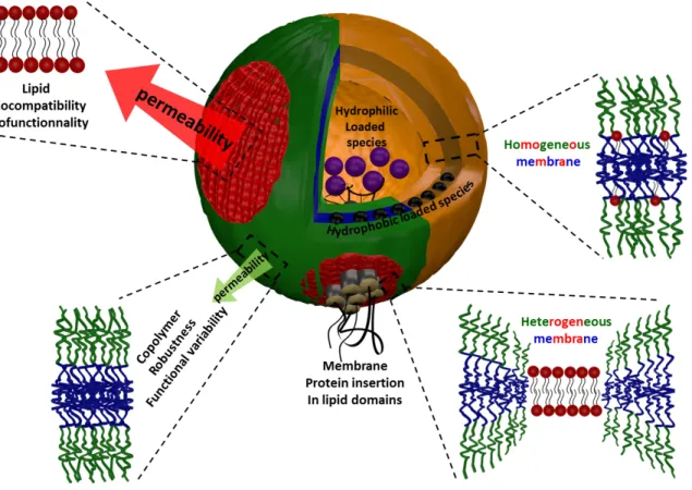
18 at the lipid/copolymer boundaries arising from the exposure of hydrophobic polymer segments to water (“hydrophobic mismatch”). To reduce this exposure and the resulting energetic cost of the boundary lines, the two opposite plausible scenarios can be considered. The first one (i) consists in a conformational adaptation through elastic deformation of the polymer chains at the boundary to decrease the line tension (Fig 27-1) in analogy to elastic deformation of membrane at lipid/lipid domain boundary in lipid bilayers (Kuzmin, Akimov et al. 2005). Another possibility (ii) is to decrease the interfacial length and therefore the interfacial energy, by coalescence into fewer domains of a larger area.
In Fig 27-1, which illustrates different properties resulting from the membrane structure and potential applications of hybrid vesicles, it is shown that the conformational adaptation of the polymer implies a collapse of the hydrophobic polymer chains near the lipid interface, therefore reducing the total number of conformations and opposing the entropic elasticity of chains. Therefore, it is clear that the molar mass (or chain length) and the rigidity (or Kühn length) of the hydrophobic polymer backbone also play a major role. If this adaptation cannot be achieved, then the domain formation is improbable (spontaneously nucleated domains
Author manuscript version of Chapter 27 published In book: The Giant Vesicle Book, Rumiana Dimova & Carlos Marques Eds. (CRC PRESS Taylor and Francis group, Oct. 7th 2019)
19 eventually collapse) and a homogeneous mixture of the components is expected (Fig. 27-1)
Figure 27-1: Illustration of the different membrane structural organization of the membrane in hGUVs and its principal characteristics.
A large hydrophobic mismatch is met in most of the studies performed so far, as the diblock or triblock copolymers most often used, form membranes whose thickness is at least 7nm, and the line tension, although not yet quantified experimentally, is expected to be high and driving the budding and fission of existing fluid domains. However the hydrophobic mismatch is certainly not the only parameter, as the budding and fission of lipid domains have been observed also in a study in which a grafted copolymer, PDMS-g-(PEO)2 well-known to
Author manuscript version of Chapter 27 published In book: The Giant Vesicle Book, Rumiana Dimova & Carlos Marques Eds. (CRC PRESS Taylor and Francis group, Oct. 7th 2019)
20 form vesicles with a membrane thickness close to liposomes (~5nm), was used (Chemin, Brun et al. 2012). In addition to the chemical nature of the hydrophobic block which obviously plays a role in the miscibility with the lipid phase and consequently on the interfacial energy, the architecture of the copolymer may also be an important factor to consider (e.g. block copolymer where the different polymers are linearly covalently coupled, vs. graft copolymer where polymers chain are distributed along the backbone of another polymer).
The fluidity of the lipid phase is also of significance in the membrane structure of hGUVs. In the case of lipid in the gel state at room temperature, and using a formation protocol described in section 3, the spontaneous formation of micron-sized domains was reported only as a rare event. For instance in one of these studies (Schulz, Glatte et al. 2011), hGUVs presenting stable micrometric domains were spontaneously obtained using DPPC and PIB87
-b-PEO17, but only in a narrow composition range (65–74 mass% polymer). It is supposed that the
large hydrophobic block in that case limited the conformational adaptation at the copolymer– lipid boundary. The large hydrophobic thickness (~10 nm) plays in favor of a statistic distribution of the lipid in the copolymer phase as sketched on Fig 27-1. Interestingly, homogeneous vesicles, at least at the micrometric scale, were observed for all copolymer contents larger than 30 mol%, (or 75 weight%). Below 60 weight%, homogeneous vesicles presenting faceted surfaces were obtained, which is the signature of the DPPC gel phase. As described above, using a copolymer presenting a membrane thickness close to that of liposomes, allows the spontaneous formation of micrometric lipid domains in hGUVs in a large polymer content range (from 10% to 93% in weight). Above 93%, the lipid is apparently
Author manuscript version of Chapter 27 published In book: The Giant Vesicle Book, Rumiana Dimova & Carlos Marques Eds. (CRC PRESS Taylor and Francis group, Oct. 7th 2019)
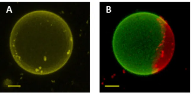
21 dispersed in the polymer phase and homogenous vesicles can be observed microscopically. Ideally the homogenous or heterogeneous character of the hGUVs can be easily observed under fluorescence microscopy by including a small amount of fluorescently tagged polymer and lipid in the initial mixture, such as Fluorescein modified polymer and Egg Liss Rhod PE for instance. In homogeneous hybrid vesicles, both signal of the fluorescent probes are observed within the entire membrane, whereas in heterogeneous hybrid vesicles, the probes preferentially partition into the polymer and lipid rich phases, as illustrated in Figure. 27-2.
Figure 27 2: Overlay of maximum intensity 3D projection images taken for A: homogeneous hGUV with the homogeneously distribution of both signals; B: de-mixed hGUV with green polymer-rich phase and red lipid rich phase. The scale bars are 5 µm. Acknowledgment to the publication Dao, T. P. T., F. Fernandes, et al. (2017). Soft Matter 13(3): 627-637. (Royal Society of Chemistry)
2.3. THE EFFECT OF THE CHEMICAL MODIFICATIONS AND ADDITIVES
Obtaining polymer-rich vesicles with stable lipid-rich domains of controlled micrometric or nanometric size would be of great interest for different kinds of functions in drug-delivery systems, bio-targeting or biophysical fundamental studies, such as the modeling of nanoparticles/membrane interactions and cellular internalization (“artificial endocytosis”). The
Author manuscript version of Chapter 27 published In book: The Giant Vesicle Book, Rumiana Dimova & Carlos Marques Eds. (CRC PRESS Taylor and Francis group, Oct. 7th 2019)
22 use of lipids in the fluid state, which is probably the most interesting, regarding the abovementioned applications, leads only to homogeneous hGUVs (i.e. at the micrometric scale, as appearing under an optical microscope). Other approaches involving the use of additional components have been used to generate a stable phase-separated membrane from a mixture of a copolymer with a mixture of several fluid lipids. For instance, with lipids presenting biotinylated head-groups, stable micrometric domains can be generated in hybrid vesicles by reacting them with streptavidin (in solution). This protein with multivalent binding sites for biotin works as a “zipper” to gather the lipid molecules together in pure lipid phases or “monodomains”. However, such a protein coating prevents further bio-functionalization of the domains (Schulz, Glatte et al. 2011). Another approach consists in using a lipid mixture containing a given amount of cholesterol, which is well-known to promote lateral phase separation into ‘‘raft-like’’ domains within liposomes for particular lipid compositions such as Chol/PC/SM mixtures (Bagatolli and Kumar 2009). This induces the same effects on hybrid copolymer/lipid vesicles in which round-shaped micrometric domains can be obtained with various phospholipids of low melting temperature Tm (DLPC, POPC, DC13PC…). Interestingly, the domain size could be modulated via the polymer/lipid/cholesterol composition. However, no domain formation can be obtained when using DOPC, even at high cholesterol content, in agreement with what is commonly observed for DOPC/cholesterol mixtures which are not known to form liquid ordered phase bilayers (Mills, Toombes et al. 2008). Regarding the hydrophobic mismatch, it has also been observed via cryo-TEM experiments on LUVs made of mixtures of PDMS-b-PMOXA and cholesterol that the vesicles obtained present lower
Author manuscript version of Chapter 27 published In book: The Giant Vesicle Book, Rumiana Dimova & Carlos Marques Eds. (CRC PRESS Taylor and Francis group, Oct. 7th 2019)
23 membrane thickness compared to pure polymersomes. This was explained by an increase of the packing density in the membrane through the support of static light scattering measurements (Winzen, Bernhardt et al. 2013). This approach could be useful to control the hydrophobic mismatch in hybrid lipid/copolymer giant vesicles.
2.4. THE EFFECT OF MACROSCOPIC PARAMETERS: TEMPERATURE, MEMBRANE TENSION
Forming hybrid copolymer/lipid vesicle containing a lipid with a melting temperature above room temperature implies a particular attention towards the process of vesicle formation. Generally, the film hydration method to prepare hGUVs is conducted at a temperature above the melting transition of the lipid. Then the vesicle suspension is cooled down to room temperature. It has been shown that the cooling rate plays a role on the resulting membrane structuration in hGUVs made of PBd-b-PEO and DPPC (Nam, Vanderlick et al. 2012). A fast cooling rate results in the formation of numerous small domains, whereas a slow cooling rate favours formation of less numerous domains but larger in size, in agreement with the classical nucleation-growth theory.
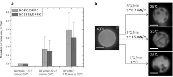
In two-component phosphatidylcholine GUVs, it has been shown that membrane tension could affect the fluid-solid phase transition of lipid and consequently the membrane structuration (Chen and Santore 2014). The same effect has been observed recently on hybrid vesicles made of PDMS-g-(PEO)2 and DPPC (Chen and Santore 2015). Using vesicles conditioned
in a way that they did not present excess membrane area, the authors were able to tune the membrane tension by using different cooling rates after vesicle formation by electro-formation
Author manuscript version of Chapter 27 published In book: The Giant Vesicle Book, Rumiana Dimova & Carlos Marques Eds. (CRC PRESS Taylor and Francis group, Oct. 7th 2019)
24 at high temperature (see section 3). The cooling causes a contraction of both the water solution and the membrane. As the membrane contracts more than the water in the compartment of the vesicle, a membrane surface tension appears, as estimated by a micropipette pulling experiment. Thereafter the membrane stress obviously relaxes as water diffuses progressively out of the vesicle. The authors were able to measure the membrane tension immediately after cooling to the temperature of interest, and a clear influence of cooling rate was shown on the membrane tension. Actually the membrane tension measured is maximal at intermediate cooling speed: A too small cooling rate allows membrane tension to relax by water diffusion by permeability, while a too fast quench induces stress relaxation through the membrane lysis and leaking by transient pore formation.
Figure 27-3: a) Illustration of the variation of membrane tension resulting from different cooling rates after preparation onto the membrane structuration: examples on vesicles containing 70% mol (39 mass%) DPPC and 30%mol (61% mass) PDMS-g-(PEO)2; b) Images of vesicles submitted to different
cooling and osmotic control. (Reproduced from Chen, D. and M. M. Santore (2015), Soft Matter 11(13): 2617-2626, with permission, copyright Royal Society of Chemistry).
Author manuscript version of Chapter 27 published In book: The Giant Vesicle Book, Rumiana Dimova & Carlos Marques Eds. (CRC PRESS Taylor and Francis group, Oct. 7th 2019)
25 At low surface tension, patchy domains were generally obtained whereas higher tension leads to striped domains. The physical state of the DPPC phase (ripple phase Pβ’ solid or gel phase Lβ)
inside these stripes or patchy domains is still under debate in literature (Gordon, Beales et al. 2006; Bernchou, Brewer et al. 2009) (Chen and Santore 2015)).
Finally, the structure of hGUVs containing high melting temperature lipids (e.g. DPPC) has been rarely commented above the transition temperature Tm. Our team has started to
evaluate the behaviour of hGUVs composed of PDMS-g-(PEO)2/DPPC previously studied at
room temperature (Chemin, Brun et al. 2012) following an electro-formation process realized at 55°C and storage conditions of the sample at 55°C. The sample was transferred as quickly as possible in a temperature-controlled stage at 55°C, in order to maintain the lipid phase in a continuous fluid (liquid disordered) state. Observations were made after different incubation times at 55°C, from 4h to 20h (Fig 27-4). L-α-phosphatidylethanolamine-N-(lissamine rhodamine B sulfonyl) (Liss-Rhod PE) was used to reveal the lipid-rich phase (0.2% mol), and copolymer grafted with fluorescein was added to reveal the polymer-rich phase (1% mol). It appeared that the fluorescent lipid (Liss Rhod PE) was excluded from the DPPC domains, although they were in the fluid state as attested by the rounded boundary between the polymer and lipid domains. After a long incubation time, the Liss Rhod PE repartitioned entirely into the more disordered polymer phase, which turned from green to yellow-orange. The most interesting feature is that the interface length between the lipid and polymer phases clearly increased, suggesting that a kind of compatibility increase occurred with time, and that such structures are strongly out-of-equilibrium in their early stage of formation.
Author manuscript version of Chapter 27 published In book: The Giant Vesicle Book, Rumiana Dimova & Carlos Marques Eds. (CRC PRESS Taylor and Francis group, Oct. 7th 2019)
26 Figure 27-4: 3D reconstructions of confocal microscopy images of PDMS-g-(PEO)2/DPPC (90/10
mass) vesicles loaded with Liss-Rhod PE (red) and Fluorescein-PDMS-g-(PEO)2 (green) observed at 55°C
after different incubation times at 55°C. Scale bar is 10µm.
3. SPECIFIC ASPECTS OF THE FORMATION OF GIANT HYBRID UNILAMELLAR VESICLES
Most of the knowledge acquired in the past ten years about hybrid copolymer/lipid vesicles result from studies performed on GUVs formed by the two well-known methods of rehydration or electro-formation (see Chapter 1) from copolymer/lipid mixtures as applied in (Ruysschaert, Sonnen et al. 2005; Cheng, Elias et al. 2011; Nam, Beales et al. 2011; Schulz, Glatte et al. 2011; Chemin, Brun et al. 2012; Nam, Vanderlick et al. 2012; Lim, de Hoog et al. 2013; Schulz, Werner et al. 2013; Schulz, Olubummo et al. 2014; Chen and Santore 2015; Scholtysek, Shah et al. 2015) and summarized in Table 27-2. A few examples report the preparation of hGUVs using mixed suspensions of giant phospholipid liposomes and polymersomes and the triggering of their adhesion followed by membrane fusion, by using temperature-responsive copolymers, or salt addition (Henderson and Paxton 2014; Morimoto, Sasaki et al. 2014). The ability of these methods to be extended to different polymer/lipid mixtures has not been confirmed so far. It has to be noted that simple film rehydration was
Author manuscript version of Chapter 27 published In book: The Giant Vesicle Book, Rumiana Dimova & Carlos Marques Eds. (CRC PRESS Taylor and Francis group, Oct. 7th 2019)
27 used by Tsourkas and coll. (Cheng and Tsourkas 2008; Cheng, Elias et al. 2011) to obtain hGUVs of HSPC phospholipid and PBd22-b-PEO14 blockcopolymer, with long incubation time (24h) in an
aqueous solution of sucrose at 65°C, but electro-formation is definitely the most commonly used technique. Practical tips about the preparation and collecting of hGUVs are described in Box 27-1.
Author manuscript version of Chapter 27 published In book: The Giant Vesicle Book, Rumiana Dimova & Carlos Marques Eds. (CRC PRESS Taylor and Francis group, Oct. 7th 2019)
28 Table 27-2 Preparation methods for obtaining hGUVs (lme: hydrophobicmembrane thickness).
Copolymer, molecular weght,
membrane thickness (phase state) Lipid Preparation technique Reference
PMOXA-b-PDMS-b-PMOXA Mn=9000 g⋅mol-1 lme= 14 nm PC (fluid) PE (fluid) lme =3 – 4 nm
Electro-formation method ITO coated glass:
5V, 10Hz, 3 hours followed by 5V, 0.5Hz 30 min Sonnen et al. 2005) (Ruysschaert,
PBd46-b-PEO30
Mn=3800 g⋅mol-1 lme= 9 nm
POPC (fluid)
Electro-formation method platinum electrodes: 3V, 11Hz several hours, at RT in sucrose solution (260
mOsm).
(Nam, Beales et al. 2011) Electro-formation method platinum electrodes:
3V, 15Hz, at 50°C, in sucrose solution (260 mOsm) (Nam, Vanderlick et al. 2012) DPPC (gel) PBd22-b-PEO14 Mn=1800 g⋅mol-1 lme ~7nm HSPC
(fluid) Film rehydration in sucrose solution (285 mM), incubating at 65 °C during 24h (Cheng, Elias et al. 2011) POPC
(fluid)
Electro-formation method ITO coated glass: 3V,10Hz, at 45°C in sucrose solution (at 300 mM)
during 120 min
(Lim, de Hoog et al. 2013)
Author manuscript version of Chapter 27 published In book: The Giant Vesicle Book, Rumiana Dimova & Carlos Marques Eds. (CRC PRESS Taylor and Francis group, Oct. 7th 2019)
29 PIB87-b-PEO17 Mn=5350 g⋅mol-1 lme = 10 nm (fluid) DOPC DPPC (gel) Electro-formation method ITO-coated glass electrodes: 1.3V, 10Hz at 70°C during 4 hours (Schulz, Glatte et al. 2011; Schulz, Werner et al. 2013; Schulz, Olubummo et al. 2014) Poly(isobutylene)-b-poly(ethylene oxide) (PIB37-b-PEO48) Mn=3970 g⋅mol-1 Poly(dimethylsiloxane)-graft-poly(ethylene oxide) PDMS12-g-(PEO)2 Mn=2700 g⋅mol-1 lme =5 nm POPC (fluid) Electro-formation method ITO coated glass:
2V, 10Hz, at RT during 20 min (Chemin, Brun et al. 2012) DPPC
(gel)
Electro-formation method ITO coated glass:
2V, 10Hz at 50°C during 20 min.
Electro-formation method platinum wire electrodes:
Author manuscript version of Chapter 27 published In book: The Giant Vesicle Book, Rumiana Dimova & Carlos Marques Eds. (CRC PRESS Taylor and Francis group, Oct. 7th 2019)
30
PROTOCOL BOX STARTS HERE
Box 27-1 Practical method for obtaining hGUVs via electro-formation Preparation of hGUVs using this method is made in three steps: Step 1: Solution preparation
Organic solution of lipids and copolymers are prepared and then mixed in order to reach the desired composition. Typically, chloroform or a mixture of chloroform and MeOH (2/1 v/v) are the solvents most often used. The concentration range (copolymer+lipid) is typically around 1mg⋅mL-1. If hybrid vesicles are meant to be studied with fluorescence or confocal microscopy
analyses, a tagged lipid should be included to reveal the lipid phase. Typically the use of Liss-Rhod PE gives good results at 0.2% molar ratio. Co-localization can also be performed using fluorescently tagged copolymers that have to be synthesized. If fluorescein or NBD is used as fluorescent moiety, a larger amount of copolymer probe should be used for visualization, e.g. 1% molar, because of the lower quantum yields and tendency of photo-bleaching of these dyes compared to rhodamine.
Step 2: Film deposition
Films are prepared by spreading around 10 µL of the lipid/copolymer solution slowly over an area of about 2 cm diameter of an electrically conductive surface. Most often used are platinum electrodes and glass plates coated with indium tin oxide (ITO glass plates of surface resistivity 15-25Ω/square). Surfaces must be cleaned with ethanol and chloroform before use. In the case of ITO plates, a Hamilton syringe with tapered needle is used to deposit the solution and also to gently scratch the film and generate irregularities that will help film swelling and
Author manuscript version of Chapter 27 published In book: The Giant Vesicle Book, Rumiana Dimova & Carlos Marques Eds. (CRC PRESS Taylor and Francis group, Oct. 7th 2019)
31 vesicle formation. No additional benefit is observed on the amount of vesicle obtained, size distribution or homogeneity of composition by forming the film by spin-coating. Then the film is dried out under dynamic vacuum for at least 5hrs. A too short drying time can lead, after the hydration step to the formation of vesicles presenting “lipid filaments” on their surface.
Step 3: Electro-swelling
Film rehydration under an alternative electric field is performed most often in a glucose or sucrose solution to set the osmolarity of the buffer, defined as the total amount of soluble species per kg of solution, and checked for instance by a freezing point osmometry measurement. As illustrated in Table 27-2, there is no real tendency that can be extracted in terms of voltage applied or duration of electro-formation. However, from our personal experience, it seems that at least 5V is needed for molar masses of hydrophobic block above 5000 g⋅mol-1. Temperature is also a parameter of great importance. Obviously
electro-formation must be realized above the main chain transition temperature of the lipid used, but even in the case of lipids fluid at room temperature (e.g. POPC), temperature have to be controlled and slightly increased (~30-40°C) when using polymers with long hydrophobic blocks >5000 g⋅mol-1. To extract a solution containing giant vesicles, it is recommended to use a syringe with at least a 0.8 mm internal diameter (gauge ≤ 21 G) of the needle in order to minimize shear stress. During electro-formation, some vesicles remain stuck on the conductive surfaces. To detach them, two methods are used depending on the type of surfaces employed. In case of platinum electrodes, gently manually shaking the electrode in the hydration medium is sufficient to detach vesicles. However, when ITO glass plates are used, GUVs are detached by
Author manuscript version of Chapter 27 published In book: The Giant Vesicle Book, Rumiana Dimova & Carlos Marques Eds. (CRC PRESS Taylor and Francis group, Oct. 7th 2019)
32 turning (or rolling) around a bubble created in the chamber by taking out a few drops of solution (see image below).
PROTOCOL BOX ENDS HERE
4. UNDERSTANDING MEMBRANE PROPERTIES FROM MEMBRANE STRUCTURE
The purpose of this section is to describe some of the properties of the hybrid giant unilamellar vesicles which were characterized so far, and try to point out a correlation between those properties and their membrane structure at macro- or nanoscale. Considering the intrinsic differences between lipid and copolymer membranes as illustrated in the Chapter 26, in terms of bio-functionality and physical properties, it can be reasonably expected that lipid polymer mixtures provide numerous ways to modulate the membrane properties, provided that a good control of membrane composition and structure is achieved. In this part, the mechanical properties, permeability, fluidity, and bio-functionality will be discussed in priority, while stability and deformability will also be cursory mentioned.
Author manuscript version of Chapter 27 published In book: The Giant Vesicle Book, Rumiana Dimova & Carlos Marques Eds. (CRC PRESS Taylor and Francis group, Oct. 7th 2019)
33 4.1. MECHANICAL PROPERTIES, FLUIDITY
Two essential features are often analyzed on giant vesicles because of their importance in biological events (resistance of cells to osmotic shock, cell fission and fusion, cell motility …) or drug delivery (resistance in the high shear rate of blood circulation through tiny vessels such as capillaries). They are related to their ability to resist to isotropic area dilation, and to deform from an initially flat surface into a curved structure, see Chapter 5. They are respectively quantified through the area compressibility modulus KA, which is linked to the interfacial tension at the junction between hydrophilic and hydrophobic moieties of the membrane, and the bending rigidity of the membrane κ (also called bending modulus) that appears in the Helfrich expression of the curvature energy that becomes non-negligible for nearly zero surface tension systems like micro-emulsions and lipid bilayers. As illustrated in Chapter 15 and Chapter 26, KA and κ from liposomes and polymersomes are rather different (see also Table 15-1 in Chapter 15) and they can be modulated to some extend by the hydrophobic block length and membrane thickness for polymersomes. This is clearly illustrated in Tables 27-3 and 27-4. Another important point to mention is the much larger lysis strain of the polymersomes, ascribed to a higher cohesive energy density between the molecules constituting the membrane. On the contrary, the excess area defined by subtracting the projected area AP from
the total true surface area of the membrane seems to be much lower for giant polymersomes than for giant liposomes, which explains why it is very difficult to extract the bending modulus κ from the very limited “entropic regime” in a micropipette pulling experiment.
Author manuscript version of Chapter 27 published In book: The Giant Vesicle Book, Rumiana Dimova & Carlos Marques Eds. (CRC PRESS Taylor and Francis group, Oct. 7th 2019)
34 Therefore, when mixing copolymer and lipid in a single membrane, a modulation of these properties can be expected, obviously by playing on the lipid/copolymer composition. Subtle modifications can also probably be obtained through precise membrane structuration. Interestingly, the area compressibility modulus has only been measured on hybrid vesicles through the help of the micropipette aspiration technique in recent studies. In their early work on hybrid vesicles in 2011,(Cheng, Elias et al. 2011) Cheng et al. reported that homogeneous hybrid vesicles composed of poly(butadiene)-b-poly(oxyethylene) (PBD22-b-PEO14) and
hydrogenated soy phosphatidylcholine (HSPC) exhibited an intermediate elastic stretching modulus between the values of pure lipid and pure polymer vesicles. Similar results for hybrid vesicles composed of PBD46-b-PEO30 and POPC were also indicated (Nam, Beales et al. 2011).
Although there was a rather large composition range where hybrid vesicles could not be formed, the remaining fractions showed distinctly a gradual decrease in KA with increasing copolymer content (Fig 27-5). It has to be noted that in the above-mentioned contributions, the hGUVs obtained presented a homogeneous membrane structure, at least at the micron scale studied by optical microscopy. A recent study has focused on hGUVs composed of poly(dimethylsiloxane)-graft-poly(ethyleneoxide) PDMS22-g-(PEO12)2 and
1,2-di-palmitoyl-sn-glycero-3-phosphocholine (DPPC) which is at the gel state at room temperature. In this system, it was shown by our group (Chemin, Brun et al. 2012) that heterogeneous membranes (presence of micrometric domains) could be obtained at room temperature in a large composition range. The main results of the Santore group have been described in Section 2.4 of this chapter. However they also performed some mechanical measurements through
Author manuscript version of Chapter 27 published In book: The Giant Vesicle Book, Rumiana Dimova & Carlos Marques Eds. (CRC PRESS Taylor and Francis group, Oct. 7th 2019)
35 micropipette aspiration on these heterogeneous membranes presenting lipid gel domains. In the case of data obtained without having lipid micrometric domains entering in the micropipettes (therefore by stretching the copolymer part of the membrane), they obtained stretching modulus similar to the pure copolymer membrane within the uncertainty of the measurement (Chen and Santore 2015) for hybrid vesicles containing 70 mol% of DPPC (39 mass%). Very interestingly, the lysis strain was still very high and similar to the lysis strain of pure polymersomes, despite the potential fragility that could result from the interfaces at the copolymer/lipid boundaries. This shows clearly that the mechanical properties of hybrid membranes can be directly linked to their lateral structure.
Author manuscript version of Chapter 27 published In book: The Giant Vesicle Book, Rumiana Dimova & Carlos Marques Eds. (CRC PRESS Taylor and Francis group, Oct. 7th 2019)
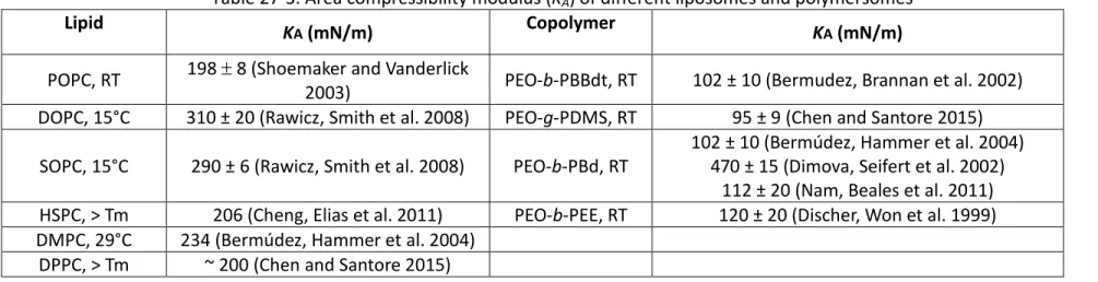
36 Table 27-3. Area compressibility modulus (KA) of different liposomes and polymersomes
Lipid KA (mN/m) Copolymer KA (mN/m)
POPC, RT 198 ± 8 (Shoemaker and Vanderlick 2003) PEO-b-PBBdt, RT 102 ± 10 (Bermudez, Brannan et al. 2002) DOPC, 15°C 310 ± 20 (Rawicz, Smith et al. 2008) PEO-g-PDMS, RT 95 ± 9 (Chen and Santore 2015)
SOPC, 15°C 290 ± 6 (Rawicz, Smith et al. 2008) PEO-b-PBd, RT 102 ± 10 (Bermúdez, Hammer et al. 2004) 470 ± 15 (Dimova, Seifert et al. 2002) 112 ± 20 (Nam, Beales et al. 2011) HSPC, > Tm 206 (Cheng, Elias et al. 2011) PEO-b-PEE, RT 120 ± 20 (Discher, Won et al. 1999) DMPC, 29°C 234 (Bermúdez, Hammer et al. 2004)
DPPC, > Tm ~ 200 (Chen and Santore 2015)
Table 27-4. Bending modulus (κ) of different liposomes and polymersomes
Lipid Bending modulus (κ) (10-19 J) Polymer Bending modulus (κ) (10-19 J) POPC, 25°C 1.58 ± 0.03 (Henriksen, Rowat et al. 2004) PDMSPMOXA60
-b-21, 101 ± 23 (Winzen, Bernhardt et al. 2013)
DOPC, 23°C 1.08 ± 0.1 (Gracia, Bezlyepkina et al. 2010) PMOXAPDMS68
-b-11, RT 70 ± 50 (Jaskiewicz, Makowski et al. 2012)
SOPC, 18°C 0.9 ± 0.06 (Evans and Rawicz 1990) PEO26-b-PBd46, RT 1.02 ± 0.46 (Bermúdez, Hammer et al. 2004)
Egg PC, RT 0.3 ± 0.1 (Evans and Rawicz 1990) PEO80-b-PBd125, RT 19.1 ± 6.4 (Bermúdez, Hammer et al. 2004)
DPPC, RT 10-15 (Jaskiewicz, Makowski et al. 2012) PEO40-b-PEE37, RT 1.37 ± 0.29 (Bermúdez, Hammer et al. 2004)
Author manuscript version of Chapter 27 published In book: The Giant Vesicle Book,
Rumiana Dimova & Carlos Marques Eds. (CRC PRESS Taylor and Francis group, Oct. 7th 2019)
37 Figure 27-5. (a) Areal stretching modulus KA (▪) and lipid lateral diffusion coefficient D (○) for varying
compositions of hGUVs. (b) Critical lysis tension (▪) and lysis strain (□) of POPC/PBd-b-PEO GUVs. HLP-GUVs: Hybrid lipid-diblock copolymer giant unilamellar vesicles. Adapted from Nam, J., P. A. Beales, et al. Langmuir 27(1): 1-6. Copyright (2011) American Chemical Society.
Regarding the bending modulus (κ) of hGUVs, quantitative data reported so far in literature are scarce compared to the abundant values for lipid GUVs which are floppier and much more flexible (see previous remark on the reduced excess area of polymersomes). There is only one alternate approach by AFM to measure the κ of hybrid vesicles hLUVs composed of poly(dimethylsiloxane)-block-poly(methyloxazoline) and 1,2-dimyristoyl-sn-glycero-3-phosphocholine (PDMS60-b-PMOXA21/DMPC) with a diameter of ~ 200 nm. The
mechanical properties of the vesicles were measured using quantitative forces versus distance curves. An intermediate value of κ between liposomes and polymersomes (62⋅10-19
Author manuscript version of Chapter 27 published In book: The Giant Vesicle Book,
Rumiana Dimova & Carlos Marques Eds. (CRC PRESS Taylor and Francis group, Oct. 7th 2019)
38 (Winzen, Bernhardt et al. 2013). The authors investigated also the modification of the membrane properties using cholesterol instead of DMPC. Interestingly, they observed that the bending modulus of a membrane containing only ~5% in mass of cholesterol was more than four times larger than that of pure polymersomes. It was suggested that this could result from an increase of the packing density in the membrane induced by cholesterol. From the preliminary results, it is clear that the flexibility of hybrid membranes can be tuned on a very large range and in a subtle way. As this parameter and other mechanical properties control many aspects of the behaviour of vesicles such as their formation, stability, fusion and budding processes, further studies need to be performed to concomitantly elucidate the membrane structure and the resulting properties.
Although there are many types of molecular individual and collective motions within membranes, the mobility of molecules inside a membrane has been mostly evaluated through the measurement of the lateral diffusion coefficient, which is directly linked to the surface shear viscosity of the membrane. Such measurement is commonly made by fluorescence recovery after photo-bleaching (FRAP) experiments, see also Chapter 21. In the case of hybrid copolymer lipid hGUVs, one can access either the mobility of the lipid molecules or the copolymer chains depending on the localization of the fluorescent probes. Numerous studies and reviews on liposomal membranes pointed out a strong influence of the lipid physical state and membrane composition on the molecular mobility. Therefore it is expected that the mobility and the fluidity of the membrane could be finely tuned by the association of copolymers and lipids in the membrane. Lateral diffusion coefficients for lipids are indeed in the range 3-5 µm2·s-1 and those of copolymer chains in the lower range
Author manuscript version of Chapter 27 published In book: The Giant Vesicle Book,
Rumiana Dimova & Carlos Marques Eds. (CRC PRESS Taylor and Francis group, Oct. 7th 2019)
39 0.0024-0.12 µm2·s-1 (Lee, Santore et al. 2002). Large differences in surface shear viscosity have also been reported between PBd-b-PEO and lipid membranes (Dimova, Seifert et al. 2002; Evans, Heinrich et al. 2003), with even higher values reported for liquid crystalline block copolymers (Mabrouk et al. 2009). Therefore large variations of such parameters are anticipated depending on the lipid / copolymer composition, but subtle modifications could result also from peculiar membrane lateral structuration (presence of domains) (Loura, Fedorov et al. 2005; Loura, Fernandes et al. 2010). Other types of effects can also be invoked, such as in the work of Nam et al. (Nam, Beales et al. 2011) who first indicated that with increasing copolymer content, the diffusion of lipid molecules in hGUVs of POPC/ PBd46-b-PEO30 becomes slower and in proportion of the copolymer amount incorporated,
except at low copolymer fractions where only a weak dependence was observed (see Figs 27-6). Since for this system, none of the hybrid vesicles showed macroscopic domains, the homogeneous insertion of copolymer chains into lipid membrane may somewhat hamper the motion of lipid chains. It is interesting to note that the authors observed a fluorescence recovery shape for hybrid vesicles different from that of the pure components (polymersomes and liposomes), suggesting that dynamics in these hybrid membranes are not ideally described by a standard diffusion model with a single diffusion coefficient.
Author manuscript version of Chapter 27 published In book: The Giant Vesicle Book,
Rumiana Dimova & Carlos Marques Eds. (CRC PRESS Taylor and Francis group, Oct. 7th 2019)
40 Figure 27-6 (Nam, Beales et al. 2011). Diffusion coefficient measurements by FRAP for different hGUVs of POPC/ PBd46-b-PEO30. Reprinted from Nam, J., P. A. Beales, et al. Langmuir 27(1): 1-6.
Copyright (2011) American Chemical Society.
Other authors (Schulz, Werner et al. 2013) obtained information about the mobility of lipid molecules in hybrid membranes composed of DPPC, in the gel state at room temperature, and PIB87-b-PEO. For that purpose, they used 1.
2-dihexadecanoyl-sn-glycero-3-phosphothanolamine-N-(lissamine rhodamine B sulfonyl) (Rh-DHPE) as a diffusion probe. This molecule fails to incorporate ordered membranes and should be localized in the most disordered phase of the hybrid vesicles (Bagatolli and Gratton 2000). Whereas no fluorescence recovery was detected for pure DPPC, above a threshold in copolymer fraction for hGUVs, the data revealed a clear increase in mobility of Rh-DHPE in the hybrid membranes with the copolymer content. The authors interpreted these observations by the breaking up of the rigid DPPC densely packed phase by the copolymer chains. The interpretation of such data is however not obvious as Rh-DHPE is known to exhibit a large preference for disordered domains. Therefore, it is likely that Rh-DHPE inserts into the copolymer phase, which presents a higher mobility compared to the DPPC gel phase.
In these preliminary results, the authors probed the lipid mobility in the entire hybrid systems, where there was no clear evidence of macroscopic domain presence. This does not rule out the existence of domains below the optical resolution, which can act as obstacles hindering the diffusion, in a similar manner as lipid rafts or membrane proteins in the so-called “mosaic membrane” model (Vaz, Goodsaid-Zalduondo et al. 1984; Saxton 1987). Liquid ordered phases within liposomal membranes are indeed suspected for a long time as a factor that significantly impacts on the dynamic of molecules in natural and reconstructed
Author manuscript version of Chapter 27 published In book: The Giant Vesicle Book,
Rumiana Dimova & Carlos Marques Eds. (CRC PRESS Taylor and Francis group, Oct. 7th 2019)
41 membranes.
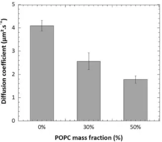
Figure 27-7 Diffusion coefficients of FITC-PDMS22-g-(PEO12)2 in hybrid PDMS22-g-(PEO12)2: POPC GUVs
were plotted versus the POPC fraction in the membranes (data originating from the PhD Thesis of T. P. Tuyen Dao, Univ. Bordeaux 2016).
We studied the consequence of the presence of POPC on the mobility of copolymer chains. From our data shown in Fig 27-7, mixtures containing either 30% or 50% of POPC in mass demonstrated a significant reduction of the copolymer lateral diffusion compared to the pure copolymer membranes. It should be noted that 30% and 50% contents of POPC in PDMS22-g-(PEO12)2, POPC mixtures were enough to drive micro-domain formation within the
hybrid membranes, and in this situation, the tracer molecule used (FITC-PDMS22-g-(PEO12)2)
was localized almost exclusively in the copolymer phase. Apart from the decrease of diffusion coefficient value, the FRAP curve of hybrid PDMS22-g-(PEO12)2: POPC vesicles also
clearly indicated the presence of an immobile fraction as shown in Fig 27-8. Indeed, even though the measurement was carried out in polymer-rich domain, we rarely got a full recovery for all fractions of POPC incorporated. These results could be explained by the
Author manuscript version of Chapter 27 published In book: The Giant Vesicle Book,
Rumiana Dimova & Carlos Marques Eds. (CRC PRESS Taylor and Francis group, Oct. 7th 2019)
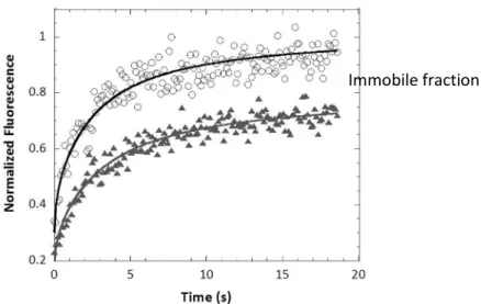
42 presence of nanoscale lipid domains which were below the resolution of confocal microscopy yet hamper the mobility of the copolymer chains. In the Figure 27-8 data were fitted with the circular spot model to extract diffusion coefficient values (Axelrod, Koppel et al. 1976; Soumpasis 1983; Ishikawa-Ankerlhod, Ankerhold et al. 2012)
Figure 27-8. Normalized fluorescence vs time of pure PDMS22-g-(PEO12)2 (○) and hybrid PDMS22
-g-(PEO12)2: POPC with 30% POPC in mass () (data originating from the PhD Thesis of T. P. Tuyen Dao)
Box 27-2 summarizes the important points which should be carefully considered for a successful FRAP measurement.
BOX STARTS HERE
Box 27-2. Practical tips for FRAP measurements on hGUVs
i) Fluorescent probes: The probes should preferably have intermediate photo-stability, since, on the one hand, they should be not too difficult to bleach and, on the other hand, they should be sufficiently photo-stable to allow imaging of the recovery process. Furthermore, the fluorophore concentration should also be considered. Indeed, the
Author manuscript version of Chapter 27 published In book: The Giant Vesicle Book,
Rumiana Dimova & Carlos Marques Eds. (CRC PRESS Taylor and Francis group, Oct. 7th 2019)
43
concentration should not be too high in order to avoid e.g., possible self-quenching effects, whereas a too low concentration will result in very weak fluorescence, poor signal/noise ratio and number fluctuations will start to dominate the signal. From our experience, fluorescein, nitrobenzoxadiazole (NBD), dichlorotriazinylamino fluorescein are normally good choices of fluorescent probes for FRAP.
ii) hGUV sample: The vesicles chosen to be measured should be sufficiently large to have a significant excess number of molecules outside the circular bleached spot (ROI - region of interest) to diffuse from. Indeed, since at least 3 µm is usually required for the minimum ROI diameter to get meaningful data, and since the precision of the measurement is improved when a larger bleached ROI is used, we recommend the use of hGUVs larger than 10 µm diameter for FRAP measurement. Another important point is that the vesicles must be fully immobilized during acquisition. The immobilization of hGUVs can be achieved by including a low ratio of biotinylated lipids (1/106 mol) into the vesicles, which allows for high affinity adhesion to an avidin-coated surface. This low amount is sufficient to fully immobilize the vesicles on the substrate, and larger amounts can lead to collapse of the vesicle on the surface. The avidin coating is realised by incubating the glass slide in an avidin solution at 0.1mg/mL. After removing all the excess amount of non-attached avidin to the glass slide, GUVs can be added in the chamber, and they will adhere to the surface in about 15 minutes. Interestingly, this method initially developed for liposomes (Puech, Askovic et al. 2006; Sarmento, Prieto et al. 2012) was also effective for hGUVs. Another approach consists in diluting suspension of vesicles that have been hydrated in sucrose 0.1M, in a glucose solution of the same concentration, to induce vesicle sedimentation.