HAL Id: hal-01459554
https://hal.archives-ouvertes.fr/hal-01459554
Submitted on 10 Dec 2019
HAL is a multi-disciplinary open access
archive for the deposit and dissemination of
sci-entific research documents, whether they are
pub-lished or not. The documents may come from
teaching and research institutions in France or
abroad, or from public or private research centers.
L’archive ouverte pluridisciplinaire HAL, est
destinée au dépôt et à la diffusion de documents
scientifiques de niveau recherche, publiés ou non,
émanant des établissements d’enseignement et de
recherche français ou étrangers, des laboratoires
publics ou privés.
with the largest genome
Aurelia Caputo, Jean-Christophe Lagier, Said Azza, Catherine Robert, Donia
Mouelhi, Pierre-Edouard Fournier, Didier Raoult
To cite this version:
Aurelia Caputo, Jean-Christophe Lagier, Said Azza, Catherine Robert, Donia Mouelhi, et al..
Mi-crovirga massiliensis sp nov., the human commensal with the largest genome. MicrobiologyOpen,
Wiley, 2016, 5 (2), pp.307-322. �10.1002/mbo3.329�. �hal-01459554�
Introduction
Culturomics was developed in 2012 in order to extend the human gut repertoire. In the first study, by multiply-ing the number of culture conditions (212 different culture conditions) with rapid identification of the colonies using MALDI- TOF, we were able to identify 340 different bac-terial species including 31 new bacbac-terial species (Lagier et al. 2012a,b,c,d). In addition, we demonstrated a com-plementarity with metagenomics (the usual gold standard for studying complex ecosystems) because only 15% of the species were concomitantly detected by culture and metagenomics applied to the same samples. We applied this strategy to diverse stool samples from healthy indi-viduals from diverse geographic origins and from patients with diverse diseases, dramatically extending our knowl-edge of the cultured human gut repertoire (Lagier et al. 2015).
The current bacterial taxonomy to define species traditionally combines phenotypic and genotypic charac-teristics (Stackebrandt and Ebers 2006; Tindall et al. 2010) such as the phylogenetic marker 16S rRNA sequence, DNA–DNA hybridization, and DNA G+C content (Rosselló- Mora 2006). These latter two tools have been considered a “gold standard” but are expensive and have poor reproducibility (Wayne et al. 1987). In recent years, the number of sequenced bacterial genomes has grown rapidly thanks to high- throughput sequencing and MALDI- TOF- mass spectrometry (MALDI- TOF- MS) analysis. These have allowed the description of new bacterial genera and species. Recently, by adapting MALDI- TOF to the routine identification of bacterial species, clinical isolated identi-fication was made possible for the first time (La Scola and Raoult 2009; Seng et al. 2009, 2013). These methods provide a wealth of proteomic and genetic information (Tindall et al. 2010; Welker and Moore 2011).
Microvirga massiliensis sp. nov., the human commensal with
the largest genome
Aurélia Caputo1, Jean-Christophe Lagier1, Saïd Azza1, Catherine Robert1, Donia Mouelhi1, Pierre-Edouard Fournier1 & Didier Raoult1,2
1Unité de Recherche sur les Maladies Infectieuses et Tropicales Émergentes, CNRS, UMR 7278 – IRD 198, Faculté de médecine, Aix-Marseille Université, 27 Boulevard Jean Moulin, 13385 Marseille Cedex 05, France
2Special Infectious Agents Unit, King Fahd Medical Research Center, King Abdulaziz University, Jeddah, Saudi Arabia
Keywords
Culturomics, large genome, Microvirga massiliensis, taxonogenomics
Correspondence
Didier Raoult, Unité de Recherche sur les Maladies Infectieuses et Tropicales Émergentes, CNRS, UMR 7278 – IRD 198, Faculté de médecine, Aix-Marseille Université, 27 Boulevard Jean Moulin, 13385 Marseille Cedex 05, France.
Tel: +33 491 385 517; Fax: +33 491 387 772; E-mail: didier.raoult@gmail.com
Received: 23 July 2015; Revised: 12 November 2015; Accepted: 19 November 2015
MicrobiologyOpen 2016; 5(2): 307–322
doi: 10.1002/mbo3.329
Abstract
Microvirga massiliensis sp. nov. strain JC119T is a bacteria isolated in Marseille
from a stool sample collected in Senegal. The 16S rRNA (JF824802) of M.
massil-iensis JC119T revealed 95% sequence identity with Microvirga lotononidis
WSM3557T (HM362432). This bacterium is aerobic, gram negative, catalase
positive, and oxidase negative. The draft genome of M. massiliensis JC119T
comprises a 9,207,211- bp- long genome that is the largest bacterial genome of an isolate in humans. The genome exhibits a G+C content of 63.28% and contains 8685 protein- coding genes and 77 RNA genes, including 21 rRNA genes. Here, we describe the features of M. massiliensis JC119T, together with
Given that the current taxonomics rules do not integrate two of the recent revolutions in clinical microbiology, we proposed a new concept named taxonogenomics including these. As genome sequence provides access to the full genomic information of a strain at a reduced cost, we proposed in addition to the classic phenotypic description, to systematically add whole- genome sequence and genome comparison with the closest species to describe a new isolate. Moreover, as MALDI- TOF became the reference identification method in most clinical microbiology labo-ratories (Clark et al. 2013), we proposed systematically adding both MALDI- TOF spectra and spectra comparison with the closest species to describe a new taxa. Using taxonogenomics, nine new bacteria have officially been considered as new genera and/or species in validation lists no. 153 and no. 155 (Kokcha et al. 2012; Lagier et al. 2012a,b,c,d, 2013a,b; Ramasamy et al. 2012; Roux et al. 2012; Hugon et al. 2013; Oren and Garrity 2014).
With both the description of the complete genomic sequencing and annotation, we present a summary clas-sification and a set of features for M. massiliensis sp. nov. strain JC119T (=CSUR P153 = DSM 26813) that was
isolated from an African stool sample from Senegal (Table 1).
Materials and Methods
Bacterial cultureSample
In 2012, a stool sample was collected from a 16- year- old Senegalese man living in Dielmo, a rural area in Sine Saloum, Senegal. Signed informed consent was obtained from the patient. The study and the assent procedure were approved by the Ethics Committees of the Institut Fédératif de Recherche 48, Faculty of Medicine, Marseille, France, under agreement number 09- 022. After receipt, the stool sample was frozen at −80°C.
Phenotypic characterization Strain isolation
The stool sample was cultivated on MOD2 medium in aerobic conditions at 37°C for 48 h. The culture condi-tions included a modified 7h10 medium supplemented with sheep blood as previously described (Ghodbane et al. 2014).
Strain identification
All the colonies were identified using MALDI- TOF MS, as described below. Colonies that exhibited a
MALDI- TOF- MS score > 1.9 were considered as correctly identified, and colonies that exhibited a MALDI- TOF- MS score < 1.9 with spectra in the database were further characterized using 16S rRNA sequencing as previously described (Seng et al. 2010). If the similarity value between the 16S rRNA sequence was lower than 98.7%, we con-sidered a new species without performing DNA–DNA hybridization as suggested by Stackebrandt and Ebers (2006).
Phenotypic properties
The main phenotypic characteristics such as Gram stain-ing, motility, sporulation, catalase, and oxidase tests were performed as previously described (Schaeffer et al. 1965; Adler and Margaret 1967; Humble et al. 1977; Gregersen 1978). The chemical characteristics of the strain JC119T
were tested using API® 20A, API® ZYM, and API® 50
CH strips (Biomerieux, Marcy- l’Etoile, France). Growth temperatures were tested at 25°C, 30°C, 37°C, 45°C, and 55°C, respectively. Strain growth was tested under aerobic conditions, with or without 5% CO2, and under anaerobic and microaerophilic conditions using GENbag anaer and GENbag microaer systems, respectively (Biomerieux). For electron microscopy, we observed colonies using a Morgani 268D (FEI, Limeil- Brevannes, France) at an operating voltage of 60 kV. Antibiotic disks of vancomycin (30 μg), rifampicin (30 μg), doxycycline (30 μg), erythromycin (15 μg), amoxicillin (25 μg), nitrofurantoin (300 μg), gentamicin (15 and 500 μg), ciprofloxacin (5 μg), amoxi-cillin (20 μg) with clavulanic acid (10 μg), peniamoxi-cillin G (10 μg), trimethoprim (1.25 μg) with sulfamethoxazole (23.75 μg), oxacillin (5 μg), imipenem (10 μg), tobramycin (10 μg), metronidazole (4 μg), and amikacin (30 μg) were purchased from Biomerieux. In vitro susceptibility testing of these antibiotics was performed by the disk diffusion method following EUCAST recommendations (Matuschek et al. 2014). Results were interpreted using Eucast 2015 clinical breakpoints (http://www.eucast.org/ clinical_breakpoints/). MALDI- TOF- MS protein analysis was carried out using a Microflex spectrometer (Bruker Daltonics, Leipzig, Germany) as previously described (Seng et al. 2009). Briefly, one isolated bacterial colony was transferred with a pipette tip from a culture agar plate and spread as a thin film on a MSP 96 MALDI- TOF target plate (Bruker). After isolation, 12 distinct deposits from 12 isolated colonies were tested for strain JC119T.
We superimposed each smear with 2 μL of matrix solu-tion (saturated solusolu-tion of α- cyano- 4- hydroxycinnamic acid) in 2.5% trifluoroacetic acid, 50% acetonitrile and allowed to dry for 5 min. We recorded the spectra in the positive linear mode for the mass range of 2000– 20,000 Da (parameter settings: ion source 1 (ISI), 20 kV; IS2, 18.5 kV; lens, 7 kV). Through variable laser power,
a spectrum was obtained after 240 shots. Per spot, the acquisition time was between 30 and 60 s. The strain JC119T spectra were imported into the MALDI BioTyper
software (version 3.0, Bruker) and analyzed by standard pattern matching (with default parameter settings) against the main spectra of 2843 bacteria. The identification method included m/z from 3000 to 15,000 Da. For every spectrum, we compared spectra in the database with a maximum of 100 peaks. From the resulting score, we
could identify tested species, where a score ≥ 1.9 enabled identification at the species level with a validly published species. For strain JC119T, no significant MALDI- TOF
score was obtained against the database, suggesting that this isolate was not a member of a known species. We added the spectrum from strain JC119T to our database
(Fig. 1A). Finally, the gel view was used to demonstrate the spectral differences with other members of the
Methylobacteriaceae family (Fig. 1B).
Table 1. Classification, general features, and project information of Microvirga massiliensis JC119T according to the MIGS recommendations (Woese et al. 1990; Field et al. 2008).
MIGS ID Property Term Evidence code1
Current classification Domain: Bacteria TAS (Woese et al. 1990) Phylum: Proteobacteria TAS (Garrity et al. 2005a)
Class: Alphaproteobacteria TAS (Oren and Garrity 2014; Garrity et al. 2005b) Order: Rhizobiales TAS (Kuykendall 2005; Oren and Garrity 2014) Family: Methylobacteriaceae TAS (Kanso and Patel 2003; Zhang et al. 2009;
Weon et al. 2010; Ardley et al. 2012; Bailey et al. 2014; Radl et al. 2014; Reeve et al. 2014) Genus: Microvirga TAS
Species: M. massiliensis IDA Type strain JC119T IDA
Gram stain Negative IDA
Cell shape Rod IDA
Motility Nonmotile IDA
Speculation Nonsporulating IDA
Temperature range Mesophile IDA
Optimum temperature 37°C IDA
MIGS- 6.3 Salinity Unknown
MIGS- 22 Oxygen requirement Aerobic IDA
Carbon source Unknown IDA
Energy source Unknown IDA
MIGS- 6 Habitat Human gut IDA
MIGS- 15 Biotic relationship Free living IDA
MIGS- 14 Pathogenicity Unknown NAS
Biosafety level 2
Isolation Human feces
MIGS- 4 Geographic location Dielmo, Senegal IDA MIGS- 5 Sample collection time September 2010 IDA
MIGS- 4.1 Latitude 13.71667 IDA
MIGS- 4.1 Longitude −16.41667 IDA
MIGS- 4.3 Depth Surface IDA
MIGS- 4.4 Altitude 34 m above sea level IDA
MIGS- 31 Finishing quality Noncontiguous finished MIGS- 28 Libraries used Six paired- end and two Shotgun
454; mate pair MiSeq
MIGS- 29 Sequencing platform 454 GS FLX Titanium and Illumina MIGS- 31.2 Fold coverage 77×
MIGS- 30 Assemblers Newbler 2.8 MIGS- 32 Gene calling method Prodigal
GenBank Date of Release November, 2014 NCBI project ID PRJEB8433 EMBL accession CDSD00000000
1Evidence codes – IDA, inferred from direct assay; TAS, traceable author statement (i.e., a direct report exists in the literature); NAS, nontraceable author statement (i.e., not directly observed for the living, isolated sample, but based on a generally accepted property for the species, or anecdotal evidence). These evidence codes are from the Gene Ontology project (Ashburner et al. 2000).
A
Genome sequencing and assembly
Genomic DNA of M. massiliensis was sequenced on a 454 sequencer with two methods: paired- end and shotgun and on a MiSeq sequencer (Illumina, Inc., San Diego, CA) with mate pair strategy; 450 μL of bacterial suspen-sion was diluted in 2 mL TE buffer for lysis treatment: a lysozyme incubation of 30 min at 37°C followed by an overnight proteinase K incubation at 37°C. The DNA was purified by three phenol–chloroform extractions and etha-nolic precipitation at −20°C overnight. After centrifugation, the DNA was resuspended in 180 μL TE buffer. The concentration was measured by the Quant- it Picogreen kit (Invitrogen) on the Genios_Tecan fluorometer at 55.70 ng/μL. This project was sequenced through two NGS technologies 454- Roche GSFLX Titanium GS and Illumina MiSeq. A shotgun and 3- kb paired- end libraries were pyrosequenced on the 454_Roche_Titanium. This project was loaded twice on a 1/4 region for the shotgun library and for the paired- end library: once on a full PTP PicoTiterPlate and four times on a 1/4 region on PTP PicoTiterPlates. The libraries were constructed according to the 454_Titanium and manufacturer protocols. The shotgun library was constructed with 500 ng of DNA as described by the manufacturer Roche with the GS Rapid library Prep kit with a final concentration at 1.35e09 by
the Quant- it Ribogreen kit (Invitrogen) on the Genios_ Tecan fluorometer. The paired- end library was constructed from 5 μg of DNA. The DNA was mechanically fragmented on the Hydroshear device (Digilab, Holliston, MA) with an enrichment size at 3–4 kb. The DNA fragments were visualized through the Agilent 2100 BioAnalyzer (Agilent Technologies Inc., Santa Clara, CA) on a DNA labchip 7500 with an optimal size of 2.950 kb. Circularization and nebulization were performed and generated a pattern with an optimal at 371 bp. After PCR amplification through 14 cycles followed by double size selection, the single- stranded paired- end libraries were then quantified on the Quant- it Ribogreen kit (Invitrogen) on the Genios_Tecan fluorometer at 140 pg/μL. The library concentration equiva-lence was calculated at 6.92e08. The two libraries were
stocked at −20°C until use. The shotgun library was clonal- amplified with 3 cpb in 3 SV emPCR and the 3- kb paired- end library was amplified with lower cpb (0.5 cpb) in 4 SV emPCR and by a bigger preparation of enriched beads at 0.25, 0.5, and 0.75 cpb with the GS Titanium MV emPCR Kit (Lib- L) v2. The yield of the emPCR was
16.83% for the shotgun and for two clonal amplification of the 3- kb paired- end library 9.65% in SV reactions and 6.79%, 14.31%, and 14.56%, respectively, in MV reactions. These yields were measured according to the quality ex-pected by the range of 5% to 20% from the Roche pro-cedure. The two libraries were loaded on the GS Titanium PicoTiterPlates PTP Kit 70 × 75 sequenced with the GS Titanium Sequencing Kit XLR70. The runs were performed overnight and then analyzed on the cluster through the gsRunBrowser and gsAssembler_Roche; 904 Mb was gen-erated through passed filters reads with a length average of 299 bp. A second DNA extraction was performed. Three petri dishes were spread and resuspended in 6 × 100 μL of G2 buffer. First, mechanical lysis was performed by glass powder on the Fastprep- 24 device (sample prepara-tion system) from MP Biomedicals for 2 × 20 s. DNA was then incubated for a lysozyme treatment (30 min at 37°C) and extracted through the BioRobot EZ 1 Advanced XL (Qiagen, Hilden, Germany)). The gDNA was quanti-fied by a Qubit assay with the high sensitivity kit (Life Technologies, Carlsbad, CA) to 118 ng/μL.
Genomic DNA of M. massiliensis was sequenced on the MiSeq Technology (Illumina) with the mate pair strat-egy. The gDNA was barcoded in order to be mixed with 11 other projects with the Nextera mate pair sample prep kit (Illumina). The mate pair library was prepared with 1 μg of genomic DNA using the Nextera mate pair Illumina guide. The genomic DNA sample was simultaneously fragmented and tagged with a mate pair junction adapter. The pattern of the fragmentation was validated on an Agilent 2100 BioAnalyzer (Agilent Technologies Inc.) with a DNA 7500 labchip. The DNA fragments ranged in size from 1 kb up to 10 kb with an optimal size at 4.81 kb. No size selection was performed and 670 ng of tagmented fragments was circularized. The circularized DNA was mechanically sheared to small fragments with an optimal at 667 bp on the Covaris device S2 in microtubes (Covaris, Woburn, MA). The library profile was visualized on a High Sensitivity Bioanalyzer LabChip (Agilent Technologies Inc.). The libraries were normalized at 2 nM and pooled. After a denaturation step and dilution at 12 pM, the pool of libraries was loaded onto the reagent cartridge and then onto the instrument along with the flow cell. Automated cluster generation and sequencing run were performed in a single 39 h run in a 2 × 251 bp. Total information of 8.3 Gb was obtained from a 947 K/mm2
cluster density with a cluster passing quality control filters
Figure 1. (A) Reference mass spectrum from Microvirga massiliensis JC119T. (B) Gel view comparing M. massiliensis strain to other Methylobacteriaceae
family. The gel view displays the raw spectra of loaded spectrum files arranged in a pseudo- gel- like look. The x- axis records the m/z value. The left y- axis displays the running spectrum number originating from subsequent spectra loading. The peak intensity is expressed by a Gray scale scheme
code. The color bar and the right y- axis indicate the relation between the color a peak is displayed with and the peak intensity in arbitrary units.
of 99% (18,112,000 clusters). Within this run, the index representation for Microvirga was determined to be 8.57%. The 1,398,916 paired reads were filtered according to the read qualities.
The total of 2,915,781 reads produced by six 454 paired- end run and two 454 shotgun of M. massiliensis JC119T
were assembled with Newbler version 2.8 which generated a genome size of 9.3 Mb with an average of 77× coverage of the genome. For increased quality of the genome, we mapped reads from MiSeq sequencer against this genome using CLC workbench software (CLC bio, Aarhus, Denmark).
Genome annotation and comparison
Open reading frames (ORFs) were predicted using Prodigal (Hyatt et al. 2010) with default parameters. However, the predicted ORFs were excluded if they were spanning a sequencing gap region (containing N). The predicted bacterial protein sequences were searched against Clusters of Orthologous Groups (COG) databases (Tatusov et al. 2001) using BLASTP (Altschul et al. 1990) (E- value 1e−03, coverage 70%, and identity percent 30%).
If no hit was found, the search was performed against the NR database using BLASTP with the same parameters as before. If the sequence lengths were smaller than 80 amino acids, we used an E- value of 1e−05. The tRNA
genes were found by the tRNAScanSE tool (Lowe and Eddy 1997), whereas ribosomal RNAs (rRNAs) were found by using RNAmmer (Lagesen et al. 2007). Lipoprotein signal peptides and the number of trans-membrane helices were predicted using Phobius (Käll et al. 2004). ORFans were identified if all the BLASTP performed did not give positive results (E- value smaller than 1e−03 for ORFs with a sequence size higher than
80 aa or E- value smaller than 1e−05 for ORFs with
se-quence lengths smaller than 80 aa). Similar parameter thresholds have already been used in previous work to define ORFans (Yin and Fischer 2006, 2008).
Based on 16S rDNA of M. massiliensis (JF824802) and closely related species, sequences were aligned using CLUSTALW, and phylogenetic tree was obtained using the maximum- likelihood method within the MEGA 6.06 software (Tamura et al. 2013). Numbers at the nodes are bootstrap values obtained by repeating the analysis 500 times to generate a majority consensus tree. We performed a BLASTP (Altschul et al. 1990) against the database of microbial polyketide and nonribosomal peptide gene clus-ters (Conway and Boddy 2013) for the predicted number of polyketide synthase (PKS)- and nonribosomal peptide synthetase (NRPS)- encoding genes, and we keep only the best hit for each protein.
For the genomic comparison, we used M. massiliensis strain JC119T (CDSD00000000), Microvirga aerilata strain
BSC39 (JPUG00000000), Microvirga flocculans strain ATCC BAA- 817 (JAEA00000000), M. lotononidis strain WSM3557 (AJUA00000000), and Microvirga lupini strain Lut6 (AZYE00000000) genomes. Orthologous proteins were identified using the Proteinortho software, version 1.4 (Lechner et al. 2011), using a 30% protein identity and 1e−05 E- value. The average genomic identity of orthologous
gene sequences (AGIOS) between compared genomes was determined using the Needleman–Wunsch algorithm global alignment technique. Artemis (Carver et al. 2005) was used for data management, and DNA Plotter (Carver et al. 2009) was used for the visualization of genomic features. The mauve alignment tool was used for multiple genomic sequence alignment and visualization (Darling et al. 2010). In silico DNA–DNA hybridization (DDH) (Richter and Rosselló- Móra 2009) was performed with the genomes previously cited. M. Massiliensis genome was locally aligned 2- by- 2 using BLAT algorithm (Kent 2002; Auch et al. 2010) against each of the selected genomes, and DDH values were estimated from a generalized linear model (Meier- Kolthoff et al. 2013).
Pulsed field gel electrophoresis (PFGE)
Plug preparation and treatment and pulsed field gel elec-trophoresis (PFGE) of bacterial DNA were performed as previously described by Raoult et al. (2004). SpeI restric-tion enzymes (Life Technologies) were used to migrate genomic DNA.
Each agarose block and molecular weight markers (Low Range PFG Marker; Biolabs, New England, New England Biolabs, Ipswich, Massachusetts, USA) were placed in the well of a 1% PFGE agarose gel (Sigma, St. Louis, Missouri, USA) in 0.5× TBE.
The pulsed field gel separation (Fig. S1) was made on a CHEF–DR II apparatus (Bio- Rad Laboratories Inc., Hercules, California, USA) with pulses ranging from 5 to 50 s at a voltage of 5 V/cm and switch angle of 120° for 20 h at 14°C. Gels were either stained with ethidium bromide and analyzed using a Gel- Doc 2000 system (Bio- Rad Laboratories Inc.) or used to prepare Southern blots.
DNA labeling and hybridization
Resolved uncut and digested genomic DNA processed by PFGE were treated and transferred onto Hybond N+ (GE Healthcare, Little Chalfont, UK) with the vacuum blotter (model 785, Bio- Rad Laboratories Inc.) and UV- cross- linked for 2 min. The blots were then hybridized against the DIG- labeled probes (scaffolds 24 and 35) as recommended by the manufacturer DIG- System (Roche Diagnostics, Meylan, France) except that the hybridized probe was detected using a horseradish peroxidase- conjugated
monoclonal mouse anti- digoxin (Jackson Immunoresearch, West Grove, Pennsylvania, USA, 1:20,000). After washings, blots were revealed by chemiluminescence assays (ECL; GE Healthcare). The resulting signal was detected on Hyperfilm™ ECL (GE Healthcare) by using an automated
film processor Hyperprocessor™ (GE Healthcare).
Results
Phenotypic characteristics
Gram staining showed gram- negative bacilli (Fig. 2A). The motility test was negative. Cells grown on agar were non-sporulated and had a mean diameter of 2.28 μm (Fig. 2B). Catalase was positive, and oxidase was negative.
Using API® 20NE (BioMerieux), positive reactions were
observed to reduce nitrite to nitrate and glucose
fermentation. Using API® ZYM strips (BioMerieux),
posi-tive reactions were observed for esterase (C4), esterase lipase (C8), leucine arylamidase, cysteine arylamidase, trypsin, acid phosphatase, and naphthol- AS- BI- phosphohydrolase. Using an API® 50 CH (BioMerieux),
positive reactions were recorded for d- trehalose hydrolysis after 48 h. These results are summarized in Tables 2–4.
Microvirga massiliensis strain JC119T was susceptible for
rifampicin, doxycycline, erythromycin, amoxicillin, gen-tamicin, ciprofloxacin, ceftriaxone, amoxicillin and clavu-lanic acid, penicillin G, imipenem, tobramycin, metronidazole, amikacin and resistant to vancomycin and nitrofurantoin.
When compared to the species M. aerophila, M. aerilata,
Methylobacterium nodulans, and Methylobacterium populi, M. massiliensis exhibits phenotypic characteristics as
de-tailed in Table 5.
Figure 2. (A) Gram straining of Microvirga
massiliensis JC119T. (B) Transmission electron microscopy of M. massiliensis JC119T, taken using a Morgagni 268D (Philips Amsterdam, Netherlands) at an operating voltage of 60 kV. The scale represents 500 nm.
A B
Table 2. Microvirga massiliensis sp. nov. reactions results with API® 20NE.
Active components Result
Potassium nitrate + l- Tryptophan − d- Glucose + l- Arginine − Urea − Esculine − Gelatin − 4- Nitrophenyl α- d- galactopyranoside − d- Glucose − l- Arabinose − d- Mannose − d- Mannitol − N- Acetyl- glucosamine − d- Maltose − Potassium gluconate − Capric acid − Adipic acid − Malic acid − Trisodium citrate − Phenylacetic acid − Oxidase −
Table 3. Microvirga massiliensis sp. nov. reactions results with API® ZYM.
Enzyme assayed for Result
Alkaline phosphatase − Esterase (C4) + Esterase lipase (C8) + Lipase (C14) − Leucine arylamidase + Valine arylamidase − Cysteine arylamidase + Trypsin + α- Chymotrypsin − Acid phosphatase +
Naphthol- AS- BI- phosphohydrolase +
α- Galactosidase − β- Galactosidase − β- Glucuronidase − α- Glucosidase − β- Glucosidase − N- Acetyl- β- glucosaminidase − α- Mannosidase − α- Fucosidase −
Genomic characteristics
Based on 16S rDNA, the M. massiliensis strain JC119T
(accessory number JF824802) exhibited, among other, 95% sequence identity with M. lotononidis WSM3557T
(acces-sory number HM362432) (Ardley et al. 2012). We
summarized the closest strains of M. massiliensis based on their sequence identity percentage in Table S1. The phylogenetic tree highlights the position of M. massiliensis JC119T (Fig. 3) in relation to other type strains within
the Microvirga genus and closely related species. The ge-nome of M. massiliensis JC119T is 9,207,211 bp long with
63.28% GC content (Fig. 4). It is composed of 50 scaf-folds (accession number LN811350–LN811399) (composed of 365 contigs) with one plasmid. Table 1 shows the project information and its association with MIGS version 2.0 compliance (Field et al. 2008). Of the 8762 predicted genes, 8685 were protein- coding genes, 56 tRNA genes, and 21 rRNA genes. A total of 5323 genes (61.29%) were assigned as putative function (by COGs or by NR blast); 1500 genes were identified as ORFans (17.27%). The re-maining genes were annotated as hypothetical proteins. We have predicted the number of rRNA by mapping. We mapped all reads against the rRNA operon and all reads against the 50 scaffolds. The average coverage of the rRNA operon is seven times higher than the average coverage of the genome so this genome has seven genes that are 16S rRNA, seven genes that are 23S rRNA, and seven genes that are 5S rRNA. The circular plasmid se-quence is complete with 73,638 bp long and was found by PFGE.
We have predicted 44 PKS/NRPS summarized Table 6. The distribution of genes into COGs functional categories is presented in Table 7. We found that the 263 genes belonging to the L category especially represented genes encoding for transposases, which are larger than M.
floc-culans (140 genes) but smaller than those of M. aerilata, M. lotononidis, and M. lupini (288, 419, and 583 genes,
respectively). The increase in transposable elements is related to the size of bacterial genome (Touchon and Rocha 2007; Chénais et al. 2012; Iranzo et al. 2014). Additionally, two scaffolds (scaffolds 43 and 48) were expected to contain a large number of transposable ele-ments (Pray 2008) because their coverage represented almost 350× and 250×, respectively, in comparison with the average coverage of the entire genome, that is, 50×, that allows us to say that the number of transposases is underestimated. We can estimate that the most real number of transposable elements would be five to seven times more. Moreover, we found twelve genes to be larger than 5000 nucleotides, one of which was larger than 14 kb and corresponded to a predicted function of a rearrange-ment hotspot (RHS) repeat protein. RHS proteins contain extended repeats, which are involved in recombination (Koskiniemi et al. 2013).
We compared the genome of M. massiliensis strain JC119T to M. aerilata strain BSC39, M. flocculans strain
ATCC BAA- 817, M. lotononidis strain WSM3557T, and
M. lupini strain Lut6T (Table 8). The draft genome
Table 4. Microvirga massiliensis sp. nov. reactions results with API® 50CH.
Active components Result
Glycerol − Erythritol − d- Arabinose − l- Arabinose − d- Ribose − d- Xylose − l- Xylose − d- Adonitol − Methyl β- d- xylopyranoside − d- Galactose − d- Glucose − F- Fructose − d- Mannose − l- Sorbose − l- Rhamnose − Dulcitol − Inositol − d- Mannitol − d- Sorbitol − Methyl α- d- mannopyranoside − Methyl α- d- glucopyranoside − N- Acetylglucosamine − Amygdaline − Arbutine − Esculine − Salicine − d- Cellobiose − d- Maltose − d- Lactose − d- Melibiose − d- Saccharose − d- Trehalose + Inuline − d- Melezitose − d- Raffinose − Amidon − Glycogene − Xylitol − Gentiobiose − d- Turanose − d- Lyxose − d- Tagatose − d- Fucose − d- Arabitol − l- Arabitol − Potassium gluconate − Potassium 2- ketogluconate − Potassium 5- ketogluconate −
sequence of M. lupini is larger than those of M.
massil-iensis, M. lotononidis, M. aerilata, and M. flocculans (9.7,
9.3, 7.1, 5.7, and 4.1 MB, respectively). The G+C content of Microvirga species ranged from 61.6 to 64.6 (Table 8). The gene content of M. massiliensis (8685) is smaller than that of M. lupini (9865), but larger than those of
M. lotononidis, M. aerilata, and M. flocculans (6991, 5571,
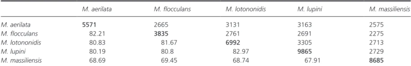
and 3835, respectively). The distribution of genes into COG categories was similar, but not identical in all com-pared genomes (Fig 5). In addition, M. massiliensis shared 2575, 2275, 2713, and 2729 orthologous genes with M.
aer-ilata, M. flocculans, M. lotononidis, and M. lupini,
respec-tively (Table 9). The AGIOS values ranged from 80.19 to 82.97 among compared Microvirga species except
M. massiliensis. When compared to other species, the
AGIOS value ranged from 67.91% with M. lupini to 69.45% with M. flocculans. The DDH was of 21.7% ±2.33 with
M. lupini, 21% ±2.33 with M. aerilata, 20.90% ±2.33
with M. flocculans, and 20.30% ±2.32 with M. lotononidis. These data confirm M. massiliensis as a unique species.
Discussion
The Methylobacteriaceae family is comprised of four genera:
Meganema, Methylobacterium, Psychroglaciecola, and Microvirga. Currently, the genus Microvirga contains nine
species (Kanso and Patel 2003; Zhang et al. 2009; Weon et al. 2010; Ardley et al. 2012; Radl et al. 2014). The first species of the Microvirga genus was M. subterranea strain Fail4T (Kanso and Patel 2003) and was first deposited as
Table 5. Differential characteristics of Microvirga massiliensis sp. nov., strain JC119T, Microvirga aerophila strain 5420S- 12T (Weon et al. 2010),
Microvirga aerilata strain 5420S- 16T (Weon et al. 2010), Methylobacterium nodulans strain ORS 2060T (Jourand et al. 2004), and Methylobacterium
populi strain BJ001T (Aken et al. 2004).
Properties M. massiliensis M. aerophila M. aerilata M. nodulans
Cell diameter (μm) 2.28 0.8–1.1 1.2–1.5 0.5–1
Oxygen requirement Aerobic Aerobic Aerobic Aerobic
Gram stain Negative Negative Negative Negative
Motility − − − ± Endospore formation − − − − Production of Alkaline phosphatase − − + NA Acid phosphatase + + + NA Catalase + NA NA NA Oxidase − NA NA NA Nitrate reductase + NA NA + Urease − NA NA + α- Galactosidase − − − NA β- Galactosidase − − − − β- Glucuronidase − − − NA α- Glucosidase − − − NA β- Glucosidase − − − − Esterase + + + NA Esterase lipase + − + NA
Naphthol- AS- BI- phosphohydrolase + + + NA
N- Acetyl- β- glucosaminidase − − − NA α- Mannosidase − − − NA α- Fucosidase − − − NA Leucine arylamidase + − + NA Valine arylamidase − − − NA Cystine arylamidase + − − NA α- Chymotrypsin − − − NA Trypsin + − + NA Acid from l- Arabinose − NA NA + d- Mannose − NA NA + d- Mannitol − NA NA − d- Trehalose + NA NA + d- Mannose − NA NA +
Habitat Human gut Air Air C. podocarpa
Figure 3. Phylogenetic tree highlighting the position of Microvirga massiliensis JC119T relative to other type strains within the Microvirga genus and closely related species. The strains and their corresponding accession number for 16S rRNA genes are indicated in parentheses. Cupriavidus taiwanensis
strain LMG 19424T was used as outgroup. The scale bar represents a 2% nucleotide sequence divergence.
Figure 4. Circular representation of the
Microvirga massiliensis strain JC119T genome. From outside to the center: contigs (red/gray), COG category of genes on the forward strand (three circles), genes on forward strand (blue circle), genes on the reverse strand (red circle), COG category on the reverse strand (three circles), GC content.
Corbulabacter subterraneus. Species of the genus Microvirga
are mostly found in soil. To the best of our knowledge, no Microvirga strain has previously been isolated from humans. M. massiliensis strain JC119T is the type strain
of M. massiliensis sp. nov., a new species of the genus
Microvirga. The genome of M. massiliensis JC119T is
9,207,211 bp long, representing the largest genome of a bacteria isolated from a human sample (Lagier et al. 2012a,b,c,d). It ranks 139th among the largest bacterial genomes according to the Genomes OnLine Database (GOLD) (Kyrpides 1999). All these bacteria whose genome is larger than M. massiliensis are found in the environment (soil, ocean, sand, and landfill). Different characteristics of this bacterium can explain a broad genome. The pro-portion of no detectable homologs (ORFans) from the gene prediction is about 17%, which corresponds to 1500 ORFans. The number of noncoding genes is important. This genome has 77 RNA: 56 tRNA and 21 rRNA. The number of RNA appears to be related to genome size (Klappenbach et al. 2000). For bacterial genomes, the copy number of rRNA operons varies from 1 to 15, for example, for Clostridium paradoxum (Rainey et al. 1996). There are
also a large number of transposases that create repeat ele-ments in this genome. The abundance of transposable elements correlates positively with genome size (Touchon and Rocha 2007; Chénais et al. 2012; Iranzo et al. 2014).
Culture remains a critical step of microbiology (Lagier et al. 2015). However, thanks to the multiplication of culture conditions, culturomics increased the knowledge of the human gut microbiota repertoire by 77% and thus helps to decipher the “dark matter” (Lagier et al. 2012a,b,c,d, 2015). Culturomics allows the generation of stable and robust data such as pure culture and genome sequencing by contrast with metagenomics, which shows a significant lack of reproducibility among laboratories (Angelakis et al. 2012).
Taxonogenomics allows the consideration of proteome and genome sequences, which must be a part of the taxonomic description of bacteria. To describe a new
Table 6. NRPS/PKS predicted for Microvirga massiliensis.
PKS/NRPS on proteome (best hit)
Streptomyces avermitilis MA- 4680 (NC_003155) 1 Streptomyces sp. FR- 008 (AY310323)
Mycobacterium avium 104 (CP000479)
Streptomyces griseoviridis (AB469822) 1 Sorangium cellulosum (AF210843)
Bacillus amyloliquefaciens FZB42 (AJ576102)
Micromonospora chalcea (EU443633) 1 Streptomyces coelicolor A3(2) (NC_003888)
Streptomyces venezuelae ATCC 10712 (FR845719) 1 Saccharopolyspora erythraea NRRL 2338 (NC_009142)
Acidobacteria bacterium A11 (JF342591) 1 Sorangium cellulosum (DQ897667)
Myxococcus xanthus DK 1622 (CP000113) 1 Mycobacterium avium subsp. paratuberculosis K- 10 (AE016958) Nocardia farcinica IFM 10152 (NC_006361) 1 Mycobacterium gilvum PYR- GCK (NC_009338)
Chitinophaga sancti (HQ680975)
Sorangium cellulosum (HE616533) 1 Mycobacterium tuberculosis CDC 1551 (NC_002755)
Mycobacterium sp. MCS (CP000384) Sorangium cellulosum (AM407731) Streptomyces sp. MP39- 85 (FJ872525) Streptomyces antibioticus (FJ545274) Tistrella mobilis KA081020- 065 (NC_017956) Streptomyces ambofaciens ATCC 23877 (AM238664) Mycobacterium smegmatis str. MC2 155 (CP000480) Thermomonospora curvata DSM 43183 (NC_013510) Microcystis aeruginosa PCC 7806 (AF183408)
1This best hit corresponded to several genes (to two from seven) in
M. massiliensis.
Table 7. Number of genes associated with the 25 general COG
func-tional categories.
Code Value % value Description J 195 2.25 Translation
A 0 0 RNA processing and modification K 359 4.13 Transcription
L 263 3.03 Replication, recombination, and repair
B 9 0.1 Chromatin structure and dynamics D 29 0.33 Cell cycle control, mitosis, and
meiosis Y 0 0 Nuclear structure V 66 0.76 Defense mechanisms
T 283 3.26 Signal transduction mechanisms M 263 3.03 Cell wall/membrane biogenesis N 67 0.77 Cell motility
Z 0 0 Cytoskeleton
W 0 0 Extracellular structures
U 80 0.92 Intracellular trafficking and secretion O 184 2.12 Post- translational modification,
protein turnover, chaperones C 347 4 Energy production and conversion G 423 4.87 Carbohydrate transport and
metabolism
E 788 9.07 Amino acid transport and metabolism
F 88 1.01 Nucleotide transport and metabolism
H 175 2.01 Coenzyme transport and metabolism
I 256 2.95 Lipid transport and metabolism P 368 4.24 Inorganic ion transport and
metabolism
Q 229 2.64 Secondary metabolites biosynthesis, transport, and catabolism R 802 9.23 General function prediction only S 525 6.04 Function unknown
bacterial species, taxonogenomics uses both phenotypic and genotypic data (Ramasamy et al. 2012; Lagier et al. 2015). On the basis of taxonogenomics, we formally pro-pose the creation of M. massiliensis sp. nov. containing the strain JC119T.
Description of Microvirga massiliensis sp nov
Microvirga massiliensis (mas.si.li.en’sis. L. masc. adj.
massil-iensis of Massilia, the Latin name of Marseille, France,
where M. massiliensis was isolated) is a gram- negative, oxidase- negative, and catalase- positive bacterium. This bac-terium is an aerobic, nonmotile rod, and non- spore- forming, individual cell exhibiting a diameter of 2.28 μm. This bacterium is positive for nitrate reductase, d- glucose, esterase, esterase lipase, leucine arylamidase, cysteine ar-ylamidase, trypsin, acid phosphatase, and naphthol- AS- BI- phosphohydrolase. This bacterium is susceptible to rifampicin, doxycycline, erythromycin, amoxicillin, gen-tamicin, ciprofloxacin, ceftriaxone, amoxicillin with clavu-lanic acid, penicillin, imipenem, tobramycin, metronidazole,
Figure 5. Distribution of functional classes of
predicted genes in the genomes from
Microvirga massiliensis, Microvirga aerilata, Microvirga flocculans, Microvirga lotononidis, and Microvirga lupini chromosomes, according
to the COG category.
Table 9. Genomic comparison of Microvirga massiliensis and four other Microvirga species.
M. aerilata M. flocculans M. lotononidis M. lupini M. massiliensis M. aerilata 5571 2665 3131 3163 2575
M. flocculans 82.21 3835 2761 2691 2275
M. lotononidis 80.83 81.67 6992 3305 2713
M. lupini 80.19 80.8 82.97 9865 2729
M. massiliensis 68.69 69.45 68.74 67.91 8685 Numbers of orthologous proteins shared between genomes (upper right) and AGIOS values (lower left); bold numbers indicate the numbers of proteins per genome.
Table 8. Comparative genomic features of Microvirga massiliensis, Microvirga aerilata, Microvirga flocculans, Microvirga lotononidis, and Microvirga
lupini.
M. massiliensis M. aerilata M. flocculans M. lotononidis M. lupini
Genome size (Mb) 9.3 5.7 4.1 7.1 9.7
DNA G+C content (%) 63.3 63.1 64.6 62.9 61.6
Protein- coding genes 8685 5571 3835 6991 9865
rRNA 21 13 5 6 3
and amikacin. The range of temperature growth is 28–55°C (with an optimum at 37°C). The potential pathogenicity of the type strain JC119T (=CSUR P153 = DSM 26813)
is unknown but was isolated from the stool specimen from a Senegalese male, in Marseille (France), using culturomics (Lagier et al. 2012a,b,c,d; Dubourg et al. 2014). The G+C content of the genome is 63.28%. The partial 16S rRNA sequence of M. massiliensis was deposited in GenBank under the accession number JF824802. The whole- genome sequence of Microvirga massiliensis strain JC119T (=CSUR
P153 = DSM 26813) has been deposited in EMBL under accession numbers CDSD01000001–CDSD01000365 for contigs and LN811350–LN811399 for scaffolds.
Acknowledgments
The authors thank the Xegen Company (www.xegen.fr) for automating the genomic annotation process.
Funding Information
No funding information provided.
Conflict of Interest
None declared.
References
Adler, J., and M. D. Margaret. 1967. A method for measuring the motility of bacteria and for comparing random and non- random motility. Microbiology 46:161–173.
Aken, B. V., C. M. Peres, S. Lafferty Doty, J. Moon Yoon, and J. L. Schnoor. 2004. Methylobacterium populi sp. nov., a novel aerobic, pink- pigmented, facultatively methylotrophic, methane- utilizing bacterium isolated from poplar trees (Populus deltoides×nigra DN34). Int. J. Syst. Evol. Microbiol. 54:1191–1196.
Altschul, S. F., W. Gish, W. Miller, E. W. Myers, and D. J. Lipman. 1990. Basic local alignment search tool. J. Mol. Biol. 215:403–410.
Angelakis, E., F. Armougom, M. Million, and D. Raoult. 2012. The relationship between gut microbiota and weight gain in humans. Future Microbiol. 7:91–109. Ardley, J. K., M. A. Parker, S. E. De Meyer, R. D.
Trengove, G. W. O’Hara, W. G. Reeve, et al. 2012. Microvirga lupini sp. nov., Microvirga lotononidis sp. nov. and Microvirga zambiensis sp. nov. are
alphaproteobacterial root- nodule bacteria that specifically nodulate and fix nitrogen with geographically and taxonomically separate legume hosts. Int. J. Syst. Evol. Microbiol. 62:2579–2588.
Ashburner, M., C. A. Ball, J. A. Blake, D. Botstein, H. Butler, J. M. Cherry, et al. 2000. Gene ontology: tool
for the unification of biology. The Gene Ontology Consortium. Nat. Genet. 25:25–29.
Auch, A. F., M. von Jan, H.-P. Klenk, and M. Göker. 2010. Digital DNA–DNA hybridization for microbial species delineation by means of genome- to- genome sequence comparison. Stand. Genomic Sci. 2:117–134.
Bailey, A. C., M. Kellom, A. T. Poret-Peterson, K. Noonan, H. E. Hartnett, and J. Raymond. 2014. Draft genome sequence of Microvirga sp. strain BSC39, isolated from biological soil crust of Moab, Utah. Genome Announc. 2:e01197-14.
Carver, T. J., K. M. Rutherford, M. Berriman, M.-A. Rajandream, B. G. Barrell, and J. Parkhill. 2005. ACT: the Artemis comparison tool. Bioinformatics (Oxford, England) 21:3422–3423.
Carver, T., N. Thomson, A. Bleasby, M. Berriman, and J. Parkhill. 2009. DNAPlotter: circular and linear interactive genome visualization. Bioinformatics (Oxford, England) 25:119–120.
Chénais, B., A. Caruso, S. Hiard, and N. Casse. 2012. The impact of transposable elements on eukaryotic genomes: from genome size increase to genetic adaptation to stressful environments. Gene 509:7–15.
Clark, A. E., E. J. Kaleta, A. Arora, and D. M. Wolk. 2013. Matrix- assisted laser desorption ionization- time of flight mass spectrometry: a fundamental shift in the routine practice of clinical microbiology. Clin. Microbiol. Rev. 26:547–603.
Conway, K. R., and C. N. Boddy. 2013. ClusterMine360: a database of microbial PKS/NRPS biosynthesis. Nucleic Acids Res. 41:D402–D407.
Darling, A. E., B. Mau, and N. T. Perna. 2010.
progressiveMauve: multiple genome alignment with gene gain, loss and rearrangement. PLoS One 5:e11147. Dubourg, G., J. C. Lagier, C. Robert, F. Armougom, P.
Hugon, S. Metidji, et al. 2014. Culturomics and pyrosequencing evidence of the reduction in gut microbiota diversity in patients with broad- spectrum antibiotics. Int. J. Antimicrob. Agents 44:117–124. Field, D., G. Garrity, T. Gray, N. Morrison, J. Selengut,
P. Sterk, et al. 2008. The minimum information about a genome sequence (MIGS) specification. Nat. Biotechnol. 26:541–547.
Garrity, G. M., J. A. Bell, and T. Lilburn. 2005a. Bergey’s manual of systematic bacteriology. 2nd ed. Vol. 2. Part B. Phylum XIV. Proteobacteria phyl. nov., Springer, New York, NY, USA, 2001–2012.
Garrity, G. M., J. A. Bell, and T. Lilburn. 2005b. Bergey’s manual of systematic bacteriology. 2nd ed. Vol. 2. Part C. Class I. Alphaproteobacteria class. nov., Springer, New York, NY, USA, 2001–2012.
Ghodbane, R., D. Raoult, and M. Drancourt. 2014. Dramatic reduction of culture time of Mycobacterium tuberculosis. Sci. Rep. 4:4236.
Gregersen, T. 1978. Rapid method for distinction of gram- negative from gram- positive bacteria. Eur. J. Appl. Microbiol. Biotechnol. 5:123–127.
Hugon, P., A. K. Mishra, J. C. Lagier, T. T. Nguyen, C. Couderc, D. Raoult, et al. 2013. Non- contiguous finished genome sequence and description of Brevibacillus massiliensis sp. nov. Stand. Genomic Sci. 8:1–14.
Humble, M. W., A. King, and I. Phillips. 1977. API ZYM: a simple rapid system for the detection of bacterial enzymes. J. Clin. Pathol. 30:275–277.
Hyatt, D., G.-L. Chen, P. F. Locascio, M. L. Land, F. W. Larimer, and L. J. Hauser. 2010. Prodigal: prokaryotic gene recognition and translation initiation site identification. BMC Bioinformatics 11:119. Iranzo, J., M. J. Gómez, F. J. López de Saro, and
S. Manrubia. 2014. Large- scale genomic analysis suggests a neutral punctuated dynamics of transposable elements in bacterial genomes. PLoS Comput. Biol. 10:e1003680.
Jourand, P., E. Giraud, G. Béna, A. Sy, A. Willems, M. Gillis, et al. 2004. Methylobacterium nodulans sp. nov., for a group of aerobic, facultatively methylotrophic, legume root- nodule- forming and nitrogen- fixing bacteria. Int. J. Syst. Evol. Microbiol. 54:2269–2273.
Käll, L., A. Krogh, and E. L. L. Sonnhammer. 2004. A combined transmembrane topology and signal peptide prediction method. J. Mol. Biol. 338:1027–1036. Kanso, S., and B. K. C. Patel. 2003. Microvirga subterranea
gen. nov., sp. nov., a moderate thermophile from a deep subsurface Australian thermal aquifer. Int. J. Syst. Evol. Microbiol. 53:401–406.
Kent, W. J.. 2002. BLAT – the BLAST- like alignment tool. Genome Res. 12:656–664. Article published online before March 2002.
Klappenbach, J. A., J. M. Dunbar, and T. M. Schmidt. 2000. rRNA operon copy number reflects ecological strategies of bacteria. Appl. Environ. Microbiol. 66:1328–1333.
Kokcha, S., D. Ramasamy, J. C. Lagier, C. Robert, D. Raoult, and P.-E. Fournier. 2012. Non- contiguous finished genome sequence and description of Brevibacterium senegalense sp. nov. Stand. Genomic Sci. 7:233–245.
Koskiniemi, S., J. G. Lamoureux, K. C. Nikolakakis, C. t’Kint de Roodenbeke, M. D. Kaplan, D. A. Low, et al. 2013. Rhs proteins from diverse bacteria mediate intercellular competition. Proc. Natl Acad. Sci. USA 110:7032–7037.
Kuykendall, L. D. 2005. Bergy’s manual of systematic bacteriology. 2nd ed. Order VI. Rhizobiales ord. nov., Springer, New York, NY, USA, 2001–2012.
Kyrpides, N. C.. 1999. Genomes OnLine Database (GOLD 1.0): a monitor of complete and ongoing genome projects world- wide. Bioinformatics (Oxford, England) 15:773–774.
La Scola, B., and D. Raoult. 2009. Direct identification of bacteria in positive blood culture bottles by matrix- assisted laser desorption ionisation time- of- flight mass spectrometry. PLoS One 4:e8041.
Lagesen, K., P. Hallin, E. A. Rødland, H.-H. Staerfeldt, T. Rognes, and D. W. Ussery. 2007. RNAmmer:
consistent and rapid annotation of ribosomal RNA genes. Nucleic Acids Res. 35:3100–3108.
Lagier, J. C., F. Armougom, M. Million, P. Hugon,
I. Pagnier, C. Robert, et al. 2012a. Microbial culturomics: paradigm shift in the human gut microbiome study. Clin. Microbiol. Infect. 18:1185–1193.
Lagier, J. C., F. Armougom, A. K. Mishra, T. T. Nguyen, D. Raoult, and P.-E. Fournier. 2012b. Non- contiguous finished genome sequence and description of Alistipes timonensis sp. nov. Stand. Genomic Sci. 6:315–324. Lagier, J. C., K. El Karkouri, T. T. Nguyen, F. Armougom,
D. Raoult, and P.-E. Fournier. 2012c. Non- contiguous finished genome sequence and description of Anaerococcus senegalensis sp. nov. Stand. Genomic Sci. 6:116–125. Lagier, J. C., G. Gimenez, C. Robert, D. Raoult, and P.-E.
Fournier. 2012d. Non- contiguous finished genome sequence and description of Herbaspirillum massiliense sp. nov. Stand. Genomic Sci. 7:200–209.
Lagier, J. C., K. El Karkouri, A. K. Mishra, C. Robert, D. Raoult, and P.-E. Fournier. 2013a. Non contiguous- finished genome sequence and description of Enterobacter massiliensis sp. nov. Stand. Genomic Sci. 7:399–412. Lagier, J. C., K. Elkarkouri, R. Rivet, C. Couderc,
D. Raoult, and P.-E. Fournier. 2013b. Non contiguous- finished genome sequence and description of
Senegalemassilia anaerobia gen. nov., sp. nov. Stand. Genomic Sci. 7:343–356.
Lagier, J. C., P. Hugon, S. Khelaifia, P.-E. Fournier, B. La Scola, and D. Raoult. 2015. The rebirth of culture in microbiology through the example of culturomics to study human gut microbiota. Clin. Microbiol. Rev. 28:237–264. doi:10.1128/CMR.00014- 14.
Lechner, M., S. Findeiss, L. Steiner, M. Marz, P. F. Stadler, and S. J. Prohaska. 2011. Proteinortho: detection of (co- ) orthologs in large- scale analysis. BMC Bioinformatics 12:124.
Lowe, T. M., and S. R. Eddy. 1997. tRNAscan- SE: a program for improved detection of transfer RNA genes in genomic sequence. Nucleic Acids Res. 25:955–964. Matuschek, E., D. F. J. Brown, and G. Kahlmeter. 2014.
Development of the EUCAST disk diffusion antimicrobial susceptibility testing method and its implementation in routine microbiology laboratories. Clin. Microbiol. Infect. 20:O255–O266.
Meier-Kolthoff, J. P., A. F. Auch, H.-P. Klenk, and M. Göker. 2013. Genome sequence- based species delimitation with confidence intervals and improved distance functions. BMC Bioinformatics 14:60.
Oren, A., and G. M. Garrity. 2014. List of new names and new combinations previously effectively, but not validly, published. Int. J. Syst. Evol. Microbiol. 64:1–6.
Pray, L. 2008. Transposons: the jumping genes. Nat. Educ. 1:1–204.
Radl, V., J. L. Simões-Araújo, J. Leite, S. R. Passos, L. M. V. Martins, G. R. Xavier, et al. 2014. Microvirga vignae sp. nov., a root nodule symbiotic bacterium isolated from cowpea grown in semi- arid Brazil. Int. J. Syst. Evol. Microbiol. 64:725–730.
Rainey, F. A., N. L. Ward-Rainey, P. H. Janssen, H. Hippe, and E. Stackebrandt. 1996. Clostridium paradoxum DSM 7308T contains multiple 16S rRNA genes with
heterogeneous intervening sequences. Microbiology (Reading, England) 142:2087–2095.
Ramasamy, D., S. Kokcha, J. C. Lagier, T. T. Nguyen, D. Raoult, and P.-E. Fournier. 2012. Genome sequence and description of Aeromicrobium massiliense sp. nov. Stand. Genomic Sci. 7:246–257.
Raoult, D., S. Audic, C. Robert, C. Abergel, P. Renesto, H. Ogata, et al. 2004. The 1.2- megabase genome sequence of Mimivirus. Science (New York, N.Y.) 306:1344–1350.
Reeve, W., M. Parker, R. Tian, L. Goodwin, H. Teshima, R. Tapia, et al. 2014. Genome sequence of Microvirga lupini strain LUT6T, a novel Lupinus alphaproteobacterial microsymbiont from Texas. Stand. Genomic Sci.
9:1159–1167.
Richter, M., and R. Rosselló-Móra. 2009. Shifting the genomic gold standard for the prokaryotic species definition. Proc. Natl Acad. Sci. USA 106:19126–19131. Rosselló-Mora, R. 2006. DNA–DNA reassociation methods
applied to microbial taxonomy and their critical evaluation. Pp.23–50 in Molecular identification, systematics, and population structure of prokaryotes. Springer, Berlin Heidelberg. http://link.springer.com/ chapter/10.1007/978-3-540-31292-5_2.
Roux, V., K. El Karkouri, J. C. Lagier, C. Robert, and D. Raoult. 2012. Non- contiguous finished genome sequence and description of Kurthia massiliensis sp. nov. Stand. Genomic Sci. 7:221–232.
Schaeffer, P., J. Millet, and J.-P. Aubert. 1965. Catabolic repression of bacterial sporulation. Proc. Natl Acad. Sci. USA 54:704.
Seng, P., M. Drancourt, F. Gouriet, B. La Scola, P.-E. Fournier, J.-M. Rolain, et al. 2009. Ongoing revolution in bacteriology: routine identification of bacteria by
matrix- assisted laser desorption ionization time- of- flight mass spectrometry. Clin. Infect. Dis. 49:543–551. Seng, P., J.-M. Rolain P.-E. Fournier M. Drancourt and D.
Raoult. 2010. MALDI- TOF- mass spectrometry applications in clinical microbiology. Future Microbiol. 5:1733–1754. Seng, P., C. Abat, J.-M. Rolain, P. Colson, J. C. Lagier,
F. Gouriet, et al. 2013. Identification of rare
pathogenic bacteria in a clinical microbiology laboratory: impact of matrix- assisted laser desorption ionization- time of flight mass spectrometry. J. Clin. Microbiol. 51:2182–2194.
Stackebrandt, E., and J. Ebers. 2006. Taxonomic parameters revisited: tarnished gold standards. Microbiol. Today 33:152–155.
Tamura, K., G. Stecher, D. Peterson, A. Filipski, and S. Kumar. 2013. MEGA6: Molecular Evolutionary Genetics Analysis Version 6.0. Mol. Biol. Evol. 30:2725–2729.
Tatusov, R. L., D. A. Natale, I. V. Garkavtsev, T. A. Tatusova, U. T. Shankavaram, B. S. Rao, et al. 2001. The COG database: new developments in phylogenetic classification of proteins from complete genomes. Nucleic Acids Res. 29:22–28.
Tindall, B. J., R. Rosselló-Móra, H.-J. Busse, W. Ludwig, and P. Kämpfer. 2010. Notes on the characterization of prokaryote strains for taxonomic purposes. Int. J. Syst. Evol. Microbiol. 60:249–266.
Touchon, M., and E. P. C. Rocha. 2007. Causes of insertion sequences abundance in prokaryotic genomes. Mol. Biol. Evol. 24:969–981.
Wayne, L. G., D. J. Brenner, R. R. Colwell, P. A. D. Grimont, O. Kandler, M. I. Krichevsky, et al. 1987. Report of the ad hoc committee on reconciliation of approaches to bacterial systematics. Int. J. Syst. Bacteriol. 37:463–464.
Welker, M., and E. R. B. Moore. 2011. Applications of whole- cell matrix- assisted laser- desorption/ionization time- of- flight mass spectrometry in systematic microbiology. Syst. Appl. Microbiol. 34:2–11.
Weon, H.-Y, S.-W. Kwon, J.-A. Son, E.-H. Jo, S.-J. Kim, Y.-S. Kim, et al. 2010. Description of Microvirga aerophila sp. nov. and Microvirga aerilata sp. nov., isolated from air, reclassification of Balneimonas flocculans Takeda et al. 2004 as Microvirga flocculans comb. nov. and emended description of the genus Microvirga. Int. J. Syst. Evol. Microbiol. 60:2596–2600.
Woese, C. R., O. Kandler, and M. L. Wheelis. 1990. Towards a natural system of organisms: proposal for the domains Archaea, Bacteria, and Eucarya. Proc. Natl Acad. Sci. USA 87:4576–4579.
Yin, Y., and D. Fischer. 2006. On the origin of microbial ORFans: quantifying the strength of the evidence for viral lateral transfer. BMC Evol. Biol. 6:63.
Yin, Y., and D. Fischer. 2008. Identification and
investigation of ORFans in the viral world. BMC Genom. 9:24.
Zhang, J., F. Song, Y. H. Xin, J. Zhang, and C. Fang. 2009. Microvirga guangxiensis sp. nov., a novel
Alphaproteobacterium from soil, and emended description of the genus Microvirga. Int. J. Syst. Evol. Microbiol. 59:1997–2001.
Supporting Information
Additional supporting information may be found in the online version of this article:
Figure S1. Pulsed field gel electrophoresis (PFGE) and
Southern blot (A) PFGE of intact genomic DNA (ND) and SpeI- digested DNA from Microvirga massiliensis. Electrophoresis was performed in 1% agarose in 0.5× TBE buffer, and the pulse time was ramped from 5 to 50 s for 20 h at a voltage of 5 V/cm for 20 h at 14°C. Gel was stained with ethidium bromide. Low Range PFG
Marker (Biolabs, New England) were used as size mark-ers. Sizes are indicated on the left in kilobase pairs. Southern blot (B and C) using DIG- labeled probes “scaffold 24” and “scaffold 35,” respectively. The probe “scaffold 24” recognized the potential plasmid DNA band observed with the uncut genomic DNA. The intact genomic DNA is recognized by the “scaffold 35” probe, suggesting that this scaffold is a part of the genomic DNA.Table S1. The percentage sequence identity and sequence coverage of the 16S rRNA of Microvirga massiliensis with other strains of Microvirga.