HAL Id: hal-02055778
https://hal.sorbonne-universite.fr/hal-02055778
Submitted on 4 Mar 2019
HAL is a multi-disciplinary open access
archive for the deposit and dissemination of sci-entific research documents, whether they are pub-lished or not. The documents may come from teaching and research institutions in France or abroad, or from public or private research centers.
L’archive ouverte pluridisciplinaire HAL, est destinée au dépôt et à la diffusion de documents scientifiques de niveau recherche, publiés ou non, émanant des établissements d’enseignement et de recherche français ou étrangers, des laboratoires publics ou privés.
heterogeneity
Pascale Richard, Flavie Ader, Maguelonne Roux, Erwan Donal,
Jean-Christophe Eicher, Nadia Aoutil, Olivier Huttin, Christine Selton-Suty,
Damien Coisne, Guillaume Jondeau, et al.
To cite this version:
Pascale Richard, Flavie Ader, Maguelonne Roux, Erwan Donal, Jean-Christophe Eicher, et al.. Tar-geted panel sequencing in adult patients with left ventricular non-compaction reveals a large genetic heterogeneity. Clinical Genetics, Wiley, 2019, 95 (3), pp.356-367. �10.1111/cge.13484�. �hal-02055778�
For Review Only
Targeted panel sequencing in adult patients with left ventricular non-compaction reveals a large genetic
heterogeneity
Journal: Clinical Genetics Manuscript ID CGE-00461-2018.R1 Manuscript Type: Original Article Date Submitted by the Author: n/a
Complete List of Authors: Richard, Pascale; Hopital Universitaire Pitie Salpetriere, UF Cardiogénétique et Myogénétique moléculaire et cellulaire
Ader, Flavie; Hopital Universitaire Pitie Salpetriere, UF Cardiogenetic and Myogenetic;
Roux, Maguelone ; Sorbonne Universités, UPMC Unive., INSERM UMR_S 1166
Donal, Erwan; CHRU Pontchaillou, Service de Cardiologie Eicher, Jean; Service de Cardiologie, CHU DIJON
Aoutil, Nadia; Hopitaux Universitaires de la Pitié Salpêtrière Charles Foix, UF Cardiogenetics
Huttin, Olivier; CHRU Nancy, Service de Cardiologie
Selton-Suty, Christine; Chu nancy - hopitaux de brabois, Cardiology COISNE, Damien; Centre Hospitalier Universitaire de Poitiers, Cardiologie JONDEAU, Guillaume; Hopital Bichat - Claude-Bernard, Cardiologie DAMY, Thibaud; Hopital Henri Mondor, Cardiology
MANSENCAL, Nicolas; Hopital Ambroise-Pare, Cardiology CASALTA, Anne Claire; Hopital de la Timone, Cardiology MICHEL, Nicolas; Hopital de la Timone, Cardiology HAENTJENS, Julie; Hopital de la Timone, Cardiology Faivre, Laurence; Hopital d'Enfants, Centre de Génétique LAVOUTE, Cécile; Hopital de la Timone, Cardiology Nguyen, Karine; APHM, medical genetics
Tregouet, David; INSERM, UMR_S 1166; ICAN Institute,
HABIB, Gilbert; Timone Hospital, Marseille , Cardiology Department, CHARRON, Philippe; Hopital Universitaire Pitie Salpetriere, Centre de référence des maladies cardiaques héréditaires
Key Words: Left ventricular non compaction, cardiomyopathy, molecular genetic, next generation sequencing
For Review Only
3 4 5 6 7 8 9 10 11 12 13 14 15 16 17 18 19 20 21 22 23 24 25 26 27 28 29 30 31 32 33 34 35 36 37 38 39 40 41 42 43 44 45 46 47 48 49 50 51 52 53 54 55 56 57For Review Only
Targeted panel sequencing in adult patients
with left ventricular non-compaction reveals a large genetic
heterogeneity
Running title: Genetic complexity of adult left ventricle non compaction
Pascale Richard1,2, Flavie Ader1, Maguelonne Roux2, Erwan Donal3, Jean-Christophe Eicher4, Nadia Aoutil1, Olivier Huttin5, Christine Selton-Suty5, Damien Coisne6, Guillaume Jondeau7, Thibaud Damy8, Nicolas Mansencal9, Anne-Claire Casalta10, Nicolas Michel10,
Julie Haentjens10, Laurence Faivre11, Cecile Lavoute10, Karine Nguyen12, David-Alexandre Tregouët2, Gilbert Habib10,13*, Philippe Charron2,14,15 *.
Conflict of interest statement: none declared for each author
1- APHP, UF Cardiogénétique et Myogénétique, Service de Biochimie Métabolique,
Hôpitaux Universitaires de la Pitié- Salpêtrière- Charles Foix, 47-83 Bd de l’Hôpital, Paris 2- Sorbonne Universités, UPMC Univ. Paris 06, INSERM, UMR_S 1166 and ICAN Institute
for Cardiometabolism and Nutrition, Paris
3- Service de cardiologie, Centre Hospitalier Régional Universitaire Pontchaillou, 2, Rue Henri le Guilloux 35000 Rennes
4- Service de cardiologie, CHU Dijon Bourgogne - Hôpital François Mitterrand, 2 bd Maréchal de Lattre de Tassigny, 21000 DIJON
5- Service de cardiologie, CHU de Nancy, Hôpitaux de Brabois, rue du Morvan, 54500 Vandœuvre-lès-Nancy
6- Service de cardiologie, CHU de Poitiers, 2, Rue de la Milétrie, 86000 Poitiers
7- APHP, Service cardiologie, CHU Paris Nord-Val de Seine - Hôpital Xavier Bichat-Claude Bernard, 46 rue Henri Huchard, 75018 Paris
8- APHP, Service cardiologie, CHU Henri Mondor, 51 Avenue du Maréchal de Lattre de Tassigny, 94010 Créteil 3 4 5 6 7 8 9 10 11 12 13 14 15 16 17 18 19 20 21 22 23 24 25 26 27 28 29 30 31 32 33 34 35 36 37 38 39 40 41 42 43 44 45 46 47 48 49 50 51 52 53 54 55 56
For Review Only
2
9- APHP, Service de Cardiologie, CHU Ambroise Paré, 9 av Charles de Gaulle, 92100 Boulogne Billancourt
10- APHM, La Timone Hospital, Cardiology Department, Marseille
11- Service de génétique, CHU Dijon Bourgogne - Hôpital François Mitterrand, 2 bd Maréchal de Lattre de Tassigny, 21000 DIJON
12- APHM, Département de génétique médicale – APHM, La Timone Hospital, INSERM UMR_S 910, Marseille
13- Aix Marseille Univ, IRD, APHM, MEPHI, IHU-Méditerranée Infection, Marseille, France 14- APHP, Centre de référence pour les maladies cardiaques héréditaires, Hôpital
Pitié-Salpêtrière, Paris
15- Université de Versailles Saint Quentin, Service de Génétique, Hôpital Ambroise Paré, Boulogne-Billancourt
*equal last author
CONFLIT OF INTEREST: None declared for any co-authors.
ACKNOWLEDGMENTS
Lab team and clinical investigators are warmly acknowledged for the technical realization of this project.
This technical steps and salary of Nadia Aoutil were supported by funds from AP-HM (PHRC 2011-A - 00987-34, coordinator Pr. G. Habib, Marseille). Genetic Labs and sequencing platform was located at APHP, Pitié Salpêtrière Hospital. M. Roux was financially supported by the Fondation pour la Recherche Médicale and the GENMED Laboratory of Excellence on Medical Genomics (ANR-10-LABX-0013). The project was also supported by BestAgeing FP7 European Community project; Leducq Transatlantic Network: "Genomic, epigenomic 3 4 5 6 7 8 9 10 11 12 13 14 15 16 17 18 19 20 21 22 23 24 25 26 27 28 29 30 31 32 33 34 35 36 37 38 39 40 41 42 43 44 45 46 47 48 49 50 51 52 53 54 55 56 57
For Review Only
and systems dissection of mechanisms underlying dilated cardiomyopathy"; PROMEX stiftung fur die forschung Charitable Foundation
ABSTRACT
Left ventricular non-compaction (LVNC) is a cardiomyopathy that may be of genetic origin, however few data are available about the yield of mutation, the spectrum of genes and allelic variations. The aim of this study was to better characterize the genetic spectrum of isolated LVNC in a prospective cohort of 95 unrelated adult patients through the molecular investigation of 107 genes involved in cardiomyopathies and arrhythmias.
Fifty-two pathogenic or probably pathogenic variants were identified in 40 patients (42%) including 31 patients (32.5%) with single variant and 9 patients with complex genotypes (9.5%). Mutated patients tended to have younger age at diagnosis than patients with no identified mutation. The most prevalent genes were TTN, then HCN4, MYH7, and RYR2. The distribution includes 13 genes previously reported in LVNC and 10 additional candidate genes.
Our results show that LVNC is basically a genetic disease and support genetic counseling and cardiac screening in relatives. There is a large genetic heterogeneity, with predominant
TTN null mutations and frequent complex genotypes. The gene spectrum is close to the one
observed in dilated cardiomyopathy but with specific genes such as HCN4. We also identified new candidate genes that could be involved in this sub-phenotype of cardiomyopathy.
KEY WORDS: Left ventricular non compaction, cardiomyopathy, molecular genetic, next generation sequencing 3 4 5 6 7 8 9 10 11 12 13 14 15 16 17 18 19 20 21 22 23 24 25 26 27 28 29 30 31 32 33 34 35 36 37 38 39 40 41 42 43 44 45 46 47 48 49 50 51 52 53 54 55 56
For Review Only
4 INTRODUCTION
Left ventricular non-compaction (LVNC, OMIM300183) is a relatively rare cardiomyopathy, with or without LV dysfunction, characterized by excessively prominent trabeculations and associated deep recesses that communicate with the ventricular cavity1. LVNC is part of unclassified cardiomyopathies according to the European Society of Cardiology2 and to genetic cardiomyopathies by the America Heart Association3.
The prevalence of LVNC was estimated at 0.014% to 1.3% depending on the age of patients4,5. Multiple imaging techniques are usually useful for the diagnosis of LVNC, with variable echocardiographic or magnetic resonance imaging diagnostic criteria but no clear consensus so that the positive diagnosis may be challenging6. The phenotypic expression and evolution of isolated LVNC is highly variable, and clinical features can range from asymptomatic to symptomatic, with a relatively stable course over several years or an evolution towards severe complications including congestive heart failure, ventricular arrhythmia and sudden cardiac death, atrial arrhythmias and systemic embolic events6. LVNC is supposed to be related to a premature arrest of compaction of the loose myocardial meshwork during fetal embryogenesis, with persistent trabeculated myocardium, but the precise pathophysiology remains poorly understood. A family history is noticed in a significant proportion of patients and predominant mode of inheritance is autosomal dominant, with some cases with an X-linked transmission7.
Several genes have been identified as LVNC disease causing. The first reported genetic cause of isolated LVNC was described by Bleyl et al. with mutations in the X-linked TAZ gene, also responsible for Barth syndrome8. The sarcomere-encoding genes (MYH7,
ACTC1, TNNT2, MYBPC3, TMP1, and TNNI3) appear to account for 17 to 30% of LVNC9,10
but other genes such as DTNA (α-dystrobrevin), NKX-2.5, Z-line protein-encoding
ZASP/LDB3, lamin A/C (LMNA) genes have been also associated with LVNC11. Recently, the
TTN gene was also reported as involved in this disease12-14, with a highly heterogeneous 3 4 5 6 7 8 9 10 11 12 13 14 15 16 17 18 19 20 21 22 23 24 25 26 27 28 29 30 31 32 33 34 35 36 37 38 39 40 41 42 43 44 45 46 47 48 49 50 51 52 53 54 55 56 57
For Review Only
prevalence, as high as 19% and the first responsible gene for LVNC in a German study of 68 index cases13 to 7% in adults but 0% in children from a Dutch cohort of 327 patients14. Several studies have analyzed the spectrum of genes in LVNC9-16 but with heterogeneous
strategies (Sanger or Next-generation sequencing), usually in retrospective cohorts without imaging core-lab, and usually with a relatively small panel of genes (45 genes in the Dutch study14, exome sequencing in the recent German study13. Therefore, the exact spectrum of
LVNC-causing variants, their prevalence and their impact in genetic counseling remain poorly understood. Furthermore, the unique specificity of LVNC as an independent nosology entity has been questioned and LVNC has been suggested as an overlapping phenotype with hypertrophic or dilated cardiomyopathy9. To explore these issues, a prospective French national research program was launched and focused on consecutive adult patients with a recent diagnosis of isolated LVNC. The general aim was to better characterize the allelic and genetic spectrum of LVNC through a large panel of genes previously reported in the various cardiac hereditary diseases. The objective was also to identify new potential candidate genes that could be involved in the phenotype of LVNC.
METHODS
The present study was conducted as part of the Programme Hospitalier de Recherche Clinique (PHRC Ref: 2011-A - 00987-34, coordinator Pr. G. Habib, Marseille) aimed at describing the clinical spectrum of LVNC and at characterizing the genetic spectrum of LVNC through a next-generation sequencing (NGS) strategy in a new prospective cohort.
Patients, inclusion criteria
The study included unrelated patients with a minimal age at inclusion of 18 years old, enrolled between 2012 and 2013 in 13 French centers for inherited cardiac diseases. Collected data included clinical data (initial presentation, first symptoms, and data from cardiac and neurologic examination), family history, and tests including ECG, echocardiography, MRI, CT scan, Holter monitoring when available, as well as follow-up 3 4 5 6 7 8 9 10 11 12 13 14 15 16 17 18 19 20 21 22 23 24 25 26 27 28 29 30 31 32 33 34 35 36 37 38 39 40 41 42 43 44 45 46 47 48 49 50 51 52 53 54 55 56
For Review Only
6
data. Only patients with a recent diagnosis of isolated LVNC (maximum in the last 6 months before inclusion) were enrolled. All echocardiographic documents were sent and reviewed by a core lab (Marseille) to confirm the diagnosis. Diagnosis of isolated LVNC was considered definite when several criteria were present in left ventricle (LV): (i) multiple trabeculations with deep endomyocardial recesses, (ii) two-layer myocardial structure with a thin compacted (C) and a thick non compacted (NC) layer, (iii) color Doppler evidence of perfused intertrabecular recesses, (iv) systolic NC / C ratio > 2 (parasternal short-axis view); (v) no associated heart disease17. Cardiac MRI was also frequently performed in our series, with a NC/C ratio of >2.3 in diastole as the recommended threshold for the diagnosis of LVNC using this technique18. However, for the purpose of the current study, only the echocardiographic criteria were used as inclusion criteria. Only patients with a diagnosis validated by an imaging core lab confirming a definite diagnosis of LVNC were included. Informed consent, blood samples, and clinical evaluations were obtained from all patients, with a protocol approved by the Ethics Committee of AP-HM (Assistance Publique- Hôpitaux de Marseille).
Genetic analysis
Targeted gene enrichment, high-throughput sequencing: Patients' DNAs were extracted from peripheral blood with QIAsymphony ® (Qiagen, Hilden, Germany) and qualitatively checked using Tape Station DNA genomic array (Agilent, Santa Clara, USA). Custom targeted gene enrichment and DNA library preparation were performed using the Nimblegen EZ choice probes® and Kappa HTP Library preparation kit® according to the manufacturer’s
instructions (Nimblegen®, Roche Diagnostics, Madison, USA). The targeted regions include all coding exons and +/- 50 base pairs of flanking intronic regions of 107 genes known to be involved in cardiomyopathies (77 genes) and arrhythmias (30 genes) (Suppl. Table 1). The targeted regions were sequenced using the Illumina MiSeq platform on a 500 cycle Flow Cell (Illumina, Santa Cruz, USA) and MiSeq Software generates FASTQ format files after
demultiplexing patients' sequences. 3 4 5 6 7 8 9 10 11 12 13 14 15 16 17 18 19 20 21 22 23 24 25 26 27 28 29 30 31 32 33 34 35 36 37 38 39 40 41 42 43 44 45 46 47 48 49 50 51 52 53 54 55 56 57
For Review Only
Bioinformatics pipeline: In presence of overlapping paired-end reads, these were merged with Flash19. Merged single reads and paired-end reads were then aligned on Hg19 human reference genome using BWA-MEM20. This was further followed by a local realignment around insertion and/or deletions and a quality base recalibration by using of the GATK program21. PCR and optical duplicates were highlighted with the MarkDuplicates Picard tool
(http://broadinstitute.github.io/picard) and were further removed using samtools22. Resulting
.bam outputs from merged single reads and properly paired-end reads were then combined into a unique .bam file. Variant calling was performed using the GATK Haplotype Caller program21 simultaneously on all sequenced samples. Detected variants were then annotated using ANNOVAR23 and CADD24 tools. Coverage statistics were produced using the HsMetrics Picard tool. Detected variants with sequencing depth greater than 30X and with at least 20% of reads supporting the alternative allele were kept for analysis. Detection of copy number variation (CNV) was performed after coverage normalization, by computing the ratio of a target's coverage of a given individual over the mean coverage of this target across all patients of the same sequencing run.
Variants interpretation: Pathogenicity of variants was determined according to current ACMG guidelines25 that recommend classifying variants into 5 categories: pathogenic, likely pathogenic, unknown significance, likely benign and benign. A recent publication dedicated to cardiomyopathiesrecommended the use of a frequency threshold of 0.01%26. Variants were filtered out according to their allele frequency as reported in the GnomAD database (http://gnomad.broadinstitute.org/). We then evaluated each variant considering a careful review of the literature, the location of the variant in the gene and the resulting corresponding protein, the in silico prediction tools (Polyphen2, SIFT, GVGD and Mutation Taster for missense variants and SpliceSiteFinder like®, MaxEntScan®, NNSPLICE®, GeneSplicer® and Human Splicing Finder® for splicing variants) and functional studies when available. Additionally, we looked at a local database of pathogenic variants related to our experience on the molecular diagnosis of cardiomyopathies. In practice, we considered as “pathogenic” 3 4 5 6 7 8 9 10 11 12 13 14 15 16 17 18 19 20 21 22 23 24 25 26 27 28 29 30 31 32 33 34 35 36 37 38 39 40 41 42 43 44 45 46 47 48 49 50 51 52 53 54 55 56
For Review Only
8
(class 5), a variant with confirmed pathogenicity criteria and already proved as responsible for cardiomyopathies or a novel nonsense variant with a frequency below 0.01%. We considered as “likely pathogenic” (class 4), unpublished variants with a frequency below 0.01% and unknown in our database, located in a functional domain of the protein and with pathogenicity prediction tools mainly (at least 3 out of 4 tools) in favor to a strong effect. “Variants of unknown significance” were new variants with no evidence for predicted deleteriousness and published variants with a frequency over 0.01%. Such variants were not considered in this work until proof of pathogenicity but are presented in supplemental data (Suppl. Table 2). In TTN gene, only null variants (consensus splice sites, stop codons, insertions and deletions leading to a shift in the reading frame) were considered as pathogenic according to a recent publication on TTN mutations in cardiomyopathies27, and we excluded other variants, especially TTN missense variants.
All variants considered as pathogenic and probably pathogenic have been confirmed by a second independent method (Sanger sequencing or MLPA)
Statistical analyses were performed with Fisher Exact test or chi2, for binary variables and Student t tests for continuous variables, when appropriate.
RESULTS
Patients
Ninety-five unrelated patients ranging from 19 to 81 years-old were included in the study with a mean age at diagnosis/inclusion of 46.3 (±15) years old. This cohort was composed of 56 males and 39 females. At the time of inclusion, 46 patients had a NYHA score >1, mean ejection fraction at inclusion was 42.5% (±14.5) and mean heart rate was 68.5 bpm (±15.6).
Performance of the custom panel
The Miseq sequencing run yielded an output of 1.8 to 2.2Gb per sample, with a mean sequencing depth per sample of 265 reads (SD:35.3). On average, 99.7% of selected targets (1740/1745) were covered over 30X and 98.5% (1720/1745) over more than 100X.
3 4 5 6 7 8 9 10 11 12 13 14 15 16 17 18 19 20 21 22 23 24 25 26 27 28 29 30 31 32 33 34 35 36 37 38 39 40 41 42 43 44 45 46 47 48 49 50 51 52 53 54 55 56 57
For Review Only
Allelic Spectrum
Cohort analysis led to the identification of 52 confirmed or highly suspected pathogenic variants (class 5 or 4) including 42 novel ones located in 23 different genes. Among these mutations, 50 were found in 22 cardiomyopathies related genes (ACTC1, BAG3, DSC2, DSP,
FLNC, HCN4, HEY2, LDB3, LMNA, MYBPC3, MYH6, MYH7, MYLK2, MYPN, NEXN, NKX2.5, PDLIM3, PKP2, RBM20, RYR2, TMEM43 and TTN) and 2 were observed in ANK2 gene, known to be responsible for long QT Syndrome (Table 1).
Among the 22 cardiomyopathy genes, the most prevalent ones were TTN (19%, 10 variants), followed by HCN4 and MYH7 genes (10 %, 5 variants each), followed by RYR2 (8%, 4 variants) then MYH6 and ACTC1 (6%, 3 variants each), then MYBPC3, LDB3, MYLK2 and
NEXN (4%, 2 variants). The 12 other genes were found mutated only once. Among the
arrhythmias genes, ANK2 was mutated in 2 patients (4%, 2 variants) (Fig. 1).
The 10 TTN truncating variants included 8 variants located in the A-band (80%) and two located at the end of the I-band at the junction with A-band. In the 22 remaining genes, 42 mutations were identified including 35 missense variants, 1 in-frame deletion, 5 null mutations (3 frame shifts, 1 splice and 1 non-sense mutations), and a CNV consisting in a complete deletion of RYR2 exon 3 (Table 2).
In the second prevalent gene HCN4, 5 different missense variants were found including one already published in LVNC. These variants were located in transmembrane domain 4 (c.1123C>T, p.Arg375Cys), in the pore (c.1403C>T, p.Ala468Val, c.1438G>T, p.Gly480Cys and c.1444G>A, p.Gly482Arg) before the transmembrane domain 5 (c.1231C>G,
p.Leu411Val). (Table 2)
In order to classify genes and variants according to their function in the cardiomyocytes, we defined 5 cellular “compartments” (Fig.1). The distribution of the 52 variants in the 23 genes showed that 52% of variants (N=27) were located in sarcomeric genes, 21% (N=11) were in ion channel or related genes, 8% (N=4) were in genes involved in the cellular structure, 6% 3 4 5 6 7 8 9 10 11 12 13 14 15 16 17 18 19 20 21 22 23 24 25 26 27 28 29 30 31 32 33 34 35 36 37 38 39 40 41 42 43 44 45 46 47 48 49 50 51 52 53 54 55 56
For Review Only
10
(N=3) were located in desmosome genes. In addition, 12% of variants (N=6) were found in transcription factors genes or genes involved in other structures or functions (eg. NKX2-5).
Finally, upon the 52 identified variants, 40 were located in the 13 already known LVNC genes (77%) and 12 were located in the 10 additional candidate genes (23%)(Tables 1 & 2).
Multiple mutations in patients
In the cohort, 9 patients (9.5%) presented a complex genotype feature with the presence of more than one pathogenic variant. Seven patients harbored two disease-causing variants in cardiomyopathy genes (Fig.2) and 2 patients carried at least 3 pathogenic variants: one with
BAG3, MYH7, and NKX2-5 variants and the second with ACTC1, ANK2, LDB3 and MYLK2.
Regarding the TTN gene, 8 patients were carrying a unique TTN variant and 2 patients carried a TTN mutation associated with another gene variant (MYH6, NEXN).
Mutations, patients and phenotype
According to our variant selection criteria, a pathogenic variant was identified in 40 patients of the cohort (20 males and 20 females; 50%), including 31 patients (32.5%) with single variant and 9 patients with complex genotypes (9.5%). TTN mutations are predominant and identified in 10 patients of the cohort (10.5%). In 55 patients (58%), no genetic cause was identified. Fifteen patients had a known family history of LVNC, 55 patients were sporadic cases and family history was not available for 25 patients. Among these groups, the mutation rate is 53%, 46% and 28% respectively. The difference between familial cases and sporadic cases is not significant (p-value : 0.77).
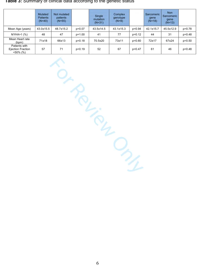
An analysis was performed regarding the age at diagnosis, ejection fraction, presence of dyspnea (NYHA>1) and heart rate comparing the groups of mutated patients vs patients with no identified genetic cause. Mutated patients tended to be younger at diagnosis (43.0 vs 48.7 years, p=0.07) but systolic dysfunction showed no significative difference between groups 3 4 5 6 7 8 9 10 11 12 13 14 15 16 17 18 19 20 21 22 23 24 25 26 27 28 29 30 31 32 33 34 35 36 37 38 39 40 41 42 43 44 45 46 47 48 49 50 51 52 53 54 55 56 57
For Review Only
(Table 3). Interestingly, the mutation yield was higher in youngest patients <65 years old (38/84, 45%) as compared to oldest patients >65 years (2/11, 18.2%, p-value: 0.11).
Patients with complex genotypes (≥2 mutations), as compared to patients with single variants, tended to be younger at diagnosis and to have a decreased ejection fraction although differences were not significant (Table 3). Finally, in patients carrying a single variant, we observed that the LV mean ejection fraction in patient with a mutation in sarcomeric genes (N=18) was lower than in patients mutated in non-sarcomeric genes (N=13) (43.8% vs 51.6%, p-value: 0.26).
DISCUSSION
We present here the results of the genetic analysis of a cohort of 95 independent patients (index cases) with LVNC in order to evaluate the yield of mutation screening and to assess the allelic and genetic spectrum of the disease. The design of our study has some characteristics that may differ from previous studies since our project was a prospective study performed in newly diagnosed consecutive patients (diagnosis less than six months) with a validation of the cardiac diagnosis by an expert centralized imaging core-lab. This design was conceived to limit potential inclusion bias and strengthen the representativeness of the cohort. We also focused on isolated LVNC, without associated congenital heart defects, in adult patients in order to have a more homogeneous population. Next generation sequencing was performed with a panel of 107 genes involved in cardiomyopathies and some arrhythmias, which represents the most comprehensive genetic analysis published (exome sequencing was performed by Sedagast-Hamedani et al but only selected genes known to be involved in the phenotype were reported) 13.
When considering pathogenic or likely pathogenic variants, we identified a mutation in 42% of the patients, which represents a proportion slightly higher than previously reported in this disease9,10,13,14,16, but relatively similar to features observed in other cardiomyopathies28. The distribution of genes revealed a high degree of genetic heterogeneity with putative or 3 4 5 6 7 8 9 10 11 12 13 14 15 16 17 18 19 20 21 22 23 24 25 26 27 28 29 30 31 32 33 34 35 36 37 38 39 40 41 42 43 44 45 46 47 48 49 50 51 52 53 54 55 56
For Review Only
12
confirmed pathogenic mutations identified in 23 different genes. The distribution includes 13 genes previously published as associated with the phenotype of LVNC (77% of variants) and 10 additional candidate genes (9 cardiomyopathy and 1 arrhythmia genes) that were never reported before as associated with LVNC (Table 1). Despite the stringent selection criteria of variants, TTN represents the most prevalent gene in the cohort (19% of variants or 10.5% of patients) including 8 variants located in the A-band (80%). As previously observed in patients with DCM, TTN truncating mutations in the A-band region of the protein were over represented27.
The following most prevalent genes were HCN4 and MYH7, followed by MYH6, RYR2 and ACTC1. Considering others published reports13,14, some discrepancies were observed in the gene distribution especially regarding TTN, MYH7, HCN4 and LMNA. Differences in distribution may be related in part to the variable characteristics of the cohorts (isolated LVNC or not, age at diagnosis, incident or prevalent cases). We observed a relatively high proportion of patients with HCN4 pathogenic variants as we found 5 different variants, located in S4, S5 and pore domains of the protein (Table 2). Among these patients 3/5 presented bradycardia (one patient was implanted by a pace maker) but no valvular disease has been reported in any of them29,30. This suggests that this recently published gene29 constitutes an important disease-causing gene in LVNC. The prevalence of HCN4 did not appear as such in previously published cohorts, possibly due to the absence of this gene in some studied panels9-11,14,15 or a difference in the cohort recruitment 13,16. For other genes, a higher rate of MYH7 variants and a lower rate of LMNA variants were found in our cohort as compared with the study of Sedaghat-Hamedani et al. 13 while frequency of TTN and
MYBPC3 variants were consistent. In the work of van Waning et al. 14, the proportion of
MYH7 and MYBPC3 in the group of adult patients were consistent with us but a lower rate of TTN variants was observed. In older publications9,10 in which only sarcomeric genes were analyzed, the spectrum of genes showed that MYH7 was the most prevalent gene then
TNNT2, TMP1, TNNI3. These last three genes were not found mutated in our cohort, which
3 4 5 6 7 8 9 10 11 12 13 14 15 16 17 18 19 20 21 22 23 24 25 26 27 28 29 30 31 32 33 34 35 36 37 38 39 40 41 42 43 44 45 46 47 48 49 50 51 52 53 54 55 56 57
For Review Only
could be due to the fact that our cohort is composed by patients with an adult onset of the disease. Interestingly, our results also strengthen the involvement of recently published genes such as HCN429,30 and RYR231 and help to better estimate their prevalence. As a whole, our finding about the large genetic and allelic spectrum is helpful in refining the genes of interest for routine molecular diagnostic of patients with LVNC.
Additionally, we tried to determine if patients reporting a familial history of LVNC, were more frequently found with a mutation than patients presenting as sporadic cases. The cohort includes 15 familial form and 55 sporadic cases, the 25 remaining patients had no information’s about their relatives. Quite the same rate of mutation identification was found in the group of familial forms and sporadic cases (53 % and 46% respectively, difference not significant)”
Apart from the report of mutations in genes previously associated with LVNC, an important finding of our study is that we identified mutations in 10 genes known to be involved in cardiac inherited diseases but not described until now as associated with this specific phenotype. Among these genes, 9 were previously reported as associated with other sub-types of cardiomyopathy such as HCM, DCM and ARVC (Table 1), suggesting an overlap between the various cardiomyopathies. Among these 10 genes, MYLK2 and NEXN were identified each in 2 patients and 7 genes (BAG3, FLNC, HEY2, MYPN, PDLIM3,
TMEM43 and DSC2) were involved each in only one patient. Although these genes could be good candidates for being pathogenic, the definitive role of these genes for causing LVNC will require confirmation in further studies, especially through segregation analyses in
families or functional studies. Similarly, in 2 patients with no particular ECG abnormalities, we identified variants in ANK2 gene known to be involved in long QT syndrome. As ANK2 was never reported before in LVNC, cautious is necessary before any conclusion about the causal role of these variants. However, HCN4 was initially described in channelopathies and then involved in LVNC29,30, with a significant proportion in our cohort, illustrating the fact that a given gene involved in arrhythmias does not preclude the potential role in LVNC.
3 4 5 6 7 8 9 10 11 12 13 14 15 16 17 18 19 20 21 22 23 24 25 26 27 28 29 30 31 32 33 34 35 36 37 38 39 40 41 42 43 44 45 46 47 48 49 50 51 52 53 54 55 56
For Review Only
14
Another observation emerging from this study is the high level of patients (9.5%) presenting complex genotypes with causative variants in two (or more) different genes. This feature about double mutated patients was not previously described as so high in adult patients with LVNC. In patients with complex genotypes, cumulative effect of variants have been associated with a higher severity of the disease in one study14 but was not analyzed in details in most of other studies. The hypothesis of a gene-dose- effect can be suspected as well as in other sub- morphotypes of cardiomyopathies, particularly in HCM. In the present cohort, patients with complex genotypes tended to have a same age at diagnosis (43.1 vs. 43.5 years old) but more symptoms (dyspnea > NYHA1: 67% vs 53%) and a lower ejection fraction (36% vs. 47%). However these differences were not statistically significant and must be confirmed in larger cohorts.
The global analysis of the distribution of genes observed in our cohort of adult patients with LVNC also provides useful information regarding the debated issue of whether or not LVNC is an independent nosological entity or a phenotype overlapping with other cardiomyopathy sub-types such as HCM or DCM28, 32-34. The biggest cohorts published so far about HCM patients (including 3267 HCM patients sequenced on 16 genes and 874 patients sequenced on 20 genes) reported sarcomeric genes (especially MYBPC3) as the major genes26,32 . In patients with DCM, the TTN gene has been consistently reported as the most frequent mutated gene33. In a study of 639 DCM patients sequenced on 84 genes, the highest prevalence observed was for TTN (13%), PKP2, MYBPC3, DSP, RYR2, DSC2 and SCN5A genes34. In the present cohort, we observed that the most prevalent genes are TTN, then MYH7, HCN4, MYH6, and RYR2. This distribution, and the fact that TTN is by far the most frequent gene we observed in LVNC, as well as the high level of complex genotypes, suggests that the genetic profile of LVNC patients is relatively similar to patients with DCM but not similar to patients with HCM26,28,32-37. However, the distribution of LVNC patients presents some specific findings, such as the relatively high rate of HCN4 gene mutations, which favor the possible specific role of some particular genes in this disorder.
3 4 5 6 7 8 9 10 11 12 13 14 15 16 17 18 19 20 21 22 23 24 25 26 27 28 29 30 31 32 33 34 35 36 37 38 39 40 41 42 43 44 45 46 47 48 49 50 51 52 53 54 55 56 57
For Review Only
Limitations. Our results were derived from a cohort of adult-onset patients with
isolated LVNC. Therefore, results may not be applicable to a pediatric population or a population with syndromic LVNC. Even though probably pathogenic variants completed all the criteria for pathogenicity, we don’t provide family segregation and functional analysis for now.
In conclusion, molecular analysis of 107 genes in 95 adult patients with isolated
LVNC shows a mutation detection rate of 42%. These data, coming from the most comprehensive study available until now in terms of genes that were analyzed, show that LVNC is basically a genetic disease in most cases, with a large genetic heterogeneity. The global distribution of genes appears quite close to the profile observed in DCM patients, with
TTN as the most frequent mutated gene, but with some specific genes such as HCN4. We
found 9.5% of patients presenting a complex genotype with a disease causing variant in two different genes located on different chromosomes. This observation could explain part of intra-familial variable expressivity in case of bi-lineal inheritance, as some relative should be carrier of a single variant with a moderate phenotype. We also described mutations in 10 genes not described until now as associated with LVNC. Although these genes are putative good candidates for causing LVNC, the definitive causal role of these genes in this phenotype will require confirmation in further studies.
3 4 5 6 7 8 9 10 11 12 13 14 15 16 17 18 19 20 21 22 23 24 25 26 27 28 29 30 31 32 33 34 35 36 37 38 39 40 41 42 43 44 45 46 47 48 49 50 51 52 53 54 55 56
For Review Only
16
REFERENCES
1- Towbin JA, Lorts A, Jefferies JL. Left ventricular non-compaction cardiomyopathy. Lancet. 2015;386:813-25.
2- Elliott P, Andersson B, Arbustini E, et al. Classification of the cardiomyopathies: A position statement from the European Society Of Cardiology Working Group on Myocardial and Pericardial Diseases. Eur Heart J 2008; 29:270–276.
3- Maron BJ, Towbin JA, Thiene G, et al. American Heart Association; Council on Clinical Cardiology, Heart Failure and Transplantation Committee; Quality of Care and Outcomes Research and Functional Genomics and Translational Biology Interdisciplinary Working Groups; Council on Epidemiology and Prevention. Contemporary definitions and classification of the cardiomyopathies: An American Heart Association Scientific Statement from the Council on Clinical Cardiology, Heart Failure and Transplantation Committee; Quality of Care and Outcomes Research and Functional Genomics and Translational Biology Interdisciplinary Circulation Journal. Circulation 2006;113:1807–1816.
4- Sandhu R, Finkelhor RS, Gunawardena DR, Bahler RC. Prevalence and characteristics of left ventricular non compaction in a community hospital cohort of patients with systolic dysfunction. Echocardiography. 2008;25:8-12.
5- Pignatelli RH, McMahon CJ, Dreyer WJ, et al. Clinical characterization of left ventricular non compaction in children: a relatively common form of cardiomyopathy. Circ J. 2003;108:2672–2678.
6- Habib G, Charron P, Eicher JC, et al; Working Groups 'Heart Failure and Cardiomyopathies' and 'Echocardiography' of the French Society of Cardiology.. Isolated left ventricular non-compaction in adults: clinical and echocardiographic features in 105 patients. Results from a French registry. Eur J Heart Fail 2011; 13:177–85. 3 4 5 6 7 8 9 10 11 12 13 14 15 16 17 18 19 20 21 22 23 24 25 26 27 28 29 30 31 32 33 34 35 36 37 38 39 40 41 42 43 44 45 46 47 48 49 50 51 52 53 54 55 56 57
For Review Only
7- Sasse-Klaassen S, Gerull B, Oechslin E, Jenni R, Thierfelder L. Isolated non compaction of the left ventricular myocardium in the adult is an autosomal dominant disorder in the majority of patients. Am J Med Genet A. 2003; 119: 162–167.
8- Bleyl SB, Mumford BR, Thompson V, et al. Lethal non compaction of the left ventricular myocardium is allelic with Barth syndrome. Am J Hum Genet. 1997;61:868-72.
9- Klaassen S, Probst S, Oechslin E, et al. Mutations in sarcomere protein genes in left ventricular non compaction. Circulation. 2008;117:2893–2901.
10- Probst S, Oechslin E, Schuler P, et al. Sarcomere gene mutations in isolated left ventricular non compaction cardiomyopathy do not predict clinical phenotype. Circ Cardiovasc Genet. 2011;4:367-74
11- Xing Y, Ichida F, Matsuoka T, et al. Genetic analysis in patients with left ventricular non compaction and evidence for genetic heterogeneity. Mol Genet Metab. 2006; 88:71-7. 12- Hastings R, de Villiers CP, Hooper C, et al. Combination of Whole Genome
Sequencing, Linkage, and Functional Studies Implicates a Missense Mutation in Titin as a Cause of Autosomal Dominant Cardiomyopathy With Features of Left Ventricular Non compaction. Circ Cardiovasc Genet. 2016;7 :426-435.
13- Sedaghat-Hamedani F, Haas J, Zhu F, et al. Clinical genetics and outcome of left ventricular non-compaction cardiomyopathy. Eur Heart J. 2017 ;38:3449-3460.
14- van Waning JI, Caliskan K, Hoedemaekers YM, et al. Genetics, Clinical Features, and Long-Term Outcome of Non compaction Cardiomyopathy. J Am Coll Cardiol. 2018 20;71:711-722
15- Tian T, Wang J, Wang H, et al. A low prevalence of sarcomeric gene variants in a Chinese cohort with left ventricular non-compaction. Heart Vessels. 2015; 30:258-64. 3 4 5 6 7 8 9 10 11 12 13 14 15 16 17 18 19 20 21 22 23 24 25 26 27 28 29 30 31 32 33 34 35 36 37 38 39 40 41 42 43 44 45 46 47 48 49 50 51 52 53 54 55 56
For Review Only
18
16- Wang C, Hata Y, Hirono K, et al. A Wide and Specific Spectrum of Genetic Variants and Genotype-Phenotype Correlations Revealed by Next-Generation Sequencing in Patients with Left Ventricular Non compaction. J Am Heart Assoc. 2017 Aug 30;6(9). 17- Jenni R, Oechslin E, Schneider J, Attenhofer Jost C, Kaufmann PA.
Echocardiographic and pathoanatomical characteristics of isolated left ventricular non-compaction: a step towards classification as a distinct cardiomyopathy. Heart 2001;86:666–67.
18- Petersen SE, Selvanayagam JB, Wiesmann F, et al. Left ventricular non-compaction: insights from cardiovascular magnetic resonance imaging. J Am Coll Cardiol. 2005; 46:101–105.
19- Magoc T. and Salzberg S. FLASH: Fast length adjustment of short reads to
improve genome assemblies. Bioinformatics, 2011; 27:21, 2957-63.
20- Li H and Durbin R. Fast and accurate short read alignment with Burrows-Wheeler Transform. Bioinformatics. 2009; 25:1754-60.
21- McKenna A, Hanna M, Banks E, et al. The Genome Analysis Toolkit: a MapReduce framework for analyzing next-generation DNA sequencing data. Genome Research. 2010; 20:1297-303.
22- Li H., Handsaker B, Wysoker A, et al. The Sequence alignment/map (SAM) format and SAMtools. Bioinformatics, 2009; 25: 2078-9.
23- Wang K, Li M, Hakonarson H. ANNOVAR: Functional annotation of genetic variants from next-generation sequencing data. Nucleic Acids Research. 2010; 38:16, e164. 24- Kircher M, Witten DM, Jain P, O'Roak BJ, Cooper GM, Shendure J. A general
framework for estimating the relative pathogenicity of human genetic variants. Nature Genetics, 2014; 46: 310-5. 3 4 5 6 7 8 9 10 11 12 13 14 15 16 17 18 19 20 21 22 23 24 25 26 27 28 29 30 31 32 33 34 35 36 37 38 39 40 41 42 43 44 45 46 47 48 49 50 51 52 53 54 55 56 57
For Review Only
25- Richards S, Aziz N, Bale S, et al. Standards and guidelines for the interpretation of sequence variants: a joint consensus recommendation of the American College of Medical Genetics and Genomics and the Association for Molecular Pathology. Genet Med. 2015; 17: 405-424.
26- Walsh R, Buchan R, Wilk A, et al. Defining the genetic architecture of hypertrophic cardiomyopathy: re-evaluating the role of non-sarcomeric genes. Eur Heart J. 2017;0:1-8
27- Ware JS, Cook SA. Role of titin in cardiomyopathy: from DNA variants to patient stratification. Nat Rev Cardiol. 2018 ;15:241-252
28- Walsh R, Thomson KL, Ware JS, et al. Reassessment of Mendelian gene pathogenicity using 7,855 cardiomyopathy cases and 60,706 reference samples. Genet Med. 2017;19:192-203.
29- Milano A, Vermeer AM, Lodder EM, et al. HCN4 mutations in multiple families with bradycardia and left ventricular non compaction cardiomyopathy. J Am Coll Cardiol 2014;64:745–56.
30- Schweizer PA, Koenen M, Katus HA, Thomas D. A Distinct Cardiomyopathy: HCN4 Syndrome Comprising Myocardial Noncompaction, Bradycardia, Mitral Valve Defects, and Aortic Dilation.J Am Coll Cardiol. 2017;69:1209-1210.
31- Ohno S, Omura M, Kawamura M, et al. Exon 3 deletion of RYR2 encoding cardiac ryanodine receptor is associated with left ventricular non-compaction. Europace. 2014;16:1646-54.
32- Lopes LR, Syrris P, Guttmann OP, et al. Novel genotype-phenotype associations demonstrated by high-throughput sequencing in patients with hypertrophic cardiomyopathy. Heart. 2015;101:294-301.
33- Herman DS, Lam L, Taylor MR, et al. Truncations of titin causing dilated cardiomyopathy. N Engl J Med. 2012;366: 619-28.
3 4 5 6 7 8 9 10 11 12 13 14 15 16 17 18 19 20 21 22 23 24 25 26 27 28 29 30 31 32 33 34 35 36 37 38 39 40 41 42 43 44 45 46 47 48 49 50 51 52 53 54 55 56
For Review Only
20
34- Haas J, Frese KS, Peil B, et al. Atlas of the clinical genetics of human dilated cardiomyopathy. Eur Heart J. 2015 May 7;36:1123-35a.
35- Waldmüller S, Schroeder C, Sturm M, et al. Targeted 46-gene and clinical exome sequencing for mutations causing cardiomyopathies. Mol Cell Probes. 2015;29: 308-14.
36- Charron P, Elliott P, Gimeno JR, et al. The Cardiomyopathy Registry of the EURObservational Research Programme of the European Society of Cardiology: baseline data and contemporary management of adult patients with cardiomyopathies. Eur Heart J. 2018 Jan 24. [Epub ahead of print]
37- Mademont-Soler I, Mates J, Yotti R, et al. Additional value of screening for minor genes and copy number variants in hypertrophic cardiomyopathy. PLoS One. 2017;12:e0181465.
38- K.R. Egan, J.C. Ralphe, L. Weinhaus, et al. Just sinus bradycardia or something more serious? Case Rep Pediatr, 2013, p. 736164.
39- L. Shan, N. Makita, Y. Xing, et al. SCN5A variants in Japanese patients with left ventricular noncompaction and arrhythmia Mol Genet Metab,2008;93:468-474.
40- Parent JJ, Towbin JA, Jefferies JL. Fibrillin-1 Gene Mutations in Left Ventricular Non-compaction Cardiomyopathy. Pediatr Cardiol. 2016;37:1123-6.
41- Arndt AK, Schafer S, Drenckhahn JD, et al. Fine Mapping of the 1p36 Deletion Syndrome Identifies Mutation of PRDM16 as a Cause of Cardiomyopathy. Am J Hum Genet 93:67–77.
42- Ramond F, Janin A, Filippo SD, et al. Homozygous PKP2 deletion associated with neonatal Left Ventricule Non Compaction. Clin Genet. 2017;91:126-130.
43- Williams T, Machann W, Kühler L, et al. Novel desmoplakin mutation: juvenile biventricular cardiomyopathy with left ventricular non-compaction and acantholytic palmoplantar keratoderma. Clin Res Cardiol. 2011;100 :1087-93.
3 4 5 6 7 8 9 10 11 12 13 14 15 16 17 18 19 20 21 22 23 24 25 26 27 28 29 30 31 32 33 34 35 36 37 38 39 40 41 42 43 44 45 46 47 48 49 50 51 52 53 54 55 56 57
For Review Only
1
Table 1: List of genes published in LVNC cardiomyopathy and potentially new genes identified in the present work.
Cellular structure Gene (NM) Protein Phenotype Ref. in LVNC This Cohort
Sarcomere
MYH7 (NM_000257.2) Myosin heavy chain HCM, DMC, LVNC 9-10 yes
TNNT2(NM_001001430.1) Troponin T2 HCM, DMC, LVNC 9-10 no
ACTC1 (NM_005159.4) Cardiac Actin HCM, DMC, LVNC 9-10 yes
MYBPC3(NM_000256.3) Cardiac C protein HCM, DMC, LVNC 9-10 yes
TPM1(NM_001018005.1) Alpha-tropomyosin HCM, DMC, LVNC 9-10 no
TNNI3(NM_000363.4) Troponin I3 HCM, DMC, LVNC 9-10 no
DTNA(NM_001390.4) Alpha-Dystrobrevin DCM, LVNC 11-15 no
MYH6 (NM_002471.3) Myosin light chain DCM, HCM, LVNC 15 yes
ACTN2 (NM_001103.2) Actinin HCM, LVNC 32 no
TTN (NM_001256850.1) Titin DMC, HCM, LVNC 12 yes
MYLK2 (NM_033118.3) Myosin Light chain kinase DCM This work yes
MYPN (NM_032578.2) Myopalladin DCM This work yes
NEXN (NM_144573.3) Nexilin HCM, DCM This work yes
Structure
FLNC (NM_001458.4) Filamin-C HCM, DCM This work yes
LDB3 (NM_007078.2) LIM Domain Binding 3 DCM, LVNC 11-15 yes
LMNA (NM_170707.2) Lamine A/C DMC, LVNC 11-15 yes
Ion channel and related genes
ANK2 (NM_001148.4) Ankyrin 2 LQT This work yes
HCN4 (NM_005477.2) Hyperpolarization Activated Cyclic Nucleotide Gated
Potassium Channel 4
ARVC, LVNC 29,30 yes
RYR2 (NM_001035.2) Ryanodin receptor 2 ARVC, LVNC, CPVT 31 yes
CASCQ2 (NM_001232.3) Calsequestrin 2 QTL, LVNC 38 no
SCN5A (NM_198056.2) Sodium channel, voltage-gated, type V, alpha subunit LQT, Brugada, DCM, LVNC 39 no
3 4 5 6 7 8 9 10 11 12 13 14 15 16 17 18 19 20 21 22 23 24 25 26 27 28 29 30 31 32 33 34 35 36 37 38 39 40 41 42 43 44 45 46 47 48 49 50 51 52 53 54 55 56
For Review Only
2
Other
Nkx2.5 (NM_004387.3) NK2 Homeobox 5 DCM, LVNC 11-15 yes
TAZ (NM_000116.3) Taffazin Barth Syndrom, LVNC 8 no
FBN1 (NM_000138.4) Fibrillin Marfan Syndrom, LVNC 40 no
ABCC9 (NM_020297) ATP Binding Cassette Subfamily C Member 9 DCM, LVNC 35 no
PDRM16 (NM_022114) PR/SET Domain 16 LVNC 41 no
BAG3 (NM_004281.3) BCL2 Associated Athanogene 3 DCM This work yes
HEY2 (NM_012259)
Hairy-Related Transcription Factor 2 DCM This work yes
PDLIM3 (NM_014476.4) PDZ And LIM Domain 3 ARVC, HCM This work yes
RBM20 (NM_001134363.1) RNA Binding Motif Protein 20 DCM 13 yes
TMEM43 (NM_024334.2) TransmembraneProtein 43 ARVC This work yes
Desmosome
PKP2 (NM_004572.3) Plakophilin 2 ARVC, LVNC 42 yes
DSP (NM_004415.2) Desmoplakin ARVC, LVNC, DCM 43 yes
DSC2 (NM_024422.3) Desmocollin ARVC This work yes
3 4 5 6 7 8 9 10 11 12 13 14 15 16 17 18 19 20 21 22 23 24 25 26 27 28 29 30 31 32 33 34 35 36 37 38 39 40 41 42 43 44 45 46 47 48 49 50 51 52 53 54 55 56 57
For Review Only
3
Table 2: List of pathogenic and probably pathogenic variants identified in the cohort.
Position c., cDNA position; Position p., protein effect; Published: No or Yes; Associated phenotype for published variants; GnomAD correspond to the allelic frequency, and Htz corresponds to the allele count in GnomAD in all populations; GVGD, SIFT, Mutation taster and polyphen are algorythms corresponding to in silico Predictive Algorithms used for evaluation of missense variants. Range of scores for each are indicated in the title column; “Type” indicated the nature of the variant; MS: missense, NS; Nonsense, Del; deletion, Splice; mutation affecting splicing site. Column “interpretation”, indicates conclusions about the pathogenicity of the variant: class 5 ; certainly pathogenic, Class 4; probably
pathogenic. NA: not applicable
Gene Position c. Position p. Published
No/Yes Associated phenotype GnomAD, Htz GVGD (C65-C0) SIFT (0-1) Mutation Taster (1-0) Polyphen (1-0) Type Interpretation
ACTC1 c.670G>T p.Asp224Tyr N NA / 65 0 1 0.994 MS Class 4
ACTC1 c.281A>G p.Asn94Ser N NA 4.061e-6, 1 45 0 1 0.615 MS Class 4
ACTC1 c.623G>A p.Arg208His N NA 4.061e-5, 10 25 0 1 0,01 MS Class 4
ANK2 c.11150T>A p.Ile3717Asn N NA 1.083e-5, 3 45 0 0,995 0.865 MS Class 4
ANK2 c.9145C>T p.Arg3049Trp N NA 8.155e-6, 2 65 0 0,93 0.999 MS Class 4
BAG3 c.465_466insGCG p.Ala155delinsAlaAla N NA / NA NA NA NA MS Class 4
DSC2 c.1448A>T p.Asn483Ile N NA / 15 0 NA 0.905 MS Class 4
DSP c.3035delA p.Asp1012fs N NA / NA NA NA NA Del Class 5
FLNC c.1933_1935del p.645del N NA 3.969e-5, 11 NA NA NA NA Del Class 4
HCN4 c.1403C>T p.Ala468Val N NA 4.065e-6, 1 65 0 1 0,95 MS Class 4
HCN4 c.1123C>T p.Arg375Cys N NA 4.061e-6, 1 65 0 1 0,99 MS Class 4
HCN4 c.1231C>G p.Leu411Val N NA / 25 0 1 0,99 MS Class 4
HCN4 c.1444G>A p.Gly482Arg Y NCVG / 65 0 1 1 MS Class 5
HCN4 c.1438G>T p.Gly480Cys N NA / 65 0 1 1 MS Class 4
HEY2 c.683C>T p.Thr228Met N NA 3.231e-5, 2 0 0,05 1 0,45 MS Class 4
LDB3 c.625G>C p.Glu209Gln N NA / 25 0 1 0,94 MS Class 4
LDB3 c.608C>T p.Ser203Leu Y CMD 2.538e-5, 7 65 0 1 0,88 MS Class 5
3 4 5 6 7 8 9 10 11 12 13 14 15 16 17 18 19 20 21 22 23 24 25 26 27 28 29 30 31 32 33 34 35 36 37 38 39 40 41 42 43 44 45 46 47 48 49 50 51 52 53 54 55 56
For Review Only
4
LMNA c.738delG p.Gln246fs N NA / NA NA NA NA Del Class 5
MYBPC3 c.532G>A p.Val178Met Y HCM / 0 0,01 1 0,992 MS Class 5
MYBPC3 c.1504C>T p.Arg502Trp Y HCM 5.411e-5, 15 65 0 1 0,484 MS Class 5
MYH6 c.1793dupA p.Asn598fs N NA 8.122e-6, 2 NA NA NA NA Dup Class 5
MYH6 c.4828C>T p.Arg1610Cys N NA 3.247e-5, 1 0 0 1 0,988 MS Class 4
MYH6 c.50G>T p.Arg17Leu Y Cardiac
septal defect / 0 0 1 0,55 MS Class 5
MYH7 c.379C>A p.Pro127Thr N NA / 0 0 1 0,98 MS Class 4
MYH7 c.3830G>C p.Arg1277Pro N NA / 35 0 1 0,842 MS Class 4
MYH7 c.3586C>T p.His1196Tyr N NA / 15 0 1 0,613 MS Class 4
MYH7 c.2419C>G p.Arg807Gly N NA / 25 0,02 1 0,85 MS/Splice Class 4
MYH7 c.4588C>T p.Arg1530X N NA / NA NA NA NA NS Class 4
MYLK2 c.1754T>A p.Ile585Asn N NA / 45 0 1 0,921 MS Class 4
MYLK2 c.1658G>A p.Arg553His N NA 1.276e-5, 3 0 0,15 0,92 0,004 MS Class 4
MYPN c.3457G>A p.Gly1153Arg N NA 1.219e-5, 3 65 0 1 1 MS Class 4
NEXN c.2012T>C p.Ile671Thr N NA 2.541e-5, 7 0 0 1 0,936 MS Class 4
NEXN c.1396A>C p.Lys466Gln N NA / 0 0 1 0,996 MS Class 4
NKX2-5 c.604C>G p.Leu202Val N NA / 25 0 1 0,07 MS Class 4
PDLIM3 c.742C>T p.Arg248Cys N NA 8.153e-6, 2 0 0 1 1 MS Class 4
PKP2 c.2018G>A p.Gly673Asp Y ARVC / 65 0 1 1 MS Class 5
RBM20 c.1907G>A p.Arg636His Y DCM / 0 0 1 0,99 MS Class 5
RYR2 c.13936G>C p.Asp4646His N NA / 0 0 1 0,99 MS Class 4
RYR2 c.6180G>T p.Gln2060His N NA / 15 0 1 0,89 MS Class 4
RYR2 c.878A>C p.Gln293Pro N NA / 65 0 1 0,756 MS Class 4
RYR2
c.169-?_c.273+?del ? / Y / NA NA NA NA Del Class 5
TMEM43 c.317A>G p.Tyr106Cys N NA 2.031e-5, 5 65 0 1 1 MS Class 4
TTN c.93376delA p.Arg31126fs N NA / NA NA NA NA Del Class 4
TTN c.93376_93377de l p.Arg31126fs N NA / NA NA NA NA FS Class 4 3 4 5 6 7 8 9 10 11 12 13 14 15 16 17 18 19 20 21 22 23 24 25 26 27 28 29 30 31 32 33 34 35 36 37 38 39 40 41 42 43 44 45 46 47 48 49 50 51 52 53 54 55 56 57
For Review Only
5
TTN c.82724delA p.Asn27575fs N NA / NA NA NA NA FS Class 4
TTN c.98039_98040in
sTCAA p.Asn32680fs N NA / NA NA NA NA Ins Class 4
TTN c.64100_64101in
sTTGA p.Asp21368X N NA / NA NA NA NA Ins Class 4
TTN c.53947C>T p.Arg17983X N NA / NA NA NA NA NS Class 4 TTN c.61961G>A p.Trp20654X N NA / NA NA NA NA NS Class 4 TTN c.44248C>T p.Arg14750X N NA / NA NA NA NA NS Class 4 TTN c.80845C>T p.Arg26949X Y CMD / NA NA NA NA NS Class 4 TTN c.43360C>T p.Arg14454X Y CMD / NA NA NA NA NS Class 4 3 4 5 6 7 8 9 10 11 12 13 14 15 16 17 18 19 20 21 22 23 24 25 26 27 28 29 30 31 32 33 34 35 36 37 38 39 40 41 42 43 44 45 46 47 48 49 50 51 52 53 54 55 56
For Review Only
6
Table 3: Summary of clinical data according to the genetic status
Mutated Patients (N=40) Not mutated patients (N=55) Single mutation (N=31) Complex genotype (N=9) Sarcomeric gene (N=18) Non Sarcomeric gene (N=13)
Mean Age (years) 43.0±15.5 48.7±15.2 p=0.07 43.5±14.5 43.1±15.3 p=0.94 42.1±15.7 45.5±12.9 p=0.78 NYHA>1 (%) 48 47 p=1.00 41 77 p=0.12 44 31 p=0.48 Mean Heart rate
(bpm) 71±18 66±13 p=0.18 70.5±20 73±11 p=0.60 72±17 67±24 p=0.50 Patients with Ejection Fraction <50% (%) 57 71 p=0.19 52 67 p=0.47 61 46 p=0.48 3 4 5 6 7 8 9 10 11 12 13 14 15 16 17 18 19 20 21 22 23 24 25 26 27 28 29 30 31 32 33 34 35 36 37 38 39 40 41 42 43 44 45 46 47 48 49 50 51 52 53 54 55 56 57
For Review Only
FIGURE LEGENDS
Figure 1: Distribution of genes according to their number of identified pathogenic variants
and visualization of their cellular location and function.
Figure 2: Representation of genes association in patients carrying two pathogenic variants.
Gene symbol were indicated on the right and left scales. For each of the seven patients carrying 2 mutations, the two mutated genes are connected by a straight line.
3 4 5 6 7 8 9 10 11 12 13 14 15 16 17 18 19 20 21 22 23 24 25 26 27 28 29 30 31 32 33 34 35 36 37 38 39 40 41 42 43 44 45 46 47 48 49 50 51 52 53 54 55 56
For Review Only
3 4 5 6 7 8 9 10 11 12 13 14 15 16 17 18 19 20 21 22 23 24 25 26 27 28 29 30 31 32 33 34 35 36 37 38 39 40 41 42 43 44 45 46 47 48 49 50 51 52 53 54 55 56 57For Review Only
3 4 5 6 7 8 9 10 11 12 13 14 15 16 17 18 19 20 21 22 23 24 25 26 27 28 29 30 31 32 33 34 35 36 37 38 39 40 41 42 43 44 45 46 47 48 49 50 51 52 53 54 55 56For Review Only
Suppl Data- Table 1: List of genes analyzed in this cohort.
Gene Reference Sequence Chromosome AARS2 NM_020745.2 chr6 ABCC9 NM_020297 chr12 ACAD9 NM_014049 chr3 ACTA1 NM_001100_ chr1 ACTC1 NM_005159.4 chr15 ACTN2 NM_001103.2 chr1 AGK NM_018238.3 chr7 AKAP9 NM_005751.4 chr7 ANK2 NM_001148.4 chr4 ANKRD1 NM_014391.2 chr10 BAG3 NM_004281.3 chr10 C2orf64(COA5) NM_001008215.1 chr2 CACNA1B NM_000718.2 chr9 CACNA1C NM199460.2 chr12 CACNA2D1 NM_000722.2 chr7 CACNB2 NM_201596.2 chr10 CALR3 NM_145046.3 chr19 CASQ2 NM_001232.3 chr1 CAV3 MN033337.2 chr3 COX10 NM_001303.3 chr17 COX15 NM_078470.4 chr10 CSRP3 NM_003476.3 chr11 CTNNA3 NM_013266.2 chr10 DES NM_001927.3 chr2 DSC2 NM_024422.3 chr18 DSG2 NM_001943.3 chr18 DSP NM_004415.2 chr6 DTNA NM_001390.4 chr18 EMD NM_000117.2 chrX EYA4 NM_004100.4 chr6 FBN1 NM_000138.4 chr15 FHL1 NM_001159702 chrX FLNC NM_001458.4 chr7 GAA NM_000152.3 chr17 GJA5 NM_005266.5 chr1 GLA NM_000169.2 chrX GPD1L MN_015141.3 chr3 HCN4 NM_005477.2 chr15 HEY2 NM_012259 chr6 JPH2 NM_020433.4 chr20 JUP NM_002230.2 chr17 3 4 5 6 7 8 9 10 11 12 13 14 15 16 17 18 19 20 21 22 23 24 25 26 27 28 29 30 31 32 33 34 35 36 37 38 39 40 41 42 43 44 45 46 47 48 49 50 51 52 53 54 55 56 57
For Review Only
KCNA5 NM_002234.2 chr12 KCND3 NM_004980.4 chr1 KCNE1 NM_000219.3 chr21 KCNE1L NM_012282.2 chrX KCNE2 NM_172201.1 chr21 KCNE3 NM_005472.4 chr11 KCNH2 NM000238.2 chr7 KCNJ2 NM000891.2 chr17 KCNJ5 NM_000890.3 chr11 KCNJ8 NM_004982.2 chr12 KCNQ1 NM 000218.2 chr11 KRAS NM_004985.3 chr12 LAMP2 NM_002294.2 chrX LDB3 NM_007078.2 chr10 LMNA NM_170707.2 chr1 MRPL44 NM_022915.3 chr2 MYBPC3 NM_000256.3 chr11 MYH6 NM_002471.3 chr14 MYH7 NM_000257.2 chr14 MYL2 NM_000432.3 chr12 MYL3 NM_000258.2 chr3 MYLK2 NM_033118.3 chr20 MYOM1 NM_003803.3 chr18 MYOZ2 NM_016599.3 chr4 MYPN NM_032578.2 chr10 NEBL NM_006393.2 chr10 NEXN NM_144573.3 chr1 NKX2-5 NM_004387.3 chr5 NPPA NM_006172 chr1 PDLIM3 NM_014476.4 chr4 PKP2 NM_004572.3 chr12 PLN NM_002667.3 chr6 PRDM16 NM_022114 chr1 PRKAG2 NM_016203.3 chr7 PSEN1 NM_000021.3 chr14 PSEN2 NM_000447.2 chr1 PTPN11 NM_002834.3 chr12 RAF1 NM_002880.3 chr3 RANGRF NM_016492.4 chr17 RBM20 NM_001134363.1 chr10 RYR2 NM_001035.2 chr1 SCN1B NM_199037.3 chr19 SCN2B NM_004588.4 chr11 SCN3B NM018400.3 chr11 SCN4B NM174934.3 chr11 3 4 5 6 7 8 9 10 11 12 13 14 15 16 17 18 19 20 21 22 23 24 25 26 27 28 29 30 31 32 33 34 35 36 37 38 39 40 41 42 43 44 45 46 47 48 49 50 51 52 53 54 55 56For Review Only
SCN5A NM_198056.2 chr3 SCO2 NM_001169109.1 chr22 SDHA NM_004168.2 chr5 SGCD1 NM_000337.5 chr5 SLC25A4 (ANT1) NM_001151.3 chr4 SNTA1 NM_003098.2 chr20 SOS1 NM_005633.3 chr2 SYNPO2 NM_133477.2 chr4 TAZ NM_000116.3 chrX TCAP NM_003673.3 chr17 TGFB3 NM_003239.2 chr14 TMEM43 NM_024334.2 chr3 TMEM70 NM_017866.5 chr8 TMPO NM_003276.2 chr12 TNNC1 NM_003280.2 chr3 TNNI3 NM_000363.4 chr19 TNNT2 NM_001001430.1 chr1 TPM1 NM_001018005.1 chr15 TTN NM_001256850.1 chr2 TTR NM_000371.3 chr18 VCL NM_014000.2 chr10 3 4 5 6 7 8 9 10 11 12 13 14 15 16 17 18 19 20 21 22 23 24 25 26 27 28 29 30 31 32 33 34 35 36 37 38 39 40 41 42 43 44 45 46 47 48 49 50 51 52 53 54 55 56 57For Review Only
Gene name HGVSc. HGVSp. AARS2 c.1752+13C>T --- AARS2 c.44C>G p.Ala15Gly ABCC9 c.2424+10A>G --- ABCC9 c.4212-31T>G --- ABCC9 c.1981C>T p.Arg661Cys ABCC9 c.1165-7_1165-6delTT --- ABCC9 c.-38C>A --- ACAD9 c.244+7A>G ---ACTN2 c.2057T>A p.Ile686Asn
AGK c.21G>A p.Thr7Thr
AKAP9 c.11546+11G>C ---
AKAP9 c.11229G>A p.Met3743Ile
AKAP9 c.11384A>G p.Asn3795Ser
AKAP9 c.4246-4G>T --- AKAP9 c.7212C>T p.Thr2404Thr ANK2 c.11032+45_11032+50delGTGTGT --- ANK2 c.1288-40C>A --- ANK2 c.2179-11A>G --- ANK2 c.2024C>G p.Thr675Arg
ANK2 c.5072A>G p.Gln1691Arg
ANK2 c.7915C>G p.His2639Asp
ANK2 c.2662C>A p.Arg888Arg
ANK2 c.4710C>T p.Thr1570Thr ANK2 c.7161T>C p.Ala2387Ala CACNA1B c.2268-23dupG --- CACNA1B c.3413+22T>C --- CACNA1B c.3711-41A>T --- CACNA1B c.4473+20G>C --- CACNA1B c.5777+34G>A ---
CACNA1B c.274A>G p.Thr92Ala
CACNA1B c.282G>T p.Trp94Cys CACNA1B c.4497C>T p.Tyr1499Tyr CACNA1B c.5052C>T p.Ala1684Ala CACNA1B c.6936C>T p.Asn2312Asn CACNA1C c.1218-48C>T --- CACNA1C c.2531-39G>T --- CACNA1C c.5823-16G>A --- CACNA1C c.5930-33G>T ---
CACNA1C c.3280A>G p.Ile1094Val
CACNA1C c.5519A>G p.Glu1840Gly
CACNA1C c.2460G>C p.Lys820Asn
CACNA2D1 c.1516-10delT ---
CACNA2D1 c.728+29A>G ---
CACNB2 c.121_122insTTTTTT p.Gln40_Ser41insPhePhe
CACNB2 c.1302+51_1302+52insCTTTTTTTTTTT ---
CACNB2 c.886-36dupT ---
CACNB2 c.1550A>C p.Glu517Ala
CACNB2 c.1880G>A p.Arg627His
CACNB2 c.1650C>T p.Ser550Ser
COX15 c.507C>T p.Tyr169Tyr
COX15 c.876C>G p.Ser292Ser
COX15 c.999A>G p.Ser333Ser
DES c.1245-39G>A --- DES c.736-19G>A --- DSC2 c.630+45G>A --- DSC2 c.943-27A>G --- DSC2 c.1448A>T p.Asn483Ile DSG2 c.1173C>A p.Ser391Arg
DTNA c.1138G>A p.Ala380Thr
DTNA c.549A>G p.Glu183Glu
EMD c.399+50C>T --- FBN1 c.1148-33G>C --- FBN1 c.1571C>T p.Thr524Met FBN1 c.3026C>T p.Pro1009Leu FBN1 c.165-7G>A --- FBN1 c.1530G>A p.Ser510Ser FBN1 c.6402C>T p.Pro2134Pro FLNC c.1048-11C>T --- FLNC c.1210+14delC --- FLNC c.4951+45T>C --- FLNC c.4301G>T p.Arg1434Leu FLNC c.7546G>A p.Glu2516Lys FLNC c.2733G>A p.Lys911Lys FLNC c.492G>T p.Arg164Arg GAA c.546+23C>G ---
GAA c.2786G>A p.Ser929Asn
GAA c.2482-1G>T ---
GLA c.427G>A p.Ala143Thr
GLA c.639+31C>G ---
HCN4 c.3081C>T p.Pro1027Pro
HEY2 c.565T>A p.Phe189Ile
HEY2 c.438G>T p.Ser146Ser HEY2 c.843C>G p.Ser281Ser JPH2 c.1836C>A p.Pro612Pro JPH2 c.1963C>A p.Arg655Arg 3 4 5 6 7 8 9 10 11 12 13 14 15 16 17 18 19 20 21 22 23 24 25 26 27 28 29 30 31 32 33 34 35 36 37 38 39 40 41 42 43 44 45 46 47 48 49 50 51 52 53 54 55 56