HAL Id: cea-00169528
https://hal-cea.archives-ouvertes.fr/cea-00169528
Submitted on 4 Sep 2007
HAL is a multi-disciplinary open access
archive for the deposit and dissemination of
sci-entific research documents, whether they are
pub-lished or not. The documents may come from
teaching and research institutions in France or
abroad, or from public or private research centers.
L’archive ouverte pluridisciplinaire HAL, est
destinée au dépôt et à la diffusion de documents
scientifiques de niveau recherche, publiés ou non,
émanant des établissements d’enseignement et de
recherche français ou étrangers, des laboratoires
publics ou privés.
exposed to cobalt.
Veronique Malard, Frederic Berenguer, Odette Prat, Sylvie Ruat, Gerard
Steinmetz, Eric Quemeneur
To cite this version:
Veronique Malard, Frederic Berenguer, Odette Prat, Sylvie Ruat, Gerard Steinmetz, et al.. Global
gene expression profiling in human lung cells exposed to cobalt.. BMC Genomics, BioMed Central,
2007, 8, pp.147. �10.1186/1471-2164-8-147�. �cea-00169528�
Open Access
Research article
Global gene expression profiling in human lung cells exposed to
cobalt
Veronique Malard*, Frederic Berenguer, Odette Prat, Sylvie Ruat,
Gerard Steinmetz and Eric Quemeneur
Address: Service de Biochimie et Toxicologie Nucléaire, DSV/iBEB, CEA VALRHO, B.P. 17171, 30207 Bagnols-sur-Cèze, France Email: Veronique Malard* - [email protected]; Frederic Berenguer - [email protected]; Odette Prat - [email protected]; Sylvie Ruat - [email protected]; Gerard Steinmetz - [email protected]; Eric Quemeneur - [email protected]
* Corresponding author
Abstract
Background: It has been estimated that more than 1 million workers in the United States are
exposed to cobalt. Occupational exposure to 59 Co occurs mainly via inhalation and leads to various lung diseases. Cobalt is classified by the IARC as a possible human carcinogen (group 2B). Although there is evidence for in vivo and in vitro toxicity, the mechanisms of cobalt-induced lung toxicity are not fully known. The purpose of this work was to identify potential signatures of acute cobalt exposure using a toxicogenomic approach. Data analysis focused on some cellular processes and protein targets that are thought to be relevant for carcinogenesis, transport and biomarker research.
Results: A time course transcriptome analysis was performed on A549 human pulmonary cells,
leading to the identification of 85 genes which are repressed or induced in response to soluble 59 Co. A group of 29 of these genes, representing the main biological functions, was assessed by quantitative PCR. The expression profiles of six of them were then tested by quantitative RT-PCR in a time-dependent manner and three modulations were confirmed by Western blotting. The 85 modulated genes include potential cobalt carriers (FBXL2, ZNT1, SLC12A5), tumor suppressors or transcription factors (MAZ, DLG1, MYC, AXL) and genes linked to the stress response (UBC,
HSPCB, BNIP3L). We also identified nine genes coding for secreted proteins as candidates for
biomarker research. Of those, TIMP2 was found to be down-regulated and this modulation was confirmed, in a dose-dependent manner, at protein level in the supernatant of exposed cells.
Conclusion: Most of these genes have never been described as related to cobalt stress and
provide original hypotheses for further study of the effects of this metal ion on human lung epithelial cells. A putative biomarker of cobalt toxicity was identified.
Background
In the United States, more than a million workers are potentially exposed to cobalt or its compounds [1]. Cobalt is massively used in the steel industry, being a
major constituent of hard metal alloys, in combination with tungsten carbides. Other industrial uses include dia-mond polishing with Co-containing disks and the pro-duction of drying agents, pigments, and catalysts [2]. Published: 6 June 2007
BMC Genomics 2007, 8:147 doi:10.1186/1471-2164-8-147
Received: 24 October 2006 Accepted: 6 June 2007 This article is available from: http://www.biomedcentral.com/1471-2164/8/147
© 2007 Malard et al; licensee BioMed Central Ltd.
This is an Open Access article distributed under the terms of the Creative Commons Attribution License (http://creativecommons.org/licenses/by/2.0), which permits unrestricted use, distribution, and reproduction in any medium, provided the original work is properly cited.
Radioactive isotopes of cobalt are used in industry, medi-cine and nuclear research. In nuclear power plants, 59 Co-containing alloys can be activated into radioactive 60Co oxides, dispersed in the cooling water and then contami-nate workers [3,4]. A study measuring the ambient air in cobalt powder production reported concentrations of cobalt ranging from 0.675 to 10 mg/m3 [5]. Airborne concentrations measured in the working environment from a factory producing hard-metal inserts ranged from 14.6 to 37.4 mg/m3 [6]. Occupational exposure to Co occurs mainly via inhalation leading to various lung dis-eases, such as pneumonitis, fibrosis and asthma [7,8]. As with human exposure, animal exposure to cobalt-contain-ing aerosols causes pronounced respiratory effects. A sin-gle 30-minute exposure of rats to relatively high levels (26–236 mg cobalt hydrocarbonyl/m3), resulted in lung congestion, oedema, and haemorrhage [9]. Necrosis and inflammation of the respiratory tract epithelium were reported in rats exposed to 19 mg cobalt/m3 and mice exposed to 1.9 mg cobalt sulfate/m3 over 16 days [10,11]. Some acute effects have been observed concerning general public exposure. Lethal cardiomyopathy was reported in people who consumed large quantities of beer containing cobalt as a foam stabilizer (0.04–0.14 mg cobalt/kg/day), and acute mortality accounted for 18% of the deaths [12]. A 19-month-old boy who swallowed an unknown amount of cobalt chloride solution died 6.5 hours after ingestion [13].
Following absorbtion by inhalation, cobalt is eliminated in the urine. Biological monitoring of accidental exposure mainly involves measuring the concentration of metal in the urine. This might be inadequate for several reasons. Firstly, the quantity of metal excreted (exposure marker) does not necessarily reflect organ damage, which varies from one person to another. Secondly, depending on the chemical form, excretion does not necessarily reflect the level of metal in the body because some forms are retained in the lungs. Thirdly, depending on its solubility, clearance can be very rapid and the cobalt may have left the body by the time samples can be taken. Therefore, a key issue in monitoring occupational exposure is the availability of adequate biomarkers.
Although the chemical toxicity of cobalt has been proven, the molecular mechanisms of its toxicity are not all known. Cobalt is genotoxic [14,15], and an oxidizing stress inducer [16]. It also induces apoptosis [17]. Cobalt is used as a hypoxia-simulating agent [18], leading to increased apoptosis, glycolysis, angiogenesis and erythro-poiesis[19].
Since the lung is the main target organ of cobalt toxicity, the human A549 lung cell line was chosen as a model for this study, to evaluate cobalt toxicity. Noteworthy, this
cell line has been widely documented in molecular toxi-cology, including hypoxia mimicked by cobalt [16,20]. Microarrays are currently used for large scale gene profil-ing, measuring sensitive cell changes in response to xeno-biotic exposure. Such investigative studies may help identify new molecular targets for toxicants or provide new hypotheses about their mechanisms of action [21]. We used toxicogenomic tools to detect biomolecular tar-gets of acute cobalt exposure and identify candidates as biomarkers of cobalt toxicity.
Results and discussion
Cobalt cytotoxicityA549 is a stable tumor cell line, obtained from human lung carcinoma, with properties of type II alveolar epithe-lial cells [22]. The response of A549 cells to increasing concentrations of cobalt (CoCl2) after 24 h exposure was first analysed using the intracellular ATP measurement that reflects early metabolic modification [23]. We deter-mined that the concentration needed to decrease ATP con-centration to 50% was reached at 2 mM cobalt (figure 1). At this concentration, we measured an average load of 2.4 pg of cobalt per cell, using flame atomic absorption spec-troscopy (data not shown). Some toxicologists, including one reviewer of this paper, assume that cytotoxic doses are too high to study specific cellular response to a toxicant [31]. The consequence, on gene modulation, could be the activation of large numbers of nonspecific pathways of toxicity, particularly a predominance of apoptosis and stress related genes. However, as the purpose of this study was to simulate accidental acute exposure, we nevertheless chose to expose A549 cells to an acute dose of cobalt (2 mM).
Differential gene expression and functional classification
The global transcriptional response was monitored using CEA microarrays (GPL4263) [24]. A549 cells were treated with 2 mM cobalt in a time course experiment. For each experiment, control cells were grown in parallel and col-lected simultaneously. At 30 min. and 2 h, six microarrays (three dyeswaps) were hybridized with RNA from two dif-ferent cell exposures. At 4 h, four microarrays (two dye-swaps) were hybridized with RNA from two exposures and at 24 h, 14 microarrays (seven dyeswaps) were hybridized with RNA from three cell exposures. Details of the experimental design and microarray data were submit-ted to the Gene Expression Omnibus repository at the National Center for Biotechnology Information [24] and are accessible via number GSE5892. Data analysis was performed as described in the methods section. After applying our selection criteria to the data (ratio > 1.5 and p < 0.05), we obtained a list of 173 modulated clones cor-responding to 85 known genes. The modulation trend was up-regulation since 53 genes were up-regulated and
32 were down-regulated (table 1 and 2). The results showed that the main effect was obtained after 24 h of cobalt treatment (50 genes), and to a lesser extent after 30 min (16 genes). At 2 h and 4 h, 11 and 8 genes respectively were found to be modulated. Since 24 h was the main cat-egory in terms of modulated gene numbers, we reinforced the study for this time point by performing a total of 14 microarrays.
We used DAVID [25] web-accessible software, to obtain information from the Gene Ontology Database. We then sorted the modulated genes manually into 8 major func-tional classes (table 1 and 2), combining information from DAVID, SWISS-PROT and additional elements taken from the literature. The classifications of genes modulated after 30 min and 24 h of cobalt treatment were compared. At 30 min, the main category regulated was signal trans-duction and trafficking (38%) and half of the modulated genes coded for membrane proteins. This suggests strong involvement of membrane transport in the early response to cobalt exposure. The later response (24 h) mainly con-cerned protein expression and turnover (34%), metabo-lism and energy (20%), and finally signal transduction and trafficking (16%). This different pattern indicates that after 24 h, the cell response is established, with metabolic adaptation and strong involvement of protein translation. It can be noticed that in spite of the cytotoxic dose we used, the genes modulation trend was rather increase than decrease, indicating an active metabolic response of the
cell, and also that apoptotic and stress related genes are not over represented.
Quantitative RT-PCR
We selected genes to be validated by quantitative RT-PCR (qRT-PCR) in some specific functional classes: develop-ment, differentiation and proliferation, signal transduc-tion and trafficking, cell defense and finally gene transcription and modification. We also selected genes coding for secreted proteins as potential hits for biomar-ker research. Tables 1 and 2 show the results of quantita-tive RT-PCR validation (lines in bold) and the primers used are described in table 3. Of the 29 genes tested, most confirmed the variation observed in the microarrays (83%). Five genes appeared to be modulated in the oppo-site direction by qRT-PCR (AXL, IFNA4, DLG1, TNFRSF9 and SIN3A). Some discrepancies between the two tech-niques may be due to technical artefacts even if the prim-ers were designed within the microarray cDNA sequence. For further analysis of gene modulation, we performed time-course quantitative RT-PCR on several genes: AXL,
UBC, FBXL2 and SLC12A5 (figure 2). The results show that modulation varies over time following various pro-files: either diminishing at first, then increasing up to 24 h (AXL, SLC12A5), or being induced from 2 h (UBC), or finally, early and continuous diminishing (FBXL2). For genes displaying temporal modulation, down-fluctuation quickly followed by up-regulation, as with AXL (figure 2), a slight difference in the kinetics of different exposures could explain the opposing results we found between microarray and qRT-PCR ratios, because RNAs from dif-ferent exposures were used to perform the microarray experiments and qRT-PCR. These results, revealing an oscillatory gene expression profile, suggest complex regu-lation pathways in response to cobalt.
Hypoxia
Cobalt is known as a "hypoxia-simulating" agent[26] and most of the literature therefore relates to this property. Hypoxia-inducible factor (HIF-1) is a transcription factor which acts as the main regulator of oxygen homeostasis and the genes it induces have mostly been described [27]. We compared this list with our results to identify modula-tions due to hypoxia. Strikingly, very poor overlap was observed, as only 7 of the 85 modulated genes (8 %) in our study matched HIF1 target genes. These 7 genes are: aldolase A (ALDOA), glucose transporter 1 (SLC2A1), glyceraldehyde 3-phosphate dehydrogenase (GAPD), lac-tate dehydrogenase A (LDHA), proapoptotic factor (BNIP3L), phosphoglycerate kinase 1 (PGK1) and trans-ferrin receptor (TFRC), (table 1 and 2). Like HIF1 regu-lated genes, they were all induced at 24 h by cobalt (except
TFRC which was repressed at 30 min), therefore our results agree with those already published and validate our system relative to previous reports, since they
repre-ATP viability test on A549 cells exposed to cobalt
Figure 1
ATP viability test on A549 cells exposed to cobalt.
The cells were cultured for 24 h with increasing concentra-tions of cobalt. Cell viability was determined by ATP Cell-titer-Glow Luminescence Cell Viability Assay (Promega). Measurements were made on the Lumistar Galaxy (BMG) luminometer. The viability (V) was determined as the ATP ratio of treated cells versus control cells in %. The results are presented as mean ± SD (n = 4 to 8).
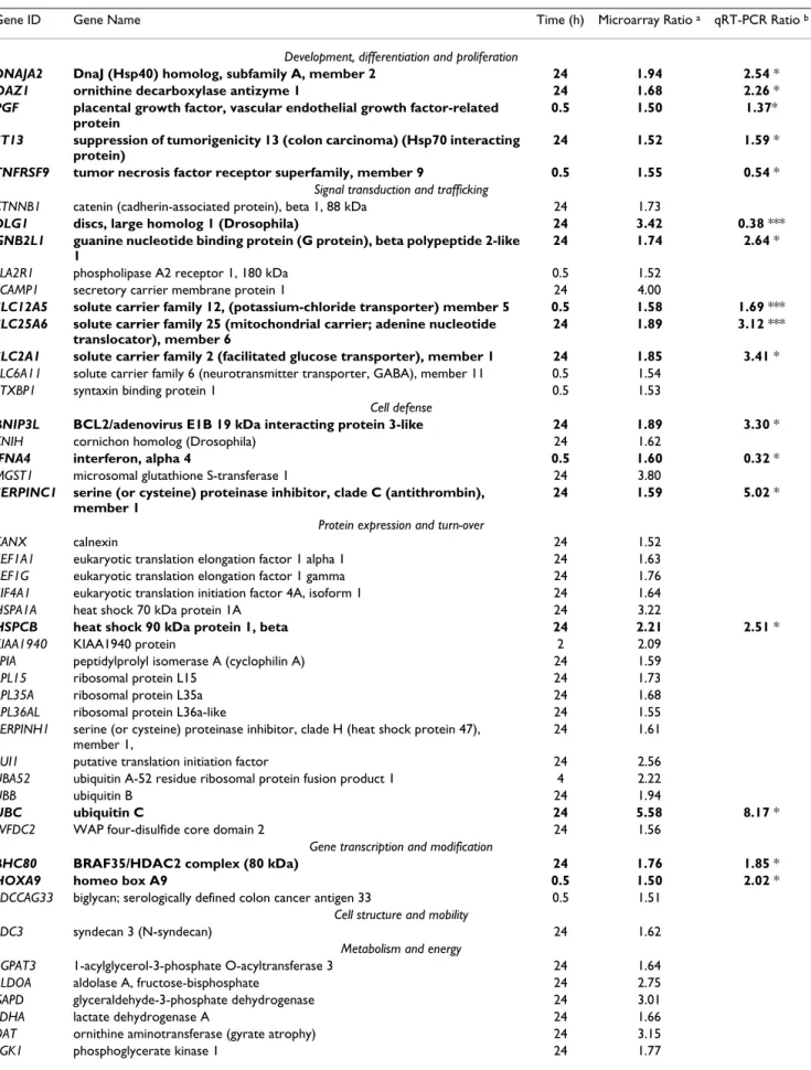
Table 1: Functional classification and ratios of genes up-regulated (> 1.5-fold) by exposure to cobalt across all time points
Gene ID Gene Name Time (h) Microarray Ratio a qRT-PCR Ratio b
Development, differentiation and proliferation
DNAJA2 DnaJ (Hsp40) homolog, subfamily A, member 2 24 1.94 2.54 *
OAZ1 ornithine decarboxylase antizyme 1 24 1.68 2.26 *
PGF placental growth factor, vascular endothelial growth factor-related protein
0.5 1.50 1.37*
ST13 suppression of tumorigenicity 13 (colon carcinoma) (Hsp70 interacting protein)
24 1.52 1.59 *
TNFRSF9 tumor necrosis factor receptor superfamily, member 9 0.5 1.55 0.54 *
Signal transduction and trafficking
CTNNB1 catenin (cadherin-associated protein), beta 1, 88 kDa 24 1.73
DLG1 discs, large homolog 1 (Drosophila) 24 3.42 0.38 ***
GNB2L1 guanine nucleotide binding protein (G protein), beta polypeptide 2-like 1
24 1.74 2.64 *
PLA2R1 phospholipase A2 receptor 1, 180 kDa 0.5 1.52
SCAMP1 secretory carrier membrane protein 1 24 4.00
SLC12A5 solute carrier family 12, (potassium-chloride transporter) member 5 0.5 1.58 1.69 *** SLC25A6 solute carrier family 25 (mitochondrial carrier; adenine nucleotide
translocator), member 6
24 1.89 3.12 ***
SLC2A1 solute carrier family 2 (facilitated glucose transporter), member 1 24 1.85 3.41 *
SLC6A11 solute carrier family 6 (neurotransmitter transporter, GABA), member 11 0.5 1.54
STXBP1 syntaxin binding protein 1 0.5 1.53
Cell defense
BNIP3L BCL2/adenovirus E1B 19 kDa interacting protein 3-like 24 1.89 3.30 *
CNIH cornichon homolog (Drosophila) 24 1.62
IFNA4 interferon, alpha 4 0.5 1.60 0.32 *
MGST1 microsomal glutathione S-transferase 1 24 3.80
SERPINC1 serine (or cysteine) proteinase inhibitor, clade C (antithrombin), member 1
24 1.59 5.02 *
Protein expression and turn-over
CANX calnexin 24 1.52
EEF1A1 eukaryotic translation elongation factor 1 alpha 1 24 1.63
EEF1G eukaryotic translation elongation factor 1 gamma 24 1.76
EIF4A1 eukaryotic translation initiation factor 4A, isoform 1 24 1.64
HSPA1A heat shock 70 kDa protein 1A 24 3.22
HSPCB heat shock 90 kDa protein 1, beta 24 2.21 2.51 *
KIAA1940 KIAA1940 protein 2 2.09
PPIA peptidylprolyl isomerase A (cyclophilin A) 24 1.59
RPL15 ribosomal protein L15 24 1.73
RPL35A ribosomal protein L35a 24 1.68
RPL36AL ribosomal protein L36a-like 24 1.55
SERPINH1 serine (or cysteine) proteinase inhibitor, clade H (heat shock protein 47), member 1,
24 1.61
SUI1 putative translation initiation factor 24 2.56
UBA52 ubiquitin A-52 residue ribosomal protein fusion product 1 4 2.22
UBB ubiquitin B 24 1.94
UBC ubiquitin C 24 5.58 8.17 *
WFDC2 WAP four-disulfide core domain 2 24 1.56
Gene transcription and modification
BHC80 BRAF35/HDAC2 complex (80 kDa) 24 1.76 1.85 *
HOXA9 homeo box A9 0.5 1.50 2.02 *
SDCCAG33 biglycan; serologically defined colon cancer antigen 33 0.5 1.51
Cell structure and mobility
SDC3 syndecan 3 (N-syndecan) 24 1.62
Metabolism and energy
AGPAT3 1-acylglycerol-3-phosphate O-acyltransferase 3 24 1.64
ALDOA aldolase A, fructose-bisphosphate 24 2.75
GAPD glyceraldehyde-3-phosphate dehydrogenase 24 3.01
LDHA lactate dehydrogenase A 24 1.66
OAT ornithine aminotransferase (gyrate atrophy) 24 3.15
SMAP1 stromal membrane-associated protein 1 24 1.65
TKT transketolase (Wernicke-Korsakoff syndrome) 24 1.86
Unknown function
GPM6A glycoprotein M6A 2 1.62
LOC115704 24 1.63
LTBP3 latent transforming growth factor beta binding protein 3 2 1.71 2.37 *
a: p value < 0.05 with 1.5 fold change.
b: validation with quantitative qRT-PCR (lines in bold) : *: p value < 0.005, ** p value < 0.01, *** p value < 0.05.
Microarray p-values were calculated using a t-test statistical analysis (Benjamini-Hochberg FDR) on Genespring software. Pair Wise Fixed Reallocation Randomized Test was used to calculate qRT-PCR p values with REST software.
Table 1: Functional classification and ratios of genes up-regulated (> 1.5-fold) by exposure to cobalt across all time points (Continued)
sent an appropriate internal positive control. Recently, two transcriptomic studies of hypoxia compared the action of low oxygen with that of metal ions such as cobalt or nickel on embryonic mouse fibrobasts [28] and human liver carcinoma [29]. 21% and 28% of the genes respec-tively were found to be modulated by both hypoxia and cobalt. This shows that the toxic effect of cobalt is not lim-ited to hypoxia. It should also be noted that nickel had an action closer to that of low oxygen than cobalt, with 65% common modulated genes [29]. Comparing these results with ours showed that, apart from the hypoxia genes, we had no overlap. This discrepancy might be due to differ-ences between cell models, species and conditions of exposure (one non-lethal dose of 0.1 mM cobalt for 24 h).
Stress response – apoptosis
The HSPCB gene coding for heat shock protein 90, was induced by a factor of 2.2 at 24 h (table 1). HSP90 protein plays a major role in the stress response by preventing irre-versible protein aggregation. In our previous study of the toxicogenomics of uranium [30], we noted that both
HSPCB gene and HSP90 protein were strongly repressed. This protein can even be thought of as a metal "sensor", since the HSPCB gene was also reported to be modulated by other metals (Ni, As, Cr, Cd) [31]. However, we did not see any change in the quantity of HSP90 in Western blot-ting experiments (figure 3). This proteome/transcriptome discordance can be explained by an increase in protein turnover associated with gene induction, resulting in a constant amount of protein being maintained in the cell. Another explanation may be that, as Gerner observed for apoptosis, HSP90 is aggregated and accumulates in the insoluble fraction of the cytosol and is not collected dur-ing protein extraction [32]. Two other genes described as HSP mediators are also induced by cobalt: ST13 and
DNAJA2.
Ubiquitin is a key element in eliminating proteins via pro-teasome because polyubiquitinylation is the recognition signal for protein elimination [33,34]. The results show that ubiquitin genes were up-regulated after 24 h of toxic exposure (see table 1, UBB, ratio 1.94, UBC, ratio 5.6) while UBC gene induction was observed in the kinetics
from 4 h with qRT-PCR (figure 2), showing that it is not an early event in cell response. The Western blot revealed protein ubiquitinylation, with the ubiquitin anti-body, shown by a smear at high molecular weight in the cobalt extract, indicating an accumulation of ubiquiti-nylated protein (figure 3). This demonstrates increased turnover of proteins involved in defense against stress, or the elimination of proteins partly damaged by the metal, or the start of apoptosis.
Cobalt has been described as an apoptosis inducer [17] and this is confirmed here by BAG1 repression and
BNIP3L induction (table 1 and 2).
Tumor suppressors – transcription factors
The protein encoded by MAZ (Myc-associated zinc finger protein), is a transcription factor involved in both the ini-tiation and termination of target gene transcription. It binds zinc and can act as a tumor suppressor [35]. This gene is repressed early at 2 h. Other cobalt-modulated genes code for zinc-binding proteins involved in tran-scription or DNA metabolism: BHC80 (BHC80 protein),
NT5E (CD73) and SDCCAG33 (Teashirt homolog 1). Cobalt ions have been shown to substitute for zinc in zinc finger protein domains which control the transcription of several genes and also in zinc-DNA repair proteins, inhib-iting DNA repair [14,36,37].
DLG1 (disk large homolog 1) has been identified as a tumor suppressor gene in drosophila, with its mutation inducing the loss of cell polarity and neoplastic tissue growth. This gene is highly conserved. The molecular mechanism which regulates cell proliferation by DLG1 is not well known but there are arguments for implicating epithelial cell polarization in this regulation [38]. The
DLG1 (disk large homolog 1) gene was strongly repressed in qRT-PCR. The anti-DLG1 antibody revealed clear depletion of DLG1 protein in Co+ samples (figure 3). This confirmed the results of the qRT-PCR.
Two tumor suppressors (MAZ and DLG1) were repressed by cobalt, and in one case (DLG1), this repression was also observed at protein level.
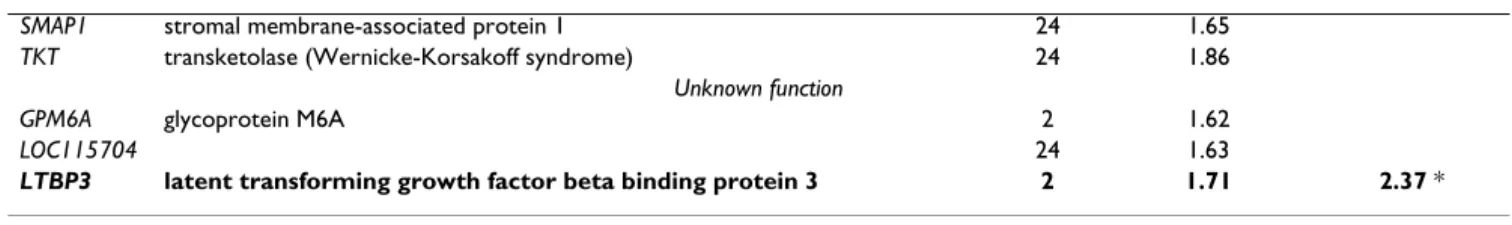
Table 2: Functional classification and ratios of genes down-regulated (< 1.5 fold) by exposure to cobalt across all time points
Gene ID Gene Name Time (h) Microarray Ratio a qRT-PCR Ratio b
Development, differentiation and proliferation
NDRG4 NDRG family member 4 24 0.58 0.29 ***
SEZ6L seizure related 6 homolog (mouse)-like 4 0.62
Signal transduction and trafficking
ATP6V1A ATPase, H+ transporting, lysosomal 70 kDa, V1 subunit A 2 0.66
AXL AXL receptor tyrosine kinase 4 0.60 1.36 ***
GFRA2 GDNF family receptor alpha 2 0.5 0.63 0.09 *
ICAM1 intercellular adhesion molecule 1 (CD54), human rhinovirus receptor 2 0.60
IFNAR2 interferon (alpha, beta and omega) receptor 2 24 0.61 0.55 ***
MAPK10 mitogen-activated protein kinase 10 24 0.47 0.31 ***
S100A4 S100 calcium binding protein A4 2 0.64
TFRC transferrin receptor (p90, CD71) 0.5 0.50 0.65 *
Cell defense
BAG1 BCL2-associated athanogene 2 0.65 0.55 **
TNFSF6 tumor necrosis factor (ligand) superfamily, member 6 4 0.54
Protein expression and turn-over
CNDP2 CNDP dipeptidase 2 (metallopeptidase M20 family) 4 0.57
FBXL2 F-box and leucine-rich repeat protein 2 0.5 0.55 0.75 ****
TIMP2 tissue inhibitor of metalloproteinase 2 24 0.63 0.54 ***
Gene transcription and modification
MAZ MYC-associated zinc finger protein (purine-binding transcription factor) 2 0.28 0.59 **
NT5E 5'-nucleotidase, ecto (CD73) 24 0.50
PAI-RBP1 PAI-1 mRNA-binding protein 2 0.62
SFRS1 splicing factor, arginine/serine-rich 1 (splicing factor 2, alternate splicing factor) 0.5 0.52
SIN3A SIN3 homolog A, transcriptional regulator (yeast) 24 0.63 2.21 *
Cell structure and mobility
FAT2 FAT tumor suppressor homolog 2 (Drosophila) 4 0.56
TUBB2 tubulin, beta, 2 4 0.62
VIL2 villin 2 (ezrin) 0.5 0.64
Metabolism and energy
AKR1C3 aldo-keto reductase family 1, member C3 24 0.53
ALDH1A1 aldehyde dehydrogenase 1 family, member A1 24 0.60
GLCE glucuronyl C5-epimerase 0.5 0.46
GPD2 glycerol-3-phosphate dehydrogenase 2 (mitochondrial) 4 0.55
MTHFS 5,10-methenyltetrahydrofolate synthetase 2 0.52
Unknown function
C20orf30 chromosome 20 open reading frame 30 2 0.48
C20orf30 chromosome 20 open reading frame 30 24 0.60
FLJ12806 hypothetical protein FLJ12806 0.5 0.64
KIAA158 2
KIAA1582 protein 24 0.58
a: p value < 0.05 with 1.5 fold change.
b: validation with quantitative qRT-PCR (lines in bold) :
*: p value < 0.005, ** p value < 0.01, *** p value < 0.05, **** p value < 0.1.
Microarray p-values were calculated using a t-test statistical analysis (Benjamini-Hochberg FDR) on Genespring software. Pair Wise Fixed Reallocation Randomized Test was used to calculate qRT-PCR p values with REST software.
We noted the modulation of three genes related to MYC, a key element in oncogenic response (table 1 and 2). The first, SIN3A, known as an MYC suppressor, is induced by cobalt. The two others: NDRG4, a gene negatively regu-lated by MYC and MAZ, a MYC transcription inhibitor, are down-regulated by cobalt. These results are not clear regarding MYC up- or down-regulation, but MYC regula-tion pathways are complex. These results led us to
hypoth-esize that MYC might be modulated by cobalt, so we tested MYC temporal modulation with qRT-PCR. Interest-ingly, we noted significant MYC inhibition from 30 min to 24 h (figure 2) indicating that cobalt modulates regula-tion pathways controlling or controlled by MYC. Liu has recently shown that MYC is involved in the carcinogenic response of mouse liver to arsenic, another toxic metal [39].
The AXL gene is positively modulated. Members of the AXL/UFO family of tyrosine kinases are prone to tran-scriptional regulation and perform various functions including regulation of cell adhesion, migration, phago-cytosis, and survival. The biological consequences of AXL activation are complex. AXL was initially identified as a transforming gene product, and AXL expression is indeed up-regulated in human tumors [40,41].
Concerning the potential effects of cobalt on cancer devel-opment, it will be interesting to carry out further studies of the mechanisms by which cobalt induces these responses. These genes have never been linked to cobalt effects before.
Transporters
Two cobalt transporters are described in yeast: COT1 and
COT2 [42]. Eleven cobalt-tolerant mutants were obtained with a common COT2 mutation, suggesting that it is involved in cobalt uptake [43]. The mutants were resistant to cobalt toxicity and cobalt was not incorporated in yeast cells. The human homolog of COT2, FBXL2, was strongly repressed in our study as early as 2 h and with a ratio of 0.1 at 24 h; this is the strongest inhibition observed in time-course qRT-PCR analysis (figure 2). FBXL2 function is not very well described, but it is potentially involved in ubiquitinylation. Conklin's observations on the yeast homolog of FBXL2 [43] and our results, lead us to suggest that FBXL2 gene repression could be a cellular defense mechanism and this protein might be associated with cobalt uptake in humans.
The product of the COT1 gene is involved in the uptake of cobalt ions [44]. COT1, has a human homolog in ZNT1, coding for Zinc transporter 1 protein, probably involved in zinc transport out of the cell. ZNT1, not spotted on the microarray, was also tested in kinetics with qRT-PCR and was found to be slightly modulated with a biphasic response; first down- then up-regulated (figure 2). The SLC12A5 gene, coding for an integral membrane K-Cl cotransporter, was repressed at 4 h then strongly increased at 24 h (qRT-PCR ratio : 4.3). The three genes, FBXL2,
ZNT1 and SLC12A5, are therefore appropriate candidates for research into cobalt transport proteins in human cells and will be studied further using targeted biological approaches.
Potential biomarkers and secreted proteins
Biological monitoring of exposure to cobalt is mainly based on its concentration in urine. This method may be inadequate for two reasons. Firstly, measuring the quan-tity of excreted metal (exposure marker) does not neces-sarily reflect organ damage, which varies from one person to another. Furthermore, depending on the chemical
form, excretion does not necessarily reflect the concentra-tion of metal in the body; indeed, certain forms are elim-inated in several phases; only 40% of cobalt oxides inhaled are excreted within 72 h [45]. In this context, only an effective biomarker could evaluate the damage and provide a valuable tool for monitoring occupational con-tamination in the event of accidental acute exposure or chronic exposure. We selected nine modulated genes cod-ing for secreted proteins: ICAM1 (CD54), SERPIN C1 (antithrombin-III), IFNA4 (alpha-4 interferon), TFRC (transferrin receptor), PLA2R1 (phospholipase receptor A2), TNFSF6 (FAS ligand), WFDC2 (WAP four-disulfide core domain protein 2), LTBP3 (latent transforming growth factor beta binding protein 3) and TIMP2 (tissue inhibitor of metalloproteinase 2). To further evaluate the interest of these genes, we checked the secretion modula-tion of the corresponding proteins. A549 cells were exposed to cobalt in a dose-response manner, to 0.2, 1 and 2 mM, and the supernatants analysed using available immunoassays. The levels of FAS ligand, WFDC2 [46], ICAM1, antithrombin-III, alpha-4 interferon, and soluble transferrin receptor were found to be invariant or below the kit detection limit. These results indicate that most of these immunoassays were not sensitive enough to detect faint modulations. TIMP2 protein (tissue inhibitor of metalloproteinase 2) was measured using ELISA and we confirmed the modulation recorded at gene level (micro-array ratio: 0.63, qRT-PCR ratio: 0.69). A strong decrease in TIMP2 supernatant concentration was noted at 1 or 2 mM of cobalt (figure 4). The changes in TIMP2 levels in cell supernatants between cobalt and the control were -5% for 0.2 mM, -57% for 1 mM and -72% for 2 mM. The reason for this reduction in TIMP2 can be explained as a consequence of cobalt exposure. Matrix metalloprotein-ases (MMPs) are a family of endopeptidmetalloprotein-ases that can degrade all the components of the extracellular matrix [47]. Endogenous protease inhibitors, known as tissue inhibitors of metalloproteinase (TIMP), provide critical extracellular regulation of MMP proteolytic activities [48,49], regulating tumor growth, progression, and angio-genesis in a variety of experimental cancer models and human malignancies. TIMP2 mRNA is described as being suppressed by hypoxia in endothelial cells [50]. Exposure of cells to cobalt up-regulates indirectly hypoxia-inducible genes by a mechanism which has recently been explained [51]. Therefore, TIMP-2 down-regulation might be a con-sequence of cobalt-simulated hypoxia. Down-regulation of TIMP2 mediated by ROS production has been observed by Rezzani in rat cardiac tissue exposed to cyclosporin [52]. Since it is known that cobalt triggers ROS production [16], TIMP2 modulation by cobalt could also be explained by this pathway.
TIMP2 protein modulation in cobalt-exposed cell super-natant corroborated the gene down-regulation observed
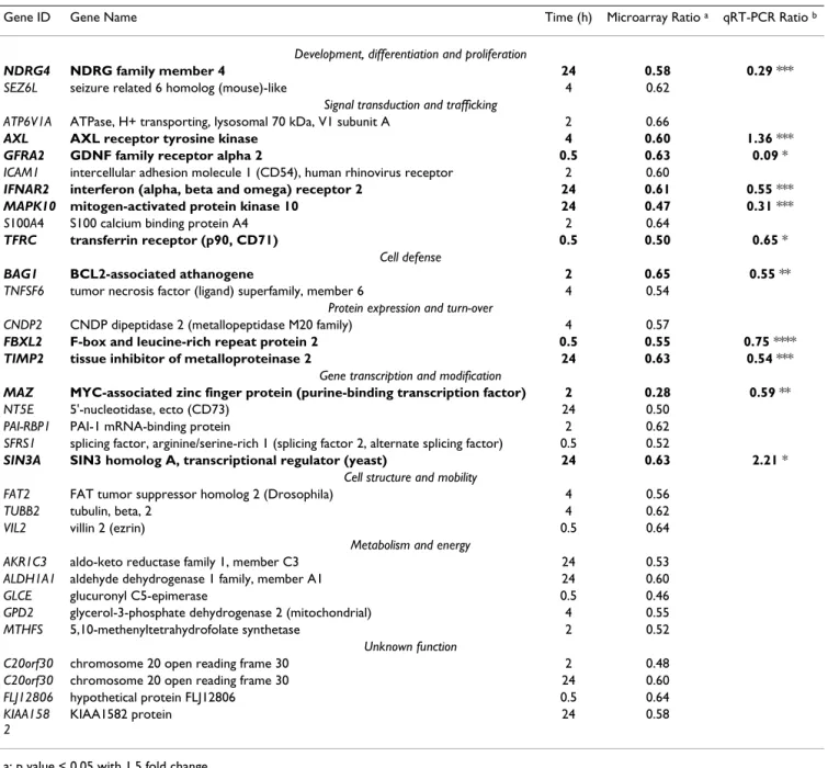
Table 3: List of primers used in qRT- PCR
Gene ID Gene Name GenBank ID Forward primer Reverse primer Amplicon (bp)
AXL AXL receptor tyrosine kinase NM_001699 GACCGGCCAAGTTTTACAGA ATAACCTCCACCCTCATCCA 117 BAG1 BCL2-associated athanogene NM_004323 GCAGCAGTGAACCAGTTGTC CGGTGTTTCCATTTCCTTCA 119 BHC80 BRAF35/HDAC2 complex (80 kDa) NM_016621 CCGAGCCGTTTGTTTAGGTA CACTGGGGTTGGTGAAATCT 117
BNIP3L BCL2/adenovirus E1B 19 kDa interacting protein 3-like
NM_020221 ATGTTTGGCTTTGGGGCTA CTTCACAGGTCACACGCATT 109
DLG1 discs, large homolog 1 (Drosophila)
NM_004087 CAGCCAGATACTCCCCAGTT TGAGCCACGATGAAGAACAA 89
DNAJA2 DnaJ (Hsp40) homolog, subfamily A, member 2
NM_005880 CCAGGGTGTGTTCGTGTAGTT TGGGTTGATCCAGTTGTTTTC 120
FBXL2 F-box and leucine-rich repeat protein 2
NM_012157 CAGAACTGCCGAAACATTGA CACACAGGAGGTCAGATCCA 123
FIGF c-fos induced growth factor (vascular
endothelial growth factor D)
NM_004469 GAACACCAGCACCTCGTACA TGGCAAGCACTTACAACCTG 118
GFRA2 GDNF family receptor alpha 2
NM_001495 GAGACACACGGTCACTGGAA TCGAGGACGAGAGACTGGAG 126
GNB2L1 guanine nucleotide binding protein (G protein), beta polypeptide 2-like 1
NM_006098 GGTGTCTTGTGTCCGCTTCT CAATGTGGTTGGTCTCAGC 119
HOXA9 homeo box A9 NM_152739 CACCAGACGAACAGTGAGGA ACTCCGTTACAATCAGCATTCA 111
HSPCB Heat shock protein HSP 90-beta
NM_007355 GGAGAGGAGGAGGTGGAGAC GAGGGTTGGGGATGATGTC 217
IFNA4 interferon, alpha 4 NM_021268 GAAGAAATACAGCCCTTGTGC TGAACCAGTTTTCAATCCTTCC 114
IFNAR2 interferon (alpha, beta and omega) receptor 2
NM_000874 GTCTCGCTAAGGGCTGGAAT AGGCAGGACGACTGTTTGAG 94
LTBP3 latent transforming growth factor beta binding protein 3
NM_021070 CCAGGGCTACAAGAGGCTTA GGCAGACACAGCGATAGGAG 119
MAPK10 mitogen-activated protein kinase 10
NM_002753 CTTCCCAGATTCCCTCTTCC GTAAGGCGTCGTCCACTGAT 128
MAZ MYC-associated zinc finger protein (purine-binding transcription factor)
NM_002383 CGGATCACCTCAACAGTCAC ATGGCACTTTCTCCTCGTGT 135
MYC v-myc myelocytomatosis viral oncogene homolog (avian)(MYC)
NM_002467 AAAGGCCCCCAAGGTAGTTA TTTCCGCAACAAGTCCTCTT 103
NDRG4 NDRG family member 4 NM_020465 ATGCTTTCCATCCACTCACC TTCACTGCTCTCTCCCGTTT 115
OAZ1 ornithine decarboxylase antizyme 1
NM_004152 GAGCCGACCATGTCTTCATT CCCGGTCTCACAATCTCAAA 100
PGF placental growth factor, vascular endothelial growth factor-related protein
NM_002632 ACCCCTTGGAGGAGAGAGAC GCATTCAGCAGGGAAACAGT 119
SERPINC1 serine (or cysteine) proteinase inhibitor, clade C (antithrombin), member 1
NM_000488 CAATCGCCTTTTTGGAGACA TGGACACCCATTTGTTGATG 146
SIN3A SIN3 homolog A, transcriptional regulator (yeast)
NM_015477 CTCCCAACTGCAAGCACATA TCCCAACGAGATTGTCACTG 115
SLC12A5 solute carrier family 12, (potassium-chloride transporter) member 5
NM_020708 CAAGGGTCCAACTTTTCCTG GCCTCTCGGTTTCTTCCTCT 152
SLC25A6 solute carrier family 25 (mitochondrial carrier; adenine nucleotide translocator), member 6
on the microarrays and with qRT-PCR. TIMP2 modula-tion has never been associated with metal stress. This result is innovative and will be studied further in the bio-logical fluids of rats exposed to different chemical forms of cobalt by inhalation.
Conclusion
This study provides the first toxicogenomic analysis of human lung cell response to acute cobalt exposure. We have confirmed that genes involved in the cobalt hypoxia response and apoptosis are modulated. We have also revealed genes linked to heat shock response and proteas-ome function that have already been described in other metal stress responses. Newly identified genes linked to cobalt acute toxicity include potential cobalt carriers (FBXL2, ZNT1, SLC12A5) and tumor suppressors or tran-scription factors (MAZ, DLG1, MYC, AXL). Some of these genes provide new hypotheses for elucidating the mecha-nisms of cobalt intracellular chemical toxicity. Targeted biological approaches might confirm their biochemical role in the cobalt response.
Regarding biomarkers, we have highlighted the down-reg-ulation of TIMP2, a gene coding for a secreted protein. TIMP2 modulation was confirmed at protein level, in a dose-dependent manner, in the supernatant of exposed A549 cells. TIMP2 provides a putative biomarker of cobalt toxicity that will be further studied on animal models.
Methods
Cell culture
The human type II epithelial cell line A549 from ATCC, was cultured as described previously [53]. At midlog phase, a medium without FCS, containing cobalt (CoCl2, Sigma) or not, was added for 24 hours. A CoCl2 stock solution (1 M in MilliQ water) was prepared
extempora-neously, filtered through a sterile 0.22 μm membrane and then diluted in the cell culture medium (final concentra-tion 2 mM). After 30 mn, 2 h, 4 h or 24 h, the cells were harvested with trypsin and washed in PBS containing 1 mM EDTA. Culture supernatants for ELISA and cells for Western blot confirmations were collected after 24 h of exposure.
Cytotoxicity studies
Cobalt cytotoxicity was determined by measuring the intracellular ATP after 24 h using the Promega Celltiter-GlowTM Luminescence Cell Viability Assay (four to eight replicates per concentration). The viability rate was deter-mined as the ratio between the ATP in treated cells and control cells. Cobalt concentration in cell pellets was measured using flame atomic absorption spectroscopy (FAAS, CERECO laboratory, Nimes, France).
RNA extraction
Total RNA was isolated using the Quiagen RNeasy mini-prep kit according to the manufacturer's instructions. mRNAs were prepared using the Oligotex mRNA Quiagen kit, the purification process being repeated once to elimi-nate any contamination. RNA concentration was deter-mined by OD measurement (260 nm/280 nm), purity and integrity were assessed using an Agilent 2100 Bioana-lyser.
Microarrays and data analysis
Microarrays were obtained from the Service de Genom-ique Fonctionnelle (CEA, Evry, France). Two types of DNA collections were used for preparing the cDNA arrays: a col-lection of 5760 full length cDNA clones from the human infant brain 1NIB library, kindly provided by Genethon, and a collection of 2304 human PCR products (400–600 bp) corresponding to specific key-words selected by direct
SLC2A1 solute carrier family 2 (facilitated glucose transporter), member 1 NM_006516 GTGGAGACTAAGCCCTGTCG CATAGCCACCTCCTGGGATA 128 ST13 suppression of tumorigenicity 13 (colon carcinoma) (Hsp70 interacting protein) NM_003932 AGGCAGACGAACCATCAAGT TCCGTTATCTCCGCATTTTC 115 TFRC transferrin receptor (p90, CD71) NM_003234 CGCTGGTCAGTTCGTGATTA TCAGGCCCATTTCCTTTATG 134
TIMP2 tissue inhibitor of metalloproteinase 2
NM_003255 TTCATTCGTCTCCCGTCTTT ACCAACGTGTGTGGATCAAA 113
TNFRSF9 tumor necrosis factor receptor superfamily, member 9
NM_001561 AGGGCTGTTGGGACTTTCTT GGATGGTGTTCTTGCTTTTGA 83
TUBA3 tubulin, alpha 3 NM_006009 CCTACAACTCCATCCTCACCA GTCAACATTTCAGGGCTCCA 203
UBC ubiquitin C NM_021009 GGAACAGGCGAGGAAAAGTA AACAAGAACTGCGACCCAAA 146
ZNT1 solute carrier family 30 (zinc transporter), member 1
NM_021194 ACCCAGAAAACCCCAGAAGT CACTGAACCCAAGGCATCTC 158 Table 3: List of primers used in qRT- PCR (Continued)
query in the Unigene database. A complete description of the microarrays used in this study, including the protocols for slide production, has been submitted to the GEO data-base [24] under accession number GPL4263.
For each microarray experiment, 20 μg of total RNA were reverse transcripted and indirectly labelled using the Fair-Play Microarray Labelling Kit (Stratagene). Amino reactive Cy3- and Cy5- dyes (Cy™ Dye Post-Labelling Reactive Dye
Time-course qRT-PCR analysis
Figure 2
Time-course qRT-PCR analysis. The temporal expression pattern of genes regulated by cobalt was analysed by qRT-PCR
in sextuplets for each time point. Mean results are given in Log2 ratios ± SD. *: significant modulation calculated by pairwise testing with p < 0.05
Pack, Amersham) were then chemically bound to cobalt and control cDNA respectively. Reverse labelling (or dye-swap) was performed for each experiment. Microarrays were pre-treated for 20 min with an N-methyl-pyrrolidi-none solution containing 20 g/L succinic anhydride and 20 mM sodium borate, pH 8, immersed quickly in etha-nol and dried by centrifugation for 6 min at 500 rpm. Fol-lowing a quick rinse in RNase-free water, the slides were prehybridized in 30 ml 5X SSC, 1% SDS, 1% BSA (w/v) at 50°C for 40 min. The microarrays were then rinsed in RNAse-free water, immersed quickly in isopropanol and dried by centrifugation for 6 min at 500 rpm. The hybrid-ization solution was prepared using 100 μl formamide, 10 μl SDS 10% and 30 μl RNase-free water. Labelled cDNA was solubilized in 17.5 μl of this solution plus 7.5 μl of 20 × SSPE and heat-denatured. Hybridization was performed overnight at 42°C. The microarrays were quickly rinsed in 0.1 × SSC, 0.01% SDS, washed twice for 10 min in 0.01 × SSC, then dried quickly in a stream of nitrogen and scanned with GenePix 4000B (Axon Instrument Inc., For-ster City, CA). Cy3 and Cy5 spot fluorescence intensities were quantified after local subtraction of background using Genepix Pro 4.0 software (Axon Instrument Inc.). For each time point the result files were submitted to GeneSpring software 6.2 (Agilent Technologies) as fol-lows. The data were first converted to take into account the results of the dyeswap reverse labelling, then normal-ized using the Lowess method, applying robust locally-weighted regression to smooth the intensity-dependence of the log ratios. The normalized data were then filtered on a quality test basis. This involved selecting spots detected on at least half of the microarrays with at least 70% pixels above threshold intensity (set to the median background plus two standard deviations).
From these remaining spots, we selected those with fluo-rescence ratios (representing cobalt-treated cells versus control) above 1.5 fold with p-value < 0.05 using a t-test
statistical analysis on Genespring software and perform-ing a Benjamini and Hochberg false discovery rate multi-ple testing correction.
Quantitative real-time polymerase chain reaction (qRT-PCR)
Specific primers were designed with Primer3 [54] using cDNA sequences spotted on microarrays, and amplicons were controlled using Mfold [55]. The list of primers is given in table 3. Before differential analysis, good primer efficiency (80% -120 %) was checked as a mandatory test. One μg of total RNA (for 30 min, 2 h and 4 h) or 50 ng of mRNA (for 24 h) were reverse transcribed with oligo dT (12–18) using Omniscript reverse transcriptase (Qiagen) following the manufacturer's instructions. cDNA was diluted at least 10-fold with DNase-free water to 10 ng/μL. PCR was performed on 15 μl using a DyNAmoTM HS SYBR Green qPCR Kit (Finnzymes) on a VWR DNA Engine Opticon® 2 system. The amplification program consisted of 1 cycle at 95°C with 10 min-hold followed by 40 cycles at 95°C with 15 sec-hold, 60°C with 1 min-hold, and a reading step at 60°C for 1 sec. Amplification was followed by melting curve analysis between 65°C and 95°C. RNA was used as a negative template for the absence of residual genomic DNA and a negative control without cDNA was used to control overall specificity. House- keeping genes, which are generally used as refer-ence genes, are often modulated by stress. For example, GAPDH is strongly induced by cobalt (ratio 3). To find an invariant gene at each time point, several candidates were selected for their invariance from all the microarray data, and tested as invariant genes in qRT-PCR. FIGF (c- fos induced growth factor) was selected for time points 0.5 h, 2 h and 4 h. For samples at 24 h, mRNA had to be purified
TIMP2 ELISA
Figure 4
TIMP2 ELISA. Concentrations of TIMP2 in supernatants
were determined by ELISA following treatment of A549 cells with cobalt over 24 h. The changes in level of TIMP2 in cell supernatants between cobalt and the control were -5% for 0.2 mM, -57% for 1 mM and -72% for 2 mM (n = 2).
Western blot confirmation
Figure 3
Western blot confirmation. Western blot analyses were
performed on A549 extracts following exposure to 2 mM cobalt for 24 h. Treated (Co+) and control (Co-) extracts were probed with anti-ubiquitin, anti-DLG1, or anti-HSP90 antibodies. An anti-actin antibody was used as a control.
from total RNA to detect TUBA3 as an invariant reference gene. Differential analysis was performed on cDNA tem-plates obtained from cobalt-treated or untreated cells in sextuplets for each gene, in the same way as for the refer-ence gene (5 ng for cDNA from total RNA or 0.1 ng for cDNA from mRNA). The results were processed using REST-MCS software [56] and tested for significance using a Pair Wise Fixed Reallocation Randomized Test by calcu-lating a p value. Modulation was considered to be signifi-cant when p calculated by REST software was < 0.05.
Western blot
Western blots were performed as previously described [53] after 24 h of cobalt exposure. Cells were lysed with 7 M urea, 2 M thiourea, 4% CHAPS, 20 mM spermine base, anti proteases (Roche cocktail), 40 mM DTT or 50 mM Tris-HCl, NaCl 150 mM ph7.5, 20 mM spermine base, anti proteases, 0.5% NP40 and 1% DOC. 10 to 50 μg of proteins were loaded onto a 4–12% or 12% NuPAGE gel in a MOPS or MES buffer (Invitrogen). Primary antibodies were: mouse monoclonal anti-DLG1 (sc-9961, 1/100, Santa Cruz), rabbit polyclonal anti-hsp90β (Ab1, 1/1666, NeoMarkers), mouse monoclonal anti-βactin (AC-15, 1/ 2000, Sigma), or mouse monoclonal anti-ubiquitin (P4D1, 1/400, Santa Cruz). A VECTASTAIN ABC kit (Vec-tor labora(Vec-tories) or a goat anti-mouse IgG coupled to HRP (1/10000, Novagen) was used for detection.
Immunoassays
Immunoassays were performed on culture supernatants from cells exposed to various concentrations of cobalt over a 24 h period. The supernatants were tested crude or at 10 fold concentration. TIMP2 and FASL proteins were detected using ELISA kits (Raybiotech and R&D systems respectively) according to the manufacturer's instructions. The WFDC2 protein, was analysed in Dr Hellström's lab-oratory because they developed the WFDC2 ELISA [46]. Other immunoassays were performed by the CERBA lab-oratory (Cergy Pontoise, France).
Authors' contributions
VM designed the study and drafted the manuscript. FB carried out data acquisition and analysis and prepared the microarray results for tables and figures.
OP was involved in the design stage, qRT-PCR analysis and microarray experiments.
SR carried out qRT-PCR.
GS carried out the RNA extraction, labelling and micro-array hybridization.
EQ was involved in the design stage and revised the man-uscript
All authors read and approved the final manuscript.
Acknowledgements
This research was supported in part by "Electricité De France". We thank the Service de Genomique Fonctionnelle (CEA Evry, France) for providing us with microarrays and Dr I.Hellström (Washington University) for per-forming the WFDC2 ELISA test. We thank Philippe Guérin and Nicole Sage for their technical support. We thank Elisabeth Darrouzet for critical review of this manuscript and for helpful discussions.
References
1. ATSDR: toxicological profile for cobalt. 2004 [http:// www.atsdr.cdc.gov/toxprofiles/tp33.pdf].
2. Barceloux DG: Cobalt. J Toxicol Clin Toxicol 1999, 37(2):201-206. 3. Hartmann P: Inventaire des radioexpositions [in French]. EDF
- Service de radioprotection 2000, 17(Radiotoxicologie):2-5.
4. Le Guen B, Ansoborlo E: Le cobalt et ses isotopes [in French]. In Toxicologie-Pathologie professionnelle Elsevier; 2005:1-11. 5. Chlorinated drinking-water; chlorination by-products; some
other halogenated compounds; cobalt and cobalt com-pounds. International Agency for Research on Cancer (IARC) Working Group, Lyon, 12-19 June 1990. 1991, 52:1-544.
6. Goldoni M, Catalani S, De Palma G, Manini P, Acampa O, Corradi M, Bergonzi R, Apostoli P, Mutti A: Exhaled breath condensate as a
suitable matrix to assess lung dose and effects in workers exposed to cobalt and tungsten. Environ Health Perspect 2004, 112(13):1293-1298.
7. Rosenberg N: Allergie respiratoire professionnelle au cobalt.
INRS 1989, 40TR15:.
8. INERIS: Cobalt et ses dérivés [in French]. fiche de données
toxi-cologiques et environnementales des substances chimiques 2004, version1(3- décembre 04):1-52.
9. Palmes ED, Nelson N, Laskin S, Kuschner M: Inhalation toxicity of
cobalt hydrocarbonyl. Am Ind Hyg Assoc J 1959, 20:453-468.
10. Bucher J: NTP technical report on the toxicity studies of
Cobalt Sulfate Heptahydrate in F344/N Rats and B6C3F1 Mice (Inhalation Studies) (CAS No. 10026-24-1). Toxic Rep Ser
1991, 5:1-38.
11. Bucher JR, Elwell MR, Thompson MB, Chou BJ, Renne R, Ragan HA:
Inhalation toxicity studies of cobalt sulfate in F344/N rats and B6C3F1 mice. Fundam Appl Toxicol 1990, 15(2):357-372.
12. Alexander CS: Cobalt-beer cardiomyopathy. A clinical and
pathologic study of twenty-eight cases. Am J Med 1972, 53(4):395-417.
13. Jacobziner H, Raybin HW: Poison control...accidental cobalt
poisoning. Arch Pediatr 1961, 78:200-205.
14. De Boeck M, Kirsch-Volders M, Lison D: Cobalt and antimony:
genotoxicity and carcinogenicity. Mutat Res 2003, 533(1-2):135-152.
15. Lison D, De Boeck M, Verougstraete V, Kirsch-Volders M: Update
on the genotoxicity and carcinogenicity of cobalt com-pounds. Occup Environ Med 2001, 58(10):619-625.
16. Salnikow K, Su W, Blagosklonny MV, Costa M: Carcinogenic
met-als induce hypoxia-inducible factor-stimulated transcription by reactive oxygen species-independent mechanism. Cancer Res 2000, 60(13):3375-3378.
17. Pulido MD, Parrish AR: Metal-induced apoptosis: mechanisms.
Mutat Res 2003, 533(1-2):227-241.
18. Bruick RK: Oxygen sensing in the hypoxic response pathway:
regulation of the hypoxia-inducible transcription factor. Genes Dev 2003, 17(21):2614-2623.
19. Harris AL: Hypoxia--a key regulatory factor in tumour
growth. Nat Rev Cancer 2002, 2(1):38-47.
20. Li Q, Chen H, Huang X, Costa M: Effects of 12 metal ions on iron
regulatory protein 1 (IRP-1) and hypoxia-inducible factor-1 alpha (HIF-1alpha) and HIF-regulated genes. Toxicol Appl Phar-macol 2006, 213(3):245-255.
Publish with BioMed Central and every scientist can read your work free of charge "BioMed Central will be the most significant development for disseminating the results of biomedical researc h in our lifetime."
Sir Paul Nurse, Cancer Research UK
Your research papers will be:
available free of charge to the entire biomedical community peer reviewed and published immediately upon acceptance cited in PubMed and archived on PubMed Central yours — you keep the copyright
Submit your manuscript here:
http://www.biomedcentral.com/info/publishing_adv.asp
BioMedcentral 21. Lettieri T: Recent applications of DNA microarray technology
to toxicology and ecotoxicology. Environ Health Perspect 2006, 114(1):4-9.
22. Lieber M, Smith B, Szakal A, Nelson-Rees W, Todaro G: A
continu-ous tumor-cell line from a human lung carcinoma with prop-erties of type II alveolar epithelial cells. Int J Cancer 1976, 17(1):62-70.
23. Crouch SP, Kozlowski R, Slater KJ, Fletcher J: The use of ATP
bio-luminescence as a measure of cell proliferation and cytotox-icity. J Immunol Methods 1993, 160(1):81-88.
24. Andrew AS, Warren AJ, Barchowsky A, Temple KA, Klei L, Soucy NV, O'Hara KA, Hamilton JW: Genomic and proteomic profiling
of responses to toxic metals in human lung cells. Environ Health Perspect 2003, 111(6):825-835.
25. National Center for Biotechnology Information [http://
www.ncbi.nlm.nih.gov/geo]
26. DAVID: DAVID. [http://david.abcc.ncifcrf.gov/].
27. Maxwell P, Salnikow K: HIF-1: an oxygen and metal responsive
transcription factor. Cancer Biol Ther 2004, 3(1):29-35.
28. Semenza GL: Targeting HIF-1 for cancer therapy. Nat Rev
Can-cer 2003, 3(10):721-732.
29. Vengellur A, Woods BG, Ryan HE, Johnson RS, LaPres JJ: Gene
expression profiling of the hypoxia signaling pathway in hypoxia-inducible factor 1alpha null mouse embryonic fibroblasts. Gene Expr 2003, 11(3-4):181-197.
30. Vengellur A, Phillips JM, Hogenesch JB, LaPres JJ: Gene expression
profiling of hypoxia signaling in human hepatocellular carci-noma cells. Physiol Genomics 2005, 22(3):308-318.
31. Prat O, Berenguer F, Malard V, Tavan E, Sage N, Steinmetz G, Quemeneur E: Transcriptomic and proteomic responses of
human renal HEK293 cells to uranium toxicity. Proteomics
2005, 5(1):297-306.
32. Gerner C, Frohwein U, Gotzmann J, Bayer E, Gelbmann D, Bursch W, Schulte-Hermann R: The Fas-induced apoptosis analyzed by
high throughput proteome analysis. J Biol Chem 2000, 275(50):39018-39026.
33. Bachmair A, Varshavsky A: The degradation signal in a
short-lived protein. Cell 1989, 56(6):1019-1032.
34. Hershko A, Leshinsky E, Ganoth D, Heller H: ATP-dependent
deg-radation of ubiquitin-protein conjugates. Proc Natl Acad Sci U S A 1984, 81(6):1619-1623.
35. Stubbs MC, Min I, Izzo MW, Rallapalli R, Derfoul A, Hall DJ: The
ZF87/MAZ transcription factor functions as a growth sup-pressor in fibroblasts. Biochem Cell Biol 2000, 78(4):477-485.
36. Kopera E, Schwerdtle T, Hartwig A, Bal W: Co(II) and Cd(II)
sub-stitute for Zn(II) in the zinc finger derived from the DNA repair protein XPA, demonstrating a variety of potential mechanisms of toxicity. Chem Res Toxicol 2004, 17(11):1452-1458.
37. Witkiewicz-Kucharczyk A, Bal W: Damage of zinc fingers in
DNA repair proteins, a novel molecular mechanism in car-cinogenesis. Toxicol Lett 2006, 162(1):29-42.
38. Humbert P, Russell S, Richardson H: Dlg, Scribble and Lgl in cell
polarity, cell proliferation and cancer. Bioessays 2003, 25(6):542-553.
39. Liu J, Xie Y, Ducharme DM, Shen J, Diwan BA, Merrick BA, Grissom SF, Tucker CJ, Paules RS, Tennant R, Waalkes MP: Global gene
expression associated with hepatocarcinogenesis in adult male mice induced by in utero arsenic exposure. Environ Health Perspect 2006, 114(3):404-411.
40. Nakano T, Tani M, Ishibashi Y, Kimura K, Park YB, Imaizumi N, Tsuda H, Aoyagi K, Sasaki H, Ohwada S, Yokota J: Biological properties
and gene expression associated with metastatic potential of human osteosarcoma. Clin Exp Metastasis 2003, 20(7):665-674.
41. Janssen JW, Schulz AS, Steenvoorden AC, Schmidberger M, Strehl S, Ambros PF, Bartram CR: A novel putative tyrosine kinase
receptor with oncogenic potential. Oncogene 1991, 6(11):2113-2120.
42. Kobayashi M, Shimizu S: Cobalt proteins. Eur J Biochem 1999,
261(1):1-9.
43. Conklin DS, Kung C, Culbertson MR: The COT2 gene is required
for glucose-dependent divalent cation transport in Saccharo-myces cerevisiae. Mol Cell Biol 1993, 13(4):2041-2049.
44. Conklin DS, McMaster JA, Culbertson MR, Kung C: COT1, a gene
involved in cobalt accumulation in Saccharomyces cerevi-siae. Mol Cell Biol 1992, 12(9):3678-3688.
45. Kreyling WG, Cox C, Ferron GA, Oberdorster G: Lung clearance
in Long-Evans rats after inhalation of porous, monodisperse cobalt oxide particles. Exp Lung Res 1993, 19(4):445-467.
46. Hellstrom I, Raycraft J, Hayden-Ledbetter M, Ledbetter JA, Schummer M, McIntosh M, Drescher C, Urban N, Hellstrom KE: The HE4
(WFDC2) protein is a biomarker for ovarian carcinoma. Cancer Res 2003, 63(13):3695-3700.
47. Coussens LM, Fingleton B, Matrisian LM: Matrix
metalloprotein-ase inhibitors and cancer: trials and tribulations. Science 2002, 295(5564):2387-2392.
48. Jiang Y, Goldberg ID, Shi YE: Complex roles of tissue inhibitors
of metalloproteinases in cancer. Oncogene 2002, 21(14):2245-2252.
49. Baker AH, Edwards DR, Murphy G: Metalloproteinase inhibitors:
biological actions and therapeutic opportunities. J Cell Sci
2002, 115(Pt 19):3719-3727.
50. Ben-Yosef Y, Lahat N, Shapiro S, Bitterman H, Miller A: Regulation
of endothelial matrix metalloproteinase-2 by hypoxia/reoxy-genation. Circ Res 2002, 90(7):784-791.
51. Salnikow K, Donald SP, Bruick RK, Zhitkovich A, Phang JM, Kasprzak KS: Depletion of intracellular ascorbate by the carcinogenic
metals nickel and cobalt results in the induction of hypoxic stress. J Biol Chem 2004, 279(39):40337-40344.
52. Rezzani R, Giugno L, Buffoli B, Bonomini F, Bianchi R: The
protec-tive effect of caffeic acid phenethyl ester against cyclosporine A-induced cardiotoxicity in rats. Toxicology 2005, 212(2-3):155-164.
53. Malard V, Prat O, Darrouzet E, Berenguer F, Sage N, Quemeneur E:
Proteomic analysis of the response of human lung cells to uranium. Proteomics 2005, 5(17):4568-4580.
54. Primer3: Primer3. Primer3 [http://frodo.wi.mit.edu/cgi-bin/primer3/ primer3_www.cgi].
55. Mfold: Mfold. [http://www.bioinfo.rpi.edu/applications/mfold/]. 56. Pfaffl MW, Horgan GW, Dempfle L: Relative expression software
tool (REST) for group-wise comparison and statistical analy-sis of relative expression results in real-time PCR - http:// www.gene-quantification.de/rest.html. Nucleic Acids Res 2002, 30(9):e36.