HAL Id: pasteur-02545979
https://hal-pasteur.archives-ouvertes.fr/pasteur-02545979
Submitted on 27 Apr 2020
HAL is a multi-disciplinary open access archive for the deposit and dissemination of sci-entific research documents, whether they are pub-lished or not. The documents may come from teaching and research institutions in France or abroad, or from public or private research centers.
L’archive ouverte pluridisciplinaire HAL, est destinée au dépôt et à la diffusion de documents scientifiques de niveau recherche, publiés ou non, émanant des établissements d’enseignement et de recherche français ou étrangers, des laboratoires publics ou privés.
Distributed under a Creative Commons Attribution| 4.0 International License
2012–2016
Jean-Marc Reynes, Damien Carli, Damien Thomas, Guillaume Castel
To cite this version:
Jean-Marc Reynes, Damien Carli, Damien Thomas, Guillaume Castel. Puumala Hantavirus Geno-types in Humans, France, 2012–2016. Emerging Infectious Diseases, Centers for Disease Control and Prevention, 2019, 25 (1), pp.140-143. �10.3201/eid2501.180270�. �pasteur-02545979�
Jean-Marc Reynes,1 Damien Carli,
Damien Thomas,2 Guillaume Castel
The analysis of the nucleoprotein gene of 77 Puumala hantavirus strains detected in human samples in France during 2012–2016 showed that all belonged to the Central European lineage. We observed 2 main clusters, geograph-ically structured; one included strains with the Q64 signa-ture and the other strains with the R64 signasigna-ture.
P
uumala virus (PUUV) is the main hantavirus detected in Europe. Several variants of this enveloped triseg-mented RNA virus species have been reported, compris-ing PUUV stricto sensu, detected in humans, and the Asian Hokkaido and Muju viruses, not yet detected in humans. PUUV variant, hosted in the wild by the bank vole (Myodes glaerolus), is responsible for a mild hemorrhagic fever with renal syndrome called nephropathia epidemica (1,2). With-in the variant PUUV, 8 lWith-ineages have been described, ac-cording to phylogenetic analysis of the small (S) RNA seg-ment coding domain sequence (CDS) of the strains. Each lineage is constituted by well-supported and geographical-ly structured clusters of variants, supporting the hypothesis of a hantavirus/host co-evolution (3,4). Furthermore, it has been shown that this genetic diversity may affect the mo-lecular detection of PUUV in humans (5). Consequently, the genotyping of local PUUV strains is also essential for laboratory diagnostics.PUUV is also the main hantavirus detected in France. Approximately 100 human hospitalized cases are detected annually, all of them located in the northeastern part of the country (6). Sequences of 7 PUUV strains have been
stud-human hantavirus cases from 2012–2016 in France, in-cluding Tula virus (n = 1), Seoul virus (n = 6), and PUUV (n = 228) infections (6). We did not obtain strains from all samples for several reasons, including the absence of mo-lecular testing because of lack of samples, samples being taken too long after the date of onset, and inadequate sam-ple storage temperature before reception in our laboratory. We detected 162 PUUV strains using both real-time PCR and nested reverse transcription PCR (RT-PCR) (9,10), 33 using real-time PCR, and 33 using nested RT-PCR. The discordance of results between the 2 techniques could be explained by a viral load in the sample close to our limit of detection. We identified these 228 PUUV strains in 28 of the 34 hantavirus-endemic departments (administrative divisions) in France (6). We used 3 overlapping heminest-ed RT-PCRs to obtain the S segment sequences. After se-quencing by Sanger method, we obtained the entire S CDS from 77 strains coming from 17 departments (Appendix Tables 1,2, https://wwwnc.cdc.gov/EID/article/25/1/18-0270-App1.pdf). We deposited PUUV sequences in GenBank (accession nos. MG923598–MG923674). We mapped strains according to the municipality of exposure. We used ClustalW and Muscle implemented in MEGA7 (https://www.megasoftware.net) (8) to align the S seg-ment CDS and to deduce aa sequences from these strains and from 115 other PUUV strains published and available in GenBank (as of December 1, 2017). We then ordered the sequences according to aa similarity. We conducted maximum-likelihood phylogenetic analysis with 1,000 bootstrap replicates by using PhyML version 3.0 (http:// www.atgc-montpellier.fr/phyml) implemented in Seaview version 4.6.1 (8), on the basis of the S segment CDS using
Puumala Hantavirus Genotypes in
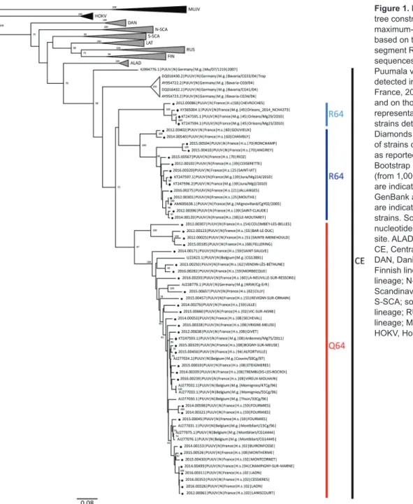
The phylogenetic analysis showed that the PUUV S CDS were grouped into the 8 lineages as previously scribed (3,4). All of the PUUV sequences from France de-tected in humans belonged to the Central European lineage (Figure 1). We distinguished 2 main sublineages, the first harboring the aa signature Q64 and including strains from Belgium and Germany (3), and the second with strains har-boring an arginine (R) amino acid at position 64. Within this cluster, strains isolated from bank voles in the Loiret department (coded 45) cluster together with branch support of 100; they share the K258 signature (8) with the human
strain 2012.00086 isolated in the Nièvre department (coded 58), neighbor of the Loiret department (Figure 1). Of note, strains from the Q64 cluster were from the northeast part of the endemic region, whereas the strains from the R64 clus-ters were detected in the south of the endemic area, except for strains carrying the I276 residue; those came from the northwest part of the endemic region in the Oise depart-ment (coded 60), where the Q64 cluster was co-circulating (Figure 2).
Several real-time RT-PCRs targeting the PUUV S seg-ment have been impleseg-mented to diagnose PUUV infection
Figure 1. Phylogenetic
tree constructed using the maximum-likelihood approach based on the complete small-segment RNA nucleotide coding sequences of representative Puumala virus (PUUV) strains detected in human cases in France, 2012–2016 (circles), and on those published and representative of PUUV strains detected in Europe. Diamonds indicate sequences of strains detected in rodents as reported elsewhere (7,8). Bootstrap percentages >70% (from 1,000 resamplings) are indicated at each node; GenBank accession numbers are indicated for reference strains. Scale bar indicates nucleotide substitutions per site. ALAD, Alpe-Adrian lineage; CE, Central European lineage; DAN, Danish lineage; FIN, Finnish lineage; LAT, Latvian lineage; N-SCA, north-Scandinavian lineage; S-SCA; south-Scandinavian lineage; RUS, Russian lineage; MUJV, Muju virus; HOKV, Hokkaido virus.
in humans (5,9,11–14). We detected some mismatches between the sequences of the France strains and those of the primers and probes (for example, at the 3′ end of the forward primer designed by Garin D et al. [12]), which could jeopardize the detection of some PUUV strains from France (Appendix Figure). However, we did not perform assays to evaluate this hypothesis.
Conclusions
Genomic findings from this study revealed that the 77 PUUV strains studied and detected in humans in France during 2012–2016 all belonged to the Central European lineage. Within this lineage, the strains were clustered within 2 sublineages, with marked geographical
pat-our dataset. The lack of amplification could be a result of improper storage of the specimens for several days at 4°C by peripheral laboratories for serologic diagnostic testing before sending to the National Reference Center; the low viral load observed in the specimens at reception (crossing point >35); the limit of detection of our S CDS overlapping heminested RT-PCR (reduced 10-fold, com-pared with detection limit using our molecular diagnostic methods); or mismatch between the sequences of PUUV strains and those of our primers used for S segment CDS amplification. Therefore, other clusters may be present in France, and we cannot clearly determine the geographic overlap of the clusters we described. Furthermore, our study was limited to the S segment CDS, whereas it has
Figure 2. Location of the
Puumala virus small segment RNA coding domain sequence sublineages Q64 and R64 detected in human cases, by municipality of exposure, France, 2012–2016. Gray shading indicates the hantavirus-endemic area; red circles indicate Q64 sequences, by size; blue diamonds indicate R64 sequences.
Acknowledgment
We thank the members of the laboratory network involved in first-line hantavirus diagnostics and in providing samples for expertise and surveillance.
Santé Publique France supports the Centre National de Référence des Hantavirus.
About the Author
Dr. Reynes is a veterinarian and a medical virologist at Institut Pasteur, Paris, France. He is responsible for the National Reference Center for Hantavirus in France. His research interests focus on zoonotic infectious diseases.
References
1. Vaheri A, Henttonen H, Voutilainen L, Mustonen J, Sironen T, Vapalahti O. Hantavirus infections in Europe and their impact on public health. Rev Med Virol. 2013;23:35–49. http://dx.doi.org/ 10.1002/rmv.1722
2. International Committee on Taxonomy of Viruses (ICTV). 2016.023a-cM Hantavirus approved proposal. 2017 March 19. [cited 2018 Feb 9]. https://talk.ictvonline.org/ICTV/
proposals/2016.023a-cM.A.v2.Hantavirus_sprev.pdf
3. Sironen T, Vaheri A, Plyusnin A. Molecular evolution of Puumala hantavirus. J Virol. 2001;75:11803–10. http://dx.doi.org/10.1128/ JVI.75.23.11803-11810.2001
4. Razzauti M, Plyusnina A, Niemimaa J, Henttonen H, Plyusnin A. Co-circulation of two Puumala hantavirus lineages in Latvia: a Russian lineage described previously and a novel Latvian lineage. J Med Virol. 2012;84:314–8. http://dx.doi.org/10.1002/ jmv.22263
5. Evander M, Eriksson I, Pettersson L, Juto P, Ahlm C, Olsson GE, et al. Puumala hantavirus viremia diagnosed by real-time reverse transcriptase PCR using samples from patients with hemorrhagic fever and renal syndrome. J Clin Microbiol. 2007;45:2491–7. http://dx.doi.org/10.1128/JCM.01902-06
6. Reynes JM, Carli D, Renaudin B, Fizet A, Bour JB, Brodard V, et al. Surveillance of human hantavirus infections in metropolitan France 2012–2016. [in French]. Bull Epidemiol Hebd (Paris). 2017;23:492–9.
7. Plyusnina A, Deter J, Charbonnel N, Cosson JF, Plyusnin A. Puumala and Tula hantaviruses in France. Virus Res. 2007;129:58– 63. http://dx.doi.org/10.1016/j.virusres.2007.04.023
8. Castel G, Couteaudier M, Sauvage F, Pons JB, Murri S, Plyusnina A, et al. Complete genome and phylogeny of Puumala hantavirus isolates circulating in France. Viruses. 2015;7:5476–88. http://dx.doi.org/10.3390/v7102884
9. Kramski M, Meisel H, Klempa B, Krüger DH, Pauli G,
Nitsche A. Detection and typing of human pathogenic hantaviruses by real-time reverse transcription-PCR and pyrosequencing. Clin Chem. 2007;53:1899–905. http://dx.doi.org/10.1373/ clinchem.2007.093245
10. Bowen MD, Gelbmann W, Ksiazek TG, Nichol ST, Nowotny N. Puumala virus and two genetic variants of Tula virus are present in Austrian rodents. J Med Virol. 1997;53:174–81. http://dx.doi.org/10.1002/(SICI)1096-9071(199710) 53:2<174::AID-JMV11>3.0.CO;2-J
11. Aitichou M, Saleh SS, McElroy AK, Schmaljohn C, Ibrahim MS. Identification of Dobrava, Hantaan, Seoul, and Puumala viruses by one-step real-time RT-PCR. J Virol Methods. 2005;124:21–6. http://dx.doi.org/10.1016/j.jviromet.2004.10.004
12. Garin D, Peyrefitte C, Crance JM, Le Faou A, Jouan A, Bouloy M. Highly sensitive Taqman PCR detection of Puumala hantavirus. Microbes Infect. 2001;3:739–45. http://dx.doi.org/10.1016/ S1286-4579(01)01424-1
13. Korva M, Saksida A, Kejžar N, Schmaljohn C, Avšič-Županc T. Viral load and immune response dynamics in patients with haemorrhagic fever with renal syndrome. Clin Microbiol Infect. 2013;19:e358–66. http://dx.doi.org/10.1111/1469-0691.12218 14. Lagerqvist N, Hagström Å, Lundahl M, Nilsson E, Juremalm M,
Larsson I, et al. Molecular diagnosis of hemorrhagic fever with renal syndrome caused by Puumala virus. J Clin Microbiol. 2016;54:1335–9. http://dx.doi.org/10.1128/JCM.00113-16 15. Razzauti M, Plyusnina A, Sironen T, Henttonen H, Plyusnin A.
Analysis of Puumala hantavirus in a bank vole population in northern Finland: evidence for co-circulation of two genetic lineages and frequent reassortment between strains. J Gen Virol. 2009;90:1923–31.http://dx.doi.org/10.1099/vir.0.011304-0 Address for correspondence: Jean-Marc Reynes, Centre National de Référence des Hantavirus, Unité de Recherche et d’Expertise
Environnement et Risques Infectieux, Institut Pasteur, 25-28 rue du Dr Roux, 75724 Paris CEDEX 15, France; email: [email protected]